Research Article
Prognostic Biomarkers of Sarcoidosis: A Comparative Study of
Serum Chitotriosidase, ACE, Lysozyme, and KL-6
Laura Bergantini
,
1Francesco Bianchi,
1Paolo Cameli,
1Maria Antonietta Mazzei
,
2Annalisa Fui,
1Piersante Sestini,
1Paola Rottoli,
1and Elena Bargagli
11Department of Clinical Medicine and Immunological Sciences, Respiratory Disease and Lung Transplant Unit, Respiratory Diseases
and Transplant Unit, Siena University, Siena, Italy
2Department of Clinical Medicine and Immunological Sciences, Radiology Unit, Siena University, Siena, Italy
Correspondence should be addressed to Laura Bergantini; [email protected]
Received 26 November 2018; Revised 19 January 2019; Accepted 31 January 2019; Published 3 March 2019 Academic Editor: Maria Dalamaga
Copyright © 2019 Laura Bergantini et al. This is an open access article distributed under the Creative Commons Attribution License, which permits unrestricted use, distribution, and reproduction in any medium, provided the original work is properly cited. Purpose. Sarcoidosis is a systemic granulomatous disease with unknown etiology. Many clinical presentations have been reported, and acute disease needs to be distinguished from subacute and chronic disease. The unpredictable clinical course of the disease prompted us to evaluate the clinical utility of biomarker serum detection in sarcoidosis follow-up. Methods. Serum
concentrations of chitotriosidase, ACE, KL-6, and lysozyme were analyzed by different methods in a population of 74
sarcoidosis patients (46 on steroid therapy at sampling) regularly monitored at Siena Sarcoidosis Regional Referral Centre and in a group of controls with the aim of comparing their contribution to clinical management of sarcoidosis patients. Results. KL-6 concentrations were significantly elevated in sarcoidosis patients with lung fibrosis and were significantly correlated with
DLco and CPI score, while chitotriosidase was significantly higher in patients with extrapulmonary localizations. With a cut-off
value of 303.5 IU/ml, KL-6 showed the best sensitivity (78%), while chitotriosidase reported the best specificity (85%) among the
biomarkers. Conclusions. KL-6 is a reliable biomarker offibrotic lung involvement in sarcoidosis patients. Among biomarkers,
KL-6 showed the best sensitivity and serum chitotriosidase the best specificity, even in patients on chronic steroid therapy, and seemed to correlate with extrapulmonary localizations.
1. Introduction
Sarcoidosis is a systemic granulomatous disease associated with T lymphocyte and macrophage activation and migra-tion into affected organs. The interaction between antigens and APC polarizes T lymphocytes to T helper 1 phenotype (Th1), leading to the formation of sarcoid granulomas consisting of T cells, macrophages, and epithelioid and giant cells [1, 2]. The course of sarcoidosis is unpredictable: remis-sion occurs in most cases, while persistent granuloma inflam-mation may lead to fibrotic lung disease [3–6]. Specific biomarkers with good sensitivity and specificity are therefore needed to predict clinical outcome and guide clinical decisions.
Human chitotriosidase is a biomarker secreted by acti-vated macrophages and neutrophils. Although its physiolog-ical role is not clear, it may play a role in the hydrolysis and
degradation of chitin and chitin-like substrates [7]. Increased concentrations of chitotriosidase have been reported in serum and BAL of patients with active sarcoid-osis [8]. Chitotriosidase shows a correlation with radiolog-ical stages. It may predict clinradiolog-ical course, steroid responsiveness, and potential relapses of the disease [9]. However, it is not clear whether chitotriosidase is reliable in the management of sarcoidosis patients with extrapul-monary organ involvement.
Angiotensin-converting enzyme (ACE) is an acid glyco-protein that converts angiotensin I into angiotensin II. It is produced by lung endothelial cells, mainly activated alveolar macrophages [10]. It is elevated in serum and BAL of sar-coidosis patients and its concentrations correlate with radio-logical stages [11–13]. However, its utility as a diagnostic and prognostic tool is limited by its low sensitivity and specificity. ACE may be elevated in certain granulomatous disorders Volume 2019, Article ID 8565423, 7 pages
(including berylliosis and silicosis), hyperthyroidism, diabe-tes, and other diseases [14, 15].
Lysozyme is produced by monocyte-macrophage system and epithelioid cells involved in granuloma formation can release this enzyme. Sarcoidosis patients show increased concentration of lysozyme at onset. Lysozyme is regarded more as a prognostic indicator rather than a diagnostic tool. It has a low specificity being elevated in lung diseases such as tuberculosis and pneumoconiosis [16, 17].
Krebs von den Lungen-6 (KL-6) is a human high-molecular weight MUC1 mucin protein derived from AEC. Its serum concentrations are elevated in several ILDs, including idiopathic pulmonary fibrosis and sarcoidosis [18–20]. In sarcoidosis, KL-6 correlates with ACE activity: it is mainly increased in stage 2 and 3 patients with scinti-graphic evidence of positive pulmonary accumulation [21]. High levels of serum KL-6 in sarcoidosis reflect produc-tion of KL-6 derived from damaged or regenerating type 2 pneumocytes [22].
In this study, we compared serum levels of different biomarkers in a cohort of patients with chronic sarcoidosis looked for correlations with specific phenotypes, clinical presentation, and localizations.
2. Materials and Methods
2.1. Study Population. We retrospectively enrolled 74 sarcoidosis patients (27 males (36.5%), with a mean age of 44 6 ± 7 7 years) regularly monitored at Siena Regional Referral Centre for Sarcoidosis and ILDs. All patients were diagnosed according to international ATS/ERS/WASOG cri-teria. All had been in follow-up at our center for more than 2 years since diagnosis and showed a persistent chronic dis-ease. The exclusion criteria included patients with Lofgren syndrome, acute disease onset, spontaneous resolution, or a follow-up of less than 2 years.
Serum sampling was performed at the enrollment: in the same day, pulmonary function test (PFT) and chest X-ray, with radiological staging according to Scadding criteria [23], were performed. The radiological classification was linked to sample detection in a standard manner according to widely accepted criteria: stage 0, normal; stage 1, bilateral hilar adenopathy without parenchymal involvement; stage 2, bilateral adenopathy and parenchymal infiltration; stage 3, parenchymal infiltration; and stage 4, pulmonary fibrosis associated with sarcoidosis. All patients underwent high-resolution CT scan of the chest (HRCT) to check for pulmonaryfibrosis. All subjects gave their informed consent to the study, approved by the Local Ethic Committee. In order tofind a cut-off value of serum KL-6 in our population, we collected serum samples from 25 healthy volunteers (6 males, mean age 48 ± 21 years), with no history of respiratory diseases and not taking any drugs
2.2. Pulmonary Function Tests. The following lung function measurements were conducted according to ATS/ERS standard parameters [24–26], using a Jaeger Body Plethys-mograph with corrections for temperature and barometric pressure: forced expiratory volume in the first second
(FEV1), forced vital capacity (FVC), and diffuse lung carbon monoxide (DLCO). All parameters were expressed as per-centages of predicted values [27].
2.3. Clinical Phenotyping. Patients were classified on the basis of the radiological lung involvement and sarcoidosis localiza-tions, according to recent Genotype-Phenotype Relationship in Sarcoidosis document (GenPhenReSa) [28], as well as composite physiologic index (CPI) score. We calculated CPI in every patient according to the following formula, as previously described [29]: 91− (0 65 × percent predicted DLCO)− (0 53 × percent predicted FVC) + 0 34 × percent predicted FEV1 .
Extrapulmonary localizations of disease were assessed with organ-specific diagnostic pathways: involvements of the liver, spleen, bone, bone marrow, extrathoracic lymph nodes, and skin were all biopsy proven, while cardiac locali-zation was assessed with MR imaging.
2.4. Chitotriosidase Assay. Chitotriosidase activity was determined by a fluorimetric method using 22 μM 4-methylumbelliferyl β-D-N,N′,N″-triacetylchitotriosidase (Sigma Chemical Co.) in citrate-phosphate buffer, pH 5.2;
100μl substrate was incubated for 1 h at 37°C and the
reaction was stopped with 1.4 ml 0.1 M glycine-NaOH buffer, pH 10.8. Fluorescence was read at 450 nm with a PerkinElmer Victor X4 fluorimeter (excitation wavelength 365 nm). Serum activity of chitotriosidase was expressed in nmol/ml/h. Normality values were calculated according to our previous studies [9].
2.5. ACE Assay. ACE activity was measured using a colori-metric method (FAR srl, Verona, Italy), for determination of ACE activity in serum. The normal range of ACE concen-trations was 30-80 IU/l [30].
2.6. Lysozyme Assay. Lysozyme activity was measured using a colorimetric method (FAR kit, FAR srl, Verona, Italy). The reference value for serum lysozyme was 2.5-8 mg/l [17]. 2.7. Krebs von den Lungen-6 Assay. Serum KL-6 was mea-sured by NANOPIA® KL-6 reagents assay (Sekisui Diagnos-tics, UK). The principle of the assay is agglutination of sialylated carbohydrate antigen in samples with KL-6 mono-clonal antibody through the antigen-antibody reaction. The change in absorbance is measured to determine serum KL-6 levels. KL-6 concentrations are expressed in IU/ml. 2.8. Statistical Analysis. The data was expressed as mean ± standard deviation M ± SD . Comparisons between groups were performed by Mann-Whitney test and Kruskal-Wallis test with significance set at p < 0 05. The Spearman test was used to look for correlations between variables. Statistical analysis and ROC curves were performed using Statistica v 7.0 software; graphic representations of data were conducted using GraphPad Prism 4.0 software.
3. Results
3.1. Clinical, Radiological, and Functional Parameters. Demographic features, pulmonary function test values, and Scadding radiological stages of population are reported in Table 1. As expected [31], onset mainly occurred in the 5th decade (44 6 ± 7 7 years), prevalent in never-smoker females. Concerning medical history, 46 patients (62%, 17 males) were on steroid therapy at the time of serum sampling, with an average dose of 7 07 ± 9 74 mg of prednisone. Of these, all but one were taking oral steroids for more than one year. 13 patients (3 males) were taking immunosuppressive drugs in combination with oral steroids (12 with methotrexate, 1 with azathioprine). Among steroid-free subgroup (28 patients, 10 males), only two patients had never taken specific therapy for sarcoidosis.
In our population, PFT parameters were in the normal range with no significant alteration of lung volumes: 63 patients managed to perform an acceptable maneuver for DLCO that was mildly impaired, on average.
3.2. Comparison of Biomarkers. Serum KL-6 concentration was calculated in all patients and healthy volunteers; ACE and lysozyme were detected in serum of all patients. Six patients with chitotriosidase activity <10 nmol/ml/h were suspected to have CHIT1 polymorphism and were excluded from the analysis.
Sarcoidosis patients reported significantly higher KL-6 levels than healthy controls (573 ± 480 IU/ml vs. 267 7 ± 147 7 IU/ml, U = 344, p < 0 0001). ROC curve analysis revealed an area under the curve (AUC) of 0.788, 78% sensi-tivity and 73% specificity, with a cut-off of 303.5 IU/ml
(p < 0 0001). Table 1 shows the mean concentrations of
serum biomarkers in our population: the prevalence of ele-vated KL-6, chitotriosidase, ACE, and lysozyme was 78%, 58.1%, 36.5%, and 12.1%, respectively.
No significant differences of biomarkers’ levels were found between patients treated with steroids and those treated with steroids and immunosuppressive therapy.
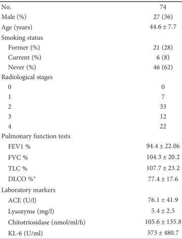
KL-6 was significantly correlated with ACE (r = 0 5;
p < 0 0001), lysozyme (r = 0 35; p = 0 001), and
chitotriosi-dase activity (r = 0 32; p = 0 004) (Figure 1).
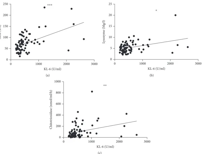
KL-6 was significantly correlated with DLCO percentages
(r =-0.34; p = 0 006) (Figure 2(a)), but not with FVC, FEV1,
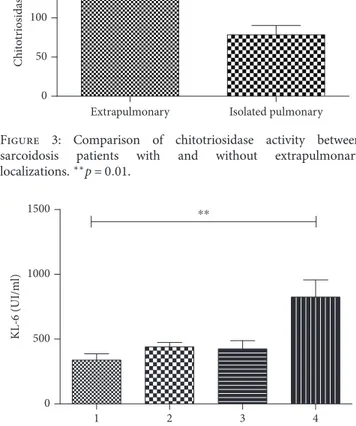
and TLC. CPI score (13 6 ± 17 4) showed a significant direct correlation with KL-6 levels (r = 0 4; p = 0 001) (Figure 2(b)). 3.3. Organ Involvement and Phenotypes. The percentage of sarcoidosis patients showing extrapulmonary involvement was 43.2%. The most common extrapulmonary localiza-tions were the skin (16 patients, 21.6%), spleen (11 patients, 14.8%), and liver (5 patients, 6.7%). Less com-mon localizations were the eyes (3 patients, 4% of cases) and extrathoracic lymph nodes (3 patients, 4%); CNS (2 patients 2.7% of cases) and cardiac, bone, or bone marrow involvement (1.3% of cases). Sarcoidosis patients with extrapulmonary involvement had significantly higher chit-otriosidase activity than those with limited pulmonary disease (146 6 ± 175 9 vs 72 7 ± 80 1 nmol/h/ml; p = 0 01)
(Figure 3). No other significant differences were observed in relation with organ localizations.
In relation to GenPhenReSa phenotypes, patients with abdominal involvement (n = 9) showed significantly higher chitotriosidase activity than those with ocular-cardiac-cutaneous-central nervous system (OCCC) (n = 7), musculoskeletal-cutaneous (n = 11), and isolated pulmonary involvement (n = 34) (221 ± 272 vs 126 ± 162 vs 162 ± 98 vs 86 ± 81, respectively; p < 0 05). No statistically significant differences were found between KL-6, ACE, and lysozyme levels in these subgroups.
According to radiological score, patients with fibrotic sarcoidosis (stage 4) showed the highest concentrations of serum KL-6 compared to the other radiological stages of disease (p = 0 01) (Figure 4).
4. Discussion
Many biomarkers have been proposed for sarcoidosis, but none has shown satisfactory prognostic value for identifying relapse under therapy or vital organ localization. In this study, we compared different sarcoidosis biomarkers. We selected chronic disease patients in treatment for more than 1 year with clinical and demographic data compatible with the general features of sarcoidosis reported in the literature
Table 1: Demographic features, pulmonary function test values, radiological assessment, and biomarker assessment in sarcoidosis population. No. 74 Male (%) 27 (36) Age (years) 44 6 ± 7 7 Smoking status Former (%) 21 (28) Current (%) 6 (8) Never (%) 46 (62) Radiological stages 0 0 1 7 2 33 3 12 4 22
Pulmonary function tests
FEV1 % 94 4 ± 22 06 FVC % 104 3 ± 20 2 TLC % 107 7 ± 23 2 DLCO %∗ 77 4 ± 17 6 Laboratory markers ACE (U/l) 76 1 ± 41 9 Lysozyme (mg/l) 5 4 ± 2 5 Chitotriosidase (nmol/ml/h) 105 6 ± 135 8 KL-6 (U/ml) 573 ± 480 7
0 1000 2000 3000 0 50 100 150 200 250 KL-6 (U/ml) A CE (U/l) ⁎⁎⁎ (a) 0 1000 2000 3000 0 5 10 15 20 25 KL-6 (U/ml) L ys o zyme (Mg/l) ⁎ (b) 0 1000 2000 3000 0 200 400 600 800 1000 KL-6 (U/ml) Chi to tr iosidas e (nmo l/ml/h) ⁎⁎ (c)
Figure 1: KL-6 correlations with ACE, chitotriosidase, and lysozyme. ∗∗∗r = 0 5154, p < 0 0001; ∗r = 0 3238, p = 0 0049; ∗∗r = 0 3750,
p = 0 001. 0 1000 2000 3000 0 5 10 15 KL-6(U/ml) D L co (ml/min/mmH g) (a) 1000 2000 3000 −20 0 20 40 60 KL-6 CPI (b)
Figure 2: (a) Correlation of KL-6 and DLCO percentages. R =-0.34, p = 0 0006. (b) Correlation between CPI score and KL-6 levels. CPI:
[1, 2]. We included KL-6 in the list of biomarkers, although this protein has been studied in IPF and little data are avail-able on its role in sarcoidosis. Our interest in KL-6 was related to its reported correlation with severe fibrotic lung involvement and poor survival [32]. Chitotriosidase, ACE, and lysozyme showed a lower prevalence in our population than previously observed, probably because most patients were on long-term systemic treatment with steroids and did not show signs of active disease at the time of serum sam-pling. On the contrary, KL-6 showed a good sensitivity and specificity in our population, with a cut-off value of 303.5 IU/ml. To our knowledge, this is the first study to investigate a specific cut-off value of KL-6 in chronic sarcoid-osis versus a group of healthy controls; however, this was not the main aim of the study and the data needs to be validated in a wider cohort of patients. Our findings confirmed the predictive value of KL-6 for fibrotic lung involvement and functional impairment in ILD sarcoidosis patients already reported by Miyoshi et al. [20]; however, our study is thefirst one that shows a significant correlation between a serum biomarker, KL-6, and pulmonaryfibrotic lung involvement in sarcoidosis patients, quantified by CPI index. Moreover,
thisfinding is further confirmed by a significant correlation with DLCO percentages, sustaining the potential utility of KL-6 measurements to evaluate sarcoidosis severity in the clinical management of these patients. These data were consistent regardless therapy status, suggesting a chronic macrophage activation unresponsive to steroid therapy, in particular in those patients with fibrotic lung disease. No significant differences of KL-6 expression were found among clinical phenotypes and/or extrapulmonary localizations, indicating that KL-6 can be considered reliable only for pulmonary involvement of disease. Positive correlations were detected among sarcoidosis biomarkers, including ACE, lysozyme, chitotriosidase, and KL-6 that can therefore be included in the list of prognostic indicators.
On the other hand, chitotriosidase proved more sensitive than ACE, lysozyme, and KL-6 even in patients under sys-temic treatment; it also seems to be the only biomarker that can reflect extrathoracic involvement, according to the recent GenPhenReSa clinical phenotype classification. In particular, its expression is more pronounced in patients with abdomi-nal organ localizations (the liver and spleen, in our popula-tion), probably due to an overexpression by activated resident macrophages; this data suggests that chitotriosidase may be a reliable marker of activation of reticuloendothelial system in sarcoidosis, as previously reported in patients with lysosomal storage disease [33]. No studies are available on chitotriosidase prognostic value in splenic and hepatic sar-coidosis localizations and this is a relevant novelty of this study. Unlike KL-6, chitotriosidase activity seems not to be correlated with the severity of sarcoid pulmonary involve-ment or functional impairinvolve-ment, suggesting that different pat-terns of macrophage activation may be present in respiratory system of sarcoidosis patients and influence biomarkers’ expression [34].
In conclusion, patients with chronic sarcoidosis, severe extrapulmonary involvement, or stage 4fibrotic impairment represent an important target requiring the identification of reliable prognostic biomarkers to predict disease relapse and response to systemic treatment.
Based on these preliminary results, high levels of chitotriosidase and KL-6 (and not ACE or lsysozyme) were observed in chronic sarcoidosis patients, regardless therapy. Chitotriosidase activity was significantly higher in patients with a multiple organ involvement, showing a promising reli-ability for evaluation multisistemic sarcoidosis. On the con-trary, KL-6 levels correlated withfibrotic lung involvement, CPI score, and reduced DLCO percentages suggest a poten-tial role as a marker of severity of pulmonary sarcoidosis. Interestingly, these findings were reported in a population of chronic multiorgan sarcoidosis patients, including those on therapy, expanding the reliability of chitotriosidase and KL-6 also in this setting.
Data Availability
The datasets generated during and/or analyzed during the current study are available from the corresponding author on reasonable request. Data are available from Laura Bergantini ([email protected]) and Elena 0
Extrapulmonary Isolated pulmonary
50 100 150 200 250 Chi to tr iosidas e (nmo l/ml/h) ⁎⁎
Figure 3: Comparison of chitotriosidase activity between
sarcoidosis patients with and without extrapulmonary
localizations.∗∗p = 0 01. 0 1 2 3 4 500 1000 1500 RX stages KL -6 (UI/ml) ⁎⁎
Figure 4: Comparison of KL-6 concentration among RX stages in
Bargagli ([email protected]) for researchers who meet the criteria for access to confidential data.
Conflicts of Interest
The authors declare that they have no conflicts of interest.
Authors
’ Contributions
Laura Bergantini and Francesco Bianchi equally contributed to the study.
Acknowledgments
The present work was performed at Siena University.
References
[1] U. Costabel, G. W. Hunninghake, and on behalf of the
Sarcoidosis Statement Committee, “ATS/ERS/WASOG
statement on sarcoidosis. Sarcoidosis Statement Committee. American Thoracic Society. European Respiratory Society. World Association for Sarcoidosis and Other Granulomatous
Disorders,” European Respiratory Journal, vol. 14, no. 4,
pp. 735–737, 1999.
[2] K. C. Patterson and E. S. Chen,“The pathogenesis of
pulmo-nary sarcoidosis and implications for treatment,” Chest,
vol. 153, no. 6, pp. 1432–1442, 2018.
[3] V. Kouranos, A. Wells, and S. Walsh,“Why do people die from
pulmonary sarcoidosis?,” Current Opinion in Pulmonary
Medicine, vol. 24, no. 5, pp. 527–535, 2018.
[4] M. Bonifazi, S. Gasparini, V. Alfieri, and E. A. Renzoni,
“Pulmonary sarcoidosis,” Seminars in Respiratory and Critical
Care Medicine, vol. 38, no. 4, pp. 437–449, 2017.
[5] S. G. West,“Current management of sarcoidosis I: pulmonary,
cardiac, and neurologic manifestations,” European Respiratory
Journal, vol. 30, no. 3, pp. 243–248, 2018.
[6] T. E. Wessendorf, F. Bonella, and U. Costabel,“Diagnosis of
sarcoidosis,” Clinical Reviews in Allergy & Immunology,
vol. 49, no. 1, pp. 54–62, 2015.
[7] E. Bargagli and P. Rottoli,“Serum chitotriosidase activity in
sarcoidosis patients,” Rheumatology International, vol. 27,
no. 12, p. 1187, 2007.
[8] E. Bargagli, C. Maggiorelli, and P. Rottoli,“Human
chitotriosi-dase: a potential new marker of sarcoidosis severity,”
Respira-tion, vol. 76, no. 2, pp. 234–238, 2008.
[9] E. Bargagli, D. Bennett, C. Maggiorelli et al.,“Human
chito-triosidase: a sensitive biomarker of sarcoidosis,” Journal of
Clinical Immunology, vol. 33, no. 1, pp. 264–270, 2013.
[10] D. Coates,“The angiotensin converting enzyme (ACE),” The
International Journal of Biochemistry & Cell Biology, vol. 35,
no. 6, pp. 769–773, 2003.
[11] J. Lieberman, “Elevation of serum
angiotensin-converting-enzyme (ACE) level in sarcoidosis,” The American Journal of
Medicine, vol. 59, no. 3, pp. 365–372, 1975.
[12] P. Ungprasert, E. M. Carmona, C. S. Crowson, and E. L.
Matteson, “Diagnostic utility of angiotensin-converting
enzyme in sarcoidosis: a population-based study,” Lung,
vol. 194, no. 1, pp. 91–95, 2016.
[13] J. Duan, Y. Xu, H. Zhu et al.,“Relationship between CT activity
score with lung function and the serum angiotensin converting
enzyme in pulmonary sarcoidosis on chest HRCT,” Medicine,
vol. 97, no. 36, article e12205, 2018.
[14] Y. Nakamura, T. Takeda, M. Ishii et al.,“Elevation of serum
angiotensin-converting enzyme activity in patients with
hyperthyroidism,” The Journal of Clinical Endocrinology and
Metabolism, vol. 55, no. 5, pp. 931–934, 1982.
[15] S. K. Zorn, C. A. Stevens, E. N. Schachter, and J. B. Gee,“The
angiotensin converting enzyme in pulmonary sarcoidosis and the relative diagnostic value of serum lysozyme,” Lung,
vol. 157, no. 2, pp. 87–94, 1980.
[16] H. Koskinen, H. Nordman, and B. Fröseth,“Serum lysozyme
concentration in silicosis patients and workers exposed to silica dust,” European Journal of Respiratory Diseases, vol. 65,
no. 7, pp. 481–485, 1984.
[17] H. Tomita, S. Sato, R. Matsuda et al.,“Serum lysozyme levels
and clinical features of sarcoidosis,” Lung, vol. 177, no. 3,
pp. 161–167, 1999.
[18] N. Ishikawa, N. Hattori, A. Yokoyama, and N. Kohno,“Utility
of KL-6/MUC1 in the clinical management of interstitial lung
diseases,” Respiratory Investigation, vol. 50, no. 1, pp. 3–13,
2012.
[19] F. Bonella, X. Long, S. Ohshimo et al.,“MUC1 gene
polymor-phisms are associated with serum KL-6 levels and pulmonary dysfunction in pulmonary alveolar proteinosis,” Orphanet Journal of Rare Diseases, vol. 11, no. 1, p. 48, 2016.
[20] S. Miyoshi, H. Hamada, T. Kadowaki et al., “Comparative
evaluation of serum markers in pulmonary sarcoidosis,” Chest,
vol. 137, no. 6, pp. 1391–1397, 2010.
[21] R. Janssen, H. Sato, J. C. Grutters et al.,“Study of Clara cell 16,
KL-6, and surfactant protein-D in serum as disease markers in
pulmonary sarcoidosis,” Chest, vol. 124, no. 6, pp. 2119–2125,
2003.
[22] B. Menon, M. Tiwari, A. Gopi, P. Raj, and K. Panwar,“Serum
Krebs von den Lungen-6 (KL-6): a promising biomarker in sarcoidosis,” MOJ Current Research & Reviews, vol. 1, no. 2, p. 1, 2018.
[23] J. G. Scadding, “Prognosis of intrathoracic sarcoidosis in
England,” British Medical Journal, vol. 2, no. 5261, pp. 1165–
1172, 1961.
[24] M. R. Miller, J. Hankinson, V. Brusasco et al.,“Standardisation
of spirometry,” European Respiratory Journal, vol. 26, no. 2,
pp. 319–338, 2005.
[25] J. Wanger, J. L. Clausen, A. Coates et al.,“Standardisation of
the measurement of lung volumes,” European Respiratory
Journal, vol. 26, no. 3, pp. 511–522, 2005.
[26] N. Macintyre, R. O. Crapo, G. Viegi et al.,“Standardisation of
the single-breath determination of carbon monoxide uptake in
the lung,” European Respiratory Journal, vol. 26, no. 4,
pp. 720–735, 2005.
[27] P. J. Sterk, L. M. Fabbri, and P. H. Quanjer,“Standardized lung
function testing: official statement of the European Respiratory
Society,” European Respiratory Journal, vol. 16, pp. 1–100,
1993.
[28] J. C. Schupp, S. Freitag-Wolf, E. Bargagli et al.,“Phenotypes of
organ involvement in sarcoidosis,” European Respiratory
Journal, vol. 51, no. 1, article 1700991, 2018.
[29] A. U. Wells, S. R. Desai, M. B. Rubens et al.,“Idiopathic
pul-monaryfibrosis: a composite physiologic index derived from
disease extent observed by computed tomography,” American
Journal of Respiratory and Critical Care Medicine, vol. 167, no. 7, pp. 962–969, 2003.
[30] S. Rothkrantz-Kos, M. P. van Dieijen-Visser, P. G. Mulder, and
M. Drent,“Potential usefulness of inflammatory markers to
monitor respiratory functional impairment in sarcoidosis,”
Clinical Chemistry, vol. 49, no. 9, pp. 1510–1517, 2003.
[31] E. V. Arkema and Y. C. Cozier,“Epidemiology of sarcoidosis:
currentfindings and future directions,” Therapeutic Advances
in Chronic Disease, vol. 9, no. 11, pp. 227–240, 2018.
[32] K. Honda, F. Okada, Y. Ando et al.,“Comparison of
pulmo-nary thin section CT findings and serum KL-6 levels in
patients with sarcoidosis,” The British Journal of Radiology,
vol. 84, no. 999, pp. 229–235, 2011.
[33] L. van Dussen, E. J. Hendriks, J. E. M. Groener, R. G. Boot,
C. E. M. Hollak, and J. M. F. G. Aerts, “Value of plasma
chitotriosidase to assess non-neuronopathic Gaucher disease severity and progression in the era of enzyme replacement
therapy,” Journal of Inherited Metabolic Disease, vol. 37,
no. 6, pp. 991–1001, 2014.
[34] M. Shamaei, E. Mortaz, M. Pourabdollah et al.,“Evidence for
M2 macrophages in granulomas from pulmonary sarcoidosis:
a new aspect of macrophage heterogeneity,” Human
Stem Cells
International
Hindawi www.hindawi.com Volume 2018 Hindawi www.hindawi.com Volume 2018 INFLAMMATIONEndocrinology
International Journal ofHindawi www.hindawi.com Volume 2018 Hindawi www.hindawi.com Volume 2018
Disease Markers
Hindawi www.hindawi.com Volume 2018 BioMed Research InternationalOncology
Journal of Hindawi www.hindawi.com Volume 2013 Hindawi www.hindawi.com Volume 2018Oxidative Medicine and Cellular Longevity
Hindawi
www.hindawi.com Volume 2018
PPAR Research
Hindawi Publishing Corporation
http://www.hindawi.com Volume 2013 Hindawi www.hindawi.com
The Scientific
World Journal
Volume 2018 Immunology Research Hindawi www.hindawi.com Volume 2018 Journal ofObesity
Journal of Hindawi www.hindawi.com Volume 2018 Hindawi www.hindawi.com Volume 2018 Computational and Mathematical Methods in Medicine Hindawi www.hindawi.com Volume 2018Behavioural
Neurology
Ophthalmology
Journal of Hindawi www.hindawi.com Volume 2018Diabetes Research
Journal ofHindawi
www.hindawi.com Volume 2018
Hindawi
www.hindawi.com Volume 2018 Research and Treatment
AIDS
Hindawi
www.hindawi.com Volume 2018
Gastroenterology Research and Practice
Hindawi www.hindawi.com Volume 2018