Research Article
CO
2
Pneumoperitoneum Preserves
𝛽-Arrestin 2 Content and
Reduces High Mobility Group Box-1 (HMGB-1) Expression in
an Animal Model of Peritonitis
Angela Simona Montalto,
1Alessandra Bitto,
2Letteria Minutoli,
2Pietro Impellizzeri,
1Gaetano Costa,
3Natasha Irrera,
2Gabriele Pizzino,
2Francesco Squadrito,
2Domenica Altavilla,
1and Carmelo Romeo
11Department of Pediatric, Gynecological, Microbiological and Biomedical Sciences, University of Messina,
Azienda Ospedaliera Universitaria Policlinico “G. Martino”, Via Consolare Valeria 1, 98125 Messina, Italy
2Department of Clinical and Experimental Medicine, University of Messina, Azienda Ospedaliera
Universitaria Policlinico “G. Martino”, Via Consolare Valeria 1, 98125 Messina, Italy
3Department of Public Health, University of Messina, Azienda Ospedaliera Universitaria Policlinico “G. Martino”,
Via Consolare Valeria 1, 98125 Messina, Italy
Correspondence should be addressed to Pietro Impellizzeri; [email protected] Received 10 October 2014; Revised 11 January 2015; Accepted 26 January 2015 Academic Editor: Angel Catal´a
Copyright © 2015 Angela Simona Montalto et al. This is an open access article distributed under the Creative Commons Attribution License, which permits unrestricted use, distribution, and reproduction in any medium, provided the original work is properly cited.
Laparoscopy (LS) has been shown to decrease the inflammatory sequelae of endotoxemia.𝛽-arrestin 2 plays an important function in signal transduction pathway of TLR4. High mobility group box-1 (HMGB-1) is involved in the delayed systemic inflammatory response. We investigated the effects of CO2insufflation on liver, lung, and kidney expression of both𝛽-arrestin 2 and HMGB-1 during sepsis. Cecal ligation and puncture (CLP) was performed in male rats and 6 h later the animals were randomly assigned to receive a CO2pneumoperitoneum or laparotomy. Animals were euthanized; liver, lung, and kidney were removed for the evaluation of𝛽-arrestin 2 and HMGB-1 expression. Immunohistochemical detection of myeloperoxidase (MPO) was investigated in lung and liver and bacterial load was determined in the peritoneal fluid. CO2pneumoperitoneum reduced peritoneal bacterial load, increased the expression of𝛽-arrestin 2, and blunted the expression of the potent proinflammatory HMGB-1 in liver, lung, and kidney compared with laparotomy. Liver and lung MPO was markedly reduced in rats subjected to LS compared with laparotomy. We believe that CO2exerts an early protective effect by reducing bacterial load and likely toll-like receptor activation which in turn leads to a preserved𝛽-arrestin 2 expression and a reduced HMGB-1 expression.
1. Introduction
The better preservation of immune function during laparo-scopy compared with open surgery seems to be maintained under septic conditions but the underlying mechanisms that explain such results are still not completely understood. Sev-eral experimental evidences suggest that CO2 pneumoperi-toneum reduces the severity of sepsis and prolongs survival, compared with laparotomy in animal models of peritonitis [1–3]. However, little is known about the CO2 pneumoperi-toneum impact on early acute phase response in sepsis.
Recently, we demonstrated that CO2 pneumoperitoneum reduces inflammatory response by reducing early liver and lung interleukin (IL) 6 and tumor necrosis factor-alpha (TNF-𝛼) expression in cecal ligation and puncture rat model [4]. Other studies have reported that CO2insufflation could also influence intra-abdominal bacterial contamination in murine models of abdominal sepsis [5, 6]. In this regard, 𝛽-arrestins play a critical role as modulators of inflam-matory response.𝛽-arrestins are peculiar adaptor proteins involved in G-protein-coupled receptor (GPCR) desensi-tization and are also implicated in regulation of Toll-like
Volume 2015, Article ID 160568, 7 pages http://dx.doi.org/10.1155/2015/160568
receptor signalling and proinflammatory gene expression. Both𝛽-arrestins 1 and 2 differentially seem to regulate TLR4 signalling pathways. In particular, it has been suggested that 𝛽-arrestin 2 is a negative regulator of inflammatory response in CLP-induced mortality [7–9].
HMGB-1 is another potent proinflammatory cytokine involved in the delayed endotoxin lethality and systemic inflammatory response [10]. The majority of HMGB-1 secreted by activated monocyte/macrophages is from a pre-informed cellular pool within the first 16 h [11]; HMGB-1 mRNA levels are increased in various tissues such as muscle, liver, and lung following endotoxemia [12]. Subsequently, increased cellular synthesis of HMGB-1 further reinforces their release into extracellular milieu [11]. In addition, cells undergoing unprogrammed cell death can passively release HMGB-1, which worsens and prolongs inflammation [13]. It has been reported that HMGB-1 can induce cellular acti-vation and generate inflammatory responses by interaction of both TLR2 and TLR4 [14]. Finally, HMGB-1 seems to function as an “alarmin” signal to recruit, alert, and acti-vate innate immune cells, sustaining a potentially injurious inflammatory response [15]. In light of these considerations and supported by our previous data, we performed this study, in order to better understand for the first time whether the role of CO2 pneumoperitoneum might be related to a reduced bacterial count and consequently to a decreased activation of the inflammatory cascade via TLR activation in an experimental model of CLP.𝛽-arrestin 2 and HMGB-1 protein expressions are investigated in liver, lung, and kidney as target sites of septic injury.
2. Materials and Methods
2.1. Cecal Ligation and Puncture. For this study, a total of
21 animals purchased from Charles River (Calco, Lecco, Italy) were used. All procedures complied with the standards for the care and use of animal subjects, as stated in the Guide for the Care and Use of laboratory Animals, and were approved by the Committee on Animal Health and Care of the University of Messina. CLP was performed in 14 male Sprague-Dawley rats as previously described [8]. Specifically, rats were anaesthetized with ether, and a midline incision was made below the diaphragm to expose the cecum. The cecum was ligated at the colon juncture with a 2-0 silk ligature suture without interrupting intestinal continuity. The cecum was punctured twice with an 18-gauge needle. The cecum was returned to the abdomen, and the incision was closed in layers with a 2-0 silk ligature suture. Six hours following CLP, animals were randomly selected to undergo CO2pneumoperitoneum (LS) at a pressure of 5–7 mmHg or laparotomy (LT) for 1 hour under general anaesthesia with sodium pentobarbital (50 mg/kg/i.p.). Pneumoperitoneum was achieved by introducing a Veress needle into the peri-toneal cavity and by insufflating the abdomen with CO2using the Wisap CO2gas insufflator (Wisap, Sauerlach, Germany). The remaining seven animals did not undergo CLP, were left untreated, and were used as controls (na¨ıve). At the end of the procedures, all animals were sacrificed: peritoneal fluid was collected and liver, lung, and kidney were excised and stored at−20∘C or in 10% buffered formalin for further analysis.
2.2. Bacterial Load. To determine the bacterial load in the
peritoneum, the peritoneal cavity was washed with 5 mL of sterile phosphate-buffered saline (PBS) and diluted with sterile PBS. One hundred microlitres of each dilution was then plated on chocolate agar plates (Fisher Scientific, Pitts-burgh, PA) and incubated at 37∘C for 24 hours under aerobic conditions. CFU were counted and results were expressed as CFU/mL of peritoneal fluid.
2.3. Isolation of Cytoplasmic Proteins and Western Blot Anal-ysis for𝛽-Arrestin 2 and HMGB-1. Liver, lung, and kidney
samples were homogenized in 1 mL lysis buffer (25 mM tris/ HCl, pH 7.4, 1.0 mM ethylene glycol tetra-acetic acid, 1.0 mM ethylenediamine tetra-acetic acid, 0.5 mM phenylmethylsul-fonyl fluoride, aprotinin, leupeptin, pepstatin A (10𝜇g/mL each), and Na3VO4100 mM). The homogenate was subjected to centrifugation at 15,000×g for 15 min. The supernatant was collected and stored at−80∘C. The concentration of total proteins was determined using a Bio-Rad protein assay kit (Milan, Italy).
𝛽-arrestin 2 and HMGB-1 were evaluated by western blot, according to the methods described previously [9]. Primary antibodies for𝛽-arrestin 2 and HMGB-1 were purchased by Abcam (Cambridge, UK). Secondary antibodies peroxidase conjugated were obtained by Pierce (Rockford, Illinois, USA). The protein signals were quantified by scanning using a bioimage analysis system (Bio-Profil Celbio, Milan, Italy) and were expressed as integrated intensity in comparison with those of control animals measured with the same batch. 𝛽-actin (Cell Signaling) antibody was used on stripped blots to confirm equal protein loading.
2.4. Immunohistochemistry. An immunohistochemical study
was performed to evaluate the presence (or absence) of an inflammatory reaction in liver and lung specimens. All specimens were tested with myeloperoxidase (Thermo, Fre-mont, CA) as an index of neutrophil accumulation. Slices of 5𝜇m were rehydrated in graded alcohol and antigen retrieval was performed with pH 6.0 buffer citrate and endogenous peroxidase (i.e., from red blood cells) blocking with 1% H2O2 in PBS. Primary antibody was incubated overnight at 4∘C in a moisturized chamber and the day after secondary antibody was added and reaction visualized with DAB. Counterstain-ing was performed to identify nuclei in the specimens.
2.5. Statistical Analysis. All data are expressed as mean ±
standard deviation (SD). Significance of difference was assessed by two-way repeated-measures analysis of variance (ANOVA) followed by post hoc analyses where indicated. A value of 𝑃 < 0.05 was considered statistically significant. Graphs were produced using GraphPad Prism (version 4.0 for Windows).
3. Results
3.1. CO2Pneumoperitoneum Reduces Bacterial Load in Peri-toneal Fluid. The bacterial load in periPeri-toneal cavity was
deter-mined by CFU count. Cecal ligation and puncture following LT produced a marked bacterial load in the peritoneal fluid
1 2 3 4 5 6 7 8 9 ×10⁶ Na¨ıve LT LS Na¨ıve LT LS ∗∗ P er it o ne al fl uid (CFU/mL) #
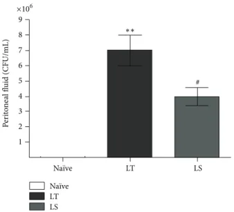
Figure 1: Bacterial count analysis in peritoneal fluid cultures (CFU/mL) from animals subjected to CLP followed by LT or LS procedures and na¨ıve animals. Data represent the mean± SD of seven animals.∗∗𝑃 < 0.001 compared with na¨ıve; #𝑃 < 0.05 compared with LT.
compared with na¨ıve animals (Figure 1). CO2 pneumotoneum (LS) significantly reduced bacterial loads in peri-toneal cavity compared with animals subjected to laparotomy (LT) (Figure 1; 4× 10⁶versus 7× 10⁶CFU/mL, resp.,𝑃 < 0.05) suggesting that an antimicrobial effect may concur to the protective effect of CO2.
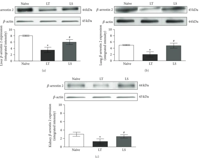
3.2. CO2Pneumoperitoneum Regulates Liver, Lung, and Kid-ney Expression of Arrestin 2. To determine whether
𝛽-arrestin 2 plays a key role in modulating TLR signalling pathway in CLP-induced inflammation, we first evaluated CO2 effects on 𝛽-arrestin 2 expression in liver, lung, and kidney. Animals that underwent laparotomy (LT) showed significantly reduced𝛽-arrestin 2 protein expression in these organs compared with na¨ıve group (𝑃 < 0.05). CO2 pneu-moperitoneum preserved 𝛽-arrestin 2 expression in liver, lung, and kidney (LS), displaying higher levels of the protein compared with laparotomy (Figures2(a),2(b), and2(c), 𝑃 < 0.05).
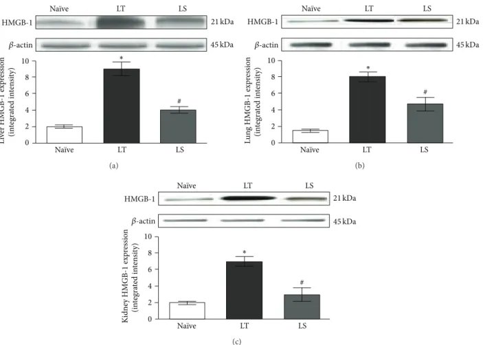
3.3. CO2Pneumoperitoneum Reduces Liver, Lung, and Kidney Expression of HMGB-1. HMGB-1 is known as late mediator in
sepsis, but it can also be released early after a traumatic insult. Therefore, we studied the CO2pneumoperitoneum effects on CLP-induced HMGB-1 expression in liver, lung, and kidney of rats. Animals subjected to laparotomy showed a significant increase of HMGB-1 protein expression in liver, lung, and kidney (𝑃 < 0.05) compared with na¨ıve animals (Figures3(a),
3(b), and3(c)). CO2pneumoperitoneum markedly reduced the expression of this cytokine in liver, lung, and kidney tissues (LS) with respect to the LT (𝑃 < 0.05), suggesting a CO2protective role during polymicrobial sepsis.
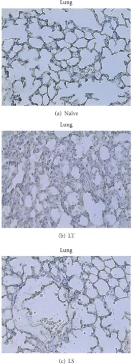
3.4. CO2 Pneumoperitoneum Reduces Liver and Lung Neu-trophil Accumulation. MPO staining for the evaluation of
neutrophil infiltrate showed a strong inflammatory reaction in liver and lung of LT animals (Figures 4(b) and 5(b)) compared with na¨ıve animals (Figures 4(a) and 5(a)). By contrary, rats subjected to CO2 pneumoperitoneum (LS) revealed a reduced accumulation of inflammatory cells in liver and lung (Figures4(c)and5(c)), suggesting a protective role of this procedure regarding CLP.
4. Discussion
In today’s surgical practice, laparoscopic treatment represents a regular part of therapeutic approach in septic surgical patients with localized and diffuse peritonitis [16]. Parallely, increasing studies delineate the physiopathological effects of pneumoperitoneum in septic conditions, where CO2 seems to have a relevant biologic role. The immunological changes induced by pneumoperitoneum appear to contribute favourable outcomes to laparoscopy with increased sur-vival in experimental model of sepsis [1–3]. However, this encouraging data is reflected on the final step of a common and complicated pathway. The upstream mechanisms by which CO2can affect the inflammatory process preserving immune function in septic state are not identified and not fully investigated. In sepsis, the massive deterioration of the immune response results in fact from an inappropriately amplified systemic inflammatory response to infection [17]. Exaggerated proinflammatory mediators production from macrophages, monocytes, or other host cells governs this crucial phase and can lead to the development of tissue dam-age until multiple organ failure and even death [18]. TLRs, expressed on the host cells, play a critical and primary role in the inflammatory responses [19]. Binding to TLR4 primes a series of signalling proteins that activate transcription factors and proinflammatory gene expression [20]. Recent studies have shown that 𝛽-arrestin 2, a member of the arrestin family, plays an important function in signal transduction pathway of TLR4 [7, 21, 22]. Similarly, HMGB-1, that has proinflammatory cytokine-like properties, can interact also with both TLR4 and TLR2 and prime inflammatory response that is similar to that initiated by LPS [14].
In a previous study, we have demonstrated an early and significant reduction of liver and lung TNF-𝛼 and IL-6 expression in CLP rat model undergoing CO2 pneu-moperitoneum. We also showed the correlation between early cytokine expression and histological alterations. In fact, in liver and lung specimens, these results were accompanied by a reduction in the inflammatory infiltrate, edema, venular ectasia, and cellular degeneration in animals subjected to CO2 pneumoperitoneum compared with laparotomy. These data supported the possible protective role of CO2 pneumoperi-toneum in reducing the inflammatory cascade associated with sepsis via reduced expression of liver and lung proin-flammatory cytokines [4]. Therefore, the main focus of the present study was to further investigate the possible upstream mechanisms of our preliminary findings consequent on CO2 pneumoperitoneum in a murine model of polymicrobial
0 2 4 6 8 10 Na¨ıve LT LS Na¨ıve LT LS ∗ # Li ve r (inte gra ted in te n si ty) 𝛽-actin 44 kDa 45 kDa 𝛽-arrestin 2 𝛽 -a rr estin 2 exp re ssio n (a) ∗ 0 2 4 6 8 10 Na¨ıve LT LS Na¨ıve LT LS Lu n g (in te gra ted in te n si ty) 𝛽-actin 44 kDa 45 kDa # 𝛽-arrestin 2 𝛽 -a rr estin 2 exp re ssio n (b) # ∗ 0 2 4 6 8 10 Na¨ıve LT LS Na¨ıve LT LS Ki d n ey (in te gra ted in te n si ty) 𝛽-actin 44 kDa 45 kDa 𝛽-arrestin 2 𝛽 -a rr estin 2 exp re ssio n (c)
Figure 2: Representative western blot analysis of𝛽-arrestin 2 protein in liver (a), lung (b), and kidney (c) from animals subjected to CLP followed by LT or LS procedures and na¨ıve animals. Data represent the mean± SD of seven animals.∗𝑃 < 0.05 compared with na¨ıve;#𝑃 < 0.05 compared with LT.
sepsis that better reproduces human peritonitis. The current study is the first to explore the influence of CO2 pneumoperi-toneum on early 𝛽-arrestin 2 and HMGB-1 expressions in target organs of sepsis in correlation with peritoneal bacterial load and myeloperoxidase in a well-standardized rat model of abdominal sepsis. The data obtained by us demonstrate that CO2 pneumoperitoneum exerts a dual beneficial effect both locally and on the organ level in CLP-induced inflam-mation.
Balagu´e et al. reported that the number of bacterial CFU obtained in peritoneal fluid was significantly lower in the laparoscopic groups than in the open group in a defined bac-terial inoculum model [5]. In a similar experiment, Pitombo and coworkers demonstrate, 18 h after bacterial inoculum into the peritoneum, a significant increase of CFU/mL for the laparotomy group compared with CO2 and control groups [6]. In our study, the microbiological evaluation of peritoneal fluid collected after CLP and pneumoperitoneum revealed that CO2significantly reduced the number of colony forming units in this group of animals compared to the LS group.
These intriguing data induce thinking primarily that CO2can have a specific and local action. Its prominent role in bacterial control in the storage and preservation of food products is known [23–25]. The potential antimicrobial effect of the gas if translated in the current study could explain the correlation between microbial inactivation (reduced bacterial count) and intra-abdominal exposure to CO2.
Looking at 𝛽-arrestin 2 results, we found a significant high expression in liver, lung, and kidney of animals sub-jected to laparoscopy following CLP compared with laparo-tomy animals and, in previous experiment, CLP-induced TNF-alpha and IL-6 expression turned out to be significantly higher in LT group with respect to the LS [4]. These data suggest that𝛽-arrestin 2 is, in agreement with the literature, an essential negative regulator of inflammatory response in polymicrobial sepsis [7–9, 26, 27] via TLR signalling. In particular, our investigation reveals that CO2 insufflation preserves𝛽-arrestin 2 expression in the organs studied. This result if correlated with blunted CLP-induced increase in TNF-alpha and IL-6 in liver and lung tissues [4] supports
0 2 4 6 8 10 Na¨ıve LT LS Na¨ıve LT LS ∗ # L iv er HM GB-1 exp re ssio n (in te gra ted in te n si ty) 𝛽-actin 45 kDa HMGB-1 21 kDa (a) ∗ 0 2 4 6 8 10 Na¨ıve LT LS Na¨ıve LT LS L u n g HM GB-1 exp re ssio n (in te gra ted in te n si ty) 𝛽-actin 45 kDa HMGB-1 21 kDa # (b) # ∗ 0 2 4 6 8 10 Na¨ıve LT LS Na¨ıve LT LS K idne y HM GB-1 exp re ssio n (in te gra ted in te n si ty) HMGB-1 𝛽-actin 21 kDa 45 kDa (c)
Figure 3: Representative western blot analysis of HMGB-1 protein in liver (a), lung (b), and kidney (c) from animals subjected to CLP followed by LT or LS procedures and na¨ıve animals. Data represent the mean± SD of seven animals.∗𝑃 < 0.05 compared with na¨ıve;
#𝑃 < 0.05 compared with LT.
the hypothesis that𝛽-arrestin 2 improved expression is pro-tective in a model of abdominal sepsis.
Regarding HMGB-1, we demonstrated, according to lit-erature, an increase in liver, lung, and kidney expression of this proinflammatory protein in rats subjected to laparotomy following CLP. In contrast, after CO2 insufflation, we had a significant reduction of this cytokine.
Our data also showed an increased neutrophil infiltra-tion in liver and lung of LT animals. On the contrary, CO2pneumoperitoneum significantly reduced inflammatory cells, confirming its beneficial role in this animal model of peritonitis.
Altogether, our data might be taken into account in order to explain additional mechanism(s) of CO2beneficial effects in experimental sepsis. It is in fact likely that the reduced bacterial load induced by antimicrobial action of CO2leads to a reduced release of bacterial components as lipopolysac-charides. This could downregulate the Toll-like receptors acti-vation that mediate antimicrobial responses. Consequently, the potential 𝛽-arrestin 2 and HMGB-1 interactions with TLRs could allow both a preservation of 𝛽-arrestin 2 and a reduction of HMGB-1 expression. We speculate that all
these composite changes can correlate with a better anti-inflammatory effect exerted by 𝛽-arrestin 2 and in addi-tion also justify the subsequent reduced proinflammatory cytokine expressions and tissue damage contributing to corroborating the previously reported results.
In conclusion, our study contributes to reinforcing the hypothesis that laparoscopic surgery better controls abdom-inal infection and favourably modifies the immune response reducing the inflammatory septic cascade. In particular, by our results, it can be evicted that CO2exerts a precocious and protective effect by reduced bacterial load with (and) likely Toll-like receptors activation which in turn lead to a pre-served𝛽-arrestin 2 expression, reduced HMGB-1 expression, and myeloperoxidase in this animal model of peritonitis.
Conflict of Interests
Angela Simona Montalto, Alessandra Bitto, Letteria Min-utoli, Pietro Impellizzeri, Gaetano Costa, Natasha Irrera, Gabriele Pizzino, Francesco Squadrito, Domenica Altavilla, and Carmelo Romeo have no conflict of interests to disclose.
Liver (a) Na¨ıve Liver (b) LT Liver (c) LS
Figure 4: Immunohistochemical detection of myeloperoxidase in liver from na¨ıve, LT, or LS animals. Samples collected from LT rats (b) show an increased inflammatory infiltrate and little, but still representative, edematous interstitial areas compared to na¨ıve tissue samples (a). In contrast, samples from animals subjected to CO2 pneumoperitoneum revealed a reduced accumulation of inflammatory cells (c). Original magnification×40.
Authors’ Contribution
Angela Simona Montalto, Alessandra Bitto, and Letteria Minutoli contributed equally to this paper.
Lung (a) Na¨ıve Lung (b) LT Lung (c) LS
Figure 5: Immunohistochemical detection of myeloperoxidase in lung from na¨ıve, LT, or LS animals. Samples collected from LT rats (b) show an increased inflammatory infiltrate and some grade of fibrosis of interalveolar septa compared to na¨ıve tissue samples (a). In contrast, samples from animals subjected to CO2 pneumoperi-toneum revealed a reduced accumulation of inflammatory cells, as well as a substantial reduction of pulmonary fibrosis (c). Original magnification×40.
Acknowledgment
This study was supported by departmental funding of Department of Pediatric, Gynecological, Microbiological and
Biomedical Sciences and Department of Clinical and Exper-imental Medicine of University of Messina.
References
[1] E. J. Hanly, J. M. Fuentes, A. R. Aurora et al., “Carbon dioxide pneumoperitoneum prevents mortality from sepsis,” Surgical Endoscopy and Other Interventional Techniques, vol. 20, no. 9, pp. 1482–1487, 2006.
[2] M. Metzelder, J. F. Kuebler, A. Shimotakahara, D.-H. Chang, C. Vieten, and B. Ure, “CO2pneumoperitoneum increases survival in mice with polymicrobial peritonitis,” European Journal of Pediatric Surgery, vol. 18, no. 3, pp. 171–175, 2008.
[3] G. Chatzimavroudis, T. E. Pavlidis, I. Koutelidakis et al., “CO2 pneumoperitoneum prolongs survival in an animal model of peritonitis compared to laparotomy,” Journal of Surgical Research, vol. 152, no. 1, pp. 69–75, 2009.
[4] A. S. Montalto, A. Bitto, N. Irrera et al., “CO2 pneumoperito-neum impact on early liver and lung cytokine expression in a rat model of abdominal sepsis,” Surgical Endoscopy and Other Interventional Techniques, vol. 26, no. 4, pp. 984–989, 2012. [5] C. Balagu´e, E. M. Targarona, M. Pujol, X. Filella, J. J. Espert, and
M. Trias, “Peritoneal response to a septic challenge: comparison between open laparotomy, pneumoperitoneum laparoscopy, and wall lift laparoscopy,” Surgical Endoscopy, vol. 13, no. 8, pp. 792–796, 1999.
[6] M. B. Pitombo, O. H. Lupi, R. N. Gomes et al., “Inflammatory response and bacterial dissemination after laparotomy and abdominal CO2insufflation in a murine model of peritonitis,” Surgical Endoscopy and Other Interventional Techniques, vol. 20, no. 9, pp. 1440–1447, 2006.
[7] H. Fan, L. M. Luttrell, G. E. Tempel, J. J. Senn, P. V. Halushka, and J. A. Cook, “𝛽-Arrestins 1 and 2 differentially regulate LPS-induced signaling and pro-inflammatory gene expression,” Molecular Immunology, vol. 44, no. 12, pp. 3092–3099, 2007. [8] H. Fan, A. Bitto, B. Zingarelli et al., “Beta-arrestin 2 negatively
regulates sepsis-induced inflammation,” Immunology, vol. 130, no. 3, pp. 344–351, 2010.
[9] A. Bitto, L. Minutoli, A. David et al., “Flavocoxid, a dual inhib-itor of COX-2 and 5-LOX of natural origin, attenuates the inflammatory response and protects mice from sepsis,” Critical Care, vol. 16, no. 1, article R32, 2012.
[10] W. Huang, Y. Tang, and L. Li, “HMGB1, a potent proinflamma-tory cytokine in sepsis,” Cytokine, vol. 51, no. 2, pp. 119–126, 2010. [11] H. Wang, O. Bloom, M. Zhang et al., “HMG-1 as a late mediator of endotoxin lethality in mice,” Science, vol. 285, no. 5425, pp. 248–251, 1999.
[12] C. H. Lang, C. Silvis, N. Deshpande, G. Nystrom, and R. A. Frost, “Endotoxin stimulates in vivo expression of inflamma-tory cytokines tumor necrosis factor alpha, interleukin-1beta, -6, and high-mobility-group protein-1 in skeletal muscle,” Shock, vol. 19, no. 6, pp. 538–546, 2003.
[13] P. Scaffidi, T. Misteli, and M. E. Bianchi, “Release of chro-matin protein HMGB1 by necrotic cells triggers inflammation,” Nature, vol. 418, pp. 191–195, 2002.
[14] J. S. Park, F. Gamboni-Robertson, Q. He et al., “High mobility group box 1 protein interacts with multiple Toll-like receptors,” The American Journal of Physiology—Cell Physiology, vol. 290, no. 3, pp. C917–C924, 2006.
[15] H. Wang, M. F. Ward, and A. E. Sama, “Novel HMGB1-inhib-iting therapeutic agents for experimental sepsis,” Shock, vol. 32, no. 4, pp. 348–357, 2009.
[16] S. Sauerland, F. Agresta, R. Bergamaschi et al., “Laparoscopy for abdominal emergencies: evidence-based guidelines of the Euro-pean Association for Endoscopic Surgery,” Surgical Endoscopy and Other Interventional Techniques, vol. 20, no. 1, pp. 14–29, 2006.
[17] R. S. Hotchkiss and I. E. Karl, “The pathophysiology and treat-ment of sepsis,” The New England Journal of Medicine, vol. 348, no. 2, pp. 138–150, 2003.
[18] R. C. Bone, “Why sepsis trials fail,” The Journal of the American Medical Association, vol. 276, no. 7, pp. 565–566, 1996. [19] S. Akira, S. Uematsu, and O. Takeuchi, “Pathogen recognition
and innate immunity,” Cell, vol. 124, no. 4, pp. 783–801, 2006. [20] S. Akira and K. Takeda, “Toll-like receptor signalling,” Nature
Reviews Immunology, vol. 4, no. 7, pp. 499–511, 2004.
[21] L. M. Luttrell and R. J. Lefkowitz, “The role of𝛽-arrestins in the termination and transduction of G-protein-coupled receptor signals,” Journal of Cell Science, vol. 115, no. 3, pp. 455–465, 2002. [22] R. J. Lefkowitz and S. K. Shenoy, “Transduction of receptor signals by beta-arrestins,” Science, vol. 308, no. 5721, pp. 512–517, 2005.
[23] C. O. Gill and K. M. DeLacy, “Growth of Escherichia coli and Salmonella typhimurium on high-pH beef packed under vac-uum or carbon dioxide,” International Journal of Food Microbi-ology, vol. 13, no. 1, pp. 21–30, 1991.
[24] C. D. Hart, G. C. Mead, and A. P. Norris, “Effects of gaseous environment and temperature on the storage behaviour of Listeria monocytogenes on chicken breast meat,” Journal of Applied Bacteriology, vol. 70, no. 1, pp. 40–46, 1991.
[25] L. Lee, L. Arul, R. Lencki, and F. Castaigne, “A review on modi-fied atmosphere packaging and preservation of fresh fruits and vegetables: physiological basis and practical aspects: part II,” Packaging Technology and Science, vol. 9, no. 1, pp. 1–17, 1996. [26] F. Basher, H. Fan, B. Zingarelli et al., “𝛽-Arrestin 2: a negative
regulator of inflammatory responses in polymorphonuclear leukocytes,” International Journal of Clinical and Experimental Medicine, vol. 1, no. 1, pp. 32–41, 2008.
[27] Y. Wang, Y. Tang, L. Teng, Y. Wu, X. Zhao, and G. Pei, “Asso-ciation of𝛽-arrestin and TRAF6 negatively regulates Toll-like receptor-interleukin 1 receptor signaling,” Nature Immunology, vol. 7, no. 2, pp. 139–147, 2006.