UNIVERSITÀ DEGLI STUDI DELLA TUSCIA DI VITERBO
DIPARTIMENTO DAFNE
Corso di Dottorato di Ricerca in Protezione delle Piante – XXIV Ciclo
“Chestnut gall wasp (Dryocosmus kuriphilus Yasumatsu): antennal morphology
and natural control”
AGR/11
Tesi di dottorato di: Tutore
Dott.ssa Francesca Riga Prof. Bruno Paparatti
Coordinatore del corso Co-tutore
Prof. Leonardo Varvaro Prof. Nunzio Isidoro
Contents
1 General introduction 1
1.1 The genus Castanea 1
1.2 Chestnut cultivation 2
1.2.1 The chestnut orchard in Europe and Italy 2
1.2.2 Chestnut orchards in Marche Region 4
1.3 Cinipid gall wasps 4
1.4 Galls 5
1.5 Dryocosmus kuriphilus Yasumatsu (Hymenoptera: Cynipidae) 7
1.5.1 Origin and diffusion 7
1.5.2 Morphology 10
1.5.3 Life cycle 11
1.5.4 Host plants 11
1.5.5 Symptoms and damages 12
1.5.6 Control measures 12 1.5.6.1 Resistant varieties 12 1.5.6.2 Chemical control 13 1.5.6.3 Agronomical control 13 1.5.7 Preventive measures 13 1.5.8 Native parasitoids 14
1.5.9 Exotic Parasitoid – Torymus sinensis 14
1.5.9.1 - Technical notes on the release of Torymus sinensis in Italy 16
2 Aim 17 3 Antennal morphology 18 3.1 Introduction 18 3.1.1 Antenna 18 3.1.2 Definition of sensillum 18 3.1.3 Type of sensilla 19 3.1.3.1 Aporous sensilla 20 3.1.3.2 Uniporous sensilla 21 3.1.3.3 Multiporous sensilla 21 3.2 Aim 23
3.3 Materials and methods 24
3.3.1.1 Scanning Electron Microscopy 24
3.3.1.2 Transmission electron microscopy 24
3.4 Result and discussion 25
3.4.1 Sensilla Trichoidea (ST) 26
3.4.2 Sensilla chaetica (SCH) 27
3.4.3 Multiporous Plate Sensilla (MPS) 29
4.4.4 Coeloconic Sensilla (CS) 30
4.4.5 Styloconic Sensilla (SS) 32
3.5 Conclusions 33
4 Natural control 34
4.1 Introduction 34
4.1.1 Natural enemies of cynipid gallwasps: chalcid parasitoid wasps 34
4.1.2 Natural control of D. kuriphilus in Asia 36
4.1.3 Natural control of D. kuriphilus in Italy 38
4.1.4 Notes on Calcidoidea families recruited in Italian chestnuts 40
4.2 Aim 42
4.3 Materials and methods 43
4.3.1 Description of sites 43
4.3.2 Collection of D. kuriphilus galls on plants of C. sativa - Year 2010 46
4.3.3 Collection of galls on wild vegetation – Year 2010 47
4.3.4 Collection galls of D. kuriphilus on plants of C. sativa - Year 2011 47
4.3.5 Collection of galls on wild vegetation- Year 2011 47
4.3.6 Vegetational surveys 48
4.3.7 Infestation rate – Year 2011 49
4.3.8 Statistical analysis 50
4.4 Results and discussion 51
4.4.1 Collection of chestnut galls - Year 2010 51
4.4.2 Collection of chestnut galls - Year 2011 52
4.4.3 Collection of galls from wild vegetation - Year 2010 60
4.4.4 Collection of galls from wild vegetation - Year 2011 60
4.4.5 Infestation rate – Year 2011 61
4.4.6 Vegetational surveys 64
4.5 Conclusions 67
6 References 96
1
1 General introduction
1.1
The genus Castanea
The chestnut tree Castanea sativa Miller is native of South-Eastern Europe and Asia Minor. During the Roman Empire, its cultivation was widespread both for the edible fruits, and for the excellent wood (AA. VV., 2001a).
In the Middle Ages, chestnut cultivation has been associated with various monastic orders that have contributed to its spread and care and, even today, the memories of this type of association remain, like a symbiosis between the trees and ancient monasteries.
Chestnut distribution affects temperate and warm-temperate mountain areas, below the beech altitudinal range. The altitudinal range of chestnut (Castanetum) is between 200-300 m and 800-1000 m. At lower altitudes the vegetation is characterized by oak woods, while the band above is mainly submontane beech (Fagetum, from 800-900 m, in the Apennines its relevance band goes up to 1000 m).
Normally, due to its low tolerance to limestone (the limestone is sustained only with high seasonal rainfall), chestnut grows over arenaceous-pelitic or arenaceous substrates, where they find the conditions of neutrality or sub-acid environment required by the species. Sandy and mineralized soils are appreciated, ensuring ventilation to root system (AA.VV., 2010).
The Genus Castanea belongs to the Fagaceae family, and is a monoecious plant with male and female flowers carried by the same plant.
This genus includes a dozen species of shrubs and trees; the most important species belonging to the genus Castanea, are the following:
• European species: sweet chestnut (C. sativa) (also called "Spanish chestnut" in the US) is
the only European species of chestnut, though successfully introduced to the Himalayas and other temperate parts of Asia.
• Asiatic species: Castanea crenata (Japanese chestnut), Castanea mollissima (Chinese
chestnut), Castanea davidii (China), Castanea henryi (Chinese chinkapin, also called Henry's chestnut – China) and Castanea seguinii (also called Seguin's chestnut - China).
• American species: Castanea dentata (American chestnut - Eastern states), Castanea pumila
(American- or Allegheny chinkapin, also known as "dwarf chestnut" - Eastern states), Castanea
alnifolia (Southern states), Castanea ashei (Southern states), Castanea floridana (Southern
2
C. sativa is a medium-sized to large deciduous tree, that reaches a height of 20–35 m. The
oblong-lanceolate, boldly toothed leaves are 16–28 cm (centimeters) long and 5–9 cm broad. The flowers of both sexes are borne in 10–20 cm long upright catkins, the male flowers in the upper part and female flowers in the lower part. In the Northern hemisphere, they appear in late June to July, and in autumn the female flowers develop into spiny cupules containing 3-7 brownish nuts that are shed during October. The female flowers eventually form a spiky sheath that deters predators from the seed. Some cultivars produce only one large nut per cupule, rather than the average two to four nuts. The bark often has a net-shaped (retiform) pattern with deep furrows or fissures running spirally in both directions up the trunk.
1.2
Chestnut cultivation
1.2.1 - The chestnut orchard in Europe and Italy
Chestnut is economically and environmentally important in Italy. Chestnut orchards are part of traditional farming in many Italian regions and provide a non-negligible additional income to farmers. The widespread distribution of chestnuts, cultivated or naturalized, is crucial for the maintenance of a local biodiversity but also to maintain steep slopes in mountain areas. (Quacchia
et al., 2008)
Woodland management are mainly chestnut coppice, and meadow chestnut, a sort of high forest
with a few scattered individuals (70-150/ha) large and constantly mowed grass undergrowth to make easy fruits collection. The meadow chestnut can be regarded as a kind of orchard, in which cultural practices are typical of this management: pruning, mowing grass, etc.. (AA.VV., 2001a). Chestnut wood represents an excellent building material, while meadow chestnut produces fruits that processed in different ways can provide a wide range of food products (fresh chestnuts, flour, cream, etc...)
• Chestnut for fruit production: world production of chestnuts is concentrated in two large
areas, Asia and Europe, representing respectively 80% and 16% of world production (FAO, 2008) . It should be stressed that the Asian production is obtained from chestnut species (C. crenata . - Japanese chestnut, C. mollissima - Chinese chestnut, and hybrids thereof) different from European one (C. sativa ) and with different and lower organoleptic characteristics.
3 In Europe (from Turkey to Portugal) production is largely based on European chestnut (Castanea
sativa Miller) but in a few areas, such as the South-Western France, also hybrids are cultivated.
Italian production is estimated between 50 000 and 70 000 tons. The part of world production has decreased from 11% to 4% due to China increased production.
European production, after a drastic decline in the 1960s and 1970s, has reached around 170,000 tons. The main European producers are Italy, Turkey and Portugal, which account for 30%, 29% and 15% of European chestnut
Italy is the largest exporter of chestnuts for the quality of trade, while is second for quantity traded; the export price of Italian chestnut is, in fact, higher than that of Asian chestnuts.
The ISTAT data of 2007 show that chestnut production is mainly concentrated in the Central and Southern regions, especially in Campania (13.3 thousand ha), Calabria (10.7 thousand ha), Tuscany (7.8 thousand ha) and Lazio (5.2 thousand ha), while in the Northern regions Piedmont (5.4 thousand ha) and Emilia-Romagna (2.2 thousand ha) are the main producers.
The Italian chestnut farms are small to medium size. In fact, on average, 80% of the farms and 40% of the area is included within SAU 0-5 ha, while the chestnut fruit average area is about 1 ha. (AA.VV., 2010)
• Chestnut for wood production - The number of timber industry
In the national scene, where 10.5 out of 30 total million ha are occupied by forests, the fraction invested in chestnut takes a very prominent role reaching 2.62% of the Italian land area and 7.53% of the forest.
The extensions of Piedmont, Tuscany and Liguria are no less than 50% of the national heritage. Including the regions that have assets of more than 30,000 ha (Lombardy, Calabria, Campania, Emilia Romagna and Lazio), we get to 90% of the entire national surface, it follows that over 50% of the regions have very low chestnut surface.
4
1.2.2 - Chestnut orchards in Marche Region
The presence of chestnut in Marche Region is limited by soil characteristics, infact C. sativa is a calcifuge plant.
Except of a chestnut area of about 100 hectares is in Pesaro-Urbino province, in particular in high Montefeltro district, we can say that chestnut areas are concentrated in the southernmost provinces of the Region.
According to data shown by IPLA (AA.VV.,2001b), more than half of the chestnut areas of Marche are located on the “Monti della Laga”, in high Tronto Valley.
In the Piceno district chestnut distribution is further concentrated in the municipalities of Acquasanta Terme, Arquata del Tronto, Montegallo, Montemonaco and Roccafluvione, for about
2100 ha
(
about 90% ) (AA.VV., 2010).The organoleptic characteristics of the cultivars widespread on the territory make regional chestnuts particularly precious.
Chestnut areas in the Piceno district are partly subject to recurrent cultural practices.
According to data of "Regional Inventory”, chestnut in Marche Region involves 4.6 ha, 1.8% of the total regional surface.
1.3
– Cinipid gall wasps
Cynipid gall wasps consist of six tribes (Tab. 1.1): Aylacini, Diplolepidini, Eschatocerini, Pediaspidini and Cynipini are gall inducers, and Synergini are gall-associated inquilines. They comprise around 1,300 species worldwide, predominantly in temperate regions of the Holarctic.The Eschatocerini are restricted to the Nearctic, but the other five tribes are found in Asia (Abe et al., 2007).
The Cynipini includes around 1,000 species, and most of them induce galls on oak Quercus trees. Heterogony, cyclical parthenogenesis occurs in many species of this tribe. Female adults of the bisexual and unisexual generations of the same species differ considerably, not only in their morphology but also in the shape of galls that their offspring induce. Therefore, the two generations of the same species have often been described as different species, which confuses the taxonomy of oak gall wasps (Abe, 2010)
5
Tribes No. of genera No. of
species
Hosts
Aylacini 18 122 Asteraceae, Rosaceae,
Lamiaceae Papaveraceae, Apiaceae, Valerianaceae, Brassicaceae, Smilax (Smilaceae)
Diplolepidini 2 50 Rosa (Rosaceae)
Eschatocerini 1 3 Acacia, Prosopis
(Fabaceae)
Pediaspidini 3 3 Acer (Aceraceae)
Cynipini 27 c. 1,000 Fagaceae (mostly
Quercus, also
Castanea,
Chrysolepis and
Lithocarpus)
Synergini 8 159 Inquilines in galls
induced by other
insects
Tab. 1.1 - Classification, diversity and host associations of Cynipinae (after Abe et al., 2007)
1.4
- Galls
Galls are structures composed of plant tissue within which the insect feeds; they can form on
roots, fruits, buds, leaves and flowers (Fig. 1.1).
Many groups of insects induce the production of galls in plants.
Associations of galling insect an host are often species-specific, with the insect inducing a highly characteristic type of host differentiation. Some such associations are so specific that they have been used as taxonomic criteria for some insects (Hewett, 1977).
Insect galls are dramatic examples of extended phenotypes: although composed of host plant tissues, their development is largely controlled by insect genes (Stone and Schönrogge, 2003).
It is likely that the altered host differentiation results from a changed metabolism in response to a chemical stimulus from the insect (Hewett, 1977).
6
Fig. 1.1 - Distribution of tissues within galls Diplolepsis rosaefolii (Hymenoptera: Cynipidae) (Stone and Schönrogge,
2003).
Three general mutually compatible hypotheses for the adaptive significance of insect galls have been proposed: (a) The nutrition hypothesis, which states that gall inducers regulate the nutritive value of the plant tissues on which they feed to their own benefit; (b) the microenvironment hypothesis, which states that gall occupation protects the gall inducer from external fluctuations in microclimate; and (c) the enemy hypothesis, which states that gall structures have been selected to reduce mortality imposed on the gall inducer by natural enemies (Stone et al., 2002).
It’s well known, for example, that the gall can act as a metabolic sink for photosynthate, attracting food for the sedentary parasite. One of the physiological mechanisms is mediated by cytokinin, that have also been extracted from the larvae of D. kuriphilus. This evidence indicates that, although the hormone may arise from the host, it is being concentrated in the body of the insect, then being excreted and having its effect (Hewett, 1977).
Another physiological process that occours in galling cynipids is manipulation of tannins, both in the inner nutritive tissue and in the external protective one (Ikai and Hijii, 2007).
7
1.5
- Dryocosmus kuriphilus Yasumatsu (Hymenoptera: Cynipidae)
The chestnut gall wasp, Dryocosmus kuriphilus Yasumatsu (Hymenoptera, Cynipidae), is the most widespread insect pest of chestnut (Castanea spp.) (Aebi et al., 2006).
In Europe, it is an exotic species, which induces gall formation on petioles and leaves in chestnut.
Dryocosmus species are known to induce gall formation on Quercus spp., except D.
castanopsidis (Beutenmueller), widespread only in the United States (Melika et al., 2004), which
induces gall formation only on Castanopsis sp., and D.kuriphilus, which causes galls formation only on Castanea spp..
1.5.1- Origin and diffusion
The orienta1 chestnut gall wasp is indigenous to China.
In 1941 it was reported in Japan, in the Okayama Prefecture where it was considered as an un-named Biorhiza species but later described as a new species (Yasumatsu, 1951).
In the following years it caused serious damages to Japanese chestnut orchards: it spread throughout Japan within 25 years, with a dramatic impact on chestnut production (Aebi et al., 2006).
In 1958 it was observed in Korea while in 1974 it was found for the first time in Peach County, Georgia (Payne et al., 1975).
In the United States within 10 years it gravely affected local chestnut orchards, based on Chinese and Japanese chestnut varieties (Aebi et al., 2006).
8
Fig. 1.2- Distribution and dispersal pattern of D. kuriphilus in North-Eastern America (Rieske, 2007)
D. kuriphilus was reported from Nepal in 1999 (Abe et al., 2007).
In Europe, D. kuriphilus was first recorded in 2002, in Piedmont, Italy (Brussino et al. 2002). It’s believed that the actual introduction date is 1995- 1996, when eight Chinese chestnut cultivars were introduced in the Piedmont region (Aebi et al., 2006).
Besides Italy, the gall wasp has been reported in France in 2005, in an orchard (Saint-Dalmas Valdéblore, region of Provence-Alpes-Còte d'Azur) of young plants coming from Cuneo province. Other infestation hotspots have been observed in 2007 in municipal districts of Roya Valley (Tende, La Brigue, Fontan and Saorge) (EPPO RS, 2007). The spread in this area is probably due to the traffic through the Tenda tunnel. In 2005, the gall wasp has been found in Slovenia, after the importation of young infested plants from Piedmont (EPPO RS, 2006). Here, after attempts of eradication, the presence of D. kuriphilus has been confirmed in 2007. During the spring of 2009, the insect has been recorded in Switzerland, in chestnut areas in Canton Ticino (www.ti.ch/DT).
9
Tab. 1.2 - A summary of the spread of Dryocosmus kuriphilus in Europe (EFSA, 2010)
Spread of D. kuriphilus into new countries occurs by introduction of infested twigs or shoots. Local spread occurs through the movement of infested twigs and young plants, or by adult
females during their flight period (end of May to the end of July) (http://archives.eppo.org).
Mathematical models have estimated short distance dispersal of adult gall wasps at a rate of 8 km year-1, with a variation comprised in a range of 3–12 km year-1 (EFSA, 2010)
D. kuriphilus could potentially spread throughout the range of C. sativa in Europe, but the
most threatened areas are Northern Portugal, Northern Spain and South-Western France (EFSA, 2010).
Year of first report Italy France Slovenia Hungary
2002 Piemonte 2005 Abruzzo, Campania, Lazio, Toscana Valdéblore (Provence- Alpes-Côte d'Azur region) Zgornja Pohanca (Spodnjeposavska region), Znojile pri Krki
(Osrednjeslovenska region), Renče- Merljaki and Bilje (Goriška region) 2006 Lombardia 2007 Liguria, Trentino Alto-Adige, Sardegna, Veneto Roya valley (4 contaminated communes of Provence-Alpes-Côte d'Azur region); Frouzins (Midi-Pyrénées region) (movement of plants prior to implementation of emergency measures Sabotin mountain (Goriška region) (trees planted in 2004) 2008 Emilia-Romagna, Friuli Venezia Giulia
Roya valley (4 + 2 more contaminated communes Provence-Alpes-Côte d'Azur region)
9 other foci (Goriška and Obalno-kraška regions)
2009 Calabria, Marche,
Umbria
Roya valley (4 + 2 more contaminated communes Provence-Alpes-Côte d'Azur region); Maxillysur- Léman (Rhône-Alpes region) 53 locations (Goriška and Obalno-kraška regions 1 tree in Üröm (Budapest)
10
1.5.2 – Morphology
Eggs
D. kuriphilus eggs are deposited by females into the buds of current shoots in June and July. Eggs
are oval, milky white, 0.1–0.2 mm long, with a long stalk. Larva
The larva is 2.5 mm long when fully grown, milky white, without eyes and legs. Pupa
The pupa is 2.5 mm long, black or dark brown. Adult
The adult female of D. Kuriphilus is 2.5–3 mm long on average, body is black; legs, scapus and pedicels of antennae, apex of clypeus and mandibles are yellow brown; head is finely sculptured; scutum, mesopleuron and gaster are highly polished, smooth; propodeum with 3 distinct longitudinal carinae; propodeum, pronotum (especially above) strongly sculptured; scutum with 2 uniformly impressed and pitted grooves (notaulices) that converge posteriorly; radial cell of forewing opened; antennae 14-segmented with apical segments not expanded into a club (Bosio and Vettorazzo, 2005)
Fig. 1.3 - Pictures of different stages of D. kuriphilus: a) eggs - b), c) larvae within the gall
niches open longitudinally. d) newly formed pupa, e) pupae in more advanced stage e) adults (Foto: a Melika (2004); c,d F. Riga, b,e E. Moretti).
a
b c11
1.5.3- Life cycle
D. kuriphilus is univoltine and thelytokus (only female develop from unfertilized eggs);
male individuals have never been reported. Thanks to this reproductive way D. kuriphilus had a real demographic explosion causing extensive damage to crops.
The adults emerge from the late spring until early summer (the figures vary according to site conditions and weather from May to June, from June to July, etc.) and is immediately ready for oviposition.
The females fly in search of chestnut gems and are able to lay groups of 3 to 5 eggs. Each female may lay more than 100 eggs, with 20-30 eggs found in one bud (Bosio and Vettorazzo,
2005; Ôtake, 1980, Tamura, 1960).
After about 35 to 40 days the eggs hatch. The first larval stage grows slowly and overwinters in the buds. Throughout the fall and winter the presence of the insect is asymptomatic.
Symptoms begin to appear at at bud burst in spring when larval feeding induces the formation of green or rose-coloured galls. The larvae go on developing through the various stages within the galls until, next to flowering of the male catkins of the chestnut tree, they turn into pupae. At the end of chestnut flowering occurs in the adult stage. The females are short-lived (2-10 days) (Yasumatsu, 1951).
1.5.4 - Host plants
D. kuriphilus attacks Castanea crenata (Japanese chestnut), Castanea dentata (American
chestnut), Castanea mollissima (Chinese chestnut) and Castanea sativa (European chestnut) and their hybrids. It infests also Castanea seguinii in China, but is not known to attack the wild North American secies Castanea pumila and Castanea alnifolia , which are very often grown adjacent to infested chestnuts (Bosio and Vettorazzo, 2005).
12
1.5.5 - Symptoms and damages
Symptoms are stem, petiole, or leaf galls. These galls provide the developing wasp protection throughout the larval and pupal stages (Cooper and Rieske, 2011b).
Heavy infestations reduce the vigor of the plants, which occur with very sparse foliage, and can cause death. The reductions in production seem to reach 50-70% (Bosio and Vettorazzo, 2005).
Attack by D. kuriphilus reduces fruit yield by 50-75% (Payne et al., 1983), and heavy attack reduces tree vigor and wood production (Kato and Hijii, 1997) and can kill the tree (Moriya et al., 2003).
To date in Italy, neither in chestnut orchards that were experiencing heavy attacks since 2002, no death of trees has been reported (Bosio et al., 2009).
Therefore it’s very difficult to evaluate the responsibility of D. kuriphilus in yield reductions. Infact, if the climatic and agronomic conditions, especially water availability, are favorable to the increase of shoots after the female flight period, the new buds will not be affected by eggs laying. In this situation chestnuts would normally bear fruits in the next spring (Bosio et al., 2009).
1.5.6 - Control measures
1.5.6.1 Resistant varieties
Japanese researchers, who faced this problem for the first time in the 1940s, began controlling the pest by means of insecticides and natural enemies without any result, so they focussed their efforts on breeding and propagating resistant varieties, and the gall wasp became a less worrying problem (Quacchia et al., 2008).
Subsequent breeding of resistant chestnut varieties enabled chestnut growers to control D.
kuriphilus for about 20 years, until the appearance of a strain able to attack resistant chestnut
varieties (around 1960) (Aebi et al.2006).
Moreover, developing resistant varieties of Castanea spp. could potentially be a viable management option, but this will only be beneficial for new planting and will not help existing chestnut populations (EFSA, 2010).
13
1.5.6.2 Chemical control
Since the larval and pupal stages of D. kuriphilus are protected within their galls, conventional chemical control is regarded as largely ineffective (EFSA, 2010).
In some cases it has been demonstrated that the use of systemic insecticides is effective on the adult stage of the gall wasp (Tarcali and Radocz, 2009). Anyway, given that these treatments induce side effects on the environment, they have to be avoided in natural environment, especially in forest.
1.5.6.3 Agronomical control
Infestations in small chestnut orchards may be reduced by pruning and destroying the infested shoots, but commercial growers cannot rely on this strategy because of the cost (Bosio and Vettorazzo, 2005).
Even a proper fertilization may be useful, infact manuring treatment at vegetative restart in treated plants showed an evident improvement in the canopy vegetation and efficacy of this kind of treatments for infested chestnut stands (Turchetti et. al., 2012)
1.5.7 - Preventive measures
D. kuriphilus is considered one of the most serious pest of chestnut worldwide but despite
this fact it was not included in the European list of quarantine pests (Directive 2002/89/CE and subsequent integrations). In 2003, after the arrangement of a specific Pest Risk Assessment (PRA), it was added to EPPO (European and Mediterranean Plant Protection Organization) A2 action list, which includes pests yet introduced in EU. EPPO member countries are thus recommended to consider it as a quarantine pest to prevent the introduction and the spread in other areas (Bosio et
al., 2009) .
Control measures such as the regulation of nursery activities and commercialization of chestnut young plants were underlined by a Ministerial Decree issued by the italian Govemment on October 30, 2007, outlining compulsory measures for pest control.
Infact human exchange of infected cultivars and material for grafting among chestnut growers is thought to be the main factor facilitating its dispersal (Aebi et al., 2006)
14
1.5.8 – Native parassitoids
D. kuriphilus does not represent a threaten for chestnut in its native country: this suggests
that in China insect populations are controlled by natural enemies.
When it was accidentally introduced in Italy, D. kuriphilus found no competitors in its niche but, in a few years, many native parasitoids are gradually moving to the new host and becoming associated with it in its invaded ranges, as best illustrated in Chapter 4.1.
1.5.9 – Exotic Parassitoid – Torymus sinensis
Classical insect biological control has been successfully used for more than 120 years, and deployment of more than 2000 species of natural enemies has resulted in the control of at least 165 pest species worldwide (Van Lenteren et al., 2006).
T. sinensis Kamijo (Hymenoptera: Torymidae), is univoltine like its host; adults emerge
from withered galls in early spring and, after mating, the female lays eggs into newly formed galls, either onto the body surface of the host larva or on the wall of the larval chamber. The parasitoid’s larva feeds ectoparasitically on the pest’s mature larva and pupates during late winter (Quacchia et
al., 2008). Among the native parasitoids that control D. kuriphilus in China T. sinensis was chosen
to be introduced by Japanese researchers.
In 1979 and 1981, a total of 260 mated T. sinensis females (reared from approximately 5000
D. kuriphilus galls imported from China) were released for biocontrol on Japanese chestnut trees at
the Fruit Tree Research Station in Ibaraki prefecture (reviewed in Aebi et al., 2006).
By 1989 this T. sinensis population had grown by 25 times and had become the most common parasitoid reared locally from D. kuriphilus (Aebi et al., 2006).
Hybridization between introduced parasitoid T. sinensis and indigenous congener T.
beneficus has drawn attention since T. sinensis was first introduced because the biological control
would fail if the hybrids were sterile. Such apprehension disappeared as the damage caused by D.
kuriphilus decreased. Since the initial introduction of T. sinensis, however, morphologically
intermediate individuals between the two Torymus species have appeared in the field. This has led to investigation of the hybridization in the form of examining the non-target effect of biological control in recent years. Molecular markers have been used for hybridization surveys in the field not only because T. sinensis and T. beneficus are exceedingly similar morphologically but also because at least two emergence types (early- and late-spring) are recognized in T. beneficus, for which emergence periods are not defined clearly. Our surveys show that the hybridization between T. sis
15 and early-spring T. beneficus occurred at low frequency (around 1%). On the other hand, the hybrid
F1 between T. sinensis and late-spring T. beneficus were detected at higher frequency (around 20%).
Furthermore, individuals with the F1 genotype were detected even after T. sinensis displaced T.
beneficus and were the dominant species, which suggests that hybrids (including descendants of F1)
interbreed successively with T. sinensis. (Yara, 2009)
In 1977, three years after D. kuriphilus was first reported in North America, the parasitic wasps T. sinensis, T. tubicola, and Megastigmus sp. (Hymenoptera: Torymidae) which had successfully suppressed damaging gall wasp populations in Japan, were introduced into gall wasp infested orchards in Byron, Georgia. Within a short period, gall wasp populations in central Georgia declined and the incidence of galling dropped below acceptable levels (Rieske, 2007).
Following success in Japan and North America, a classical biological control was initiated in Italy too, in order to reduce the rapid colonisation of D. kuriphilus in all the chestnut areas (Bosio et
al., 2009).
This program was coordinated by entomologists of DIVAPRA, Torino University.
Also in this case the candidate, as a beneficial insect to be inoculated, was T. sinensis (Stone et al., 2002; Aebi et al., 2007; Quacchia et al., 2008).
From 2005 on, the parasitoid was released in open fields every spring. A total of 95 release points were achieved in 2009. Establishment of the parasitoid was verified in some sites by the collection of galls during winter. Although the percentage of parasitization by T. sinensis varied among sites (from 0.5% to 25.5%), the establishment has been confirmed in each site and the parasitization trend continues (Quacchia et. al, 2010).
The introduction of an exotic organism always involves huge risks and must be conducted after carrying out the necessary tests. Preliminary studies on the release of T. sinensis in Europe were conducted in 2003 and 2004 in Italy (Quacchia et al., 2008). Introduced T. sinensis has been placed in contact with galls taken from oak; none of the females put in contact with galls of M. fagi, C.
quercusfolii, and A. kollari showed the behavioural components looked for and no oviposition was
registered. Five galls of each species were tested (Quacchia et al., 2008).
However, this range of alternative hosts was very limited, and other more logical alternative host galls on other plants (such as Diplolepis galls on Rosa spp.) were not considered (Gibbs et al., 2011). Another aspect to be considered when assessing the effectiveness of T. sinensis as a biological control agent is its uncertain systematic status (Quacchia et al., 2008).
As suggested by the EFSA Panel on Plant Health (2010) regarding the T. sinensis: “further research is needed, particularly on a) the host range of the parasitoid to determine the direct and
16 indirect non-target effects on closely related oak gall wasps of the Cynipidae; b) the taxonomy and phylogenetic analysis of T. sinensis and closely related species and c) the potential of T. sinensis for hybridisation with other Torymus species”.
Actually, about the complex interactions that can be established in the chestnut between native and introduced insects, in the USA it has recently been observed that the native parasitoid Ormyrus
labotus, in addition to parasitising D. kuriphilus, also attacks the introduced biological control agent
T. sinensis, acting as a hyperparasitoid (Cooper & Rieske, 2011a).
1.5.9.1 - Technical notes on the release of Torymus sinensis in Italy
T. sinensis is currently being introduced in most regions across Italy (Quacchia et al., 2008).
In the “Chestnut Sector Project 2010/2013” (AA.VV., 2010), a biological control protocol for the introduction of T. sinensis is described.
The description of methods of biological control protocol rises from the experience developed by the DIVAPRA of the University of Turin.
The biological control of the chestnut gall wasp is realized through release in the open field of couples of T.sinensis obtained from parasitized galls collected in ‘areas of multiplication’.
The “multiplication” areas are well-managed chestnut areas in which T. sinensis, intentionally introduced, acclimatizes, establishes and multiplies, providing a self-perpetuating supply for the following years. The area can be obtained from a pre-existing chestnut or it can be made ex novo. The main feature of the area must be isolation. A distance of at least 2 km away from other chestnut orchard is recommended. The isolation promotes the concentration of the parasitoid and slows the dispersion. Furthermore, when selectioning and setting up a new area, is also very important to consider the Chestnut varieties, infact Castanea varieties should be the most susceptible to D.
17
2 Aim
The purpose of this work was to investigate two different aspects of the cynipid D. kuriphilus that can allow to achieve data in a plant protection strategy: from one side, the study of the sensory receptors of the antenna, and, on the other, the community of parasitoids related to the gall maker.
First, an ultrastructure study was carried out on the different types of sensilla. The results of this study associated with future electrophysiological and behavioral tests will allow to understand attraction or avoidance behaviors toward plant pheromones and to develop alternative measures of control and monitoring of D. kuriphilus such as or attraction/repulsion.
Then, it was carried out a survey to value the presence of native parasitoids associated to D.
kuriphilus, in order to value the composition and the amount of potential limiters as biological
18
3 Antennal morphology
3.1 Introduction
3.1.1 Antenna
Among the appendages characterizing the different body parts of insects, antennae are considered those involved primarily in the perception of stimuli from the environment.
Insect antennae occur in a large variety of shapes and sizes, but a general scheme can be outlined considering these appendages divided into three parts: scape (or scapus), pedicel, and flagellum.
Scape, the basal segment, is articulated with the head capsule through the torulus, where it connects via an elastic joint membrane. The base of the scape is inserted into a socket where extrinsic muscles are attached.
Pedicel is articulated proximally with the scape and distally with the rest of the antenna. In most insects, the pedicel houses the Johnston’s organ, an auditory organ.
Flagellum, all of the remaining antennal segments, is the main part of the antenna. The flagellum is composed by a different number of segments, termed “flagellomeres”.
Historically, insect antennae have been considered the sensory center of the smell and taste, as well as the perception of airborne or contact vibration (Zacharuk, 1985; Keil, 1999). These sensory functions are made possible by the presence of small organs, known as sensilla.
Antennae are not only receivers of various types of signal, but sometimes also emitters of chemical messages (Bin et al, 1999).
3.1.2 Definition of sensillum
A sensillum is defined as a well-defined complex of bipolar receptor neurons, auxiliary cells, and cuticular elements (Keil, 1999) that is able to receive and transfer environmental stimuli from the extreme periphery of the insect’s body to the main processing centers, located within the head capsule, mainly the protocerebrum and deutocerebrum.
A chemoreceptor receives or senses chemical information in the environment, such as taste and smell, while mechanoreceptors receive mechanical information, such as movement, touch, pressure and vibration.
19
3.1.3 Type of sensilla
Insect sensilla are based on a stereotyped scheme, and according to this basis, they have been defined as “kleinorgan” (Henke, 1953) or “organule” (Lawrence, 1966). Each sensillum is made up of an external cuticular part and several cellular components.
The cuticular part, that in most cases houses the sensory neurons projections, can have different shapes, although the differentiation in hair or hair-like structures seems to be the most common feature.
The first attempts of classification for insect sensilla were based on light microscopy, and the shape of the external cuticular part was exclusively considered.
More recently, using scanning electron microscopy (SEM) and trasmision electron microscopy (TEM), pore structure and position has been proposed as criteria for sensillar classification, because in many cases these characteristics have been correlated with sensillar function (Altner, 1977).
Based on the wall pore structure, as it can be observed through transmission electron microscope, Altner classifed insect sensilla as:
– No-pore (aporous) sensilla: these sensilla have no permeable pores on the sensilla wall. Different morphological types have been reported, and their functional significance is related with mechanosensitivity.
– Tip-pore (uniporous) sensilla: mostly of these sensilla appear as hairs or pegs, and possess a single pore in apical position. Their function is usually chemosensitivity related with taste stimuli. When they occur in form of hair or pegs, they are inserted on the antenna wall through a flexible socket, thus acting as a bi-modal mechano-chemosensory structure.
– Wall-pore (multiporous) sensilla: the sensilla wall is perforated by numerous pores that are all localized on only some parts of the sensillum or are evenly distributed all over the sensory cuticle. Although the main function of these sensilla is the olfaction, in some cases there have been reports they acted as bi-modal receptors, combining olfaction and thermo-perception (Altner et al. 1983, Hansson et al. 1996).
20
Fig. 3.1 Schematic drawing of different type of sensilla. (a) multiporous olfactory sensillum; (b) uniporous
gustatory sensillum; (c) mechanosensory hair. CU – cuticle, DB – dendritic branches, DS – dendrite sheath, IDS – inner dendritic segments, JM – joint membrane, N – nucleus, ODS – outer dendritic segments, P – pores, SN – sensory neurons, TB – tubular body, TH – thecogen cell, TO – tormogen cell, TR – trichogen cell (Romani et al, 2010)
3.1.3.1 Aporous sensilla
Aporous sensilla can be represented according to different morphological types, i.e. mechanosensory hairs, styloconic sensilla, campaniform sensilla, and coeloconic sensilla.
Mechanosensory hairs possess a cuticular shaft of varied length that, trough a modified base (socket), allows mechanical distortion and stimulation of the sensory neuron (Keil, 1997); this socket is characterized by specialized cuticular elements that connect to the external hair, a joint membrane, a socket septum and suspension fibers (Gaffal et al. 1975).
These sensilla are composed of a single sensory neuron and a number of two to three accessory cells (McIver 1985). The dendritic projection of the sensory neuron is typically modified into a tubular body (Thurm 1964), i.e. a variable number of microtubules closely packed together and
P
DB
DS
ODS
IDS
TB
TB
P
JM
JM
TR
N
CU
TO
SN
P
DB
DS
ODS
IDS
TB
TB
P
JM
JM
TR
N
CU
TO
SN
a b c21 arranged in parallel. Typically, the tubular body is located on the distal end of the outer dendritic segment and it appears circular in a cross section, and very electrondense because of the material lying between the microtubules. The tubular body is accepted as the site of sensory transduction (Gnatzy and Tautz 1980, Thurm 1983).
Mechanosensory hairs usually represent the most abundant class of antennal sensilla, and their function is related to the touching of different surfaces scanned by insect’s antennae.
eloconic sensilla ( Altner et al. 1981, Yokohari 1983, Hansson et al. 1996).
3.1.3.2 Uniporous sensilla
Uniporous sensilla are characterized by the presence of a single, in most cases apical, cuticular pore, which opens onto a cuticular shaft usually appearing as a hair or a peg of various lengths. These sensilla are usually innervated by four to six sensory neurons that run unbranched inside the peg lumen, reaching the apical pore (Zacharuk 1985). The lumen is sometimes divided into two chambers: the first chamber (the dendritic chamber) is occupied by the dendrites enveloped by the dendrite sheath, while the other (the sensillar chamber) sends pore tubules or filaments to the external pore. Usually, uniporous sensilla are bi-modal sensilla, since they combine mechanosensory and chemosensory functions in a single sensory unit. In this case one of the sensory neurons does not enter the peg lumen, thus ending at the base of the sensillum where a typical mechanosensory tubular body is formed.
These sensilla are only located in areas that are associated with touching the substrate, host or opposite sex (Isidoro et al., 1996). Uniporous sensilla are consider “gustatory” units, i.e. chemical signals are perceived through contact with the substrate.
3.1.3.3 Multiporous sensilla
Multiporous sensilla are characterized by the presence of numerous, well evident cuticular pores opening on the sensory cuticle.
Based on the external appearance multiporous sensilla usually occur in the form of hairs (of various lengths), pegs or plates.
22 Altner (1977) classified these sensilla as wall-pore sensilla, further divided into single-walled and double-walled.
In function of this, the sensory transduction system in olfactory sensilla can be of two different modalities, depending on the specialization of cuticular structures allowing the transport of the volatile molecules from the external aerial environment to the receptor cells.
In single-walled sensilla, the cuticle is perforated by numerous tiny pores. Ultrastructural details of the pores revealed that they consist of a small chamber opening below the external pore, from which several small cuticular channels (pore channels) develop throughout the antennal wall. These pore channels house specialized stimulus-transport structures, known as pore-tubules that reach the sensillar lumen and sometimes establish contact with the olfactory neurons (Steinbrecht and Müller 1971, Keil 1982).
In double-walled sensilla, the sensillum lymph cavity is divided in an innermost and an outermost space, which arise from the external cuticular finger of the sensilla, and are partly fused together (Hunger and Steinbrecht 1998). The sensory neurons are located exclusively in the innermost space, and volatile molecules reach the outer dendritic segment passing through “spoke channels”, i.e. small canals crossing both walls and typically arranged like spokes of a wheel (Altner and Prillinger 1980). In this case, pore-tubules were never observed.
23
3.2 Aim
The aim of this study is to carry out a detailed morphological investigation, using both scanning and transmission electron microscopy, of the antennae of Dryocosmus kuriphilus; this analysis may be important to understand how the perception of chemical or mechanical stimuli takes place and to examine the inner workings of host location.
This can be useful to focus future techniques of lotta/controllo, for example by the use of attractants or repellents in plant/insect interaction.
24
3.3 Materials and Methods
3.3.1 Sample preparation
3.3.1.1 Scanning Electron Microscopy
Twenty females were immersed in 60% ethanol water solution. After dehydration in a graded ethanol series, the heads with the antennae were dried, gold coated in a Balzer Union SCD 040 unit, and finally examined with a Philips XL 30.
3.3.1.2 Transmission electron microscopy
Ten females each were anesthetized with CO2 and immersed in 2,5% glutaraldehyde in 0.1 M cacodylate buffer + 5 % sucrose, pH 7.2-7.3. Then the antennomeres were detached, to aid fixative penetration, and left at 4°C for 2 hours. After rinsing overnight in cacodylate buffer, the specimens were postfixed in 1% osmium tetroxide at 4°C for 1 hour and rinsed in the same buffer. Dehydratation in a graded ethanol series was followed by embedding in Epon-Araldite with propylene oxide as bridging solvent. Thin sections were taken with a diamond knife on a L.K.B. “Nova” ultramicrotome, and mounted on collodium-coated 50 mesh grids. Finally the sections were examined with a Philips EM 400T, after staining with uranyl acetate (20 minutes, room temperature) and lead citrate (5 minutes, room temperature).
25
3.4 Result and discussion
D. kuriphilus female antenna is geniculate, composed by 14 antennomeres, and can be divided
into three parts (Fig. 3.2): scape, pedicel and flagellum.
Scape is the basal segment articulated with the head capsule through the trorulus, where it connects via an elastic joint membrane.
The second antennal segment is the pedicel, articulated proximally with the scape and distally with the rest of the antenna.
All the remaining 12 antennal segments, termed “flagellomeres”, form the flagellum that bears most of the sensilla.
While the structure of scape and pedicel is characterized by a reduced length and a greater development in diameter, flagellomeres appear isodiametric, so that the antenna seems threadlike.
However, total of six proximal flagellomeres have a greater development in length than the distal six. The overall length of the antenna is about 1.5 mm. SEM observation of the antennal surface allowed the identification of 5 types of sensilla (chaetica, trichoidea, placoidea, caeloconica and styloconica). These sensilla are described below in detail: their ultrastructural characterization is achieved through SEM and transmission electron microscopy (TEM) techniques.
Fig. 3.2. General structure of D. kuriphilus antenna. F= flagellum; P=pedicel. Scale bar:
26
3.4.1 Sensilla Trichoidea (ST)
They are uniformly present over the entire surface of the antenna (Fig. 3.3a). They are characterized by a elongated cuticular shaft decreasing in diameter toward the apex. ST are characterized by the absence of evident cuticular pores. Their cuticular shaft is 25 µm long. The basal part of the sensillum is inserted through a visible socket on the wall; how suggested by Keil (1997), the cuticolar shaft allows the perception of the flexible basal socket relative distortion through which it is inserted into the antennal wall. The cuticular shaft appear empty (Fig. 3.3c).
TEM investigations show that these sensilla are composed of a single sensory neuron and three accessory cells. As described by Thurm (1964) the dendritic projection of the sensory neuron is typically modified into a tubular body (Fig. 3.3b). The tubular body is located on the distal end of the outer dendritic segment and it appears circular in a cross section and very electrondense due to the material lying between the microtubules. The tubular body is accepted as the site of sensory transduction (Gnatzy and Tautz, 1980; Thurm, 1983). All these features suggest a mechanosensory function for these sensilla.
Sensilla trichoidea were reported on the antennae of several species belonging to different families (Amornsak et al., 1998; Cônsoli et al., 1999; Van Baaren et al., 1999) where represent the most abundant class of antennal sensilla, and their function is related to the touching of the different surfaces scanned by insect’s antennae.
27
Figure 3.3 Sensilla trichoidea (ST). a) SEM pictures of the last antennomere; b) TEM oblique section at
socket level showing tubular body (TB) and suspension fibres (SF); c) TEM shaft cross section close to the apex. Scale bar: a=50 µm; b=1 µm; c=500 nm.
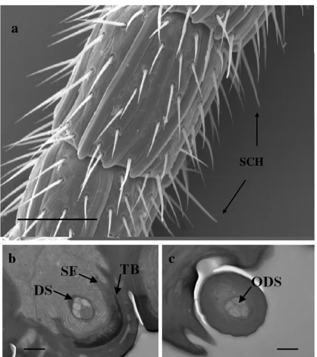
3.4.2 Sensilla chaetica (SCH)
They are long sensilla, ending in a fine, sharp tip (Fig. 3). The SCH are positioned radially in the tip of the A14, in number of 5, and distally from A3 to A13, in number of 4. They are characterized by a elongated cuticular shaft decreasing in diameter toward the round apex pierced by few pores not visible in SEM pictures. The length of the sensilla is about 28-30 µm. They show a flexible socket. These sensilla usually possess a thick wall.
b
c
SF
TB
28 TEM investigations show that the cellular components consist of five sensory neurons and three accessory cells (Fig. 3.4b). The bundle of four dendrites is surrounded by a common dendritic sheath. The dendritic projection of one sensory neuron is modified into a tubular body located on the distal end of the outer dendritic segment, connected to suspension fibers of the socket. The others four dendrites, after entering the peg lumen reach unbrunched the tip of the shaft (Fig. 3.4c).
Sensilla with similar ultrastructural features were reported in several groups of insects (Romani
et al, 2010; Zacharuk,1985). Mechanosensory associated to a gustatory function is hypothesized for
them.
Figure 3.4 Sensilla chaetica (SCH). a) SEM pictures of the two last antennomere; b) TEM oblique
section at socket level showing five dendrites enveloped in dendritic sheat (DS); c) TEM shaft cross section showing four dendrite inside (ODS). TB=tubular body; SF=suspension fibres. Scale bar: a=50 µm; b=1 µm; c=500 nm.
a
29 3.4.3 Multiporous Plate Sensilla (MPS)
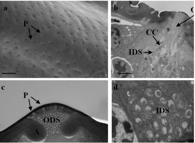
This sensory structures are found on all antennal segments, with exception for scape and pedicel. MPS extend longitudinally, often occupying the entire length of antennomere, with the distal part slightly free. The number of MPS decreases from apical to basal antennomere, from A6 to A14 in number of eight, while from A3 to A5 in number of four. Their distribution is uniform for all the entire surface of the segments. The length of MPS varies according to antennomere’s length and ranges from 70-75 in A3 and A14 to about 125 µm in the pedicel central antennomere . They are characterized by a thin cuticular structure, in the form of a long plaques slightly arising from antennal wall. The cuticle is perforated by numerous tiny pores distributed onto the entire sensillum cuticular surface (Fig.3.5a).
TEM investigations show that these sensilla are composed of 25 sensory neuron and 3 accessory cells (Fig. 4d). The bundle of dendrites is surrounded by a common dendritic sheath. In cross sections, the sensillar lumen presents two cuticular reinforcements, which divide the lumen into two chambers, with one of them occupied by the outer dendritic segments (Fig. 3.5c). After entering within the lumen, in contact with the pores, the dendrites greatly branch. A sheath cell completely envelops all the sensory cell bodies.
These sensilla perceive volatile chemical stimula, an olfactory function is attribuited.
These were mainly described in Hymenoptera (Navasero and Elzen 1991, Basibuyuk and Quicke 1998, Barlin and Vinson, 1981) and Coleoptera (Allsopp, 1990). For these sensilla, an olfactory function has been proved (Lacher, 1964).
30
Figure 3.5 Multiporous Plate Sensilla (MPS). a) SEM pictures multiporous surface; b) TEM cross section
showing cellular and cuticola components; c) TEM cross section showing the dentritic branches adjacent to the porous surface; d) cross section showing inner dendritic segments (IDS), ciliar constriction (CC) and outer dendritic segments (ODS). P=pore; A=apodema. Scale bar: a=400nm; b,d=2 µm; c=1 µm
3.4.4 Coeloconic Sensilla (CS)
As already described by Romani et al. (2010) coeloconic sensilla are usually in relatively low numbers when compared with the other antennal sensory structures, with a defined distribution on the antennal segments. In fact in this work 7 CS are counted, one each segment (Fig. 3.6a), on the distal external part from A8 to A13 and halfway of A14, where is possible to observe a suture line. The cuticular parts have a small and clavate peg with walls proximally smooth, distally grooved and multiporous along the groves except on the tip (Fig. 3.6c), and set in round, shallow pit wider at the bottom than at its opening (Fig. 3.6b). The peg, 6,25 µm long, is completely embedded within the antennal wall, in an ellipsoidal shape of maximum amplitude of 8 µm.
TEM investigations show that each sensillum is innervated by four sensory neurons. One of them, near the peg base, tapers and terminates, while the other three enter the peg lumen and, still
31 enclose in a thick dendritic sheath originating at ciliary constriction’s level and terminating where the peg grooves and porosity begin, run the peg lumen up to almost the tip without branching (Fig. 3.6d). Three accessory cells are present.
Morphologically speaking, these sensilla might be considered olfactory for their general configuration and for having a mutiporous peg (Roux et al.,2005),
Figure 3.6 Coeloconic Sensilla (CS). a) SEM pictures of the last two antennomere; b-c) detail of CS;
d) TEM longitudinal section of CS. G=groove-, ODS=outher dendritic segment. Scale bar: a=50 µm; b,d=2 µm, c=400 nm.
32
3.4.5 Styloconic Sensilla (SS)
There is one in the last seven antennomeres (Fig. 3.7), these sensilla are not articulated and their sensory portion is sunken and surrounded by the cuticular surface that comes in relief in an ellipsoidal shape of maximum amplitude of 8 µm (Fig. 3.7b). SEM observations do not show pores. A possible thermo-hygrosensitivity is associated, in agreement with other Hymenoptera previously observed (Roux et al. 2005; Altner and Prillinger, 1980).
Figure 3.7 SEM micrograph of Styloconic Sensilla. a) A11 and A12 showing SS position; b) detail of SS.
Scale bar: a=25 µm; b=2 µm.
A summary of some morphological properties of examined sensilla is shown in the following table:
o
3.5 Conclusions
Sensilla Tricoidea ST Sensilla Chetica SCH Multiporous Plate Sensilla MPS Coeloconic Sensilla CS Styloconic Sensilla CS Lenght 20-25 µm 25-30 µm 70-75 / 100-125 µm 6.25 µm - Diameter - - - 8 µm 8 µmPores aporous probably one
or few multiporous multiporous aporous
Neurons 1 5 (4+1) 25 4 (3+1) undetected
33
3.5 Conclusions
Characterization of D. kuriphilus female antenna through SEM and transmission electron microscopy (TEM) techniques permitted to highlight five kinds of sensilla:
- sensilla with a mechanosensory function (ST);
- sensilla suitable to perceive volatile chemical stimula, with an olfactory function (MPS and CS);
- sensilla with a mechano-gustatory function (SCH); - sensilla with a possible thermo-hygrosensitivity (SS).
34
4
Natural control
4.1 Introduction
4.1.1 Natural enemies of cynipid gallwasps: chalcid parasitoid wasps
The natural enemies causing the highest degree of mortality in cynipid galls are chalcid parasitoid wasps (Hymenoptera, the superfamily Chalcidoidea) (Askew, 1984; Stone et al., 2002).
Six families of chalcids attack Western Palaearctic oak galls: Pteromalidae, Eurytomidae, Eupelmidae, Eulophidae, Ormyridae and Torymidae (Askew, 1984; Stone et al., 2002; Csóka et al., 2005).
Chalcids oviposit either close to or onto their host so larvae can feed either after prohibiting further growth by stinging (idiobionts) or as the host continues to develop (koinobionts) (Godfray, 1994; Hayward and Stone, 2005).
Most chalcids reared from cynipid galls are solitary idiobiont ectoparasitoids that feed suctorially on their host (Askew, 1961; Schönrogge et al., 1999). Chalcid species commonly attack a range of possible hosts inside the galls, i.e. gall formers and inquilines, but they also act as facultative hyperparasitoids and autoparasitoids attacking other chalcids of different or sometimes the same species (Askew, 1961; Askew and Shaw, 1974).
Some exceptions to these rules include Sycophila biguttata and the gregarious Baryscapus
berhidanus, both idiobionts (Askew, 1984; Schönrogge et al., 1995). A range of parasitoid species
are also at least partially herbivorous, feeding on gall tissues either before they feed on the host larva, for example, the phyto-entomophagous koinobiont, Torymus cyanaeus, or feeding on gall tissues as they move between host larvae within a single gall, for example, Eurytoma brunniventris (Askew, 1984).
The majority of chalcids exploit hosts in a wide range of oak gall types; few are restricted to just one oak gall type. Some chalcids that have broader host gall ranges over a wide geographical area can appear locally host gall specific. For example, Aulogymnus skianeuros in Britain (and probably over much of Western Europe) is specific to sexual generation galls of Biorhiza pallida, but in Eastern Europe it has also been reared from some 15 additional cynipid species, 9 of which also occur in Britain. Aulogymnus trilineatus is at the edge of its range and rare in Britain where it is known to attack only asexual generation Andricus foecundatrix, but on the continent it is recorded from a broad range of 29 types, 8 of which are found in Britain (Fulmek, 1968).
35 Most of the parasitoid species found in cynipid galls on oaks will not attack hosts even in cynipid galls on other plants, i.e. this community is relatively closed. However, a minority of species, such as Eupelmus urozonus and E. vesicularis (Eupelmidae), are exceptionally polyphagous and attack a range of endophytic hosts in a number of insects (Askew, 1984). A few chalcid species also occasionally attack cynipid galls on plants other than oak (often Rosa) (Askew, 1984).
For example, Torymus flavipes, Eurytoma pistacina, Hobbya stenonota, Mesopolobus
amaenus and Baryscapus pallidae have also been recorded from rose galls of Diplolepis mayri
(Pujade-Villar, 1992; Askew et al., 2006). Some parasitoids, particularly eulophid species, are occasionally reared from oak galls despite being more generally associated with communities centered on leaf-mining hosts (e.g. Cirrospilus species, Pediobius pyrgo, P. saulius, Closterocerus
trifasciatus, Minotetrastichus frontalis).
However, these records appear to result from rare and opportunistic ovipositions as are those of a few ichneumonid (Ichneumonoidea) species that only occur sporadically (Fulmek, 1968; Schönrogge et al., 1996).
36
4.1.2 Natural control of D. kuriphilus in Asia
D. kuriphilus in its native country is controlled by many natural enemies.
A total of 11 species, belonging to 5 chalcid families (Torymidae, Ormyridae, Eurytomidae, Eulophidae and Eupelmidae), are known parasitoids of D. kuriphilus in China (Murakami et al., 1980).
A summary of the natural enemies associated with D. kuriphilus in its native area and in the closer asian States is hereby provided. The table 2 shows the parasitoid species attacking D.
kuriphilus in Asia (China, Japan and Korea).
China Japan Korea
Chalcids Amblymerus sp. Arthrolytus usubai Caenacis peroni Cecidostibafushica Cecidostiba semifascia Cynipencyrtus flavus Cynipencyrtus sp. Eupelmus urozonus Eupelmus sp. Eurytoma brunniventris Eurytoma schaeferi Eurytoma setigera Megastigmus maculipennis Megastigmus nipponicus Mesopolobus yasumatsui Ormyrus flavitibialis
Ormyrus pomaceus (=O. punctiger) Pteromalus apantelophagus Sycophila concinna Sycophila variegata Tetrastichus sp. Torymus beneficus Torymus geranii
Torymus sinensis local strain
Torymus koreanus Torymus sp. Non-chalcid parasitoids Aspilota yasumatsui Cynipid inquiline Synergus sp. X X X X X X X X X X X X X X X X X X X X X X X X X X X X X X X X X X X X X X X X X X X X X X X X X X X
Tab. 1 Parasitoids attacking D. kuriphilus in Asia (Modified from Aebi et al., 2006 and Matsuo
37 Native T. sinensis was recorded both from Japan and from Korea (where it has never been imported). However, the local population of T. sinensis of both States differs in adult emergence phenology from wasps imported from China and it’s thought to be native to its country (Aebi et al., 2006).
In Japan and Korea this T. sisnensis local has not been effective in controlling D. kuriphilus, so as to make useful the introduction of the Chinese strain.
38
4.1.3 Natural control of D. kuriphilus in Italy
Some transpalaearctic species of parasitoids that are also effective parasitoids of D. kuriphilus in Asia are very common and widespread in oak cynipid galls in Europe.
Some of them have already shifted on the new host and infact they have been reared from galls of D. kuriphilus in Italy, as shown in the table below.
Species Family Aebi
et.al., 2007 Speranza et al., 2009 Santi and Maini, 2012) Quacchia et al., 2012 Aprostocetus sp. Eulophidae X
Aulogymnus arsames Eulophidae X
Aulogymnus sp. Eulophidae X
Baryscapus pallidae Eulophidae X
Baryscapus sp. Eulophidae X
Pediobius chilaspidis Eulophidae X
Pediobius saulius Eulophidae X
Pediobius sp. Eulophidae X
Eupelmus annulatus* Eupelmidae X
Eupelmus spongipartus* Eupelmidae X
Eupelmus splendens Eupelmidae X
Eupelmus urozonus Eupelmidae X X X
Eupelmus fulvipes Eupelmidae X
Eurytoma brunniventris Eurytomidae X X X
Eurytoma adleriae Eurytomidae X
Eurytoma pistaciae Eurytomidae X X
Sycophila variegata Eurytomidae X X
Sycophila biguttata Eurytomidae X X X
Sycophila iracemae Eurytomidae X
Ormyrus nitidulus Ormyridae X
Ormyrus pomaceus Ormyridae X X X
Cecidostiba sp. Pteromalidae X
Mesopolobus amaenus Pteromalidae X
Mesopolobus mediterraneus Pteromalidae X
Mesopolobus sericeus Pteromalidae X X X
Mesopolobus tarsatus Pteromalidae X X
Mesopolobus tibialis Pteromalidae X
Megastigmus dorsalis (sp1) Pteromalidae X X X
Megastigmus dorsalis (sp2) Pteromalidae X
Torymus auratus Torymidae X
Torymus flavipes Torymidae X X X X
Torymus scutellaris Torymidae X
Torymus erucarum Torymidae X X
Torymus geranii Torymidae X X
39 In few years parasitoid communities based around invading D. kuriphilus have developed, involving species shared with local populations of oak gallwasps - typically those with broad host ranges - and have reached a remarkable level of species richness.
In comparison with the invading oak cynipids, D. kuriphilus is recruiting natural enemies rather rapidly. For example, the asexual generation galls of the invader Andricus quercuscalicis in Britain were free of parasitoids and inquilines for about 20 years (Schonrogge et al., 2000).
In addition to the rapid recruitment of parassitoids, an other striking feature of the D.
kuriphilus community is the absence of cynipid inquilines - a major component of the communities
associated with most oak cynipid galls (Stone et al. 2002; Aebi et al., 2006).
The cynipid inquilines (tribe Synergini) feed only on gall tissue. Although unable to induce their own gall, they have some ability to modify the plant tissue that immediately surrounds them (Stone et al., 2002). If these important constituents of the oak gall community were to start utilising chestnut galls, we could also expect them to bring along greater parasitoid diversity (Schönrogge et
al., 1996) and hence tighten further the trophic links between oak and chestnut gall communities
(Quacchia et al., 2012)
While in its native distribution D. kuriphilus populations are kept at low densities by natural enemies; in regions where it has invaded (Japan, South Korea and the USA), the attack rates of indigenous parasitoid species have remained low many years after the arrival of the pest (typically <2%) (Murakami et al., 1995; Stone et al., 2002; Aebi et al., 2007).
In Italy galls of D. kuriphilus were reared to census natural enemy recruitment immediately after its discovery in 2002, 2003, 2004 and 2005, and community richness has grown steadily over the years.
However, studies of parasitoids in various Italian regions showed a low impact on the overall population dynamics of gall wasp chestnut, rarely reaching the threshold of 2% of parasitization (Aebi et al, 2007).
In Piedmont, where the percentage of parasitization (number of parasitoids / number of larval gall cells) was very low, the most abundant species were Megastigmus dorsalis (F.) and
Eupelmus urozonus Dalman (Quacchia et al, 2011).
Instead, in Emilia Romagna Torymus flavipes (Walker) soon appeared as the dominant species of parasitoids of D. kuriphilus, arriving in 2011 at a rate of parasitization checked in the gall of 31.75% (Santi and Maini, 2011).
The rapid recruitment of oak cynipid parasitoids to D. kuriphilus suggest that, despite current low attack rates, there may be some value in an augmentative or conservation biological control programme using native species (Aebi et al., 2007)