Ai miei genitori Per Tutto da Sempre
Ad Alessandro semplicemente per Tutto
Ai miei tre piccoli Anna, Luca e Marco che la curiosità e lo spirito di ricerca siano forza e guida per i loro passi nel futuro
A mio fratello Christian, a Sara e a tutti gli amici per il sostegno, per la fiducia, per esserci.
Preface and Acknowledgments
This Ph.D.-study is based on work carried out in the period from 2008 to 2011 with the support of several people and organizations. This manuscript is made up of two individual not related articles not yet submitted to a international journal.
The first research “Peripheral Nerve Stimulation of brachial plexus for treatment of chronic post traumatic Neuropathic Pain” was carried out with collaboration of
Giorgio Stevanato MD, Neurosurgical Unit, Ospedale dell’Angelo Mestre –Venice, Italy Marzio Bevilacqua MD, Pain therapy Unit, Ospedale dell’Angelo Mestre –Venice, Italy Roberto Eleopra MD, Neurological Unit, Az.Ospedaliero-Universitaria “Santa Maria della
Misericordia”, Udine, Italy
The second article “Cold painful neuropathy a peculiar phenotype of neuropathic pain” was made with collaboration of
Valeria Tugnoli MD, PhD, Neurophysiological Unit, Az.Ospedaliero-Universitaria “Sant’Anna”,
Ferrara, Italy
Jay Capone MD, Neurological Unit, Az.Ospedaliero-Universitaria “Sant’Anna”, Ferrara, Italy Sara Rinaldo, Neurological Unit, Az.Ospedaliero-Universitaria “Santa Maria della Misericordia”,
Udine, Italy
Roberto Eleopra MD, Neurological Unit, Az.Ospedaliero-Universitaria “Santa Maria della
Misericordia”, Udine, Italy
I am grateful for the personal support throughout the study, the never-failing support and the encouragement.
I would like to express my grateful and appreciation to my tutor and supervisor prof Enrico
TABLE OF CONTENTS
INTRODUCTION ... 5
Neuropathic pain definition ... 5
Neuropathic pain syndrome diagnosis ... 7
Screening tools: pain scales and questionnaries ... 8
Clinical presentation QST, EPs, skin biopsy ... 9
Treatment of NP ... 10
Peripheral neuromodulation in treatment of NP ... 11
REFERENCES ... 13
PERIPHERAL NERVE STIMULATION OF BRACHIAL PLEXUS FOR TREATMENT OF CHRONIC POST TRAUMATIC NEUROPATHIC PAIN ... 15
Background and aims ... 15
Patients and Methods ... 16
Results... 22
Reference ... 35
COLD PAINFUL NEUROPATHY A PECULIAR PHENOTYPE OF NEUROPATHIC PAIN ... 37
Introduction ... 37
Patients and Methods ... 38
Results... 41
Discussion ... 46
Figures ... 49
Introduction
Neuropathic pain (NP) is a major disability in several neurological disorders with peripheral or central nervous system involvement, such as peripheral neuropathy, spinal cord lesions, stroke, or multiple sclerosis.
Pain is a complex experience, strongly modulated by cognitive and educational influences, and understanding nociceptive function or dysfunction is a hard task for all pain specialists.
NP remains a significant challenge to diagnose and treat effectively.
Neuropathic pain is reported as neurological disorder with a high prevalence ranged from 3.3% to 8.2% (Bouhassira 2008, Gustorff 2008, Torrance 2006), is an important public health problem.
Neuropathic pain definition
Neuropathic pain has been recently redefined by the Assessment Committee of the Neuropathic Pain Special Interest Group (NeuPSIG) of the International Association for the Study of Pain (IASP) as : “pain which arises directly from a lesion or disease affecting the somatosensory pathways” (NeuPSIG, Treede 2008). The current IASP definition is: ‘‘pain initiated or caused by a primary lesion or dysfunction of the nervous system” (Merskey 1997).
In particular, the “new” definition proposed by NeuPSIG replaces the substantive ‘‘dysfunction” by the substantive ‘‘disease” to distinguish neuropathic pain from that kind of pain caused by neuroplastic changes in response to strong nociceptive stimulation (Geber et al., 2009).
of the nervous system, i.e. pain associated with muscular spasticity rising from lesions of central motor pathways.
This redefinition has strong practical diagnostic implications. First of all the diagnosis of NP requires evidence for a lesion affecting a neuroanatomically identifiable part of the peripheral or central somatosensory system, which is consistent with the distribution of the pain (Treede, 2008; Haanpaa 2010). The grading of certainty for the presence of NP consists of
- Definite NP: all (1-4);
- Probable NP: 1 and 2, plus either 3 or 4;
- Possible NP: 1 and 2, without confirmatory evidence from 3 or 4.
The levels ‘‘definite’’ and ‘‘probable’’ indicate that the presence of this condition has been established. The level ‘‘possible’’ indicates that the presence of NP has not yet been established. Notably, if a patient does not fulfil the criteria for any of these three levels, it is considered unlikely to have NP (Treede, 2008; Haanpaa 2011). As direct consequence of this a specific and detailed clinical evaluation and confirmatory testing are required to reach the NP diagnosis. The challenge is to differentiate neuropathic pain from other types of pain and to diagnose the lesion or disease causing the pain. Standard neurophysiological tests, such as nerve conduction studies and somatosensory evoked potentials, are useful to demonstrate, locate and quantify damage along the peripheral and central pathways, but they do not assess the function of nociceptive (C and A-delta) pathways (Cruccu 2004, Cruccu 2010, Haanpaa 2011).
Per visualizzare quest'immagine sono necessari QuickTime™ e un decompressore
From: Treede RD, Jensen TS, Campbell JN, Cruccu G, Dostrovsky JO, Griffin JW, Hansson P, Hughes R, Nurmikko T, Serra J. Neuropathic pain: redefinition and a grading system for clinical and research purposes. Neurology. 2008
Neuropathic pain syndrome diagnosis
We may distinguish the diagnostic tools into four major group : 1- pain
Per visualizzare quest'immagine sono necessari QuickTime™ e un decompressore
.
Per visualizzare quest'immagine sono necessari QuickTime™ e un decompressore
able to quantify the sensory thresholds using graded stimuli ; 3- extensive
neuroalgological bedside evaluation, related to either the clinician experience and the collaboration of the patient ; 4- laboratory or instrumentals tests that quantified objective responses as confirmatory tests for NP diagnosis.
Screening tools: pain scales and questionnaries
In recent years, several scales and questionnaries based on verbal pain description have been developed and validated in NP. First of all, the Leeds assessment of neuropathic symptoms and signs (LANSS) contains 5 symptom items and 2 clinical examination items (Bennet, 2001; Bennet, 2005). A score of 12 or more out of a possible 24 suggests the presence of NP. Moreover a self-report tool, the S-LANSS has also been validated (Bennet, 2005).
The neuropathic pain questionnaire (NPQ) consists of 12 items that include 10 related to sensations or sensory responses (Krause, 2003). It was reported to be power to discriminate between NP and non-NP (Haanpaa, 2011).
Douleur neuropathique en 4 questions (DN4) consists of 7 items related to
symptoms and 3 related to clinical examination (Bouhassira, 2005). The DN4 is easy to score and a total score of 4 out of 10 or higher suggests possible NP. The first 7 items can be used as a self-report questionnaire with similar results (Bouhassira, 2005). Untill now, the DN4 has been used in large epidemiological studies to estimate the prevalence of NP both in the general population and specific NP disorders as diabetic neuropathy.
The painDETECT was developed and validated in German and includes a self-report questionnaire with 9 items (Freynhagen, 2006). There are 7 sensory descriptor items and 2 items relating to the spatial and temporal characteristics of the specific pain pattern. It is available in German and English, has been translated in 22 languages (not yet validated in Italian language).
ID-Pain consists of 5 sensory descriptor items and 1 item relating to pain localization in the joints to identify nociceptive pain; it also does not require a clinical
examination (Portenoy, 2006). A recommended cut- off score of above 3 points suggestes the presence of NP.
The neuropathic pain symptom inventory (NPSI) contains 10 descriptors grouped into 5 distinct pain qualities (burning, paroxysmal, deep, evoked, paresthesia) and 2 temporal items which assess pain duration and the number of pain paroxysms. The originally validated French NPSI has been translated and linguistically validated in about 50 other languages, moreover, its conceptual adequacy has been confirmed in several languages and it has been revalidated in Italian (Padua et al., 2009).
Its structure makes it suitable to represent different aspects of NP that may have distinct pathophysiological mechanisms and may correlate to functional and neurophysiological data. For these reasons we choose this questionnaires for our research projects.
Clinical presentation QST, EPs, skin biopsy
Quantitative Sensory Testing (QST) quantifies the different modality of sensory perception in response to external graduate stimuli of controlled intensity. The detection of sensory or painful threshold can be used in clinic along with bedside testing to document the sensory profile.
Conventional neurophysiological studies such as nerve conduction studies (NCS), somatosensory-evoked potentials (SEPs), can identify and quantify a lesion along the peripheral or central sensory pathways, but they do not assess the nociceptive pathway integrity. For many years the neurophysiological diagnosis of nociceptive lesion has been a challenge. The introduction of laser evoked potential using a laser
skin layers has provided a tool to addess it. In painful peripheral neuropaties are typically involved the small fibres C and A delta. To assessed their damage also in the early phases of neuropathy the quantification of intraepidermal and dermal nerve fibers using skin biopsy is a relatively simple, mininvasive and reliable method.
Treatment of NP
Pharmacological treatment of NP
The management of NP is challenging because the response to most drugs remains unpredictable or poorly despite attempts to develop a more rationale therapeutic approach (Finnerup, 2006, Baron 2006). Recently the European Federation of Neurological Societies (EFNS) produced the revision of guidelines on
pharmacological treatment of NP (Attal, 2010).
Tricyclic antidepressants (TCA) (25– 150 mg/day) and anticonvulsants as gabapentin (1200–3600 mg/day) and pregabalin (150–600 mg/day) are recommended as the first line of treatment for various NP conditions (except for trigeminal neuralgia), while lidocaine plasters (up to 3 plasters/day) first line in postherpetic neuralgia PHN.
In painful diabetic polyneuropathies SNRI (duloxetine 60–120 mg/ day, venlafaxine 150–225 mg/day) as first line has been recommended.
Second-line treatments include other medications such as tramadol and in PHN capsaicin cream. Oxicodon and other strong opioids are recommended as second/third line because of potential risk for abuse on long-term use. Finally
Cannabinoids have a level A in MS and peripheral NPand are proposed for refractory cases.
Combination therapy is still not well established in NP however is recommended for patients who show partial response to drugs administered alone (level A for
gabapentin combined with opioids or TCA) (Attal, 2010).
Peripheral neuromodulation in treatment of NP
Peripheral nerve stimulation (PNS) is a neuromodulation technique in which electrical current is applied to the peripheral nerves to improve chronic pain. It is first described in 1967, then a variety of techniques have since been developed. In the 1970s PNS was seldom performed because the high morbidity and poor
outcomes. The reasons suggested ranged from poor patients selection and limitations related to inadequate devices.
However, growing evidences assessed that PNS is effective in particular NP syndromes characterized by peripheral nerve lesions or irritation with pain or positive phenomena with a localized peripheral nerve distribution. The most
common painful syndrome treated with PNS are trigeminal branch stimulation, the occipital nerve stimulation (Weiner 1999) and subcutaneous peripheral nerve stimulation also known as sPNS.
Cranial nerves PNS is widely used also for treatment of chronic painful syndrome like headache including migraine (Popeney, 2003), TACs and cluster headache (Broggi, 2009; Goadsby, 2007). Less commonly PNS is applied at trunk peripheral nerve disorders, tibial neuralgia, inguinal neuropathy, meralgia paresthesica.
The Patients selection has to be quite selective, and the screening work up includes general clinical and serological evaluation to assess the comorbidity, psychological screening to rule out psychological amplifiers of pain or other confounding
conditions (severe depression, psychosis, behavioural disorders, drugs and
substance abuse). Usually the use of local anesthetic injection along the peripheral nerve is thought as informative pre-test as screening tool to confirm the appropriacy of PNS.
help in diagnosis and confirm the presence of definite neuropathic pain. A trial
period with temporary electrodes generally lasts 7-14 days. If patients experienced a sufficient pain relief (usually more then 50% from the baseline), a permanent
system is implanted.
Intraoperative testing, when performed, confirms that stimulation paresthesias are in the right position able to cover the painful skin area.
Permanent leas are connected to an implantable pulse generator (IPG). The final position of IPG depends from the nerve electrocateters, i.e. the subclavicular area is used for cranial nerve electrodes, while the thigh and upper arm is choose for lower or upper harm electrocatheters.
Currently PNS procedures are performed using devices not specifically approved for this application, i.e. devices approved for spinal cord stimulation.
Most recently subcutaneous peripheral nerve stimulation (S-PNS) has been introduced. In S-PNS electrodes are placed directly in the subcutaneous space of the painful skin area.
The electrodes is supposed to elicit the small subcutaneous nerve fiber and branch. This kind of stimulation is used to treat pain condition with less defined pain
distribution. Concerning the mechanism of action, it is still unclear. The gate control mechanism is the most reported, moreover for PNS another mechanisms is due from the periphery: in such NP conditions has been suggested a dysfunction of nociceptors with alteration of the local conductance activity, of the terminal
chemistry and the efferent function of peripheral small fiber end terminals (Byers, 2001).
There are several emerging indications for S-PNS as Complex Regional Pain
Syndromes (CRPS), axial neck pain, low back pain and failed back surgery syndrome. In Literature long term efficacy of PNS is rarely described and are often reported several sides effects and complication i.e. electrocatheters dislocation and
References
Baron R. Mechanisms of disease: neuropathic pain: a clinical perspective. Nat Clin Pract Neurol, 2006; 2: 95–106.
Bennett MI. The LANSS Pain Scale: the Leeds assessment of neuropathic symptoms and signs. Pain 2001;92:147–57.
Bennett MI, Smith BH, Torrance N, Potter J. The S-LANSS score for identifying pain of predominantly neuropathic origin: validation for use in clinical and postal research. J Pain 2005;6:149–58.
Bouhassira D, Attal N, Alchaar H, Boureau F, Brochet B, Bruxelle J, Cunin G, Fermanian J, Ginies P, Grun-Overdyking A, Jafari-Schluep H, Lantéri-Minet M, Laurent B, Mick G, Serrie A, Valade D, Vicaut E. Comparison of pain syndromes associated with nervous or somatic lesions and development of a new neuropathic pain diagnostic questionnaire (DN4). Pain 2005;114:29–36.
Bouhassira D, Lanteri-Minet M, Attal N, Laurent B, Touboul C. Prevalence of chronic pain with neuropathic characteristics in the general population. Pain 2008;136:380–7.
Finnerup NB, Jensen TS. Mechanisms of disease: mech- anism-based classification of neuropathic pain-a critical analysis. Nat Clin Pract Neurol 2006; 2: 107–115.
Freynhagen R, Baron R, Gockel U, Tölle TR. PainDETECT: a new screening questionnaire to identify neuropathic components in patients with back pain. Curr Med Res Opin 2006;22:1911–20.
Geber C, Baumgaurtner U, Schwab R, Muuller H, Stoeter P, Dieterich M, Sommer C, Birklein F, Treede RD. Revised definition of neuropathic pain and its grading system: an open case series illustrating its use in clinical practice. Am J Med 2009;122:S3–S12.
Gustorff B, Dorner T, Likar R, Grisold W, Lawrence K, Schwarz F, Rieder A. Prevalence of self-reported neuropathic pain and impact on quality of life: a prospective representative survey. Acta Anaesthesiol Scand 2008;52:132–6.
Haanpää M, Attal N, Backonja M, Baron R, Bennett M, Bouhassira D, Cruccu G, Hansson P, Haythornthwaite JA, Iannetti GD, Jensen TS, Kauppila T, Nurmikko TJ, Rice AS, Rowbotham M, Serra J, Sommer C, Smith BH, Treede RD. NeuPSIG guidelines on neuropathic pain assessment. Pain. 2011 Jan;152(1):14-27.
Krause SJ, Backonja M. Development of a neuropathic pain questionnaire. Clin J Pain 2003;19:306–14.
Merskey H, Bogduk N. Classification of chronic pain. Seattle, WA: IASP Press; 1997.
Torrance N, Smith BH, Bennett MI, Lee AJ. The epidemiology of chronic pain of predominantly neuropathic origin. Results from a general population survey. J Pain 2006;7:281–9.
Treede RD, Jensen TS, Campbell JN, Cruccu G, Dostrovsky JO, Griffin JW, Hansson P, Hughes R, Nurmikko T, Serra J. Neuropathic pain: redefinition and a grading system for clinical and research purposes. Neurology. 2008
Peripheral Nerve Stimulation of brachial plexus for treatment of chronic post traumatic Neuropathic Pain
Background and aims
Chronic peripheral neuropathic pain due to peripheral nerve injury often results in significant suffering and impaired quality of life. It is poorly responsive to drugs usually used for the treatment of neuropathic pain (NP) such as anticonvulsants and membrane stabilizers and opioids, even at high dosages. Therefore, an effective treatment for neuropathic pain still remains a major clinical challenge.
Peripheral nerve stimulation (PNS) has been used by a small group of neurosurgeons for the treatment of chronic peripheral neuropathic pain since 1965. Although in Literature PNS was demonstrated to be quite successful on the short and middle term (Eisenberg 2004, Van Calenbergh 2009), PNS has never become a standard technique for treatment of NP syndrome. This may be due to technical difficulties with paddle-type electrodes inserted around the peripheral nerve and to the risk of infections or electrodes dislocation.
The mechanism by which PNS produces analgesia is still unclear.
One of the possible mechanisms is based on the assumption that direct application of low-intensity high-frequency electrical current on to a peripheral nerve can elicit the A-beta myelinated fibers and produce analgesia according to the “gate control” mechanism (Melzack and Wall, 1965). Then main mechanism proposed as key part of PNS is closely related to the brain function and the brain plasticity in particular (Stanton-Hicks, 2011).
Thus, we hypothesized that the implantation of PNS leads directly on the nerve branch mainly involved in painful syndrome, proximally to the site of injury, may produce analgesia or significant pain relief in patients with traumatic nerve injuries. For this purpose an at least partially preserved pathway is required.
In the present study, we assessed the short and middle-term outcome of PNS in a series of patients with peripheral post-traumatic neuropathic pain at the upper harm. The aim of the study was to evaluate pain relief in this selected group of patients due the direct stimulation of the nerves branches administered by means of lead inserted with a peculiar and innovative surgical technique.
Patients and Methods
Patients
Patients affected by intractable pain due to peripheral nerve injuries and referred to our neurological unit for advanced pain treatment during the years 2007–2010 were considered for PNS.
A detailed medical history and neurological examination were collected. Peripheral neuropathic pain was defined as chronic pain in an area of sensory
abnormality corresponding to the nerve lesion and with an onset less than 6 months after the lesion, according to the gradying system proposed by the NeuPSIG
guideline (Haanpaa 2011).
Patients were carefully selected for PNS after an extensive baseline assessment. Therefore, we identify the following criteria for inclusion: (1) clear identification of an isolated injured nerve (i.e., selective branch of brachial plexus, median nerve, radial nerve) by means of clinical and electroneurographic and electromyographic
evaluation as the unique cause of pain; (2) complete although transient pain relief following a diagnostic nerve block with local anesthetics; (3) poor response (<50% of pain relief) to all other treatments, including drugs assumption, surgical treatment of the injuries site such as neurolytic procedures, neuroma resection, nerve grafting and transection; (4) absence of major psychiatric disorders, such as personality disorders or major depression, as assessed by psychological examination using Minnesota Multiphasic Personality Inventory (MMPI); (5) signing of a written informed consent for the study and for surgical procedure.
The study was single-centre, open-label and received the institutional review board approval.
Neurological and algological baseline examination
All subjects underwent a complete neurological and algological examination of the upper and lower trunk, medial, lateral, and posterior cords of the brachial plexus and peripheral nerves, in order to assess the motor and sensory loss distribution and positive phenomena as following described.
Superficial and pinprick sensations were examined in order to identify negative sensory signs (sensory loss) and positive sensory signs (evoked and spontaneous pain, paraesthesias). The neurological examination was performed using cotton gauze (light touch and dynamic mechanic allodynia test) and a brush, disposable safety needle (hypoalgesia, pinprick hyperalgesia, aftersensation test), repetitive pinprick (2Hz for 30 seconds) and glass vials filled with cold and hot water (thermal sensation, allodynia test, aftersensation test). Deep tendon reflexes were classified as normal, decreased (if present with reinforcement) or absent. All muscle groups of the upper harm were evaluated and muscle strength was graded using the Medical Research Council (MRC) score.
Intensity of spontaneous pain, allodynia including static mechanical (pressure), dynamic mechanical (brush), heat or cold (thermal) and hyperalgesia were graded using the NRS (0-10) scale. We used the following descriptors for quality of pain: throbbing, lancinating, unpredictable, lightning-like, sharp, shooting, aching, burning, scalding, pruritic (Galer and Jensen, 1997; NPSI 2009). Pain attacks were described by intensity, duration, and frequency.
We collected a picture for the pain on the skin and detailed pain map and sensory abnormalities distribution on a body chart.
Finally the clinical evaluation was completed with upper harm examination in order to rule out of intrinsic joint disease, acromioclavicular pathology and in patients with lower brachial plexus involvement the elevated arm stress test (Roos, 1966) was performed in order to relieve thoracic outlet syndrome (Robert J. Schwartzman, MD, and John R. Grothusen, PhD).
Quantitative Sensory Testing
Thresholds for cold and warm sensation (CS, WS) and cold and heat pain (CP, HP) were obtained bilaterally on the thenar and hypothenar eminences, and on other sites according with the neuroanatomical distribution of pain and negative signs, with a 30x30mm thermode of the TSA-II Neuro Sensory Analyzer (Medoc, Israel). Method of limits has been performed. Each test was repeated four times for each side and the perceived threshold was defined as the average of peak temperatures. Subjects were instructed to push the response button, which recorded the
temperature and reset the thermode back to baseline, as soon as they detected a change in temperature for CS and WS, or only if the sensation changed from cold or hot to painful sensation.
Surgical procedures were performed at the General Hospital of Mestre-Venice between 2007 and 2010. All implantations were performed under general anesthesia and under strict sterile conditions by one surgeon (G.S.). The nerves selected for surgery were exposed proximal to the site of injury using the tecnique of brachial plexus exposition by the axillary approach. Patients were supine with the upper harm abducted by 90°, all the nerve and vascular structure are exposed, identified and classified as shown in figure 1.
Two On-Point leads (Medtronic Inc., Minneapolis) were used for the stimulation of the radial or/and the median nerve respectively. Each lead has 4 platinum-iridium electrodes on a silicone-rubber mesh. During the surgical procedure, the leads were placed upon the sensitive portion of the nerve according to the Sunderland’s
scheme (Sunderland, 1978) about 4cm far from the rising nerve, and carefully anchored by means of lead paddle mesh with absorbable sutures in order to avoid nerves compression. This technical solution allows the nerves protection by the surrounding fibrosis and provides stability of the overall system.
If the patient had motor function preserved at baseline, a stimulation trial was carried out by mean of a quadripolar lead in order to confirm the position near to sensory fibers and not near to motor fibers.
The electrodes were tunneled under the skin, making several loop in order to minimize traction or dislocation with the upper harm movement, and then
connected the temporary extensions. During the trial period an external stimulation (ENS, Medtronic Inc, Minneapolis) was used.
After the trial period, temporary extensions were removed and replaced by permanent extensions then connected to an implantable pulse generator (IPG, mod.Prime advanced, Medtronic Inc., Minneapolis) that was secured in the subclavicular subcutaneous pocket.
During the first trial after about 24 h from the procedure, the pulse rate, width, and voltage that produced the best response were selected. All patients has a continuos stimulation set.
Daily pain evaluation and scales
Patients were requested to evaluate their pain intensity with the use of a 11-points Likart scales (NRS), where “0” means “no pain” and “10” means “the worst
imaginable pain”. Additionally, they were asked to describe their pain with the use of the Pain Questionnaire (Galer and Jensen, 1997; NPSI 2009).
Pain and clinical follow-up evaluation
Patients were assessed during scheduled follow-up visits for efficacy evaluations after 1 week, 1 month and every 6 months thereafter for the next 2 years from the IPG implantation. QST was assessed at baseline, 1 month and 12 or 24 months with stimulation turned ON. However, patients who lived far away were followed up by telephone calls 6 and 18 months after the IPG implantation to obtain information on long-term pain intensity and analgesics consumption .
Patients were asked to fill out the pain questionnaire and clinical evaluation at follow-up evaluation in two conditions: with the stimulator switched off (“OFF”) and stimulator switched on (“ON”). The stimulator was switched “ON” or “OFF” by a technician that randomized the order of the 2 condition. The neurologist (G.D.) that performed the clinical and QST evaluation and the patient were blinded to the condition of the stimulator. In all the patients the peripheral nerve stimulation was at low intensity in order to obtain sufficient pain relief without paresthesiae
sensation. In this way the patient was blinded to the Stimulator condition (“ON” or “OFF”).
assessment periods, to minimize potential carry-over effects of PNS.
Statistics
A 2-tailed t test was used to compare NRS values and analgesic consumption prior to surgery and during the follow-up. Results at the P < 0.05 level were regarded as statistically significant.
Results
Patients.
We identify 7 patients (all men) fulfilling the inclusion criteria and suitable for PNS. All the 7 patients had a post-traumatic lesion of brachial plexus or distal peripheral nerve complaining severe intractable pain characterized by positive phenomena like allodynia, paradoxical pain and ongoing pain. The clinical data are reported in table 1 and 2. In 4 patients a traumatic peripheral nerve lesion occurred, while in 3 a traumatic brachial plexus postganglionic injury caused the peripheral NP syndrome. Their age ranged from 17 to 68 years, with a median age of 46y.o. Pain duration prior to PNS range from 19 months to 31 years. The mean baseline NRS was 9/10, indicating moderate to severe mean pain intensity before surgery. The patients described the painful syndrome with several qualities like cutting sensation, burning sensation, constriction with impressive positive phenomena like electric shock after light touch or movement, several times every day. From the history all patients had previously received neurosurgery neurosurgical treatments including neurolysis. Therefore all patients of them underwent conventional medications (analgesics, antidepressants, anticonvulsants) (see table 2) and repeated nerve blocks. None of the patients met the research diagnostic criteria for complex regional pain
syndrome II (CRPS II) (Harden, 2006).
The follow-up evaluation
The follow-up period was ranged from 18 months to–4 years, with a median value of years. The results based on pain relief were classified as good in all patients. The stimulation parameters of PNS were similar in all patients: with a 50Hz frequency of 50Hz, a 250usec of pulse width of 250µsec, and an amplitude ranging from 0.15
to-0.30 V of Amplitude.
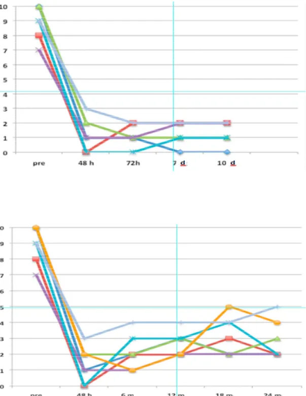
Overall, pain intensity dropped decrease from a NRS of 9±1.15 before surgery to 2.14±1.57 at 6-month of follow-up and to 2.57±1.13 at 12-months of follow-up (P < 0.001). No complications like infections, dislocation or electrodes dislocations or migrations occurred in none patient. In two patients a sudden pain sensation were reported and we observed in both a transient PNS dysfunction due from to
magnetic interference. Patients shown peripheral (hand) oedema and skin
discoloration similar to that observed in CRPS, with cold skin cyanosis without sweat dysfunction.
QST battery at baseline and at follow-up
Thermal tresholds were abnormal in all patients according to the neuroanatomical distribution of negative signs as expected by the sensory clinical evaluation, in particular we found higher cold and warm sensation in selective skin area. In 6 of 7 patients at baseline we found positive phenomena characterized by cold or warm allodynia and in 5 wrong painful sensation after cold and warm stimuli into not painful range. The abnormal painful sensation was reported as an electrick shock or burning sensation with wide skin area distribution, higher then expected by the nerve injuried distribution suggesting a peripheral or central hypersensitivity phenomena. We reported an example of sensory profile in figure 1a.
After 6 months at the follow-up evaluation thermal QST evaluation showed unchanged negative signs pattern and attenuation or complete resolution of positive phenomena.
At follow-up evaluation when the neuromodulation was blinded switch OFF after a mean ranged from 22 seconds to 54 seconds, they felt pain with NRS ranged from 8 to 10, with the same characteristics collected at the baseline pre – implant
evaluation before the trial test. During the stimulation “OFF” period, In four of seven patients we observed in this trial skin discoloration with cyanosis. During a trial of 1 h we observed a mild distal oedema. After the blinded turned “ON” of PNS the painful sensation disappeared with complete restore of pain relief in about 20-30 seconds and after a mean of 1 h all the autonomic signs disappeared.
Discussion
Peripheral nerve stimulation (PNS) is a neuromodulation technique in which electrical current is applied to the peripheral nerves to improve chronic pain. It is first described in 1967, afterwards a variety of techniques have since been
developed. In the 1970s PNS was seldom performed because the high morbidity and poor outcomes related to poor patients selection and technical limitations for
inadequate devices. However, growing evidences suggested that PNS is effective in particular NP syndromes characterized by peripheral nerve lesions or irritation with pain or positive phenomena with a localized peripheral nerve distribution (Weiner 1999).
In the present study we show a selected case series of patients complain severe neuropathic pain involving the upper arm with pain phenomena extremely
consistent to a peripheral nerve distribution. This clinical picture is usually at high risk for non-responsiveness with others neuromodulation tecniques such as motor cortex stimulation or spinal cord stimulation. However, peripheral nerve stimulation through a percutaneous approach - in particular for upper and lower limb - has poor outcome for high incidence of morbidity, related to the movement and critical
structures (i.e. tendons, nerves, vascular structures) in limited spaces.
In Literature, the use of a prognostic nerve block prior to PNS has been suggested by several groups, and from common practice it is well accepted that failure to respond to a nerve block is a bad prognostic factor for PNS. For that reason, complete
although temporary pain relief following a diagnostic nerve block with local
anesthetics was required as pre-test also in our screening evaluation. The use of the local anesthetic injection may help us in order to confirm the selection of the nerve target, and to confirm the prevalent peripheral mechanisms at the basis of the pain syndrome. Then the carefully evaluation of positive and negative signs and QST battery as screening tools defined the sensory pattern.
As reported also by other groups (Van Calenbergh, 2009) we observe that PNS did not modify neither heat or cold thresholds assessed by QST battery. Therefore the main finding for QST battery was the clear anti-allodynic or anti-hyperalgesic effect. In QST battery Z score profile the result is a reduction in positive phenomena with unchanged negative signs. The improving of positive phenomena is a direct effect of the PNS stimulation, while the negative signs in our patients are the consequence of the nerve lesion and PNS is obviously ineffective to improve a loss of peripheral nerve function like sensory sensation. However PNS can make a sort of noise in pain signals related to peripheral or central hypersensibilization, producing anti-allodynic effect.
The PNS is assessed to be a long-lasting beneficial therapy and a follow-up of more than 15 years is reported. The double-blind control test with the two condition (STIM "ON"; STIM "OFF") confirm the efficacy of PNS in our patients. Conversely to SCS, the analgesic and anti-allodynic effect is easily reached in few minutes. Notably, we observed in some of our patients peripheral autonomic dysfunction like
peripheral oedema or cyanosis with the PNS switch OFF, as collateral findings, suggesting peripheral mechanisms in axonal reflexes and vasogenic neurally mediated effects.
The follow-up of a mean of 2 years confirms the long-lasting beneficial effect of SCS, nearly unchanged between visit 1 month to visit after 2 years.
confirmed by several observations (data not shown), and above all for follow-up evaluation to clearly detect the antiallodynic effect of PNS.
In conclusion, PNS can result in long-term pain relief in the majority of carefully selected patients and has a relatively low complication rate. It should therefore be considered as a reasonable treatment of patients suffering from otherwise
Pain, referred sensations, in brachial plexus injury.
Table 1
Pain, referred sensations, in brachial plexus injury.
Table 1 Patient no. Gender, age (years) etiology Duratio n of plexus or nerve lesion (yrs, mo) Suggested nerve lesion Dermatomes or nerve area with abnormal sensation Site of lesion Concommitant injury NRS (at baseline) Ongoing pain treatment 1 M, 58 Accident at work 8 yrs Median, radial nerves Median, radial nerves Median and radial nerve lesion at third distal of forearm Partial Amputation of forearm 10 TCA, pregabalin, opioid 2 M, 68 iatrogenic 19 mo Median
nerve Median nerve
Proximal site of Median nerve 8 Pregabalin, opioid 3 M, 42 Accident at work 4yrs Median, radial nerves Median, radial nerves Nerve injuri at phalaneal level Distal Amputation (II, III, IV fingers) 10 Tramadol, gabapentin 4 M, 47 Accident at work 3yrs Median
nerve Median nerve
C6 – C7
lateral cord Amputation 7
Paracetamol , NSAID, opioid, gabapentin 5 M, 54 Motorcycl e accident 31yrs Median
nerve Median nerve
Severe injury at lateral cord 9 - 6 M, 17 Motorcycl e accident 32mo Median
nerve Median nerve
C6 – lateral cord 10 High dosage Opioid, Pregabalin, TCA 7 M, 36 Motorcycl e accident 14yrs Median
nerve Median nerve
(C5)-C6-C7-C8 postganglion 9 Opioid, Pregabalin, TCA
i lesion
Table 2
Background pain
Spontaneous pain paroxysms Evoked pain, NRS No. Of points w. Increased sensation No of points w. Referred sensation Patient no. Intensity, NRS Intensity, NRS Duration
(s) Frequency Brush Pinprick
Repetitive
Pinprick Cold Warm
1 10 7 15 4/day 10 8 5 5 8 24 5 2 8 5 30 1/h 9 5 4 3 2 1 5 3 10 10 10 1/h 10 1 6 6 9 13 0 4 7 8 160 10/h 10 10 10 10 7 11 0 5 9 9 5 1/month 7 7 8 5 8 16 2 6 10 10 30-45 1/h 5 8 10 7 6 16 4 7 9 5 2-3 3/day 6 6 5 5 6 0 0
Clinical evaluation and neuroalgological pattern.
Patient no. Intensity, NRS 48h 6 12 18 24 30 36 42 1 10 2 1 2 5 4 3 2 3 2 8 0 2 2 3 2 2 2 0 3 10 3 4 4 4 5 4 3 4 4 7 1 1 2 2 2 1 2 - 5 9 3 4 4 4 5 3 - - 6 10 0 0 1 - - - - -
7 9 2 3 3 - - - - -
Fig.2 Brachial plexus exposition at the axillary cavum, external neurolysis, insertion of a quadripolar electrode.
Reference
- Aló KM, Abramova MV, Richter EO. Percutaneous peripheral nerve stimulation. Prog Neurol Surg. 2011;24:41-57. Epub 2011 Mar 21. Review.
- Broggi G, Messina G, Franzini A. Cluster headache and TACs: rationale for central and peripheral neuromodulation. Neurol Sci. 2009 May;30 Suppl 1:S75-9.
- Buonocore M, Camuzzini N. Increase of the heat pain threshold during and after high-frequency transcutaneous peripheral nerve stimulation in a group of normal subjects. Eura Medicophys. 2007 Jun;43(2):155-60. Epub 2006 Oct 3.
- Byers MR, Bonica JJ, Peripheral pain mechanisms and nociceptor plasticity, in Bonica’s Management of Pain, ed 3 Philadelphia, Lippincott Williams & Wilkins, 2001, pp 26-72.
- Eisenberg E, Waisbrod H, Gerbershagen HU. Long-term peripheral nerve stimulation for painful nerve injuries. Clin J Pain. 2004 May-Jun;20(3):143-6.
- Ellrich J, Lamp S. Peripheral nerve stimulation inhibits nociceptive processing: an
electrophysiological study in healthy volunteers. Neuromodulation. 2005 Oct;8(4):225-32. doi: 10.1111/j.1525-1403.2005.00029.x.
- Goadsby PJ. Neurostimulation in primary headache syndromes. Expert Rev Neurother. 2007 Dec;7(12):1785-9.
- Harden RN, Bruehl SP. Diagnosis of complex regional pain syndrome: signs, symptoms, and new empirically derived diagnostic criteria. Clin J Pain. 2006 Jun;22(5):415-9.
- Huntoon MA, Burgher AH. Ultrasound-guided permanent implantation of peripheral nerve stimulation (PNS) system for neuropathic pain of the extremities: original cases and outcomes. Pain Med. 2009 Nov;10(8):1369-77.
- Melzack R., Wall PD, Pain mechanisms: a new theory. Science 1965;150:971-979. - Monti E. Peripheral nerve stimulation: a percutaneous minimally invasive approach. Neuromodulation. 2004 Jul;7(3):193-6. doi: 10.1111/j.1094-7159.2004.04195.x.
- Padua L, Briani C, Jann S, Nobile-Orazio E, et al. Validation of the Italian version of the
Neuropathic Pain Symptom Inventory in peripheral nervous system diseases. Neurol Sci. 2009 Apr;30(2):99-106.
- Popeney CA, Aló KM. Peripheral neurostimulation for the treatment of chronic, disabling transformed migraine. Headache. 2003 Apr;43(4):369-75.
- Portenoy R. Development and testing of a neuropathic pain screening questionnaire: ID Pain. Curr Med Res Opin 2006;22:1555–65.
- Roos DB. Transaxillary approach for the first rib resection to relieve thoracic outlet syndrome. Ann Surg 1966;16:354–8.
- Slavin KV. Peripheral nerve stimulation for neuropathic pain. Neurotherapeutics. 2008 Jan;5(1):100-6. Review.
- Schwartzman RJ, Grothusen JR. BRACHIAL PLEXUS traction injury: quantification of sensory abnormalities. PAIN Med. 2008 Oct;9(7):950-7.
- Stanton-Hicks M, Panourias IG, Sakas DE, Slavin KV. The future of peripheral nerve stimulation. Prog Neurol Surg. 2011;24:210-7. Epub 2011 Mar 21.
- Sunderland SS. The Brachial Plexus. Normal anatomy, In: Sunderland SS, ed., Nerves and Nerve Injuries . Churchill Livingstone, Edinburgh, pp. 854-869, 1978.
- Van Calenbergh F, Gybels J, Van Laere K, Dupont P, Plaghki L, Depreitere B, Kupers R. Long term clinical outcome of peripheral nerve stimulation in patients with chronic peripheral neuropathic pain. Surg Neurol. 2009 Oct;72(4):330-5; discussion 335. Epub 2009 Aug 7.
- van Eijs F, Stanton-Hicks M, Van Zundert J, Faber CG, Lubenow TR, Mekhail N, van Kleef M, Huygen F. Evidence-based interventional pain medicine according to clinical diagnoses. 16. Complex regional pain syndrome. Pain Pract. 2011 Jan-Feb;11(1):70-87. doi: 10.1111/j.1533-2500.2010.00388.x. Epub 2010 Aug 27.
- Weiner RL, Reed KL. Peripheral neurostimulation for control of intractable occipital neuralgia. Neuromodulation. 1999 Jul;2(3):217-21.
Cold painful neuropathy a peculiar phenotype of neuropathic pain
Introduction
It is established that cold sensation is mediated by small myelinated A delta ®bres (Yarnitsky and Ochoa, 1991), while cold pain is probably mediated by the activation of both thinly myelinated cold-specific A delta fibres and unmyelinated C fibres (LaMotte and Thalhammer, 1982; Wasner, 2004). In particolar, using
microneurographic recordings, C fibers activated by cold painful stimuli has been classify as polymodal nociceptors (Torebjork, 1974; Torebjork and Hallin, 1973). It is a common experience that in several pathological conditions like peripheral
neuropathy, innocuous skin cooling can induce pain (Woolf and Mannion, 1999; Baron, 2000).
The mechanisms of cold pain and cold positive phenomena such as cold
hyperalgesia or allodynia are less understood than of burning pain. It has been suggested a central mechanism characterized by a decrease in the inhibition
normally exerted centrally by cold sensory channels on nociceptive channels (Craig and Bushnell, 1994; Craig et al., 1996), but peripheral mechanisms has been
suggesteded (Wasner et al., 2004).
In humans a simple model for studying cold hyperalgesia is the topical application of L-menthol (C10-H20O), that is a specific agonist of cold - sensitive nociceptor of the TRP channels family, TRPM8 expressed on A delta afferent and small diameter dorsal root gangia (DRG) neurons (McCoy et al., 2010). TRPM8 is activated by by menthol and cooling, with an activation temperature ranged from 25 to 28°C.
Notably, at the DRG level TRPM8 is not co-expressed with the TRPV1, suggesting the cold-specificity of the TRPM8-positive A delta fibres (McKemy et al., 2002, Facer et al., 2008). Among the TRPM8 receptor, from the TRP channels family the TRPA1 have been identified as cold-sensitive ion channels. TRPA1 is activated by potentially painfully cold temperature (below 15°C), and by a ranged of esogenous irritants such as mustard oil, cinnamaldehyde (CA), formalin, reactive oxygen species (Obata et al., 2005) and a variety of endogenous reactive compound related to pathological conditions, including hydrogen peroxide and 4-hydroxynonenal (4-HNE). These evidences suggest that TRPA1 may play a role in several pathological pain
conditions. It is established that TRPA1 shares some pharmacological features with TRPM8, i.e. menthol clocks TRPA1 at high concentration (Karashima et al., 2007). In recent years several studies in experimental conditions in healthy humans try to clarify different pain syndromes (Wasner et al., 2004, NAmer et al., 2005, Tugnoli et al., 2007), however little is known about the mechanisms underlying the cold pain in peripheral neuropathic pain syndromes.
In small fiber neuropathy only 1-2% of patients describe a peculiar pain condition characterized by cold pain and cold paroxysms (Verdugo and Ochoa et al., 1992; Devigili et al., 2008). Cold spontaneous pain and cold hyperalgesia are common symptoms in patients with painful neuropathy, however pain phenotypes and mechanisms underlying are still poorly understood.
The aims of study was to characterize the distinct pattern of pain phenomena in patients with cold pain and to compare clinical features, neurophysiological and histological profile in order to understand the underlying pain mechanisms.
Patients and Methods
We selected 9 patients with painful small fiber neuropathy (SFN) complaining cold pain (group 1, c-SFN) during a period from 2008 to 2010. Then we selected 10 patients with the classical form of burning painful SFN (group 2, b-SFN), and a control group of 15 healthy subjects age and sex matched. All patients received the diagnosis of SFN assessed by history, clinical examination, skin biopsy and thermal thresholds QST. We selected only patients with all these tests abnormal to make a diagnosis of definite SFN. A complete neuroalgological and neurological examination were performed. Patients underwent nerve conduction studies, and if necessary EMG studies and pain questionnaire (DN4 2004, NPSI 2009 and Neuropathic Pain Questionnary 1997).
Experimental protocol
Substances.
We used as TRPM8-agonist a solution of L-menthol (40%) dissolved in 90% ethanol, and as TRPA-1 receptor agonist cinnamaldehyde (CA), while as placebo control for L-menthol we used a solution of ethanol 90% and for CA a solution of acetone
(CH3COCH3) that produces a similar smell. In different days about 1 ml of solution was placed on a gauze pad, which was applied to the target area of the skin, the distal leg (site of skin biopsy).
Application of menthol, cinnamaldehyde and control substances
A 3 x 3 cm gauze pad was placed on the distal leg 10 cm proximal to the lateral malleol for 20 min. In each of the 10 individuals, four gauze pads containing menthol, alcohol as control substance, cinnamaldehyde and acetone as control substance, respectively, were tested. The experiments were carried out in different days.
Pain and thermal sensations
rating scale (NRS) 0-10, with 0 representing `no pain' and 10 being `the maximum pain that can be imagined'.
The subjects and patients were instructed to report any sensation of pain during the 20 min of gauze pad application. The pain was quantified by rating the intensity on a numeric rating scale (NRS 0±10, with 0 representing `no pain' and 10 being `the maximum pain that can be imagined').
Areas pinprick hyperalgesia were assessed using a von Frey filament with a flat tip (diameter 0.2mm). The area of hyperalgesia was determined along two orthogonal trajectories by testing pinprick sensitivity radially each 0.5cm from the centre in both directions. Dynamic mechanical allodynia was tested using a cotton wool swab perpendicular to the two trajectories.
Thermal-Quantitative Sensory Testing (QST) battery
Thermal sensation thresholds (warm, cold, heat and cold pain) were measured by a Quantitative sensory testing (Medoc TSAII, Israel) using a Peltier type thermode (probe 3x3cm) prior to and after menthol, alcohol, CA and acetone. The Peltier thermode was applied exactly in the area of gauze pad application. The method of limits was used by applying ramp stimuli with a velocity of 1°C/s starting from32°C (baseline temperature). The subjects were asked to press a button immediately the respective thermal sensation was perceived, or for painful sensation when the sensory sensation changed in painful sensation. The period between the
achievement of the baseline temperature and the beginning of the following
stimulus (inter-stimuli interval, ISI) was 0 s for warm and cold and 20 s for heat and cold pain detection thresholds. Thresholds were determined as the average of four successive stimuli. To protect the skin from injury, the increase of the thermode temperature was limited to 50°C, even if the button was not pressed. The limit for decreasing temperatures was 0°C.
In all patients and control healthy subjects included in the present study, skin biopsies were obtained from the proximal region of the thigh (20cm below the anterior iliac spine) and the distal region of the leg (10cm above the lateral malleolus). Biopsies were taken after local anaesthesia using a 3 mm disposable punch under sterile technique. Three sections randomly chosen from each biopsy were immunoassayed with polyclonal anti-protein-gene-product 9.5 antibodies (AbD serotec; 1:800) using the free-floating protocol for bright field
immunohistochemistry previously described (Lauria et al., 2004). The linear density of IENF (IENF/mm) was calculated following the rules reported by the guidelines of the European Federation of the Neurological Societies (Lauria et al., 2010).
Finally we quantified the dermal fiber density expressed as n°fiber per total dermal area [n/mm2]; the ratio from the area of dermal nervous structures and the total dermal area [Sp nerves (mm2)/Sp dermis (mm2)]. Then three sections from each biopsy were immunoassayed with monoclonal anti-myelin basic protein MBP (AbD serotec; 1:800, IgG2b isotype) and policlonal PGP9.5 using the free-floating protocol for double-indirect immunofluorecence histochemistry previously described (Lauria et al., 2004). We quantified the number of MBP-positive dermal bundles, and their length and area. The area of dermis and nerve bundles was measured on digitized images using SPOTTM advanced software.
Results
Patients and control clinical data at baseline are collected in table 1.
Clinical findings
and all ten of the B-SFN patients had spontaneous pain, all of them had evoked pain and only one C-SFN patient had evoked pain alone. Evoked pain was characterized by static light touch allodynia in four C-SFN patients and dynamic mechanic
allodynia in two C-SFN patients. All C-SFN experienced cold hyperalgesia or cold allodynia at lower limb in a disco-proximal fashion. In the B-SFN group, eight
patients had both spontaneous and evoked pain, two patients had only spontaneous pain and none patient had evoked pain alone. We created the sensory profile (Fig 1b) that summarizes the painful phenotype of B-SFN and C-SFN.
Effects of L-menthol and cinnamaldehyde on pain and thermal sensations, thermal and sensory testing, mechanically evoked pain
Pain and thermal sensation
L-menthol induced significant cold sensations and no spontaneous pain in healthy subjects, while in seven B- SFN patients and in four C-SFN patients a spontaneous pain sensation has been reported.
Five B- SFN patients described the pain as burning, one patient as sharp pain and one as icy-cold. The pain occurred with a latency of a few minutes and reached an intensity plateau of about NRS 6 after 7 min.
While, only in four c-SFN patients L-menthol produced a spontaneous pain
described as burning and pinprick sensation, with a latency of few seconds reaching a pick of about NRS 4 after 2 minutes then a decrease till NRS 0 after about three minutes, in five patients no sensation has been produced (Fig.3b).
The pain induction was significant compared with alcohol placebo application that induced pain of a much lesser extent (NRS 2, for less then one minute) occasionally in only two healthy subjects and one B-SFN patients.
CA produced a burning sensation in five of ten controls, in three of ten B-SFN and in two of C- SFN. All subjects who felt pain after CA described its quality as burning pain. The burning pain occurred with a latency of 7 min and reached a plateau after
9 min, with a mean NRS of 6 in healthy subjects and of 3 in B-SFN and C-SFN. In all subjects CA induced a warm sensation. Acetone induced a mild sensation of warm. Neither L-Menthol nor CA induced itching sensation.
Skin flare
L-menthol produced a visible skin flare in all control subjects and in same of the B-SFN patients, with different area size (table 2). The L-menthol flare was significantly reduced or nearly abolished in the treated area in C-SFN patients (p<0.002).
CA induced a presumably mild skin flare not reliably detectable withouth a laser Doppler or scan imager for skin blood flow quantification.
Thermal testing
L-Menthol and CA induced different changes in temperature detection thresholds and pain thresholds. Topical L-menthol induced in control cold hyperalgesia by a significant decrease in cold pain threshold, with a mean of 10°C of CP threshold. Further thermal thresholds were not affected by menthol or placebo. In C-SFN patients L-Menthol induced increase in WDT, while no difference in CS or CP phenomena were detected. In B-SFN L-Menthol induced cold hyperalgesia in the flare area, while placebo induced in two patients decrease of heat pain threshold (44.2° prior; 43.5° after ethanol).
CA induced cold hypoalgesia in six controls and in six B-SFN, while it produced heat allodynia in five controls and in 8 B-SFN (Fig. 6b).
Mechanically evoked pain
L-Menthol produced pinprick hyperalgesia in the flare area in healthy subjects and in 8 of 10 B-SFN patients, while in cold SFN no positive phenomena were detected.
four patients in the area of application and in the surrounding skin. CA produced pinprick hyperalgesia in six of ten controls and in six of ten B-SFN patients.
Skin biopsy findings
The IENF density was similar in C-SFN and B-SFN group (Table 1). The dermal innervation quantification shown a more severe denervation of dermal nerve bundles in c-SFN compared with burning SFN (table 2) and MBP-positive fibers were reduced compared with burning-SFN (Fig. 7b).
Table 1.
Group C-SFN B-SFN S Control group S
Patients (n, %) 9 10 10 Male (n, %) 6 5 5 Female (n, %) 4 5 5 Age (years)* 54±7 57±12 ns 50±8 ns Height (cm)* 174±6 172±6 ns 175±5 ns BMI (kg/m2)* 26±2 28±1 ns 26±4 ns Clincal Evaluation Pinprick hypoesthesia 6 8 ns - ns Warm hypoesthesia 3 8 - Cold hypoesthesia 9 2 - Hyperalgesia 4 5 -
Static light touch allodynia 2 3 ns -
Dynamic mechanic allodynia 2 3 ns -
Warm Allodynia or hyperalgesia 7 7 ns - Thermal QST CS P P N CP P N N WS P P N HP N P N IENF density (no/mm) * Distal leg 3.6±1.2 2.9±1.4 ns 8.4±2.1 Proximal thigh 12.5±3.2 14.2±2.4 ns 26.3±4.6
Discussion
We have demonstrated the presence of a peculiar cold pain phenotype in small fiber neuropathies. Cold pain dominates their clinical picture, that is described like a icy or glacial-cold, or a ongoing burning-cold or ongoing and evoked cold pain. Some patients reported the onset of pain about two years before the cold pain onset, with paresthesia or slight feeling of bandaging. In c-SFN clinical and QST evaluation
showed a reduction of cold sensation and cold pain detection suggesting a low density cold-specific afferent fibers. Skin biopsy findings confirmed this hypothesis suggesting a more severe denervation of the dermis, related to a prominent
reduction of myelinated MBP-positive dermal bundles, relatively preserved in b-SFN. In the present study we confirm for the hairy skin of distal leg the previous findings of L-Menthol and cinnamaldeyd application assessed in healthy subjects on the forearm, usually used in experimental pain (Wasner et al., 2004, Tugnoli et al., 2007), or dorsal foot (Namer et al., 2005). In fact, we found in healthy subject after L-Menthol a strong cold sensation with several positive phenomena associated and after CA a slightly sensation of pungent pain or burning pain. Notably, L-Menthol application on distal leg induced poor or no cold sensory perception in c-SFN and the temporal sensation profile showed only in few patients a transient sensation of burning pain with onset after two minutes and a rapid decrease of the intensity of sensation into about three minutes (fig. 4b). In our opinion, it suggests an early receptor saturation related with low density TRPM8 receptors associated with presence of severe denervation. The placebo application confirm the specificity of L-Menthol actions, and unlikely to previous studies we not found presence of
significance changes after alcohol-placebo application (Namer et al., 2005), such as pinprick hyeralgesia or changes in warm sensation as a consequence of mild
distribution between the forearm and the distal leg with larger receptive field in the lower limb.
Previous studies described in different peripheral neuropathies the presence of two thermal threshold phenotypes: cold pain hyperalgesia without hypoaesthesia and cold pain hyperalgesia associated with cold hypoaesthesia (Verdugo and Ochoa, 1992; Wasner et al., 2004; ). In our patients we found a similar cold painful pattern with presence of negative signs in the cold sensation pathway represented by cold mild or severe hypoaesthesia and cold pain hyperalgesia and pinprick hyperalgesia (Fig. 1b). We think it is related to cold sensitization of cold nociceptors (Verdugo and Ochoa, 1992). When we found a cold anaesthesia related with severe denervation only a ongoing spontaneous cold pain was reported, without cold hyperalgesia. TRPA1 in an interesting receptor with less clear function and more complex proprieties (Kwan et al., 2009). Recently a gain-of-function mutation in TRPA1 (N855S) has been found in familial neuropathic pain syndrome characterized by episodic upper body pain triggered by fasting and physical stress, and punctate hyperalgesia at neurological evaluation (Kremeyer et al., 2010).
As previously reported, topical application of CA induced burning sensation instead of the cold sensation (Prescott et al., 2000, Namer et al., 2005). CA induced
sensation of slight burning with a high latency at the temporal sensation profile. In c-SFN group, CA induced sensation of slight burning suggesting a relatively integrity of C-fibers or TRPA-1 receptor afferents, however skin biopsy shown a severe dermal denervation and the intraepidermal fiber density was similarly
denervated between c-SFN and b-SFN group (table 1). A central mechanisms can be suggested or another intriguing explanation may be the involvement in sensory TRPA1-mediated transduction of keratinocytes that express several TRP channels (TRPV3, TRPV4 and TRPA1) and may play a role in thermal transduction in
well defined clinical pattern using extensive clinical evaluation, questionnaires, t-QST and skin biopsy.
SFN are often associated with burning ongoing pain in extremities and the characteristic clinical picture is usually well summarized with the expression of "burning feet syndrome". In previous studies patients with burning feet showed a heterogeneous sensory profile at sensory thresholds determination. Otherwise, in the present study we found in b-SFN group a distinct phenotype with almost
stereotypical QST-pattern. Indeed, these patients had cold and warm hypoesthesia and severe warm hyperalgesia or allodynia at baseline evaluation. The reason may be in the extreme selection of patients because we choose only patients with
isolated burning pain in order to compare a clear and representative clinical picture of burning pain, excluding patients that reported other sensory symptoms such as paraesthesia or sharp pain or cutting pain. The b-SFN group showed a peculiar pattern also after L-menthol and CA application. In particular CA induced more severe positive phenomena after warm stimuli, and we found almost in all patients warm allodynia.
In conclusion, in the present study we showed a peculiar neuropathic phenotype of cold pain that could be explained by selective or predominant dysfunction of TRPM8 receptor and A-delta thinly myelinated nerve fibers. Furthermore a selective group of patients with SFN complaining burning feet as exclusive painful syndrome shown a prevalent involvement of TRPA1 receptor afferent. More extensive studies are required to confirm these findings.
Fig.1b. An example of sensory profile from a patient of the c-SFN group compared with a patient of the b-SFN group.
A
B
c-SFN* b-SFN Controls
C
c-SFN b-SFN Controls
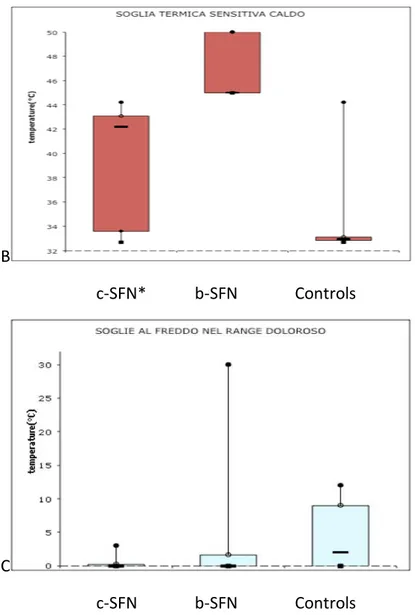
Fig. 2 b. Thermal thresholds (mean±SD) at baseline evaluation in c-SFN, b-SFN and control. *Indicates statistical significance p<0.05. At baseline, A) cold detection thresholds were higher in patients with cSFN, compared with the other groups; B) warm detection thresholds were higer in patients with b-SFN, compared with the others group and C) b-SFN group shown high variability in CPT, with positive phenomena like hyperalgesia or allodynia.
A B
Per visualizzare quest'immagine sono necessari QuickTime™ e un decompressore
.
Fig 3b.A. L-Menthol application at distal leg. B. Scheme of pinprick hyperalgesia and synamic mechanical allodynia detection, along two orthogonal trajectories each 0.5cm from the centre in both directions. See also the drawing of an example of the skin flare distribution after L-Menthol.
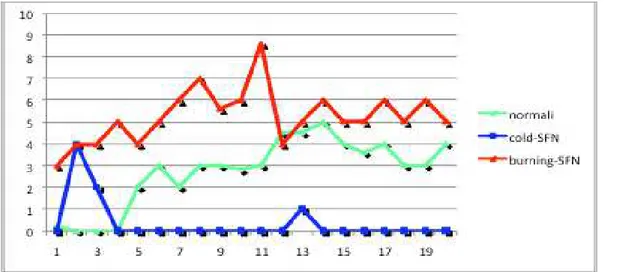
Fig 4b. Time course of sensory rating to topical L menthol stimulation, values are given as mean ratings. In blue cold-SFN group, in red burning-SFN group and in green healthy control group.
Fig. 5b. Thermal thresholds (mean±SD) after L-Menthol or placebo (alcohol 70°).
L-Menthol induced A) increase of the WDT in c-SFN compared with baseline or placebo; B) increase of cold pain thresholds in patients with b-SFN, compared with baseline or placebo.
Fig. 6b. Thermal thresholds (mean±SD) after Cinnamaldheid (CA) or placebo (acetone).
CA induced in b-SFN A) decrease of the CPT compared with baseline or placebo with hyperalgesia or allodynia; B) decrease of the heat pain thresholds HPT thresholds compared with baseline or placebo.
Fig 7b. Skin biopsy taken in a cold-SFN patient and in a burning-SFN patient at the distal leg. Immunofluorecence staining study with anti-myelin-basic protein (MBP; AbD serotec). The density of MBP-positive dermal boundles are decreased in c-SFN compared with b-SFN.
References
Altis, A Schmidtko, C Angioni, K Kuczka Analgesic efficacy of tramadol, pregabalin and ibuprofen in menthol-evoked cold hyperalgesiaK … - Pain, 2009 - Elsevier
Andrade EL , FC Meotti, et al., - TRPA1 antagonists as potential analgesic drugs Pharmacology & Therapeutics, 2011.
Baron R. Peripheral neuropathic pain: from mechanisms to symptoms. Clin J Pain 2000; 16 (2 Suppl): S12±20.
Belmonte, JA Brock Converting cold into pain C … - Experimental brain research, 2009 – Springer Birklein, C Depmeier, R Rolke, C Hansen A family-based investigation of cold pain tolerance - Pain, 2008 Campero Unmyelinated afferents in human skin and their responsiveness to low temperature M … - Neuroscience letters, 2010
Caspani, S Zurborg, D Labuz The contribution of TRPM8 and TRPA1 channels to cold allodynia and neuropathic pain O … - PloS one, 2009
del Camino, S Murphy, M Heiry, TRPA1 contributes to cold hypersensitivity … - The Journal of neurosci., 2010 - Soc Neuroscience
Chen, SK Joshi, S DiDomenico, RJ Perner…Selective blockade of TRPA1 channel attenuates pathological pain without altering noxious cold sensation or body temperature regulation - Pain, 2011
Denda M, Tsutsumi M, Denda S Topical application of TRPM8 agonists accelerates skin permeability barrier recovery and reduces epidermal proliferation induced by barrier insult: role of cold-sensitive TRP receptors in epidermal permeability barrier homoeostasis. Exp Dermatol. 2010 Sep;19(9):791-5. Epub 2010 Jul 16. Dunham JP, Leith JL, Lumb BM, Donaldson LF. Transient receptor potential channel A1 and noxious cold responses in rat cutaneous nociceptors. Dunham JP, Leith JL, Lumb BM, Donaldson LF. Neuroscience. 2010 Feb 17;165(4):1412-9. Epub 2009 Dec 1.
Facer P, Casula MA, Smith GD, Benham CD, Chessell IP, Bountra C, Sinisi M, Birch R, Anand P. Differential expression of the capsaicin receptor TRPV1 and related novel receptors TRPV3, TRPV4 and TRPM8 in normal human tissues and changes in traumatic and diabetic neuropathy. BMC Neurol. 2007 May 23;7:11. Fanger, D del Camino TRPA1 as an Analgesic Target CM Fanger, D del Camino… - Open Drug Discov. J, 2010 -
Foulkes T, Wood JN. Channels (Austin) Mechanisms of cold pain. . 2007 May-Jun;1(3):154-60. Epub 2007 Jul 6. Review.
Gentry, N Stoakley, DA Andersson…The roles of iPLA2, TRPM8 and TRPA1 in chemically induced cold hypersensitivity C - Mol Pain, 2010 - biomedcentral.com
Hondoh, Y Ishida, S Ugawa, T Ueda, Y Shibata… Distinct expression of cold receptors (TRPM8 and TRPA1) in the rat nodose-petrosal ganglion complex - Brain research, 2010 -
Karashima, Y., N. Damann, J. Prenen, K. Talavera, A. Segal, T. Voets, and B. Nilius. 2007. Bimodal action of menthol on the transient receptor potential channel TRPA1. J. Neurosci. 27:9874–9884.
Karashima, K Talavera. TRPA1 acts as a cold sensor in vitro and in vivo - Proceedings of the National Acad Sciences, 2009.
Kwan KY, et al., Burning cold: involvement of TRPA1 in noxious cold sensation … - The Journal of general physiology, 2009 - jgp.rupress.org
Kremeyer, F Lopera, JJ Cox, A Momin, F Rugiero… A gain-of-function mutation in TRPA1 causes familial episodic pain syndrome - Neuron, 2010 - Elsevier
Kono, M Satomi, M Suno, N Kimura… Oxaliplatin-induced neurotoxicity involves TRPM8 in the mechanism of acute hypersensitivity to cold sensation T - Brain and …, 2012 - Wiley Online Library
Knowlton WM , RL Daniels, R Palkar, DD McCoy… - PloS one, 2011 Pharmacological Blockade of TRPM8 Ion Channels Alters Cold and Cold Pain Responses in Mice - dx.plos.org
LaMotte RH, Thalhammer JG. Response properties of high-threshold cutaneous cold receptors in the primate. Brain Res 1982; 244: 279±87.
McCoy, WM Knowlton… Scraping through the ice: uncovering the role of TRPM8 in cold transduction DD - American Journal of Am Physiological Soc, 2011 -
Namer, IP Kleggetveit, H Handwerker, M Schmelz… Role of TRPM8 and TRPA1 for cold allodynia in patients with cold injury - Pain, 2008
Obata K, Katsura H, Mizushima T, Yamanaka H, Kobayashi K, Dai Y, Fukuoka T, Tokunaga A, Tominaga M, Noguchi K. TRPA1 induced in sensory neurons contributes to cold hyperalgesia after inflammation and nerve injury. J Clin Invest. 2005 Sep;115(9):2393-401.
Pozsgai, JV Bodkin… Evidence for the pathophysiological relevance of TRPA1 receptors in the cardiovascular system in vivo G - Cardiovascular …, 2010
Prescott J, Swain-Campbell N. Responses to repeated oral irritation by capsaicin, cinnamaldehyde and ethanol in PROP tasters and non-tasters. Chem Senses 2000; 25:239–246.
Ram, E Eisenberg, M Haddad…Oral opioid use alters DNIC but not cold pain perception in patients with chronic pain-new perspective of opioid-induced hyperalgesia KC - Pain, 2008 - Elsevier
Schmelz M Neuronal sensitivity of the skin. . Eur J Dermatol. 2011 May;21 Suppl 2:43-7. Review.
Staaf, S Oerther, G Lucas, JP Mattsson. Differential regulation of TRP channels in a rat model of neuropathic pain S - Pain, 2009
TorebjoÈrk HE. Afferent C units responding to mechanical, thermal and chemical stimuli in human non-glabrous skin. Acta Physiol Scand 1974; 92: 374±90.
Talavera K, B Nilius Neuronal TRP channels: thermometers, pathfinders and life-savers - Trends in neurosciences, 2008 –
TorebjoÈrk HE, Hallin RG. Perceptual changes accompanying controlled preferential blocking of A and C ®bre responses in intact human skin nerves. Exp Brain Res 1973; 16: 321±32.