F1000Research
Open Peer Review
, Luigi Sacco University
Fabiola Atzeni
Hospital Italy
, University of Toronto
Murray B. Urowitz
Canada, Zahi Touma, University of Toronto Canada
, Azienda
Gian Domenico Sebastiani
Ospedaliera San Camillo-Forlanini Italy Discuss this article
(0) Comments 3 2 1 REVIEW
Advanced and Conventional Magnetic Resonance
Imaging in Neuropsychiatric Lupus [v2; ref status: indexed,
http://f1000r.es/5p5]
Previously titled: Magnetic Resonance Imaging in Neuropsychiatric Lupus
Nicolae Sarbu ,
Núria Bargalló
, Ricard Cervera
3
Section of Neuroradiology, Department of Radiology, Hospital Clinic, Barcelona, Catalonia, 08036, Spain
Magnetic Resonance Imaging Core Facility, Institut d'Investigacions Biomèdiques August Pi i Sunyer (IDIBAPS), Barcelona, Catalonia, 08036, Spain
Department of Autoimmune Diseases, Hospital Clinic, Barcelona, Catalonia, 08036, Spain
Abstract
Neuropsychiatric lupus is a major diagnostic challenge, and a main cause of morbidity and mortality in patients with systemic lupus erythematosus (SLE). Magnetic resonance imaging (MRI) is, by far, the main tool for assessing the brain in this disease. Conventional and advanced MRI techniques are used to help establishing the diagnosis, to rule out alternative diagnoses, and recently, to monitor the evolution of the disease. This review explores the neuroimaging findings in SLE, including the recent advances in new MRI methods.
This article is included in the
Lupus nephritis and
channel.
neuropsychiatric lupus
1
1,2
3
1 2 3 Referee Status: Invited Referees version 2 published 28 Jul 2015 version 1 published 23 Jun 2015 1 2 3 report report report report 23 Jun 2015, :162 (doi: )First published: 4 10.12688/f1000research.6522.1
28 Jul 2015, :162 (doi: )
Latest published: 4 10.12688/f1000research.6522.2
v2
F1000Research
Ricard Cervera ( )
Corresponding author: [email protected] Sarbu N, Bargalló N and Cervera R.
How to cite this article: Advanced and Conventional Magnetic Resonance Imaging in
2015, :162 (doi: )
Neuropsychiatric Lupus [v2; ref status: indexed, http://f1000r.es/5p5]F1000Research 4 10.12688/f1000research.6522.2
© 2015 Sarbu N . This is an open access article distributed under the terms of the , which
Copyright: et al Creative Commons Attribution Licence
permits unrestricted use, distribution, and reproduction in any medium, provided the original work is properly cited.
This work has been founded by the Sociedad Española de Radiológia Médica (06_NBA_INVESTIGACION _SERAM_2013).
Grant information:
The funders had no role in study design, data collection and analysis, decision to publish, or preparation of the manuscript.
Competing interests: No competing interests are disclosed.
23 Jun 2015, :162 (doi: )
First published: 4 10.12688/f1000research.6522.1
16 Jul 2015, :162 (doi: )
Introduction
Despite the fact that the outcome of patients with systemic lupus erythematosus (SLE) has improved considerably over the last decades, neuropshychiatric involvement remains a main cause
of morbi-mortality1,2, being responsible for up to 19% of deaths
in SLE3,4. The real prevalence of neuropsychiatric SLE (NPSLE)
remains unknown, with significant heterogeneity between studies, from 14% to 95% depending on the inclusion criteria; an average of
40–50% is probably widely accepted5–8. Reliable methods for
diag-nosing NPSLE are also unknown, the clinical judgment remaining
the cornerstone for differentiation of these patients9,10. Therefore,
NPSLE represents a major diagnostic challenge, being essentially a diagnosis of presumption and exclusion, established after hav-ing ruled out other possible causes such as trauma, infection, drug effects, epilepsy, migraine, psychiatric disorders, multiple sclerosis, posterior reversible encephalopathy and previous nervous system
disorders5,6,11,12. Very recently, an algorithm based on a probability
score was validated to determine the relationship between
neu-ropsychiatric involvement and SLE13. On the other hand, reaching
the correct diagnosis of NPSLE is critical in terms of therapeutic decisions and outcome.
According to 1999 American College of Rheumatology (ACR) Case Definitions for NPSLE, 19 neuropsychiatric syndromes are defined,
divided into 12 central and 7 peripheral14. The central ones are
fur-ther divided into neurological (aseptic meningitis, cerebrovascular disease, demyelinating syndrome, headache including migraine and benign intracranial hypertension, movement disorders, myelopathy, epilepsy), and psychiatric (acute confusional states, anxiety disor-der, cognitive dysfunction, affective disorder). The peripheral syn-dromes are acute inflammatory demyelinating polyradiculopathy (Guillain-Barre syndrome), autonomic disorder, mononeuropathy
(single/multiplex), myasthenia gravis, cranial neuropathy, plex-opathy, and polyneuropathy. The most common syndromes which require neuroimaging studies are headache, cerebrovascular disease,
epilepsy and cognitive dysfunction8,15, and also represent four out
of five globally most prevalent NPSLE syndromes, as demonstrated
by an extensive, recent meta-analysis7. NPSLE is also divided into
primary, attributed to SLE specific mechanisms, and secondary, consequence of infections, drugs or metabolic errors, although
there are no definitive methods to differentiate between them6,10.
In spite of outstanding advances and increasing efforts into research, the physiopathology of NPSLE remains still unclear. Neural and vas-cular injuries related to antibodies and cytokines were incriminated in active NPSLE. The pathological substrate of NPSLE consists of microangiopathic disease (the most frequent neuropathological finding, typically multifocal, due to intimal hyperplasia, erythro-cytes extravasation and fibrin thrombi), macroscopic infarcts (par-tially explained by secondary coagulopathy due to antiphospholipid antibodies or by embolic phenomena due to Libman–Sacks endo-carditis), accelerated atherosclerosis (partially due to steroid treat-ment, vasculitis and microhemorrhages), direct immune mediated
alterations, demyelination and microembolisms5,16–19.
Magnetic resonance imaging (MRI) is the gold standard for study-ing the brain in SLE. The role of other imagstudy-ing modalities such as computer tomography (CT) is essentially to rule out acute compli-cations such as hemorrhage or large infarcts, or to assess
differ-ential diagnoses5,20,21. The large spectrum of clinical presentations,
laboratory and pathological findings in NPSLE made the neurora-diological findings nonspecific, a wide range of abnormalities being
described8,22. The most frequently reported findings with
conven-tional MRI in large series of NPSLE were multiple small white-matter lesions (30–75%) and cortical atrophy (15–20%), although there is a large percentage of patients (25–60%) with normal MRI
scan8,11,23,24. Advanced MRI techniques such diffusion-tensor,
magnetization-transfer and volumetric studies, which give micro-structural and functional information, could provide evidence of subtle brain changes that allow better understanding of the NPSLE mechanisms. Furthermore, the correlation of the neuroradiologi-cal, clinical and immunological biomarkers could give insights into the pathophysiology of the disease. The present review aims to describe the neuroimaging findings in conventional and advanced MRI imaging in NPSLE patients, and their importance from a practical point of view.
Conventional MRI neuroimaging findings
Around 50% of the NPSLE patients had normal MRI, especially in diffuse syndromes such as headache, mood disorder, and
psychi-atric disease8. In the other half of the patients, the most common
neuroimaging findings can be classified as vascular diseases (small
or large vessel disease), and inflammatory-type lesions (Table 1).
Vascular disease, although nonspecific and in many forms of
manifestation, is the hallmark of NPSLE8. Vascular lesions are
ill-defined hyperintensities on T2, and moderately hypointense or iso-intense on T1. Large vessel disease refers to large infarcts, which have medium-to-large size, are roughly wedge-shaped, occur with a vascular territory distribution, and involve both grey and white matter
Amendments from Version 1
In this revised version, we included the suggestions of the reviewers:
• The title was modified, emphasizing the description of the conventional and new techniques
• The recent contribution by Bortoluzzi et al. (2015), regarding the development of a new algorithm in NPSLE, was now quoted
• The meaning of DWI was explained
• Total corticosteroid was substituted by cumulative corticosteroid
• We mentioned the potential use of nanoparticles in SLE • We mentioned the use of SPECT and PET perfusion apart
of MRI perfusion
• Regarding spectroscopy, we added “The demonstration of the uptake of specific metabolites might in the future be closely associated with neuronal injury in NPSLE” • In the Conclusions paragraph, we added “A complex
diagnostic algorithm including neurophysiologic studies, laboratory tests and MRI is probably the best clinical approach”
See referee reports
(Figure 1a). With diffusion-weighted imaging (DWI) it is possible to determine if they are in the acute, subacute or chronic stage, including silent infarcts. Large vessel infarcts are one of the most debilitating complications of NPSLE, and are found in 10–15% of patients and at
a mean age of 35–40 years8,23,25,26. When infarcts occur in NPSLE, a
tendency to multiplicity was noticed, which is translated into a high
recurrence of ischemic events8. Middle cerebral artery territory is
mainly involved, as in the general population8. Many authors
associ-ated antiphospholipid antibodies with infarcts and reported a stroke
recurrence of around 50% when these antibodies were present23,25,27.
Stroke was also more commonly observed in the presence of
hyper-tension, cerebrovascular syndrome and seizures5.
Small vessel disease is typically represented by lesions smaller than 1 cm, which follow the distribution of the white matter
(periven-tricular, deep, subcortical) (Figure 1b). Recently, the definitions of
neuroimaging findings of small vessel disease have been established and consist in white-matter hyperintensities, recent small
subcorti-cal infarcts, lacunes, microbleeds and brain atrophy28. White-matter
hyperintensities (WMH) are the most widespread type of small ves-sel disease seen in SLE patients, and represents the collective term referring to small T2-hyperintensities including the white matter,
basal ganglia, cerebellum and brainstem28. They are characterized as
hyperintense on T2 and FLAIR sequences, without cavitation,
gen-erally small and ill-defined28,29. The differential diagnosis of WMH is
very wide, being associated with many conditions including ageing,
dyslipidemia, diabetes, hypertension, heart diseases and migraine28.
However, many previous reports already proved increased frequency
of WMH in SLE and NPSLE22,30–37. WMH had been shown to involve
preferentially the frontal and parietal lobes, consistent with an ante-rior to posteante-rior gradient, similar to other causes of WMH, but dif-ferent from inflammatory demyelinating etiologies such as multiple
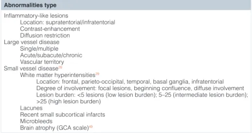
Table 1. Magnetic resonance imaging classification proposed for brain abnormalities in patients with neuropsychiatric lupus. Abnormalities type Inflammatory-like lesions Location: supratentorial/infratentorial Contrast-enhancement Diffusion restriction Large vessel disease Single/multiple Acute/subacute/chronic Vascular territory Small vessel disease28
White matter hyperintensities29
Location: frontal, parieto-occipital, temporal, basal ganglia, infratentorial Degree of involvement: focal lesions, beginning confluence, diffuse involvement Lesion burden: <5 lesions (low lesion burden); 5–25 (intermediate lesion burden); >25 (high lesion burden)
Lacunes
Recent small subcortical infarcts Microbleeds
Brain atrophy (GCA scale)49
Abbreviations: GCA-Global Cortical Atrophy scale.
a
b
Figure 1a–b. Coronal FLAIR images demonstrate both large and small vessel disease. 1a Large hyperintense cortico-subcortical area consistent with a chronic stroke involving the right middle cerebral artery territory. 1b Focal bilateral white matter hyperintensities reflecting small vessel disease. Figure origin: Department of Radiology, Hospital Clinic Barcelona.
sclerosis8,38. In a quantitative cerebral MRI assessment, Appenzeller
et al.35 showed that age, duration of neuropsychiatric manifestations and cumulative corticosteroid dosage were independent predictors for WMH in SLE. In a recent study in patients with newly
diag-nosed SLE, WMH were found in 8% of the patients39.
Neverthe-less, these lesions were observed more frequently in NPSLE when compared with SLE without neuropsychiatric manifestations, with
average ranges from 40 to 60%8,11,20,35,37,39,40. WMH were
associ-ated with cerebrovascular disease, cognitive dysfunction, seizures, antiphospholipid antibodies, low complements (C3, C4, CH50),
age, disease duration, and cumulative corticosteroid dose8,35.
Pre-vious reports demonstrated a significant association between both NPSLE activity (Neuro-SLEDAI) and injury (Neuro-SLICC) scores
with the number of WMH (high lesion burden)11,35,37,39,41.
Further-more, new lesions were noticed during onset of new neuropsychiat-ric manifestations, and resolution of lesions was found with clinical
improvement26,42,43. Quantitative methods are increasingly proposed
for the quantification and follow-up of the WMH in NPSLE, as they can function as an independent predictor for the NPSLE activity and injury, holding promise to open a new line of follow-up of NPSLE patients and their response to therapy, similar to the monitoring of
multiple sclerosis33,35,39.
Recent small subcortical infarcts, commonly known as lacunar inf-arcts, are infarctions in the territory of perforating arterioles, of less than 20 mm in its maximum diameter in the axial plane, with imaging signs or clinical symptoms consistent with a lesion occurring in the
previous few weeks28. Their natural evolution is into lacunes, WMH
without cavitation, or they might disappear44. Old lacunar infarcts
(lacunes) must be differentiated from perivascular (Virchow-Robin) spaces, which generally are smaller, located mostly around the anterior commissure and usually appear linear when imaged paral-lel to the course of the vessel. Lacunes were commonly described in elderly, asymptomatic individuals, in the presence of hypertension, and were related to dementia, gait impairment and increased risk
of stroke28. Very few studies evaluated lacunes in NPSLE and they
were found with a prevalence of 11.5–16%, higher than in the
gen-eral population8,11. Cerebral microbleeds are small (usually 2–5 mm,
but up to 10 mm) round or oval areas of signal void with associ-ated blooming on paramagnetic-sensitive sequences such as T2*-weighted gradient recalled echo (GRE) or susceptibility-T2*-weighted images (SWI). Microscopically, hemosiderin-laden macrophages in perivascular tissue are seen, indicating vascular leakage of blood cells, related to bleeding-prone microangiopathy. In the general pop-ulation, microbleeds are usually located in the cortico-subcortical junction, deep grey and white matter, brainstem and cerebellum. They were associated with hypertension, amyloid angiopathy,
cog-nitive impairment and Alzheimer disease45,46. In NPSLE,
microb-leeds were found in 14.5% of the patients on GRE/SWI sequences, and were correlated with lupus anticoagulant (antiphospholipid
antibodies) and cerebrovascular syndrome8.
Cortical atrophy is seen as generalized enlargement of peripheral cerebrospinal fluid spaces and is best evaluated on volumetric 3D-T1 or FLAIR images. In the general population, age related atrophic
changes are small prior to age 50 years, as proved by a large study47
or, similarly, by another underlying that brain volumes in females
remained stable over a span of 15 to 69 years of age48. There are
different scales to unify the radiological language, one of the most
known being the global cortical atrophy scale (GCA)49. GCA
evalu-ation at the onset of NPSLE observed cortical atrophy in 18.5% of the subjects, most commonly in a mild grade, and at a mean age of
42.5 years8. Brain atrophy occurs more frequently in the presence
of other radiological manifestations consistent with small vessel disease, such as WMH, high lesion burden, lacunes and
microb-leeds8. Brain atrophy was also correlated with lupus anticoagulant,
low complement, longer disease duration, cognitive dysfunction
and cerebrovascular disease23,37. Many authors suggested that the
atrophy might be the result of the prednisone use37,50, while others
found no association31,39,51,52, which suggests that additional
mecha-nisms, probably related to NPSLE itself, seem to be involved53–56.
Less frequently, some NPSLE patients present inflammatory-type lesions which were described as ill defined, hyperintense on T2 and FLAIR, involving the grey and white matter, generally medium or large-sized, some of them with contrast enhancement or diffu-sion restriction, without vascular territory distribution nor clinical and radiological features of infarcts, which usually resolves after aggressive corticosteroid treatment. They were reported in 5–10% of patients, and were correlated with low complement levels, indi-cating an immunological damage related to antibodies and cytokines
and supporting the immunological pathogenesis of NPSLE6. Yet
rarely present, findings related to cerebral vasculitis were described, when angiography exams (MRI or conventional) could show focal
beadings and narrowings of large and small arteries16,57–60.
Myelitis, a type of inflammatory involvement of the central nervous system, is one of the most debilitating complications of NPSLE and occurs in 1–5% of SLE patients. It usually develops early in the evolution of the disease and associates a worse outcome. In 39% of the patients with SLE related myelopathy, it constitutes the pre-senting symptom of SLE, and in another 42% it occurs during the first 5 years after the diagnosis. The most described MRI pattern in SLE myelitis is consistent with transverse myelitis: commonly long affected segment, more than 2–3 vertebral bodies in length and with
injury of both halves of the cord (Figure 2). Transverse myelitis
associates a variable swelling and focal enlargement. Enhancement is usually absent or poor, patchy in the most active presentations. The outcome of SLE myelitis is variable, ranging from complete recovery to severe disability, but the injury is typically much less
extensive on follow up MRI10,61.
Advanced MRI techniques
Up to 40–50% of NPSLE patients had no brain abnormalities on
con-ventional MRI8,11,31,33,36,62. Nonetheless, advanced MRI sequences in
NPSLE demonstrated underlying abnormalities in normal appearing white and grey matter, which shows the limitations of conventional
sequences52,63,64. Recent studies used advanced MRI techniques in
the analysis of NPSLE, as the assessment of tissue-specific atrophy
by morphometric methods38,52,65, diffusion-tensor imaging52,64,66,67,
magnetization transfer imaging52,66,67, magnetic resonance
spectros-copy67,68 and perfusion MRI. The use of nanoparticles for
molecu-lar MRI is another new technique with potential interest in SLE,
Voxel based morphometry (VBM) is a technique which allows the assessment of the focal differences in brain anatomy and, there-fore, the assessment of tissue-specific atrophy. The volume in every voxel is compared across the brain, and VBM is frequently per-formed for examining differences between populations, although it can also be used to assess asymmetries between brain hemispheres. Morphometric studies showed that decreased whole brain volume with increased lateral ventricle volume and both global gray mat-ter and white matmat-ter atrophy are present in SLE patients compared
to healthy controls70. Moreover, it was demonstrated that atrophy
evolved over a short period of time52,68,71. A number of publications
found that selective cortical atrophy was the tissue specific atrophy measure with best correlation with the presence of NPSLE, and sug-gested that cortical atrophy is more important for mediating brain
damage in NPSLE patients than the white matter lesions52,71. From a
practical point of view, the macroscopic damage of the cortical gray matter might be more important for identifying NPSLE patients
than the micro- or macrostructural damage in the white matter52,71,
yet it was reported an association of NPSLE with both cortical and
central atrophy52,65,72. Some authors compared cohorts of NPSLE
with SLE and controls. The NPSLE group exhibited decreased cor-tical thickness in left frontal and parietal lobes as well as in right parietal and occipital lobes compared to controls. Both SLE and NPSLE groups exhibited comparable thinning in frontal and
tem-poral lobes73. Automated morphometric methods were also used for
segmenting white matter lesions in patients with SLE, which could
give a more precise quantification of the focal injuries74.
Diffusion-tensor imaging (DTI) is based on the measurement of water diffusion through cellular compartments, and was demon-strated to provide better resolution than conventional sequences
regarding white matter microstructure (Figure 3a–b)75,76. Compared
to more isotropic movement of water in gray matter, the diffusion
Figure 2. Sagittal T2-weighted image shows an extensive hyperintensity involving the medulla oblongata and the cervico-toracic cord, compatible with a myelitis pattern. Figure origin: Department of Radiology, Hospital Clinic Barcelona.
a
b
Figure 3a–b. Axial maps of fractional anisotropy (FA). 3a Normal FA shows the integrity and directionality of the white matter fibers (red: right-left, green: anterior-posterior, blue: craneo-caudal). 3b Altered (low) FA seen as loss of the normal colors of the left corticospinal tract in the internal capsule and of the left longitudinal fasciculus related to ischemic infarct of the territory of the left middle cerebral artery (arrows) in a patient with neuropsychiatric lupus and stroke. Figure origin: Department of Radiology, Hospital Clinic Barcelona.
in white matter presents higher anisotropy, with preferential dif-fusion along the length of the axon. This anisotropy is due to the well-structured axonal membranes and their myelin sheaths. The diffusion can be quantified by the following parameters: apparent diffusion coefficient (ADC), fractional anisotropy (FA), mean dif-fusivity (MD), radial difdif-fusivity (RD) and axial difdif-fusivity (AD). FA is a measure of myelination and axonal integrity, and MD a measure of molecular motion. High FA and low MD suggest greater myelination and axonal integrity. Previous studies found changes in various DTI indices in SLE and NPSLE patients, in relation to
important microscopic injury of the white matter77. In patients with
SLE, white matter injury in frontal lobes, corpus callosum, and
tha-lamus has been found68,78–80. FA values were reported to be lower
and MD values to be higher in the brain of NPSLE patients than in healthy controls. Increased AD of white matter was also corre-lated with NPSLE when compared to healthy populations. It was suggested that the underlying pathological substrate of white
mat-ter changes in NPSLE may be the selective axonal damage52,66,71. A
localized injury of white matter tracts was also demonstrated in the limbic system, internal capsule, corpus callosum, forceps major and
corona radiata64,67,79,81,82. Very recent publications underline the role
of DTI as an imaging biomarker of NPSLE83.
Magnetization transfer imaging (MTI) is based on the interaction between free water protons and bound protons. The differences in the proton mobility in various macromolecules and tissues are used to generate differences in image signal. Thus, MTI is used to gener-ate contrast, and it has a variety of clinical applications. Volumet-ric MTI was used to quantify cerebral lesions in different diseases,
mainly in multiple sclerosis. Bosma et al.84 compared MTI histogram
parameters in 5 groups of patients: active NPSLE, chronic NPSLE, SLE without NPSLE, multiple sclerosis, and normal control sub-jects. The magnetization transfer ratio histograms in the group of SLE without NPSLE and the group of healthy controls were simi-lar, whereas those in chronic NPSLE and multiple sclerosis groups were flatter. The active NPSLE group showed also a flattening of the histograms, but with a higher magnetization transfer ratio. This suggests that MTI could be able to differentiate active NPSLE. It is also believed that MTI might be a good method for monitoring
treatment trials in NPSLE84. A report combining MTI with
mag-netic resonance spectroscopy (MRS) found correlation between
brain atrophy and MRS markers of axonal and myelin damage67.
Studies combining MTI with DWI, MRS and T2 relaxometry data in NPSLE suggest a common pathogenesis in NPSLE in spite of the
many differences in the neuropsychiatric presentation52,66.
MRS allows the analysis of brain metabolites. Different proton groups have different magnetic fields in relation to their valence electrons. As a result, they resonate at different frequencies of the magnetic field, which can be demonstrated by MRS, as peaks that correspond to different metabolites. N-acetylaspartate (NAA) is one of the main markers assessed on MRS and is found in higher concentrations in neurons, thus it is a marker of neuronal viability. Glutamate, a non-essential amino acid, is the most important excita-tory neurotransmitter, and prolonged neuron excitation by glutamate can be toxic to neurons. NAA and glutamine-glutamate changes were demonstrated in normal-appearing brain in SLE patients, before neurologic and imaging manifestations became apparent, which suggests that these markers might predict the early cerebral
involvement of SLE85. Lower NAA ratios were also reported in both
SLE and NPSLE patients63, and increased myo-inositol, a marker of
gliosis, was suggested as a marker of poor prognosis in NPSLE86.
The demonstration of the uptake of specific metabolites might in the future be closely associated with neuronal injury in NPSLE. Perfusion imaging such as done with single-photon emission com-puted tomography (SPECT), positron emission tomography (PET) and MRI could reveal abnormalities in SLE patients. There are three techniques of perfusion MRI, based on the administration of gadolinium (dynamic susceptibility contrast imaging and dynamic contrast enhanced imaging), or without contrast administration (arterial spin labeled imaging). The main parameters derived from them are mean transit time (MTT), time to peak (TTP), cerebral blood flow (CBF) and cerebral blood volume (CBV). The defined pathological patterns are hypoperfusion (high MTT/TTP, low CBF/
CBV) and hyperperfusion (low TTP/MTT, high CBV/CBF)87. Few
prospective studies analyzed brain perfusion in SLE patients. Some authors showed that perfusion in SLE patients was not different
from healthy controls88, while others reported a pattern of
hypop-erfusion in both SLE and NPSLE63, or even hyperperfusion in the
posterior cingulate gyrus in patients with active disease89.
Overall, advanced MRI techniques seem to be able to detect micro-structural brain damage in a very early stage when not visible on conventional sequences. There could be a temporal dissociation between the detection of damage with these sequences and its trans-lation to significant abnormalities on conventional MRI. Advanced MRI is also expected to help to better understand the underlying pathological substrate of cerebral damage in NPSLE. However, the role of advanced MRI techniques in patients with SLE is yet in its infancy and needs to be further investigated. Future longitudi-nal studies should determine whether early changes of the white and gray matter in NPSLE patients may involve a higher degree of tissue-specific brain atrophy over time and to what extent it would be possible to monitor disease progression and response to therapy.
Looking at all sides of the argument, it is questionable what patients and when should be referred for brain MRI and what is the role of MRI in the clinical management of NPSLE. Syndromes such as cerebrovascular disease, cognitive dysfunction, seizures and mye-lopathy, as well as the focal symptomatology, were often related with radiological abnormalities, and require to be comprehen-sively studied; additional sequences such as DWI, GRE/SWI and contrast-enhanced should be included when MRI is performed in these patients. Conversely, MRI is more likely to be unremarkable in some other syndromes such as headache, psychosis and, gener-ally, diffuse neurological presentations rather than focal ones. Addi-tionally, the status of antiphospholipid antibodies, complement and disease activity plays an important role. Therefore, in the current settings, the decision of when imaging a patient remains probably best reached through a case-based clinical judgment.
The other side of the argument of MRI in NPSLE regards ruling out other causes of neuropsychiatric manifestations rather than diagnosing NPSLE. Despite MRI being the imaging modality of choice and despite significant recent advances in this field, there are neither diagnostic nor specific radiological findings for NPSLE,
meaning that MRI cannot confirm nor exclude the diagnosis of NPSLE. Nevertheless, in the absence of alternative diagnoses when imaging a SLE patient, some patterns may be proposed: stroke in young patients, more than one infarct, association between large and small vessel disease, high lesion burden at young age and pre-mature cortical atrophy. All these in a subject meeting criteria for SLE and without other risk factors, probably could suggest either the presence or a possible development of NPSLE in the following period.
In conclusion, MRI is crucial for both supporting the diagnosis of NPSLE and for ruling out alternative diagnoses. A complex diag-nostic algorithm including neurophysiologic studies, laboratory tests and MRI is probably the best clinical approach. The multi-modal MRI approach including conventional and advanced tech-niques may be an important tool for monitoring the disease activity, progression and treatment response, and may provide fundamental insights into the pathological substrate. To make this possible, a common radiological terminology is a first requirement.
Author contributions
All the authors wrote and edited the manuscript and approved the final version.
Competing interests
No competing interests are disclosed.
Grant information
This work has been founded by the Sociedad Española de Radioló-gia Médica (06_NBA_INVESTIGACION _SERAM_2013).
I confirm that the funders had no role in study design, data collection and analysis, decision to publish, or preparation of the manuscript. Acknowledgements
To Pilar Toledano, MD, and Gerard Espinosa, MD, PhD, Department of Autoimmune Diseases, Hospital Clinic, Barcelona, Catalonia, Spain, for their collaboration in lupus research projects.
References
1. Cervera R, Khamashta MA, Font J, et al.: Morbidity and mortality in systemic lupus erythematosus during a 10-year period: a comparison of early and late manifestations in a cohort of 1,000 patients. Medicine (Baltimore). 2003; 82(5): 299–308.
PubMed Abstract |Publisher Full Text
2. O’Neill S, Cervera R: Systemic lupus erythematosus. Best Pract Res Clin Rheumatol. 2010; 24(6): 841–855.
PubMed Abstract |Publisher Full Text
3. Chiewthanakul P, Sawanyawisuth K, Foocharoen C, et al.: Clinical features and predictive factors in neuropsychiatric lupus. Asian Pac J Allergy Immunol. 2012; 30(1): 55–60.
PubMed Abstract
4. Zirkzee EJ, Huizinga TW, Bollen EL, et al.: Mortality in neuropsychiatric systemic lupus erythematosus (NPSLE). Lupus. 2014; 23(1): 31–38.
PubMed Abstract |Publisher Full Text
5. Joseph FG, Scolding NJ: Neurolupus. Pract Neurol. 2010; 10(1): 4–15.
PubMed Abstract |Publisher Full Text
6. Bertsias GK, Boumpas DT: Pathogenesis, diagnosis and management of neuropsychiatric SLE manifestations. Nat Rev Rheumatol. 2010; 6(6): 358–367.
PubMed Abstract |Publisher Full Text
7. Unterman A, Nolte JE, Boaz M, et al.: Neuropsychiatric syndromes in systemic lupus erythematosus: a meta-analysis. Semin Arthritis Rheum. 2011; 41(1): 1–11.
PubMed Abstract |Publisher Full Text
8. Sarbu N, Alobeidi F, Toledano P, et al.: Brain abnormalities in newly diagnosed neuropsychiatric lupus: systematic MRI approach and correlation with clinical and laboratory data in a large multicenter cohort. Autoimmun Rev. 2015; 14(2): 153–159.
PubMed Abstract |Publisher Full Text
9. Cervera R, Abarca-Costalago M, Abramovicz D, et al.: Systemic lupus erythematosus in Europe at the change of the millennium: lessons from the “Euro-Lupus Project”. Autoimmun Rev. 2006; 5(3): 180–186.
PubMed Abstract |Publisher Full Text
10. Bertsias GK, Ioannidis JP, Aringer M, et al.: EULAR recommendations for the management of systemic lupus erythematosus with neuropsychiatric manifestations: report of a task force of the EULAR standing committee for clinical affairs. Ann Rheum Dis. 2010; 69(12): 2074–2082.
PubMed Abstract |Publisher Full Text
11. Luyendijk J, Steens SC, Ouwendijk WJ, et al.: Neuropsychiatric systemic lupus erythematosus: lessons learned from magnetic resonance imaging. Arthritis Rheum. 2011; 63(3): 722–732.
PubMed Abstract |Publisher Full Text
12. Ramage AE, Fox PT, Brey RL, et al.: Neuroimaging evidence of white matter
inflammation in newly diagnosed systemic lupus erythematosus. Arthritis Rheum. 2011; 63(10): 3048–3057.
PubMed Abstract |Publisher Full Text |Free Full Text
13. Bortoluzzi A, Scire C, Bombardieri S, et al.: Development and validation of a new algorithm for attribution of neuropsychiatric events in systemic lupus erythematosus. Rheumatology (Oxford). 2015; 54(5): 891–898.
PubMed Abstract |Publisher Full Text
14. ACR Ad Hoc Committee on Neuropsychiatric Lupus Nomenclature. The American College of Rheumatology nomenclature and case definitions for neuropsychiatric lupus syndromes. Arthritis Rheum. 1999; 42(4): 599–608.
PubMed Abstract |Publisher Full Text
15. Netto TM, Zimmermann N, Rueda-Lopes F, et al.: Neuropsychiatric lupus: classification criteria in neuroimaging studies. Can J Neurol Sci. 2013; 40(3): 284–291.
PubMed Abstract
16. Ellis SG, Verity MA: Central nervous system involvement in systemic lupus erythematosus: a review of neuropathologic findings in 57 cases, 1955--1977. Semin Arthritis Rheum. 1979; 8(3): 212–221.
PubMed Abstract |Publisher Full Text
17. Sibbitt WL Jr, Brooks WM, Kornfeld M, et al.: Magnetic resonance imaging and brain histopathology In neuropsychiatric systemic lupus erythematosus. Semin Arthritis Rheum. 2010; 40(1): 32–52.
PubMed Abstract |Publisher Full Text |Free Full Text
18. Sarbu MI, Salman-Monte TC, Rubio Muñoz P, et al.: Differences between clinical and laboratory findings in patients with recent diagnosis of SLE according to the positivity of anti-dsDNA by the Crithidia luciliae method. Lupus. 2015;
pii: 0961203315573852.
PubMed Abstract |Publisher Full Text
19. Fanouriakis A, Boumpas DT, Bertsias GK: Pathogenesis and treatment of CNS lupus. Curr Opin Rheumatol. 2013; 25(5): 577–583.
PubMed Abstract |Publisher Full Text
20. Sibbitt WL Jr, Sibbitt RR, Griffey RH, et al.: Magnetic resonance and computed tomographic imaging in the evaluation of acute neuropsychiatric disease in systemic lupus erythematosus. Ann Rheum Dis. 1989; 48(12): 1014–1022.
PubMed Abstract |Publisher Full Text |Free Full Text
21. Zardi EM, Taccone A, Marigliano B, et al.: Neuropsychiatric systemic lupus erythematosus: tools for the diagnosis. Autoimmun Rev. 2014; 13(8): 831–839.
PubMed Abstract |Publisher Full Text
22. Castellino G, Govoni M, Giacuzzo S, et al.: Optimizing clinical monitoring of central nervous system involvement in SLE. Autoimmun Rev. 2008; 7(4): 297–304.
PubMed Abstract |Publisher Full Text
23. Csépány T, Bereczki D, Kollár J, et al.: MRI findings in central nervous system systemic lupus erythematosus are associated with immunoserological
parameters and hypertension. J Neurol. 2003; 250(11): 1348–1354.
PubMed Abstract |Publisher Full Text
24. Toledano P, Sarbu N, Espinosa G, et al.: Neuropsychiatric systemic lupus erythematosus: Magnetic resonance imaging findings and correlation with clinical and immunological features. Autoimmun Rev. 2013; 12(12): 1166–1170.
PubMed Abstract |Publisher Full Text
25. Joseph FG, Lammie GA, Scolding NJ: CNS lupus: a study of 41 patients. Neurology. 2007; 69(7): 644–654.
PubMed Abstract |Publisher Full Text
26. Sibbitt WL Jr, Sibbitt RR, Brooks WM: Neuroimaging in neuropsychiatric systemic lupus erythematosus. Arthritis Rheum. 1999; 42(10): 2026–2038.
PubMed Abstract |Publisher Full Text
27. Mikdashi J, Handwerger B: Predictors of neuropsychiatric damage in systemic lupus erythematosus: data from the Maryland lupus cohort. Rheumatology (Oxford). 2004; 43(12): 1555–1560.
PubMed Abstract |Publisher Full Text
28. Wardlaw JM, Smith EE, Biessels GJ, et al.: Neuroimaging standards for research into small vessel disease and its contribution to ageing and neurodegeneration. Lancet Neurol. 2013; 12(8): 822–838.
PubMed Abstract |Publisher Full Text |Free Full Text
29. Wahlund LO, Barkhof F, Fazekas F, et al.: A new rating scale for age-related white matter changes applicable to MRI and CT. Stroke. 2001; 32(6): 1318–1322.
PubMed Abstract |Publisher Full Text
30. Rovaris M, Inglese M, Viti B, et al.: The contribution of fast-FLAIR MRI for lesion detection in the brain of patients with systemic autoimmune diseases. J Neurol. 2000; 247(1): 29–33.
PubMed Abstract |Publisher Full Text
31. Sanna G, Piga M, Terryberry JW, et al.: Central nervous system involvement in systemic lupus erythematosus: cerebral imaging and serological profile in patients with and without overt neuropsychiatric manifestations. Lupus. 2000; 9(8): 573–583.
PubMed Abstract |Publisher Full Text
32. Jennings JE, Sundgren PC, Attwood J, et al.: Value of MRI of the brain in patients with systemic lupus erythematosus and neurologic disturbance. Neuroradiology. 2004; 46(1): 15–21.
PubMed Abstract |Publisher Full Text
33. Appenzeller S, Pike GB, Clarke AE: Magnetic resonance imaging in the evaluation of central nervous system manifestations in systemic lupus erythematosus. Clin Rev Allergy Immunol. 2008; 34(3): 361–366.
PubMed Abstract |Publisher Full Text
34. Katsumata Y, Harigai M, Kawaguchi Y, et al.: Diagnostic reliability of magnetic resonance imaging for central nervous system syndromes in systemic lupus erythematosus: a prospective cohort study. BMC Musculoskelet Disord. 2010; 11: 13.
PubMed Abstract |Publisher Full Text |Free Full Text
35. Appenzeller S, Vasconcelos Faria A, Li LM, et al.: Quantitative magnetic resonance imaging analyses and clinical significance of hyperintense white matter lesions in systemic lupus erythematosus patients. Ann Neurol. 2008; 64(6): 635–643.
PubMed Abstract |Publisher Full Text
36. Castellino G, Padovan M, Bortoluzzi A, et al.: Single photon emission computed tomography and magnetic resonance imaging evaluation in SLE patients with and without neuropsychiatric involvement. Rheumatology (Oxford). 2008; 47(3): 319–323.
PubMed Abstract |Publisher Full Text
37. Ainiala H, Dastidar P, Loukkola J, et al.: Cerebral MRI abnormalities and their association with neuropsychiatric manifestations in SLE: a population-based study. Scand J Rheumatol. 2005; 34(5): 376–382.
PubMed Abstract |Publisher Full Text
38. Benedict RH, Shucard JL, Zivadinov R, et al.: Neuropsychological impairment in systemic lupus erythematosus: a comparison with multiple sclerosis. Neuropsychol Rev. 2008; 18(2): 149–166.
PubMed Abstract |Publisher Full Text |Free Full Text
39. Petri M, Naqibuddin M, Carson KA, et al.: Brain magnetic resonance imaging in newly diagnosed systemic lupus erythematosus. J Rheumatol. 2008; 35(12): 2348–54.
PubMed Abstract |Publisher Full Text
40. Sibbitt WL Jr, Schmidt PJ, Hart BL, et al.: Fluid Attenuated Inversion Recovery (FLAIR) imaging in neuropsychiatric systemic lupus erythematosus. J Rheumatol. 2003; 30(9): 1983–1989.
PubMed Abstract
41. Podrazilová L, Peterová V, Olejárová M, et al.: Magnetic resonance volumetry of pathological brain foci in patients with systemic lupus erythematosus. Clin Exp Rheumatol. 2008; 26(4): 604–610.
PubMed Abstract
42. West SG, Emlen W, Wener MH, et al.: Neuropsychiatric lupus erythematosus: a 10-year prospective study on the value of diagnostic tests. Am J Med. 1995; 99(2): 153–163.
PubMed Abstract |Publisher Full Text
43. Karassa FB, Ioannidis JP, Boki KA, et al.: Predictors of clinical outcome and radiologic progression in patients with neuropsychiatric manifestations of systemic lupus erythematosus. Am J Med. 2000; 109(8): 628–634.
PubMed Abstract |Publisher Full Text
44. Doubal FN, Dennis MS, Wardlaw JM: Characteristics of patients with minor ischaemic strokes and negative MRI: a cross-sectional study. J Neurol Neurosurg Psychiatry. 2011; 82(5): 540–542.
PubMed Abstract |Publisher Full Text
45. Cordonnier C, Al-Shahi Salman R, Wardlaw J: Spontaneous brain microbleeds: systematic review, subgroup analyses and standards for study design and reporting. Brain. 2007; 130(Pt 8): 1988–2003.
PubMed Abstract |Publisher Full Text
46. Greenberg SM, Vernooij MW, Cordonnier C, et al.: Cerebral microbleeds: a guide to detection and interpretation. Lancet Neurol. 2009; 8(2): 165–174.
PubMed Abstract |Publisher Full Text |Free Full Text
47. DeCarli C, Massaro J, Harvey D, et al.: Measures of brain morphology and infarction in the framingham heart study: establishing what is normal. Neurobiol Aging. 2005; 26(4): 491–510.
PubMed Abstract |Publisher Full Text
48. Carne RP, Vogrin S, Litewka L, et al.: Cerebral cortex: an MRI-based study of volume and variance with age and sex. J Clin Neurosci. 2006; 13(1): 60–72.
PubMed Abstract |Publisher Full Text
49. Scheltens P, Pasquier F, Weerts JG, et al.: Qualitative assessment of cerebral atrophy on MRI: inter- and intra-observer reproducibility in dementia and normal aging. Eur Neurol. 1997; 37(2): 95–99.
PubMed Abstract |Publisher Full Text
50. Zanardi VA, Magna LA, Costallat LT: Cerebral atrophy related to corticotherapy in systemic lupus erythematosus (SLE). Clin Rheumatol. 2001; 20(4): 245–250.
PubMed Abstract |Publisher Full Text
51. Chinn RJ, Wilkinson ID, Hall-Craggs MA, et al.: Magnetic resonance imaging of the brain and cerebral proton spectroscopy in patients with systemic lupus erythematosus. Arthritis Rheum. 1997; 40(1): 36–46.
PubMed Abstract |Publisher Full Text
52. Zivadinov R, Shucard JL, Hussein S, et al.: Multimodal imaging in systemic lupus erythematosus patients with diffuse neuropsychiatric involvement. Lupus. 2013; 22(7): 675–683.
PubMed Abstract |Publisher Full Text
53. Lauvsnes MB, Omdal R: Systemic lupus erythematosus, the brain, and anti-NR2 antibodies. J Neurol. 2012; 259(4): 622–629.
PubMed Abstract |Publisher Full Text
54. Marcinko K, Parsons T, Lerch JP, et al.: Effects of prolonged treatment with memantine in the MRL model of CNS lupus. Clin Exp Neuroimmunol. 2012; 3(3): 116–128.
PubMed Abstract |Publisher Full Text |Free Full Text
55. Muscal E, Traipe E, de Guzman MM, et al.: Cerebral and cerebellar volume loss in children and adolescents with systemic lupus erythematosus: a review of clinically acquired brain magnetic resonance imaging. J Rheumatol. 2010; 37(8): 1768–1775.
PubMed Abstract |Publisher Full Text
56. Sled JG, Spring S, van Eede M, et al.: Time course and nature of brain atrophy in the MRL mouse model of central nervous system lupus. Arthritis Rheum. 2009; 60(6): 1764–1774.
PubMed Abstract |Publisher Full Text
57. Böckle BC, Jara D, Aichhorn K, et al.: Cerebral large vessel vasculitis in systemic lupus erythematosus. Lupus. 2014; 23(13): 1417–1421.
PubMed Abstract |Publisher Full Text
58. Kato R, Sumitomo S, Kawahata K, et al.: Successful treatment of cerebral large vessel vasculitis in systemic lupus erythematosus with intravenous pulse cyclophosphamide. Lupus. 2015; pii: 0961203315570163.
PubMed Abstract |Publisher Full Text
59. Abda EA, Selim ZI, Radwan ME, et al.: Markers of acute neuropsychiatric systemic lupus erythematosus: a multidisciplinary evaluation. Rheumatol Int. 2013; 33(5): 1243–1253.
PubMed Abstract |Publisher Full Text
60. Kizu H, Dobashi H, Kameda T, et al.: Improvement of irregularity of brain vessel walls in systemic lupus erythematosus by tacrolimus. Clin Rheumatol. 2011; 30(5): 715–718.
PubMed Abstract |Publisher Full Text
61. Espinosa G, Mendizábal A, Mínguez S, et al.: Transverse myelitis affecting more than 4 spinal segments associated with systemic lupus erythematosus: clinical, immunological, and radiological characteristics of 22 patients. Semin Arthritis Rheum. 2010; 39(4): 246–256.
PubMed Abstract |Publisher Full Text
62. Steup-Beekman GM, Zirkzee EJ, Cohen D, et al.: Neuropsychiatric
manifestations in patients with systemic lupus erythematosus: epidemiology and radiology pointing to an immune-mediated cause. Ann Rheum Dis. 2013; 72(Suppl 2): ii76–9.
PubMed Abstract |Publisher Full Text
63. Zimny A, Szmyrka-Kaczmarek M, Szewczyk P, et al.: In vivo evaluation of brain
damage in the course of systemic lupus erythematosus using magnetic resonance spectroscopy, perfusion-weighted and diffusion-tensor imaging. Lupus. 2014; 23(1): 10–19.
PubMed Abstract |Publisher Full Text
64. Hughes M, Sundgren PC, Fan X, et al.: Diffusion tensor imaging in patients with acute onset of neuropsychiatric systemic lupus erythematosus: a prospective study of apparent diffusion coefficient, fractional anisotropy values, and eigenvalues in different regions of the brain. Acta Radiol. 2007; 48(2): 213–222.
65. Appenzeller S, Bonilha L, Rio PA, et al.: Longitudinal analysis of gray and white matter loss in patients with systemic lupus erythematosus. Neuroimage. 2007; 34(2): 694–701.
PubMed Abstract |Publisher Full Text
66. Bosma GP, Steens SC, Petropoulos H, et al.: Multisequence magnetic resonance imaging study of neuropsychiatric systemic lupus erythematosus. Arthritis Rheum. 2004; 50(10): 3195–3202.
PubMed Abstract |Publisher Full Text
67. Emmer BJ, Steup-Beekman GM, Steens SC, et al.: Correlation of magnetization transfer ratio histogram parameters with neuropsychiatric systemic lupus erythematosus criteria and proton magnetic resonance spectroscopy: association of magnetization transfer ratio peak height with neuronal and cognitive dysfunction. Arthritis Rheum. 2008; 58(5): 1451–1457.
PubMed Abstract |Publisher Full Text
68. Filley CM, Kozora E, Brown MS, et al.: White matter microstructure and cognition in non-neuropsychiatric systemic lupus erythematosus. Cogn Behav Neurol. 2009; 22(1): 38–44.
PubMed Abstract |Publisher Full Text
69. Serkova N, Renner B, Larsen B, et al.: Renal inflammation: targeted iron oxide nanoparticles for molecular MR imaging in mice. Radiology. 2010; 255(2): 517–526.
PubMed Abstract |Publisher Full Text |Free Full Text
70. Xu J, Cheng Y, Chai P, et al.: White-matter volume reduction and the protective effect of immunosuppressive therapy in systemic lupus erythematosus patients with normal appearance by conventional magnetic resonance imaging. J Rheumatol. 2010; 37(5): 974–986.
PubMed Abstract |Publisher Full Text
71. Zivadinov R, Havrdová E, Bergsland N, et al.: Thalamic atrophy is associated with development of clinically definite multiple sclerosis. Radiology. 2013; 268(3): 831–841.
PubMed Abstract |Publisher Full Text
72. Appenzeller S, Carnevalle AD, Li LM, et al.: Hippocampal atrophy in systemic lupus erythematosus. Ann Rheum Dis. 2006; 65(12): 1585–1589.
PubMed Abstract |Publisher Full Text |Free Full Text
73. Jung RE, Segall JM, Grazioplene RG, et al.: Cortical thickness and subcortical gray matter reductions in neuropsychiatric systemic lupus erythematosus. PLoS One. 2010; 5(3): e9302.
PubMed Abstract |Publisher Full Text |Free Full Text
74. Scully M, Anderson B, Lane T, et al.: An Automated Method for Segmenting White Matter Lesions through Multi-Level Morphometric Feature Classification with Application to Lupus. Front Hum Neurosci. 2010; 4: 27.
PubMed Abstract |Publisher Full Text |Free Full Text
75. Pierpaoli C, Jezzard P, Basser PJ, et al.: Diffusion tensor MR imaging of the human brain. Radiology. 1996; 201(3): 637–648.
PubMed Abstract |Publisher Full Text
76. Le Bihan D: Looking into the functional architecture of the brain with diffusion MRI. Nat Rev Neurosci. 2003; 4(6): 469–480.
PubMed Abstract |Publisher Full Text
77. Schmidt-Wilcke T, Cagnoli P, Wang P, et al.: Diminished white matter integrity in patients with systemic lupus erythematosus. Neuroimage Clin. 2014; 5: 291–297.
PubMed Abstract |Publisher Full Text |Free Full Text
78. Kozora E, Filley CM: Cognitive dysfunction and white matter abnormalities in systemic lupus erythematosus. J Int Neuropsychol Soc. 2011: 1–8.
PubMed Abstract |Publisher Full Text
79. Zhang L, Harrison M, Heier LA, et al.: Diffusion changes in patients with systemic lupus erythematosus. Magn Reson Imaging. 2007; 25(3): 399–405.
PubMed Abstract |Publisher Full Text
80. Lee SP, Wu CS, Hsieh LC, et al.: Efficacy of magnetic resonance diffusion tensor imaging and three-dimensional fiber tractography in the detection of clinical manifestations of central nervous system lupus. Magn Reson Imaging. 2014; 32(5): 598–603.
PubMed Abstract |Publisher Full Text
81. Jung RE, Caprihan A, Chavez RS, et al.: Diffusion tensor imaging in neuropsychiatric systemic lupus erythematosus. BMC Neurol. 2010; 10: 65.
PubMed Abstract |Publisher Full Text |Free Full Text
82. Emmer BJ, Veer IM, Steup-Beekman GM, et al.: Tract-based spatial statistics on diffusion tensor imaging in systemic lupus erythematosus reveals localized involvement of white matter tracts. Arthritis Rheum. 2010; 62(12): 3716–3721.
PubMed Abstract |Publisher Full Text
83. Jones JT, DiFrancesco M, Zaal AI, et al.: Childhood-onset lupus with clinical neurocognitive dysfunction shows lower streamline density and pairwise connectivity on diffusion tensor imaging. Lupus. 2015; pii: 0961203315572718.
PubMed Abstract |Publisher Full Text
84. Bosma GP, Rood MJ, Huizinga TW, et al.: Detection of cerebral involvement in patients with active neuropsychiatric systemic lupus erythematosus by the use of volumetric magnetization transfer imaging. Arthritis Rheum. 2000; 43(11): 2428–2436.
PubMed Abstract |Publisher Full Text
85. Cagnoli P, Harris RE, Frechtling D, et al.: Reduced Insular Glutamine and N-acetylaspartate in systemic lupus erythematosus: a single-voxel (1)H-MR spectroscopy study. Acad Radiol. 2013; 20(10): 1286–1296.
PubMed Abstract |Publisher Full Text
86. Guillen-Del Castillo A, Alonso J, Martínez-Valle F, et al.: Increased myo-inositol in parietal white and gray matter as a biomarker of poor prognosis in neuropsychiatric lupus: a case report. Lupus. 2014; 23(10): 1073–1078.
PubMed Abstract |Publisher Full Text
87. Sarbu N, López-Rueda A, Chirife O, et al.: CT-perfusion time-maps likely disclose the earliest imaging signs of posterior reversible encephalopathy syndrome (PRES). J Neuroradiol. 2014; 41(2): 147–9.
PubMed Abstract |Publisher Full Text
88. Emmer BJ, van Osch MJ, Wu O, et al.: Perfusion MRI in neuro-psychiatric systemic lupus erthemathosus. J Magn Reson Imaging. 2010; 32(2): 283–288.
PubMed Abstract |Publisher Full Text
89. Wang PI, Cagnoli PC, McCune WJ, et al.: Perfusion-weighted MR imaging in cerebral lupus erythematosus. Acad Radiol. 2012; 19(8): 965–970.
F1000Research
Open Peer Review
Current Referee Status:
Version 2
30 July 2015 Referee Report
doi:
10.5256/f1000research.7385.r9693
Gian Domenico Sebastiani
Unità Operativa Complessa Reumatología, Azienda Ospedaliera San Camillo-Forlanini, Rome, Italy
Authors have made the suggested modifications and the paper is now suitable for indexation.
I have read this submission. I believe that I have an appropriate level of expertise to confirm that
it is of an acceptable scientific standard.
No competing interests were disclosed.
Competing Interests:
29 July 2015 Referee Report
doi:
10.5256/f1000research.7385.r9696
Murray B. Urowitz
Lupus Clinic, Centre for Prognosis Studies in the Rheumatic Diseases, Toronto Western Hospital,
University of Toronto, Toronto, ON, Canada
I have read this submission. I believe that I have an appropriate level of expertise to confirm that
it is of an acceptable scientific standard.
No competing interests were disclosed.
Competing Interests:
Version 1
17 July 2015 Referee Report
doi:
10.5256/f1000research.7001.r9164
Gian Domenico Sebastiani
Unità Operativa Complessa Reumatología, Azienda Ospedaliera San Camillo-Forlanini, Rome, Italy
F1000Research
1.
2.
3.
This review is well written and give us a comprehensive updating on magnetic resonance imaging in
NPSLE.
Only few minor suggestions:
Introduction - Authors say that "reliable methods for diagnosing NPSLE are also unknown...". This
is essentially true, but they could quote the recent contribution by
Bortoluzzi
et al
. (2015),
dealing
with the development of a new algorithm for attribution of NP events in SLE.
Conventional MRI, page 2 - "DWI sequences". Please specify the meaning of DWI.
Conventional MRI, page 3 - "total corticosteroid". Cumulative is better.
I have read this submission. I believe that I have an appropriate level of expertise to confirm that
it is of an acceptable scientific standard.
No competing interests were disclosed.
Competing Interests:
16 July 2015 Referee Report
doi:
10.5256/f1000research.7001.r9530
,
Murray B. Urowitz Zahi Touma
Lupus Clinic, Centre for Prognosis Studies in the Rheumatic Diseases, Toronto Western Hospital,
University of Toronto, Toronto, ON, Canada
Sarbu
et al.
review the utility of MRI imaging in neuropsychiatric lupus (NPL) both what is available today
in routine care and for research purposes and what might become useful in clinical care in the future. In
NPL clinicians are interested in three major questions. First, is there evidence of NPL at any time in the
past; secondly, is there evidence of active NPL currently; and thirdly can you demonstrate change over
time with or without treatment in NPL.
In terms of the first question Sarbu
et al.
review the conventional MRI findings in the cortical, subcortical
and spinal cord regions. However the nature of the morphologic changes is non specific and many could
be compatible with hypertension, amyloid, Alzheimers etc. and thus may not be helpful to the clinician.
Even the advanced techniques (not universally available) measuring brain volume and composition are
not specific for SLE.
In terms of the second question Sarbu
et al.
review Magnetic Resonance Spectroscopy (MRS) which
allows the analysis of brain metabolites. The demonstration of the uptake of specific metabolites might in
the future be associated with neuron excitation but no studies are available in NPL.
Perfusion imaging such as also done with SPECT and PET scanning can reveal areas of hyper or
hypoperfusion but thus far no studies demonstrating active NPL which could change over time exists.
In conclusion, this article is a good review of what currently exists and what may be developed over time
with MRI imaging. The authors conclude that further clinical association studies with conventional and
advanced techniques are required before MRI imaging will become the standard to understand the
mechanism, diagnosis and monitoring of NPL.
F1000Research
We have read this submission. We believe that we have an appropriate level of expertise to
confirm that it is of an acceptable scientific standard.
No competing interests were disclosed.
Competing Interests:
07 July 2015 Referee Report