INDEX
SUMMARY
4INTRODUCTION
7Frontotemporal Lobar Degeneration 11
TAR DNA binding protein-43 (TDP-43) 13
Frontotemporal Dementia 15
Molecular Genetics of Frontotemporal Lobar Degeneration 18
MAPT Gene 19
VCP Gene 20
CHMP2B Gene 21
PGRN Gene 22
Additional genes and genetic risk factors of Frontotemporal Lobar Degeneration 23
FTLD-Tauopathies 25
Microtubule Associated Protein Tau 26
Gene organization 27
Protein structure and function 30
Tau projection domain 31
Tau microtubule assembly domain 34
Post-translational modifications of tau 35
Molecular classification of Tauopathies 40
MAPT gene mutations 40
Mutation altering the tau-MT interaction 41
Mutation altering the tau mRNA splicing 42
Mutation altering Tau-MT interaction and tau RNA splicing 42 Regulation of exon 10 alternative splicing: cis-acting factors 43
Alternative splicing regulation at the Exon 10 5′ splice site: Stem loop theory 45 Alternative splicing regulation at the 5′ splice site–Linear sequence theory 47 Regulation of exon 10 alternative splicing:Trans-acting factors
Phosphorylation of SR proteins 48
Disruption of tau exon 10 splicing in tauopathies 49
AIM OF THE STUDY
51MATERIALS AND METHODS
52 Clinical description 52Genetic screening and Pedigrees 52 Neuropathology 55
Minigene constructs 56
Cells preparation and Transformation of E.coli Top10 58
Site-directed mutagenesis 58
Analyses on Thermodynamic Stability of the RNA Stem Loop Structure 59
Bioinformatic Analysis 59
Ultraviolet Melting 59 Cell cultures and transfection 60
RNA extraction and RT-PCR analysis on MAPT minigenes 61 RNA extraction and RT-PCR analyses on brain tissue 61 Statistical analysis 62
Tau Analysis 63
Immunohistochemistry 63
Biochemistry 63
Sarkosyl-insoluble tau extraction 63
Soluble tau extraction 64
Tau dephosphorilation 64
Immunoblotting 64
RESULTS
66Minigene Design 66 The two novel mutations affect E10 alternative splicing 68 Effect of the patient’s compound heterozygous condition
Minigene expression of the 5’splice site E10+3 mutation 70 Brain Tissue Study 72 Detection of increased levels of Tau3R transcripts 72 Tau Analysis 73 Immunohistochemical detection of increased levels of Tau 3R inclusions 73 Biochemical characterization of pathological tau 75 Sarkosyl-insoluble tau: detection of increased levels of Tau 3R isoforms 75 Soluble tau: detection of increased levels of fetal Tau 3R isoform 77 Thermodynamic stability of the stem loop regulatory structure increased
by the E10+4 mutation 79
DISCUSSION
82SUMMARY
Frontotemporal lobar degeneration (FTLD) is a heterogeneous syndrome encompassing different nosological entities characterized by behavioural and personality change, accompanied by deterioration of executive function, language and movement. Clinically FTLD results in at least three distinct syndromes: Frontotemporal dementia (FTD), Semantic dementia (SD) and Primary progressive aphasia (PPA), while the pathological classification is based on histopathological presence or absence of neuronal inclusions of tau and/or ubiquitin proteins accumulating in the neuronal/glial inclusions, being forms of FTLD differentiated in tau-positive, ubiquitin-positive and tau-negative.
The most common clinical manifestation of FTLD is FTD, characterized by atrophy of the frontal and temporal lobes, with neuronal loss, gliosis and spongiosis of the superficial layers. FTD is mostly a presenile disorder showing changes in personality, impaired social conduct, emotional blunting, loss of insight, disinhibition, perseverative behaviour and hyperorality; cognitive deterioration, especially in language and in executive functions, appear later. Despite most cases of FTD are sporadic, approximately 10%-50% of FTD patients have a positive family history for dementia. Familial FTDL was associated to mutations in four genes: Microtubules associated protein tau (MAPT) and Progranulin (PGRN) genes that are responsible for the most genetic forms of FTD; instead, Valosin containing protein (VCP) gene is involved in rare forms of FTD with inclusion body myopathy and Paget’s disease of the bone and Charged multivescicolar body protein 2B (CHMP2B) is mutated in some families with a combination of FTD and Amyotrophic lateral sclerosis (ALS).
Mutations in MAPT gene are responsible for 10%-20% of familial FTD. Alternative splicing of exons 2, 3 and 10 in MAPT pre m-RNA results in the expression of six isoforms. Exclusion or inclusion of Exon 10 gives rise to tau isoforms with three (tau3R, E10-) o four (tau4R, E10+) microtubule-binding repeats. In normal adult human brain the overall ratio of 3R to 4R tau is generally 1, whereas in fetal brain only the shortest tau isoform with 3R is expressed, indicating that tau expression is developmentally regulated.
To date, 44 different potential pathogenic MAPT mutations have been reported, divided into two groups depending on the primary molecular mechanism involved: missense or deletion mutations that commonly modify tau interaction with microtubules and splicing mutations that affect the alternative splicing of exon 10, leading to changes of the ratio of 3R-tau/4R-tau. However, a third group of mutations exists that might have effects at protein and RNA levels.
In the present study we report the molecular effect of two novel heterozygous MAPT gene mutations, a T to C transition at position -15 of intron 9 [T(-15)C] and an A to C transversion at position +4 of intron 10 (E10+4), identified in a patient with sporadic FTD, clinically and neuropathologically ascertained.
Considering that both mutations are located in the splicing regulatory regions surrounding Exon 10, we analyzed their molecular effect on the alternative splicing of MAPT pre-mRNA in a minigene model system and in brain tissue.
Semi-quantitative RT-PCR analyses, in minigene costructs and in brain tissue, have shown that the two novel mutations cause a novel Exon 10 splicing effect giving rise to a higher increase of mRNAs transcripts lacking Exon 10 (E10- or Tau3R) when compared with FTD-Ub+ control.
Immunohistochemical and biochemical analyses on brain tissue evidenced neuronal and oligodendroglial tau deposits mostly made of Tau3R isoforms and an increased increased availability of shorter Tau3R isoform respectively.
Data obtained with minigenes derived by the phenotipically healthy patient’s parents demonstrate that when the mutations are inherited in a non compound heterozygous condition the ratio of E10 including/E10 excluding transcripts is quite normal.
Although the molecular mechanism underlying exon 10 splicing regulation remain to be completely elucidated, the exon 10 splice donor site is predicted to give rise to a RNA stem loop structure considered crucial for the quantitative regulation of exon 10 alternative splicing. Most of previously characterized mutations identified in the upper part of the stem loop strongly alter mRNA splicing by destabilizing the secondary structure, with a corresponding increase of E10 inclusion and 4RTau expression. Considering that the E10+4 mutation is located into the exon 10 splice donor site, we
also investigated the effect of the E10+4 mutation on the thermodynamic stability of the RNA stem loop structure. Our data, based on bioinformatic prediction of the stem loop sequence thermostability and Ultraviolet Melting experiments demonstrated a strong increasing of stability in the stem-loop structure carrying the E10+4 mutation. This higher stability could be important for the skipping of exon 10, even though the E10+4 mutation alone is not able to give rise to a pathologic phenotype.
We cannot exclude that the T to C transition, localized in a regulatory region upstream of exon 10, could also alter the binding of specific trans-splicing factors increasing the effect of the E10+4 mutation, giving rise when both mutation are present in the compound heterozygous condition (namely the FTD patient) to the E10 exclusion and the altered 4R/3R tau ratio observed.
Thus, we can hypothesize a trans-acting regulatory effect of both mutations with known, or unknown splicing factors, which might have contributed to the very atypical clinical and pathological FTD phenotype of the patient.
INTRODUCTION
Neurodegenerative diseases represent a large group of sporadic and familial progressive neurological disorders affecting 5-10% of the population over the age of 65 and their prevalence rises steeply to 20-30% of the population over 80 years. Although they arise for unknown reasons and progress in a relentless manner particularly, but not exclusively, in the elderly the most consistent risk factor for developing a neurodegenerative disorder is increasing age.
Future demographic projections are predicting the number of people aged 60 years or greater to reach nearly 1.2 billion by 2025 (http://www.who.int/ageing/en/). Thus, it can be anticipated that, over the next generations, the proportion of elderly people will double, and, with this, possibly the proportion of persons suffering from some kind of neurodegenerative disorder (Serge Przedborski et al, 2003; Tanner C.M et al, 1992). It is estimated that more than 20 million people currently have dementia worldwide and that this number will double by 2020 (Ferri et al., 2005).
The number of neurodegenerative diseases is currently estimated to be a few hundred, and, among these, many appear to overlap with one another clinically and pathologically, rendering their practical classification quite challenging. Furthermore, the same neurodegenerative process, especially at the beginning, can affect different areas of the brain, making a given disease appear very different from a symptomatic standpoint. The most common classification of neurodegenerative disorders is still based on the predominant clinical feature or on the topography of the predominant lesion, or often on a combination of both. Accordingly, neurodegenerative disorders of the Central Nervous System may be first grouped, for example, into diseases of the cerebral cortex, the basal ganglia, the brainstem and cerebellum, or the spinal cord. Then, within each group, a given disease may be further classified based on its main clinical features (Bertram L. et al, 2005).
Although traditionally divided into sporadic and familial forms, there are almost certainly important influences in sporadic neurodegenerative disorders mediated through environmental and/or genetic and/or epigenetic element that contribute to the
development of neurodegeneration. Potential interaction between these elements may influence disease phenotype, onset and severity.
Among neurodegenerative disorders, dementia are most common diseases and are characterised by progressive loss of specific populations of central nervous system neurons in specific functional anatomic systems (Fig 1).
Figure 1 Anatomical location of the Macroscopic and Microscopic changes characteristic of the
neurodegenerative diseases
Neuronal degeneration results in severe cognitive decline usually presenting with disturbances in mood, behaviour and personality. The onset of the disease depends on how fast the neurodegenerative process evolves, can range from a few months to several years, although the neurodegenerative process may suddenly accelerate, especially under the influence of intercurrent deleterious factors such as infections.
Over the past two decades, significant advances in neurohistological techniques, such as immunohistochemistry, have replaced or supplemented many of the classical histological approaches and have shown that abnormal protein aggregation in the affected brain areas represents a unifying biochemical and histomorphological hallmark of a wide variety of neurodegenerative diseases.
The protein aggregates form microscopically-visible cellular deposits called “inclusion bodies” localized, in the majority of cases, in the cytoplasmic compartment (C.A. Ross et al, 2004; John Woulfe, 2008) and appear to be associated with the neurodegenerative process. The biochemical study of these neuropathological lesions led to the identification of their molecular components and provided an insight into our understanding of these neurodegenerative disorders. Furthermore, great progress was also achieved from studies of familial forms that have identified specific gene mutations which cause the inherited forms of the different disorders. In most cases, the mutated genes were found to encode altered proteins that compose the neuropathological lesions. Thus, the classification of neurodegenerative diseases is increasingly becoming based on the identity of the protein which accumulates (Tab.1).
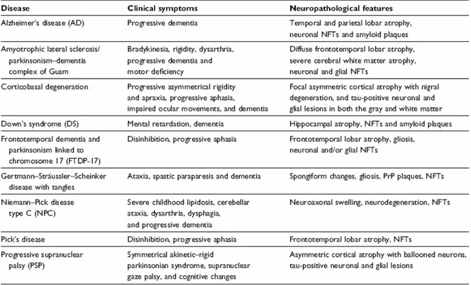
Table 1 Protein-based classification of neurodegenerative diseases Tauopathies
Alzheimer's disease
Amyotrophic lateral sclerosis/Parkinson-dementia complex Argyrophilic grain disease
Corticobasal degeneration Dementia pugilistica
Diffuse neurofibrillary tangles with calcification Down's syndrome
Frontotemporal dementia/parkinsonism linked to chromosome 17 Gerstmann–Straussler–Scheinker disease
Guadeloupian parkinsonism Myotonic dystrophy
Niemann-Pick disease type C Pick's disease
Postencephalitic parkinsonism Progressive subcortical gliosis Progressive supranuclear palsy Subacute sclerosing panencephalitis Tangle only dementia
Synucleinopathies Parkinson's disease
Dementia with Lewy bodies Multiple system atrophy
Neurodegeneration with brain iron accumulation Pure autonomic failure
Meige's syndrome Prion Proteinipathies
Creutzfeld–Jakob disease (familial, sporadic, and transmitted) Fatal insomnia (familial and sporadic)
Gerstmann–Straussler–Scheinker disease Kuru
Polyglutaminopathies Huntington's disease
Dentatorubropallidoluysian atrophy
Spinal and bulbar muscular atrophy (Kennedy's disease) Spinocerebellar ataxia 1, 2, 3, 6, 7, and 17
TDP-43 proteinopathies
Frontotemporal lobar degeneration with ubiquitin-only inclusions Amyotrophic lateral sclerosis
RNA-mediated diseases
Fragile X-associated tremor ataxia syndrome Myotonic dystrophy
Spinocerebellar ataxias 8, 10, 12 Huntington's disease-like type 2 Others
Neuronal intermediate filament inclusion body disease Neuronal intranuclear inclusion disease
Neuroferritinopathy TDP-43-negative FTLD-U
Frontotemporal Lobar Degeneration
Frontotemporal lobar degeneration (FTLD) is the third most common cause of neurodegenerative dementia, after Alzheimer disease (AD) and Dementia with Lewy bodies (DLB) (Cairns N.J. et al, 2007).
FTLD is a heterogeneous syndrome encompassing different nosological entities characterized by behavioural and personality change accompanied by deterioration of executive function, language and movement (Brun et al., 1998; Neary et al., 1998). Clinically it results in at least three distinct syndromes: Frontotemporal dementia (FTD), Semantic dementia (SD) and Primary progressive aphasia (PPA). Over the past decade, many case reports, as well as large clinicopathological studies, have recognized an overlap among these three syndromes and some extrapyramidal syndromes like cortical basal degeneration (CBD), progressive supranuclear palsy (PSP) and motor neuron disease (MND) (Josephs KA et al, 2006; Hodges JR et al 2004; Kertesz A et al 2005;
Forman M.S. et al, 2006). With the advent of immunohistochemistry it is now well
established that more than 15 different pathologies can underlie FTLD and related disorders (McKhann GM et al, 2001; Cairns N.J. et al 2007). The nomenclature of FTLD has evolved as knowledge has advanced. Much of what is FTLD descends from the findings of Arnold Pick, in the early 1890s, who described a patient presenting with childish behaviour, memory deficits and aggressiveness. At autopsy, the patient presented lobar atrophy (Pick A, 1892) ‘ballooned’ neurons, argirophilic and eosin-positive neuronal inclusions. Over time, the disease was referred to as Pick’s disease (PiD) and the intraneuronal inclusions became known as Pick bodies. In the recent years a more refined molecular characterization has been done. In 1994, The Lund-Manchester group published the first clinical and neuropathological criteria for FTD and adopted the clinical term ‘Frontotemporal Dementia’ (FTD) (Lund and Manchester Groups, 1994), successively updated in 1998 (Neary D. et al, 1998).
FTLD is pathologically heterogeneous and can be classified in two major subtypes as Tau-Positive (FTLD-τ) and Tau-Negative groups, on the basis of immunohistochemical and biochemical characterization of proteins accumulating in the neuronal and/or glial inclusions.
Tau positive group known as tauopathies, approximately one-third of FTLD cases, show inclusions made of insoluble aggregates of the microtubule-associated protein tau in the cytoplasm of neurons and glial cells (Fig 2).
Figure 2 Tau positive inclusions
Several cases of Pick’s disease have been included in this group showing that the round argyrophilic inclusions (Pick bodies) were Tau-positive (Morris H.R. et al, 2001; Spillantini M.G. et al, 1997).
Tau negative group, known as non-tauopathies, represent about 50-66% of all FTLD cases and with the advent of ubiquitin immunohistochemistry, were observed to have ubiquitin positive neuronal cytoplasmic inclusions (Fig 3) and renamed “Frontotemporal Lobar Degeneration with Ubiquitin-only–immunoreactive” changes (FTLD-U) (Kertesz A et al, 2000; K.A. Josephs et al, 2004; Shi J, Shaw CL et al, 2005).
One-third, or more, of these FTLD-U cases also have rare to occasional neuronal intranuclear inclusions (FTLD-U NII) (Mackenzie IR et al, 2006).
Dementia Lacking Distinctive Histopathology (DLDH), in which none of these kinds of inclusion are present, was defined as a third pathologic FTLD subtype.
TAR DNA binding protein-43 (TDP-43)
Recent discoveries of TARDBP gene, localized on chromosome 1q, have transformed our understanding of the FTLD-U and DLDH disorders.
TARDBP encodes TAR DNA binding protein-43 (TDP-43), a ubiquitously expressed and highly conserved nuclear protein involved in the regulation of transcription and alternative splicing (Buratti E. et al, 2001; Buratti E. et al, 2005; Ayala Y. et al 2005). Its pathology is characterised by increased phosphorylation, nucleus-to-cytoplasm translocation and co-localisation with ubiquitin in neurons and glial cells (Neumann M. et al, 2006).
TAR DNA binding protein-43 (TDP-43), on the basis of data coming from immunohistochemistry and biochemistry, is the major pathologic protein of the inclusions in majority of sporadic and familial FTLD-U cases (Cairns NJ et al, 2007; Kwong K et al, 2007;Neumann M et al, 2007) (Fig 4), even though there are FTLD-U cases without TDP-43 inclusions (Roeber S. et al, 2008; Josephs K.A. et al, 2008; Mackenzie I.R. et al, 2008).
Furthermore, a proportion of cases associated with DLDH pathology have shown TDP-43 inclusion (Fig 4). These case, reclassified as FTLD-U, are now recognized as the major pathology underlying FTLD (Samir Kumar-Singh et al 2007; Joseph K.A. et al, 2004).
Recent studies have demonstrated that abnormal TDP-43 immunoreactivity also occurs in FTLD-τ as Alzheimer’s Disease, Pick’s Disease (PiD) (Fig 4), Pure Hippocampal Sclerosis (HS), Lewy Body Disease (LBD) and in the Amyotrophic Lateral Sclerosis -Parkinsonism Dementia Complex (ALS-PDC) (Amador-Ortiz C. et al, 2007;
Figure 4 Pathological features of FTLD- τ, FTLD-U and DLDH. Tau (A–C), ubiquitin (D–F)
and TDP-43 (G,H,I) immunostaining is shown on serial sections from superior frontal cortex of Pick’s disease (left panel), FTLD-U with a PGRN IVS0 + 5G > C mutation (middle panel), and DLDH (right panel). Tau-reactive cytoplasmic inclusions (arrows) are present in Pick’s disease (A), but absent in FTLD-U (B) and DLDH (C). Ubiquitin-reactive inclusions are present in Pick’s disease (D) and FTLD-U (E), but not in DLDH (F). TDP-43 commonly stains neuronal nuclei (arrowheads in G,H,I) and also both cytoplasmic and intranuclear inclusions in FTLD-U (arrow and double-headed arrows in H).
It remains to be seen whether mutations in the TDP-43 gene can also cause FTLD-U; however, the recent discovery of pathogenic missense mutations in a highly conserved glycine-rich domain of the TARDBP gene, in autosomal dominant ALS and SALS families and sporadic cases, confirms the importance of TDP-43 in the pathogenesis and demonstrates that defects in TARDBP are sufficient to cause TDP-43 proteinopathy (Fig 5) (Gitcho MA et al, 2008; Sreedharan J et al, 2008; Kabashi E et al, 2008; Van Deerlin VM et al, 2008).
Figure 5 Mutations in the TAR DNA-binding protein 43 (TDP-43). Schematic diagram of functional
domains and mutations in the coding region are indicated using the amino acid numbering of the 414 amino acid protein. RRM1, RNA recognition motif 1; RRM2, RNA recognition motif 2; NL, nuclear localization signal; NE, nuclear export signal; hnRNP, heterogeneous nuclear ribonucleoprotein interaction domain. TARDBP mutations: autosomal-dominant FALS (black), SALS (green).
Frontotemporal Dementia
Frontotemporal dementia (FTD) is the most frequent clinical phenotype under the spectrum of Frontotemporal Lobar Degenerative Diseases (FTLD), accounting for 5% to 10% of all dementia patients and 10% to 20% of patients under 65 years of age. Mean age of onset is approximately 52 to 58 years and the mean duration of the disease is 5-15 years. Nevertheless, recent instances reported onset of FTD symptoms before 30 and later than 80 years of age (S.Spina et al, 2006; A.C. Bruni et al, 2007). The incidence of FTD is almost even between the sexes. (Graff-Radford NR and Woodruff BK, 2007).
Up to 50% of FTD have a “familial component” and a part is inherited in an autosomal dominant pattern of inheritance (Rosso SM et al, 2003; Chow TW et al, 1999; Poorkaj P et al, 2001 ). To date, it is unknown whether these familial cases are due to shared genetic factors and/or environmental factors, or a combination of both genes and environment.
It is also described a high prevalence of FTD sporadic cases; it is thought that there are certainly important genetic influences mediated through a number of different genes (polygenic influences) each of which can promote the development of FTD phenotype.
In Table 2 are listed the FTD clinical symptoms (Neary et al, 1998). The feature of the disease that allows recognition is the profound alteration in personality and social conduct, which herald the onset of the disease. In its early course the disease is often mistaken for a psychiatric disorder, because abnormalities in memory are not a feature of the illness until later.
Table 2. The Clinical Diagnostic Features of FTD
Depending on the distribution of the pathology within the frontal and temporal lobes, there are three major FTD behavioural subtypes: the disinhibited type, the apathetic type and the dysexecutive type. Anatomically, the disinhibited type is connected with neurodegeneration of the orbitofrontal and anterior temporal neocortex; the apathetic
type is associated with atrophy in the medial frontal-anterior cingulate region and the dysexecutive type is correlated with degeneration of the dorsolateral prefrontal.
The disinhibited type is characterized by personality change, impulsivity, irritability, lability and socially inappropriate behaviour. The apathetic type lacks initiative and is emotionally indifferent, while the dysexecutive type has problems with planning, self-direction and ability to shift and maintain behavioural sets (Mega MS et al, 1994). Admixtures between these types are common due to variations in the regional localisation of atrophy. Furthermore, patients often have word finding difficulties where speech is nonspontaneous, economical and often stereotyped. Progressive reduction in speech usually leads to mutism after several years of disease. Orientation in time and space, visuospatial skills and memory initially remain intact, but deteriorate with progression of the disease (Neary D et al, 2000; Lindau M et al, 2001). Due to the degeneration of extrapyramidal neurons, parkinsonian signs may also be present, sometimes already from the onset of the disease. Parkinsonism involves bradykinesia, postural instability and rigidity without resting tremor.
The combination of clinical observations, neurological examination, neuropsychological tests, information gained from laboratory investigations and morphological and functional brain imaging (e.g. CT, MRI, SPECT) are necessary to achieve accuracy of clinical diagnosis generally made according to international criteria that allow to differentiate among different types of dementia and is based on clinico-pathological features, namely, peculiar clinical profiles in combination with characteristic brain lesions (Graeber et al., 1997; Forstl, 1999; Duckett and Stern, 1999; McKhann et al., 2001).
Finally, the diagnosis of FTD should be based on neuropathological examination, that only definitively differentiates from other types of dementia.
Neuropathological features of FTD include neuronal degeneration in the frontal and temporal cortices, sometimes with subcortical changes, basal ganglia atrophy and depigmentation of the substantia nigra. (Josephs KA et al, 2006; Hodges JR et al 2004; Kertesz A et al 2005; Forman MS et al, 2006).
However the pattern of atrophy can vary involving both hemispheres in equal or differing way and sometimes also involving the parietal cortex.
Neurodegeneration usually encompasses both sides of the frontal and anterior temporal lobes symmetrically, but can also appear asymmetrically in the hemispheric or predominantly temporal lobe.
At microscopic level neuronal loss, gliosis, spongiosis and cellular inclusions are present. For instance, affected neurons frequently display intracellular inclusions, named neurofibrillary tangles (NFTs), primarily composed of hyperphosphorylated tau protein (Spillantini et al., 2000).
However, based on immunohistochemical staining and on the pattern of intracellular inclusions, FTD can be further classified into three major subtypes (Neary D et al, 1996). The most common type (Frontal Lobar Degeneration type) includes microvacuolation of the superficial neurons and spongiform changes. The second type (Pick type) is mainly characterized by widespread and abundant gliosis, in addition to swollen neurons and inclusion bodies in regions such as dentate gyrus. In the third form (MND type), patients suffer from MND in combination with pathology noticed in other subtypes. Classification into one of these subtypes is sometimes difficult when typical differentiating features are not always present.
Molecular Genetics of Frontotemporal Lobar Degeneration
The great clinical and neuropathological heterogeneity of FTLD suggest the existence of several genetic factors underlying or modifying the pathogenesis of this prevalent and untreatable disorder. Genetic studies of FTLD autosomal dominant families led to the discovery of four major genes. Micotubule Associate Protein Tau (MAPT) and Progranulin (PGRN also known as GRN) genes that when mutated cause FTLD-Tau Positive and FTLD-Tau negative pathology respectively [26,27], Valosin-Containing Protein (VCP) [28] and Charged Multivesicular Body Protein 2B (CHMP2B) genes associated to the major cases of FTLD-U [29] (Tab. 3) (O. Bugiani, 2007; Jill S et al, 2007).
Table 3 Frontotemporal Lobar Degeneration (FTLD) Causative Genes
MAPT Gene
The unraveling of FTLD genetics began in 1994 with the identification of a locus linked to chromosome 17q21.2 in 13 families with a variety of clinical and pathologic phenotypes. At a consensus conference in 1996, the clinical, pathologic, and genetic findings of these families were compared and the term “frontotemporal dementia with parkinsonism linked to chromosome 17” (FTDP-17) was introduced to best describe the predominant symptoms in these families (Foster NL et al, 1997). In 1998, as the microtubule-associated protein tau (MAPT) gene had been localized in the FTDP-17 families to the 17q21 candidate region, it becames the obvious positional and functional candidate gene (Fig 6) (Poorkaj P et al, 1998).
Figure 6 Linkage analysis for fiontotemporal dementia and parkinsonism linked to chromosome 17
(FTDP-17) families.
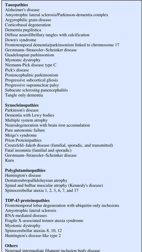
Gene name MAPT PGRN VCP CHMP2B
Gene location 17q21.1 17q21.32 9p13.3 3p11.2
Phenotype FTDL FTDL IBMPFD FTDL
Subsequently several mutations within the MAPT gene were identified (Hutton M et al, 1998; Spillantini MG et al, 1998).
This gene encodes the microtubule-associated protein tau whose transcript undergoes complex, regulated alternative splicing, giving rise to several mRNA transcripts that are differentially expressed in the nervous system, depending on stage of neuronal maturation and neuron type. The tau protein promotes microtubule assembly and stability, and might be involved in the establishment and maintenance of neuronal polarity (For further details about gene and protein see below: paragraph…)
To date, 44 different potential pathogenic MAPT mutations have been reported which result in abnormal tau aggregation and are thought to cause disease through a toxic gain
of dysfunction (AD and FTD Mutation Database
http://www.molgen.ua.ac.be/FTDMutations).
MAPT mutation frequencies in FTLD populations have been shown to vary from 0 to 50%, depending on the family history of dementia and the use of clinical or pathologic inclusion criteria (Fabre et al., 2001; Binetti et al., 2003; Rosso et al., 2003; Stanford et al., 2004).
VCP Gene
In 2004, mutations in VCP on chromosome 9p13 were associated in FTD with inclusion body myopathy and Paget disease (IBMPFD), a rare multisystem disorder with autosomal dominant inheritance (Watts, G.D. et al. 2004). The locus for IBMPFD was mapped to chromosome 9p21-p12 in 2001 by genome-wide linkage analyses in a large Illinois family (Kovach MJ et al, 2001).
VCP is a member of the AAA-ATPase gene super family (ATPase associated with diverse cellular activities) involved in multiple cellular functions including acting as a molecular chaperone in endoplasmic reticulum–associated protein degradation (ERAD), stress response, programmed cell death, and interactions with the ubiquitin-proteasome system. Most of the VCP mutations are situated within the domain involved in the ubiquitin binding. They gives rise to significant phenotypic variation and are likely to cause a loss or alteration of the normal function of VCP, leading to impaired
ubiquitin-proteasome system activity and the accumulation of ubiquitinated proteins within cells (Watts GD et al, 2004; Forman MS et al, 2006). To date, 12 missense mutations in
VCP gene have been reported. (AD and FTD Mutation Database
http://www.molgen.ua.ac.be/FTDMutations) (Guyant-Marechal L et al, 2006;
Haubenberger D et al, 2005; Schroder R et al, 2005).
One year later VCP gene identification, a large Danish family originating from the Jutland with “non specific dementia” was linked to the CHMP2B gene on chromosome 3p13 and a complex mutation was subsequently identified in this gene (Brown J et al, 1995; Skibinski G et al, 2005).
CHMP2B Gene
Human CHMP2B codes for a protein of 213 amino acids with a predicted coil-coil domain. It is a component of the endosomal secretory complex required in transport III (ESCRITIII) which is involved in the degradation of surface receptor proteins and formation of endocytic multivesicular bodies (MVB) (Babst M, 2005). Dysfunction of the ESCRT complexes results in the inability of MVBs to internalize membrane bound cargoes, leading to dysmorphic endosomes and poor protein turnover.
The CHMP2B mutation was located in the acceptor splice site of exon 6 and was shown to produce two aberrant transcripts that could give rise to a gain- or loss-of-function disease mechanism.
Overexpression of the two Danish CHMP2B mutant proteins in cell culture disrupted the CHMP2B localization and indeed resulted in the formation of dysmorphic organelles of the late endosomal pathway (Skibinski G et al, 2005). These data suggest that endosomal trafficking dysfunction might be an important factor leading to the neurodegeneration in this family.
To date 4 mutation in CHMP2B gene were reported and no other families have been
found with a segregating CHMP2B mutation (AD and FTD Mutation Database
http://www.molgen.ua.ac.be/FTDMutations) (Cannon A et al, 2006; Momeni P et al 2006).
PGRN Gene
Recently, was reported the linkage of FTDL-U families with chromosome 17 and systematic mutation analyses of positional candidate genes led, in July 2006, two groups of researchers to the identification of mutations in the Progranulin (PGRN) gene. (Cruts
M et al, 2006; Baker M et al, 2006). PGRN encodes a precursor glycoprotein of 68 kD,
composed of 7.5 tandem repeats of a highly conserved motif with 12 cysteines. Proteolytic cleavage by an elastase-like activity results in the generation of a family of 6-kD peptides called granulins. PGRN is generally thought to be a secreted mitogenic factor that is expressed in a variety of tissues (Bateman A et al 1998). In non brain tissues, progranulin is thought to function as a growth factor that regulates cell cycle progression and cell motility in multiple processes including development, inflammation, and wound repair (He Z. and Bateman A., 2003; He Z et al, 2003; Daniel R. et al, 2003). However, the function of progranulin in the brain, observed in a subset of pyramidal neurons and in activated microglial cells, has received relatively little attention prior to it being linked with FTD (Daniel R et al, 2000). The fact that
pathologic mutations in PGRN lead to a depletion of the functional protein suggests that progranulin is likely critical for neuronal survival (Baker M et al, 2006).
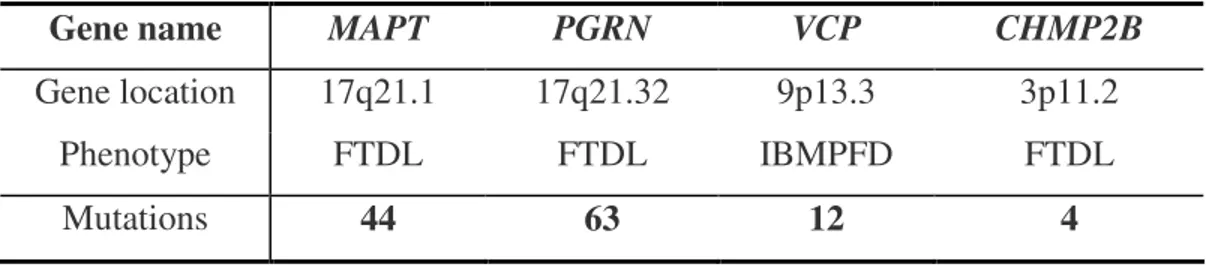
To date, 63 different pathogenetic PGRN mutations have been reported (AD and FTD Mutation Database http://www.molgen.ua.ac.be/FTDMutations) (Fig 7) (Rademakers
R et al, 2007).
Figure 7 Null mutations in the progranulin gene (PGRN) and mRNA encoding the PGRN protein,
showing all mutations identified to date. Letters in boxes refer to individual granulin peptides. Mutations are numbered relative to the largest PGRN transcript
The majority of mutations are nonsense, frameshift, and splice-site mutations that create null alleles; additionally, several missense mutations have been reported that are predicted to result in a nonfunctional protein (haploinsufficiency) although the pathogenicity of these mutations remains to be confirmed (Cruts, M. et al. 2006; Le Ber I et al, 2007).
Progranulin mutation frequencies in FTLD populations have ranged from 5 to 10%, although a frequency of 13 to 25% has been reported in the subset of FTLD population in which a positive family history is identified. PGRN mutations have also been found in individuals without any family history.
A comparison of PGRN mutations carriers showed a mean diseases age of onset is approximately 59 years with a range of 48 to 83 years and the mean age of death is 65 years with a range of 53 to 87 years. Penetrance is estimated to be 90% at age 70 (Gass J et al, 2006; Le Ber I et al, 2007). No phenotype/genotype correlation has been found but it is supposed that different genetic variants or environmental factors can influence the disease phenotype contributing to the massive range in onset age.
Additional genes and genetic risk factors of Frontotemporal Lobar Degeneration
Together, all FTLD genes, associated with significant interfamilial and intrafamilial phenotypic variation, explain the disease in less than half of the familial patients and in a minority of apparently sporadic patients, suggesting that additional genes and genetic risk factors are involved. It has now been linked to chromosome 9 an additional FTLD locus. Although candidate gene sequencing at this locus revealed the presence of a disease segregating nonsense mutation in the Intraflagellar Transport 74 (IFT74) gene in one independent family, the pathogenicity of this mutation is controversialand no causal
IFT74 mutations were identified in other FTD (Morita M et al, 2006; Vance C et al,
2006; Valdmanis PN et al, 2007). IFT74 is a 600 amino acid coiled-coil domain-containing protein that localizes to the intracellular vesicle compartment and is a component of the intraflagellar transport system responsible for vesicular transport of material synthesized within the cell body into and along dendrites and axon (Momeni P et al, 2006). Presenilin 1 (PS1) gene is another candidate gene. PS1 mutations were been
associated either with familial FTDL-τ either with FTLD-U. (Raux et al., 2000; Tang-Wai et al., 2002; Dermaut et al., 2004 Zekanowski et al., 2006;Livia Bernardi et al, 2008). Furthermore emerging evidence indicates that the tau locus itself could be a susceptibility factor playing a key role in sporadic FTLD-Tau positive. Tau gene sequencing (Andreadis A et al, 1992) has revealed several polymorphisms which are in complete linkage disequilibrium, mostly inherited as two distinct haplotypes, H1 and H2, with relatively few recombination events (Baker M et al, 1999).
Haplotype-based association studies have shown a significant association between the H1/H1 genotype and Progressive Supranuclear Palsy (PSP) [Baker M et al, 1999; Conrad C et al, 1997 Di Maria E et al, 2000]. A similar association has also been found in corticobasal degeneration (CBD) [Di Maria E et a, 2000; Houlden H et al, 1999], while the results are conflicting in both FTD [Hughes A et al, 2003; Sobrido MJ et al, 2003; Verpillat P et al, 2002; Zekanowski C et al, 2003] and Pick’s disease (PiD) (Morris HR et al, 2002). It was shown that H1 haplotype expresses significantly more 4Rtau MAPT transcripts than H2. These data suggest a mechanism for the increased susceptibility of H1 carriers to neurodegeneration. Additionally, carriage of the H2 haplotype was reported to be associated witha lower age at onset of FTD and functionally show a more severe decline of glucose utilization in frontal brain areas although no increased risk of the disease was attributed to this haplotype (Borroni et al., 2005; Simon M. et al, 2007).
In addition, few years ago Conrad and colleagues characterized a previously undescribed gene (Conrad et al., 2002), named Saitohin which is located in the intron between
exons 9 and 10 of the human MAPT gene, in a critical region for the alternative splicing of exon 10 (Hutton et al., 1998). A single nucleotide polymorphism (Q7R; CAA to
CGA), correlated with a higher incidence of Alzheimer disease patients Combarros et al., 2003; Conrad et al., 2002, was discovered in this gene; STH Q7R polymorphism is
in complete linkage disequilibrium with the MAPT haplotypes: STH Q allele is linked to H1 MAPT haplotype and the STH R allele is linked to H2 MAPT haplotype (de Silva et al., 2003; Peplonska et al., 2003; Verpillat et al., 2002b; Zekanowski et al., 2003).
The possible role of Apolipoprotein E (APOE), established as the major genetic risk factor in AD, as well as in other neurodegenerative diseases, in FTD is still debated (Verpillat P et al, 2002). There are reports of positive associations of ε2/ε 4 APOE alleles with H1 haplotype as a risk factor of developing FTD [Ingelson M et al, 2001; Verpillat P et al, 2002] but other authors have not found negative results as underlined by the results of L.Bernardi et al. in which H1 haplotype enhances the effect of the ε2 allele decreasing FTD risk (Livia Bernardi et al, 2006). This opposite effects probably accord to the population specific genetic background. In any case the genetic mechanism underlying the association of specific gene or genetic risk factor in familial and sporadic FTDL remain still unresolved.
FTLD-Tauopathies
FTLD-τ cover a significant number of FTLD and related disorders characterized by the accumulation of filamentous deposits, consisting of abnormally insoluble hyperphosphorilated tau protein, also referred as Neurofibrillary Tangles (NFTs), in neurons and glia (Fig 8) (Spillantini, M.G. and Goedert, M.,1998; Spillantini, M.G. et al;1998). This group of diseases became known as “Tauopathies”, term first used to describe a family with frontotemporal dementia and abundant tau deposits (Spillantini, M.G. et al. 1997).
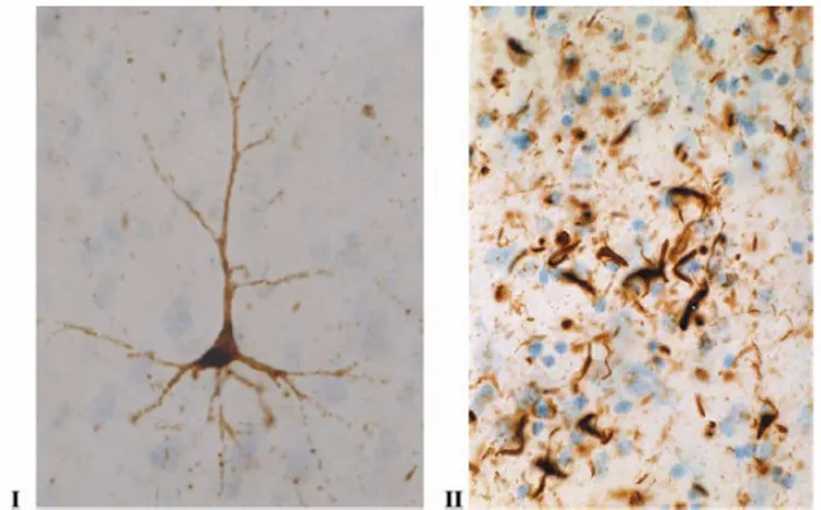
Figure 8 Examples of microscopic neuropathology in tauopathies. Photos are illustrating
immuno-histochemical staining of brain tissue, using the AT-8 antibody against hyperphosphorylated tau. (I) A pyramidal cell in pretangle state from frontal cortex from a patient with frontotemporal dementia (FTD), and (II) the characteristic oligodendroglial inclusions (coiled bodies).
Tauopathies are now referred to a group of familial and sporadic disorders with, as shown in table 4, widespread clinical symptoms and tau pathology in which tau accumulation is believed to be directly associated with neuronal death and disease development.
Tabella 4 Overview of selected tauopathies, presenting clinical and neuropathological characteristics
Microtubule Associated Protein Tau
Tau is a member of the microtubule-associated proteins (MAPs) family found in many animal species such as Caenorhabditis elegans, Drosophila, goldfish, bullfrog, rodents, bovines, goat, monkeys and human. Human cDNA for tau protein was isolated and cloned in 1989 by Goedert et al. (Goedert M, Spillantini MG, Potier MC et al, 1989; Goedert M, Spillantini MG, Jakes R et al, 1989; Goedert M, Spillantini MG, Cairns NJ et al, 1992). MAPs are abundant in the central nervous system where are normally expressed in axons of neurons. The main function of these proteins is to promote microtubule assembly, to reduce microtubule instability and to play a role in maintaining neuronal integrity and axonal transport. Furthermore, in some species the
microtubule-associated proteins can be found at low levels in astrocytes and oligodendrocytes, and can be expressed in glial cells, mainly in pathological conditions. It is possible to detect tau mRNA and proteins in several peripheral tissues such as heart, kidney, lung, muscle, pancreas, testis, as well as in fibroblasts.
Gene organization and Splicing
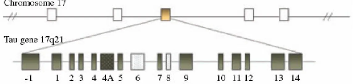
The human MAPT gene, coding for the tau protein is located over 150 kb on the long arm of chromosome 17 at position 17q21.3. It is a single copy gene composed by 16 exons (Fig 9).
Figure 9 Schematic representation of MAPT gene location and genomic structure
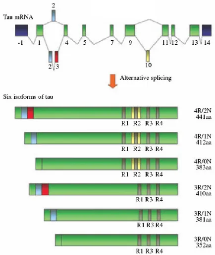
Exons 1, 4, 5, 7, 9, 11, 12 and 13 are constitutive exons whereas exons 2, 3 and 10 are alternatively spliced and adult brain specific (A. Andreadis et al, 1992). Alternative splicing of these three exons gives rise to six mRNAs transcripts [M. Goedert et al, 1989 Neron.; M. Goedert et al, 1989 EMBO; A. Himmler et al, 1989; K.S. Kosik et a, 1989]. In particular, alternative splicing of exon 10 gives rise to tau isoforms with either three (exon 10-) o four (exon 10+) microtubule-binding repeats known as 3Rtau and 4Rtau respectively. In addition, alternative splicing of exons 2 and 3 results in 3Rtau and 4Rtau isoform with zero (0N), one (1N) or two (2N) N-terminal inserts. In the adult human brain the ratio of 3R to 4R tau is generally 1 (Fig 10).
Figure 10 Schematic representation of the primary transcript and the six human tau isoforms
expressed in human brain
Exons 4A, never present in the mRNA of human brain, is specific to peripheral tau proteins. Tau mRNA with either exons 6 or 8 have not been described in human. Some transcripts with exon 8 are found in bovine and rhesus monkey brains. Exon -1 is part of the promoter, and is transcribed but not translated. Exon 14 is found in messenger RNA, but it is not translated into protein. [A. Andreadis et al, 1992; M. Goedert et al, 1989 Neron.; M. Goedert et al, 1989 EMBO; A. Sawa et al, 1994].
Restriction and sequencing analyses show that the gene contains two CpG islands, one associated with the promoter region, the other with exon 9 (A. Andreadis et al, 1992; A. Andreadis et al, 1995). Two regions homologous to the mouse Alu-like sequence are present. The sequence of the promoter region also reveals a TATA-less sequence that is
likely to be related to the presence of multiple initiation sites, typical of housekeeping genes. Three SP1-binding sites, important in directing transcription initiation in other TATA-less promoters, are also found in the proximity of the first transcription initiation site (A. Andreadis et al 1996; E. Sadot et al 1996).
In MAPT intron 9 is present a region, that behaves like a gene giving rise to the expression of a novel protein known as saithoin. Saithoin has not yet been isolated and characterized; however, by analysis of its sequence it does appear to contain some similarities to certain nucleic acid binding proteins. Saithoin contains the regions SSYEESSR and SLAWEV similar to those present in some transcription factors, and thus it may play a role in transcription or nucleic acid metabolism. It is still not clear whether saithoin is a protein that is only expressed in human cells and not in other organisms such as the mouse (Conrad C et al, 2002).
Sequence analysis of the coding region and flanking intronic sequences in the MAPT gene has identified over 200 non coding polymorphisms in exons 1, 2, 3, 9, 11 and 13 in both controls and FTDP-17 patients (MGS pag234). Analysis of the occurrence of these polymorphisms revealed that they are in complete disequilibrium (LD) with each other and constitute two extended haplotypes, H1 and H2, which cover a region of ~1.8 Mb. It is the longest region of LD identified to date in the human genome, (43) including in the centre the MAPT gene and other genes, as corticotrophin-releasing hormone receptor 1 (CRHR1), N-ethylmaleimide sensitive factor (NSF), intramembrane protease 5 (IMP5)
(44) (Fig 11).
The H1haplotype is the predominant haplotype in all ethnic groups, whereas the H2 haplotype seems to be primarily of European origin(Evans W et al, 2004). Functional differences in transcriptional activity havebeen attributed to the different haplotypes, with the H1 haplotypereported to have more activity than the H2 haplotype (Kwok JB et al, 2004).
H1 and H2 differ in nucleotide sequence and intron size, but are identical at the amino acid level. Few years ago, it has been found that a ~900 kb segment from H2 chromosomes is inverted with respect to the H1 haplotype (Fig 11).
Figure 11 Inverted MAPT H1 and H2 Haplotypes
This inversion is thought to have occurred between 3 and 3.6 million years ago (Stefansson et al., 2005) and is almost entirely absent outside of European populations (Evans et al., 2004).
Further investigations into the genomic architecture of this region have identified a number of low-copy repeat (LCR) sequences prone to Chromosomal rearrangements. There is evidence to suggest that the non-allelic homologous recombination that generated the inversion at H2 occurred between two low-copy repeat sequences, LCR A and LCR B, located 250 kb centromeric and 180 kb telomeric to MAPT respectively (Cruts et al., 2005).
Protein structure and function
In the brain, tau proteins constitute a family of six isoforms which range from 352 to 441 amino acids. Their molecular weight is ranging from 45 to 65 kDa. Tau proteins differ from each other by the presence of either three (3R) or four repeat-regions (4R) in the carboxy-terminal (C-terminal) part of the molecule and the absence or presence of one or two inserts (29 or 58 amino acids) in the amino-terminal part (N-terminal) (M.
Goedert, M.G. Spillantini, R. Jakes et al, 1989; M. Goedert, M.G. Spillantini, M.C. Potier et al, 1989; A. Himmler et al, 1989). In adult human brain the protein levels of 3R and 4R tau isoforms are similar, whereas the fetal brain only contains the shortest 3R tau isoform, characterized by the absence of N-terminal inserts and the presence of three C-terminal repeats (3R) (K.S. Kosik et al, 1989; M. Goedert and Jakes et al, 1990) . It has been demonstrated most recently that there is a transient expression of 0N3R tau isoform during adult neurogenesis (Bullmann et al, 2007). Moreover, tau isoforms may be differentially distributed in neuronal subpopulations. For istance, tau mRNAs containing exon 10 are not found in granular cells of the dentate gyrus (Goedert, M.G. Spillantini, R. Jakes et al, 1989). Differently from humans, only 4R tau isoforms are expressed in adult rodent brain.
A number of functions of tau have been characterised. Its main physiological function is the promotion of assembly and stabilization of the microtubular network. As a result of this tau protein is essential for establishing neuronal polarity and axonal outgrowth during development and for maintaining axonal morphology and axonal transport in mature neurons. Tau performs these function through the its two large domains: the “projection domain” containing the amino-terminal two-thirds of the molecule and the “microtubule binding domain” containing the carboxy-terminal one-third of the molecule.
Tau projection domain
The projection domain can be divided into two regions: the amino-terminal region, encoded by exons 2 and 3, with a high proportion of acidic residues followed by a basic proline-rich region.
The N-terminal part is referred to as the projection domain because it projects from the microtubule (MT) surface giving rise to a crucial functions such as mediating interactions between tau and the neural plasma membrane and determining the spacing between MTs in axon (R. Brandt et al, 1995; N. Hirokawa et al, 1988; J. Chen et al 1992). It should be noted that projection domain may increase axonal diameter, for istance in peripheral neurons which often project a very long axon with large diameter, an additional N-terminal tau sequence encoded by exon 4A is present, generating a
specific tau isoform called ‘big tau’ with an approximate size of 100 kDa (Couchie D et al, 1992; Goedert M et al, 1992; I.S. Georgieff et al, 1993).
Tau proteins bind to spectrin and actin filaments (M.F. Carlier et al, 1984; I. Correas et al, 1990; J.P. Henriquez et al 1995) and through these interactions may allow microtubules to interconnect with other cytoskeletal components such as neurofilaments (Y. Miyata et al, 1986) and may restrict the flexibility of the microtubules (A. Matus et al, 1990). There is also evidence that tau proteins interact with cytoplasmic organelles. Such interactions may allow for binding between microtubules and mitochondria (D. Jung et L, 1993).
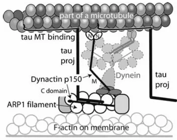
Recent findings have shown an extensive colocalizzation of tau and dynactin complex suggesting an interection between these proteins and a direct involvement of tau in axonal transport (Fig.12) (E. Magnani et al 2007).
Figure 12 Colocalization of tau and dynactin p150
Dynactin complex, essential for cytoplasmic dynein-dependent organelle transport, consists of a short filament of an actin-like protein, ARP1, and around 10 other polypeptides, including a dimer of the heavy chain, known as p150 .
Axonal MTs are organized with their fast-growing (plus) ends pointing towards the axonal tip. MTs are assembled in the cell body and then transported along the axon. Tau is also found initially in the cell body, but after the development of one of the neurites into an axon, becomes concentrated in the distal axon. Tau’s localization to the distal axon requires intact MTs and microfilaments and there is a model in which MT-bound tau is linked to the plasma membrane by a component that requires actin filaments for its
subcellular localization. Recently results suggest that the dynactin complex could be that component and support the idea that tau-stabilized MTs could be transported in the anterograde direction along the axon by dynein/dynactin motors linked to the axonal membrane.
It has been proposed a molecular model that shows a direct interaction betweentau’s N-terminal projection domain and the C-N-terminal region of p150 (Fig 13) (E. Magnani et al 2007).
Figure 13 Proposed model of the role of tau in stabilizing the interaction between axonal MTs and
the dynactin/dynein complex
In this model tau appears to facilitate and stabilize the binding of the dynactin complex with MT tracks by binding to the C-terminus of p150. Although other components of the dynactin complex bind to p150 in this extensive C-terminal region, a recent structural study has shown the presence of additional binding sites.
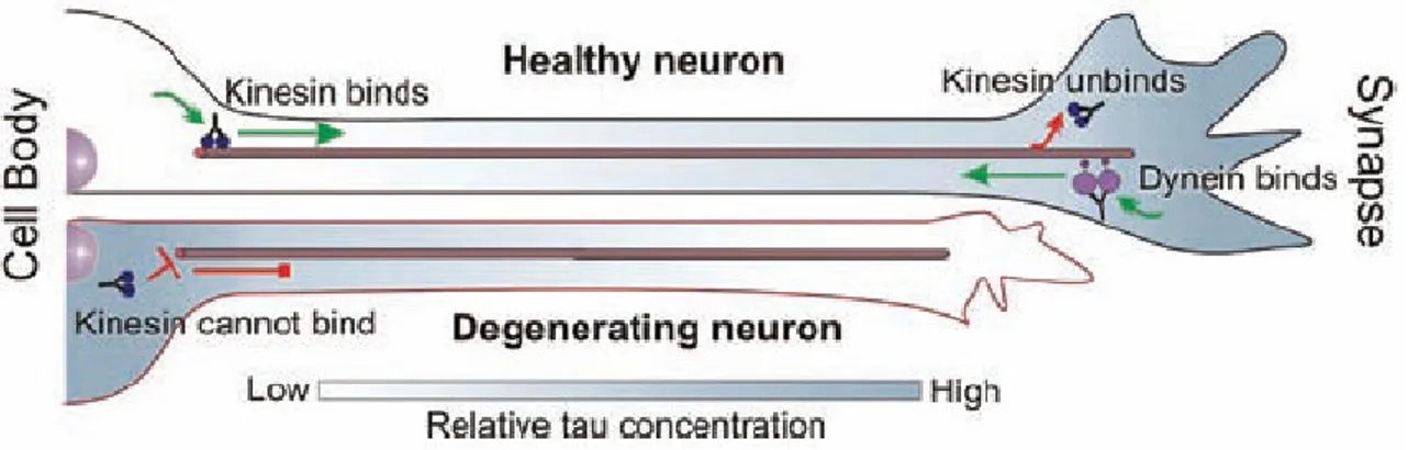
Recently, it was proposed another model in which tau protein may control the balance of microtubule-dependent axonal transport in the neuron, by locally modulating cytoplasmatic molecular motor proteins, dynein and kynesin, which transport cargo toward the minus-end (toward the cell center) and plus-end of microtubules (toward the cell periphery), respectively (Ram Dixit et al 2008). In the model is shown that in a healthy neuron, tau can be distributed in a proximal-to-distal gradient (M. Kempf et al,
1996; M. M. Black et al, 1996). Lower tau concentration at the cell body would allow kinesin to efficiently bind to microtubules and initiate anterograde transport of cargo, whereas higher tau concentration at the synapse would facilitate cargo release (Fig.14).
Figure 14 Model of the role of tau in the regulation of axonal transport
At the same time, dynein-driven retrograde transport from the distal axon would not be impeded due to dynein’s lower sensitivity to tau (Fig.14) (C. Ballatore et al, 2007; Ram Dixit et al, 2008).
• Tau microtubule assembly domain
Tau proteins bind microtubules through repetitive regions in their C-terminal part. These repetitive regions are the repeat domains (R1–R4) encoded by exons 9–12 (G. Lee et al, 1989). The three (3R) or four copies (4R) are made of a highly conserved 18-amino acid repeats separated from each other by less conserved 13- or 14-amino acid inter-repeat domains (M. Goedert, M.G. Spillantini, M.C. Potier et al, 1989; A. Himmler et al, 1989). Tau proteins, known to act as promoter of tubulin polymerization in vitro, are involved in axonal transport (R. Brandt et al, Neurochem 1993; R. Brandt et al, J. Biol. Chem 1993).
It has been demonstrated that adult tau isoforms with 4R (R1–R4) are more efficient to promote microtubule assembly than the fetal isoform with 3R (R1,R3,R4) (M. Goedert, R. Jakes, 1990; K.A. Butner et al, 1991; N. Gustke et al, 1994). Interestingly, the most potent part to induce microtubule polymerization is the inter-region between repeats 1 and 2 (R1–R2 inter-region) and more specifically peptide KVQIINKK within this sequence. This R1–R2 inter-region is unique to 4R tau, adult-specific and
responsible for a 40-fold difference in the binding affinities between 3R and 4R tau (B.L. Goode et al, 1994).
The heat schok proteins HSP70 and HSP90 can also bind to tau, and as a consequence of this interaction, the association of tau protein with microtubules increases, decreasing its self-association and hence the formation of tangles.
The nuclear tau isoforms are similar to cytoplasmic tau, but they show lower solubility, suggesting that they undergo specific modifications, either post-translational (phosphorylation) or through interactions less with other proteins. Prior to their addressing in the nucleus, tau proteins are phosphorylated in the cytoplasm. The function of nuclear tau and how it may be regulated by phosphorylation is still unknown (J.A. Greenwood et al, 1995).
Post-translational modifications of tau
Several modifications have been described for tau protein including phosphorylation, glycosylation, ubiquitinylation, deamination, oxidation, nitration, cross-linking, or glycation. The most studied of these has been phosphorylation. Tau plays a key role in regulating microtubule dynamics, axonal transport and neurite outgrowth, and all these functions are modulated by phosphorylation of numerous serine and threonine sites that are clustered in regions flanking the MT binding repeats.
The different states of tau phosphorylation result from the dynamic activity of specific kinases and phosphatases towards these sites.
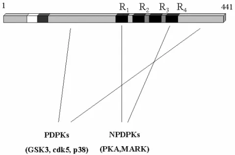
Several tau kinases have been described and they are grouped into two different types: proline (PDPK) and non-proline (NPDPK) directed protein kinases (Fig 15) (G. Drewes et al 1992; M. Goedert et al 1997; C.H. Reynolds et al 1997; R. Vulliet et al 1992; T.J. Singh et al 1996).
PDPK modified Ser-Pro or Thr-Pro Pro tau motifs. Three main PDPK have been describe to phosphorylate tau, GSK3, also known as tau kinase I, cdk5 (or tau kinase II) and stress kinases like JNK and p38. Recently it was shown that GSK-3 phosphorylation of tau modulates its axonal transport by regulating binding to kinesin-1 (Jian-Zhi Wang et al 2008; Cuchillo-Ibanez I et al, 2008).
Figure 15 Scheme of tau molecule, showing the localization of some of the sites that can be
phosphorylated by proline directed protein kinases (PDPKs) and non-proline dependent protein kinases (NPDPKs).
NPDPK modify Ser or Thr residues where not followed by prolines. Among them are: cyclic-AMP dependent kinase; Ca2+/calmodulin dependent kinase (CaMPKII), protein kinase C (PKC) or microtubule affinity regulating kinase (MARK).
There are seventy nine putative Ser or Thr phosphorylation sites on the longest brain tau isoform (441 amino-acids). Using phosphorylation-dependent monoclonal antibodies against tau, mass spectrometry and sequencing, at least thirty phosphorylation sites have been described. All of these sites are localized outside the microtubule-binding domains with the exception of Ser 262 (R1), Ser285 (R1–R2 repeat), Ser305 (R2–R3 inter-repeat), Ser324 (R3), Ser352 (R4) and Ser356 (R4). Most of these phosphorylation sites are on Ser–Pro and Thr–Pro motives. A number of consequence sites on non Ser /Thr– Pro sites have also been identified. The phosphorylation status of tau is also regulated by phosphatases such as phosphatase 1A (PP1), phosphatase 2A (PP2A) and phosphatase 2B (PP2B) (M. Goedert et al 1995; S. Goto et al 1985; H. Yamamoto et al 1988; H. Yamamoto 1990; Jian-Zhi Wang et al 2008;).
The phosphorylation of tau is developmentally regulated; it is higher in fetal neurons and decreases with age during the development possibly reflecting the high degree of plasticity in the developing CNS (Goedert et al 2001). In developing neurons, the phosphorylation of tau also seems to influence its distribution and its functions. Tau that is phosphorylated in its proline-rich region is mainly present in the somatodendritic compartment, whereas when this region becomes dephosphorylated, it can be found principally in the distal region of the axon. Additionally, tau phosphorylated in its carboxy-terminal domain is also found mainly in the distal axonal region.
The abnormal tau phosphorylation that occurs in neurodegenerative conditions not only results in a toxic loss of function (e.g. decreased microtubule binding) but probably also a toxic gain of function (e.g. increased tau-tau interactions) that perhaps even increase cell death (Jian-Zhi Wang et al 2008) (Fig.16).
The modification of tau by phosphorylation affects its interaction with microtubules, and indeed, hyperphosphorylated tau is the major component of paired helical filaments (PHF) or straight filaments that form neurofibrillary tangles (NFT’s).
Formation of NFTs is an early event in the dementia cascade, and the number of NFTs correlates with disease severity. The accumulation of abnormal tau filaments into tangles is a hallmark of the tauopathies. In a subset of these disorders, similar tau aggregates also accumulate in glial cells in structures called glial fibrillary tangles (GFPs). The most typical example of a tauopathy is Alzheimer’s disease, in which two main pathological structures form in the brains of patients: senile plaques (composed of the β-amyloid peptide), and NFT. The formation of PHF from tau molecules may follow different steps and could involve tau phosphorylation, a conformational change in the protein, and finally polymerization. The cytotoxicity mediated by tau in AD could be due to its hyperphosphorylation associated with the induction of apoptotic cell death. Aggregated tau also accumulates in progressive supranuclear palsy (PSP), corticobasal degeneration, frontotemporal dementia with parkinsonism-chromosome 17 type (FTDP-17), Pick’s disease (PiD), Down syndrome, postencephalitic Parkinsonism, Niemann Pick’s disease, and numerous other neurodegenerative diseases. NFTs are also prominent in normal aging, but to a diminished extent compared to disease states. The aggregated tau found in these different neurodegenerative diseases is hyperphosphorylated relative to tau in non-disease adult brains, and the disease phosphorylation pattern resembles that found in fetal brain. Normal tau proteins, infact, are also phosphorylated in fetal and adult brain, but they do not aggregate to form filamentous inclusions. Moreover, non-phosphorylated recombinant tau form filamentous structures under physiological conditions in vitro, when sulfated glycosaminoglycans are or other polyanions present. These data suggest that, in addition to phosphorylation, other mechanisms may be involved in the formation of pathological tau filaments (Gail V et al 2004; Jian-Zhi Wang et al 2008
)
.Molecular classification of Tauopathies
Comparative biochemistry of tau aggregates in brain tissue from patients affected by different tauopathies showed a differentiation in both phosphroylation and content of tau isoforms, wich enable a molecular classification of tauopathies. Five classes and four different electrophoretic patterns of tauopathies have been defined depending on the type of tau aggregates that constitute the “Bar Code” (Fig17).
Figure 17 The bar code of tauopathies and their classification
These are composed of pathological tau bands at 60, 64, 69 and 74 kDa which correspond to pathological tau found in aggregates (N. Sergeant et al, 1997; M. Goedert, M.G. Spillantini, N.J. Cairns et al, 1992). The τ60 is composed of shortest tau isoforms. Т64 and τ69 are a mix of tau isoforms with exon 10 or exon 2 alone, and
exon 2 +10 or exon 2+3 respectively. However, a τ68 to τ72 KDa component, according to their degree of phosphorilation, is present in only very low amounts and correspond to the longest tau isoform (2+3+10+). No tau aggregates are observed in Class 0. Type I or class I is characterized by the presence of the four pathological tau components (τ60, τ64, τ69, τ74), whereas Type II and III include two major pathological tau components at 64 and 69 kDa or 60 and 64 kDa, respectively. Finally, type IV is characterized by a strong pathological tau component at 60, 64 and 69 KDa components, is observed depending on the severity of the affected region analyzed (N. Sergeant et al, 2005)
Recent developments have shed light on the significance of tau phosphorylation and aggregation in pathogenesis. Furthermore, emerging evidence reveals the central role played by dominant mutations in the MAPT gene and tau pre-mRNA processing in tauopathies.
MAPT gene mutations
For most tauopathies, the role of tau aggregation in the initiation and progression of neurodegenerative disease is unknown. However, the discovery of missense and splice site mutations established that abnormal regulation of MAPT gene or abnormal tau protein could trigger neurodegeneration.
To date, 44 different potential pathogenic MAPT mutations have been reported in a total of 117 tauopathy families (AD and FTD Mutation Database http://www.molgen.ua.ac.be/FTDMutations). With the exception of two mutations in exon 1 (Hayashi S et al, 2002; Poorkaj P et al 2002), all coding mutations are clustered in exons 9 to 13, which encode the microtubule binding domains and its flanking regions (Fig 18). The intronic mutations are all located in the introns flanking the alternatively spliced exon 10.
MAPT mutations can be divided into two groups depending on the primary disease mechanism involved: (1) missense or deletion mutations that commonly modify tau interaction with microtubules, and (2) splicing mutations that affect the alternative splicing of exon 10, leading to changes of the ratio of 3R-tau/4R-tau. However, a third group mutations exists that might have effects at protein and RNA levels.
Figure 18 Overview of MAPT gene mutations
Mutation altering the tau-MT interaction
In vitro studies have demonstrated that coding-region mutations in exons 9, 11, 12 and 13 of MAPT gene, disrupt tau-MT interactions reducing the ability of tau to promote MT assembly. This effect is present in all six tau isoforms (Rizzini, C. et al. 2000; Grover A et al, 2003; Hogg et al, 2003; van Herpen E et al, 2003; Zarranz et al, 2005; Rosso et al, 2002; Neumann M et al, 2005; Giaccone G et al, 2005; Hong M et al, 1998; Pickering-Brown S et al, 2004; Hasegawa M et al, 1998; Lippa et al, 2000; Nicholl et al, 2003; Neumann M et al, 2001; Murrell, J.R. et al. 1999). A reduction in MT assembly has also been reported for mutations located outside the MT binding domain of tau, that is exon 1 mutations (Hayashi S et al, 2002; Poorkaj P et al 2002). The effect of exon 1 mutations on MT could possibly be mediated by a conformational change in the tau amino-terminal projection domain, leading to alteration of tau trafficking and/or compartimentalisation, affecting tau interaction with MT and possibly altering the regulation of MT dynamics.
The Q336R mutation in exon 12 represents an exception, due to its slightly increased ability to promote MT assembly (Pickering-Brown SM et al, 2004).