Review Article
Interleukin-1 as a Common Denominator from
Autoinflammatory to Autoimmune Disorders: Premises,
Perils, and Perspectives
Giuseppe Lopalco,
1Luca Cantarini,
2Antonio Vitale,
2Florenzo Iannone,
1Maria Grazia Anelli,
1Laura Andreozzi,
3Giovanni Lapadula,
1Mauro Galeazzi,
2and Donato Rigante
31Interdisciplinary Department of Medicine, University of Bari, Piazza Giulio Cesare 11, 70124 Bari, Italy
2Research Center of Systemic Autoimmune and Autoinflammatory Diseases, University of Siena, Viale Bracci 1, 53100 Siena, Italy 3Institute of Pediatrics, Universit`a Cattolica del Sacro Cuore, Largo Agostino Gemelli 8, 00168 Rome, Italy
Correspondence should be addressed to Luca Cantarini; [email protected] Received 22 October 2014; Accepted 25 December 2014
Academic Editor: Pham My-Chan Dang
Copyright © 2015 Giuseppe Lopalco et al. This is an open access article distributed under the Creative Commons Attribution License, which permits unrestricted use, distribution, and reproduction in any medium, provided the original work is properly cited.
A complex web of dynamic relationships between innate and adaptive immunity is now evident for many autoinflammatory and autoimmune disorders, the first deriving from abnormal activation of innate immune system without any conventional danger triggers and the latter from self-/non-self-discrimination loss of tolerance, and systemic inflammation. Due to clinical and pathophysiologic similarities giving a crucial role to the multifunctional cytokine interleukin-1, the concept of autoinflammation has been expanded to include nonhereditary collagen-like diseases, idiopathic inflammatory diseases, and metabolic diseases. As more patients are reported to have clinical features of autoinflammation and autoimmunity, the boundary between these two pathologic ends is becoming blurred. An overview of monogenic autoinflammatory disorders, PFAPA syndrome, rheumatoid arthritis, type 2 diabetes mellitus, uveitis, pericarditis, Behc¸et’s disease, gout, Sj¨ogren’s syndrome, interstitial lung diseases, and Still’s disease is presented to highlight the fundamental points that interleukin-1 displays in the cryptic interplay between innate and adaptive immune systems.
1. Introduction
Autoinflammatory and autoimmune diseases share many characteristics, starting with the prefix “auto” to define a pathological process directed against the self : they are sys-temic diseases, frequently involving multiple organs; both include monogenic and polygenic diseases, and both are cha-racterized by immune system overactivity. However, the spe-cific effectors of these disorders diverge, as the innate immune system directly causes tissue inflammation in the first, whereas dysregulation of both innate and adaptive immunity is operative in the latter. Mutations in the inflammasome-related genes have been associated with autoinflammation, and the role of this multiprotein-complex has been postulated
also in organ-specific autoimmunity, since a wide spectrum of endogenous danger signals can activate inflammasome prod-ucts, including interleukin-1𝛽 (IL-1𝛽), and trigger adaptive immunity pathways.
Over the last decade, genome-wide association studies, which use single-nucleotide polymorphism arrays to identify genetic variants with pathogenetic effects in large patient populations, have been conducted for many autoimmune dis-eases, and new techniques such as high-throughput proteom-ics and exome sequencing are disclosing novel key-regulators of both immune tolerance and IL-1𝛽 biosynthesis.
The identification of novel genes and pathways driving human inflammatory diseases proceeds at an accelerated rhy-thm: so, what do we know for now?
2. Monogenic Autoinflammatory Disorders
The growing progress on cellular and molecular biology has revealed that an impaired control of innate immune system generates the so-called autoinflammatory disorders (AIDs), a group of heritable diseases characterized by unprovoked attacks of systemic inflammation in the absence of autoan-tibodies and autoreactive T cells [1,2]. After the discovery of the familial Mediterranean fever-causing gene in 1997, we have witnessed an exciting revolution in the classification of monogenic AIDs with different genetic grounds and in our understanding of intrinsic mechanisms of inflammation. The unifying pathogenetic mechanism of AIDs relies in a lacking regulation of the inflammasome which leads to over-production of proinflammatory cytokines, especially IL-1𝛽 [3]. The family of AIDs (briefly listed in Table 1) includes hereditary periodic fever syndromes and pyogenic and gran-ulomatous disorders, all characterized by recurrent fever attacks accompanied by increase of acute-phase reactants and several overlapping clinical features, that is, rash, serositis, or arthritides usually starting in childhood [4–14]. The development of systemic amyloidosis, due to the deposition of a cleavage product, serum amyloid-A, one of the acute reactants produced during disease flares, is the deadly long-term complication of AIDs [15–17]. Since IL-1𝛽 plays a pivotalrole in the pathogenesis of most AIDs, monotherapy blocking IL-1 activity results in a sustained reduction of disease sever-ity, regardless of whether the therapeutic agent is anakinra, canakinumab, or rilonacept [18–21]. A checklist of papers dealing with anti-IL1 agents in AIDs is shown inTable 2, and the latest ongoing clinical trials can be found inTable 3.
3. PFAPA Syndrome
The periodic fever, aphthous stomatitis, pharyngitis, and cer-vical adenitis (or PFAPA) syndrome is an acquired disorder of unknown etiology characterized by periodically recurring episodes of high fever accompanied by at least one among aphthous stomatitis, pharyngitis, or cervical lymph node enlargement [129–132]. Disease onset is generally before 5 years, and persistence in adulthood has been described as well [133]. The exact pathogenesis of the syndrome remains enigmatic, though the brilliant response to single doses of corticosteroids [134] has led to the hypothesis that it might be an acquired autoinflammatory disorder with aberrant cytokine expression [135,136]. Indeed, at the molecular level, data from Stojanov et al. have highlighted the role of the pro-inflammatory cytokine IL-1𝛽, which was found elevated in PFAPA patients even between inflammatory attacks [137]. Kolly et al. have found an increased release of IL-1𝛽 from stimulated peripheral blood mononuclear cells of children with PFAPA syndrome during febrile episodes. Moreover, approximately 20% of them have been identified as carrying
NLRP3 gene variants, strengthening the hypothesis that
inflammasome-related genes might be involved and activated in this condition [138]. A further proof for the role of IL-1𝛽 derives from clinical responsiveness to IL-IL-1𝛽 inhibition: a small uncontrolled study has suggested that treatment with anakinra reduces the duration of acute flares in PFAPA
patients [139], and an adult case of PFAPA syndrome refrac-tory to conventional therapy who was responsive to anakinra has also been reported [140]. Since no completely satisfactory treatment options exist for PFAPA syndrome, IL-1 inhibition should be considered in the corticosteroid-resistant cases, especially if adults.
4. Rheumatoid Arthritis
A host of proinflammatory cytokines, namely, tumor necrosis factor (TNF-𝛼), IL-1𝛽, and IL-6, are involved in the pathogen-esis of rheumatoid arthritis (RA) and are crucial to determine progression of chronic joint inflammation and concomitant bone erosion [141–143]. Serum and synovial concentrations of IL-1𝛽 have been found higher in patients with active RA than those in remission [144, 145]. Furthermore, several studies have shown that IL-1𝛽 induces the expression of different proteolytic enzymes, such as the metalloproteinases collage-nase and elastase, resulting in destruction of the cartilaginous tissue. On this basis the understanding of RA pathophysi-ological mechanisms has clarified the role of IL-1𝛽 and led to the identification of new potential targets for biological therapy. In this regard anakinra, alone or in combination with methotrexate, has been evaluated in several controlled studies of patients with RA, revealing both decreased disease activity and decreased radiological progression of joint damage in the short term [146–166]. However, even though promising, anakinra seems less effective than other biologic agents in RA, like TNF-𝛼 inhibitors [167–170]. A phase II dose-finding study has investigated the favorable response of canakinumab in patients with active RA despite ongoing therapy at stable doses of methotrexate [171]. Integrated analysis from 37 phases II-III studies describing over 13.000 patients with RA showed that there was only a low probability that cana-kinumab would be better than the most effective current treatments [172].
5. Type 2 Diabetes Mellitus
Activation of the innate immune system has been shown relevant in the pathogenesis of type 2 diabetes mellitus (T2DM) [173], and caspase-1 dependent IL-1 production has been demonstrated in macrophages isolated from fat tissue of patients with T2DM [174]. High serum concentrations of glucose lead to increased IL-1𝛽 production in human 𝛽-cells, which is followed by NF-kB activation and Fas sig-nalling upregulation, inducing𝛽-cell apoptosis [175–177]. In addition, other authors have found that oligomers of islet amyloid polypeptide, a protein deposited in the pancreas of patients with T2DM, might trigger NLRP3 inflammasome, enhancing mature IL-1𝛽 production and resulting in progre-ssive decrease in𝛽-cell number, followed by insulin resistance [178–181]. Based on the hypothetic role of IL-1𝛽 in the
patho-genesis of T2DM, several studies were performed to prove that IL-1 blockade improved 𝛽-cell function and glycemic control. In this regard a double-blind clinical trial involving 70 patients with T2DM revealed that anakinra administration
T a ble 1: B ri ef summa ry o f the mo nog enic au to infla m ma to ry dis o rd er s. Dis eas e Gen e lo cu s P ro tein In h er it an ce C linical fe at ur es T re at m en t FMF [ 22 , 23 ] MEFV 16p1 3.3 Py ri n A R F ev er ,s er o si tis, ar th ralgias o r ar thr it ides, er ysi p elas-lik e er upt ion on th e le gs ,re sp ons ive n es s to co lc h ic in e proph yl ax is ,am yl oi d o si s in u n tre at ed or n o n com pl ia n t pa tie n ts C o lc hicine ,a n akinra, ca na k in u ma b TRAPS [ 24 – 30 ] TNFRS F 1A 12p13 p5 5 tumo r necr osis fac to r re cep to r typ e 1 AD F ev er ,s ev er e migra ti n g m u sc le an d jo in t in vo lv emen t, co n junc ti vi tis, p er io rb it al edema, ar th ralg ias o r ar th ri ti s, sacr o ilii tis, ser os al in vo lv emen t, st er o id re sp o n si veness o f feb ri le at tacks ,r isk o f am ylo idos is C o rt icost er o ids, et an er cep t, an akinra, ca na k in u ma b, to ci lizuma b MKD [ 31 , 32 ] MVK 12q2 4 M eva lo na te k inas e AR F ev er ,widesp re ad p o ly mo rp ho us rash, ar thralgias, ab do minal pa in, dia rrhe a, lym p h no de en la rg emen t, or al ap h th o si s NSAID s, an akinra, co rt icost er o ids CAPS [ 33 , 34 ] FCAS NLRP3 1q4 4 Cr yo p yr in A D F ev er ,co ld-ind uced ur ti ca ri a-lik e rash, co n ju nc ti vi ti s, ar th ralgias, fa tigue Ana k inra, ca na k in u ma b, ri lo nacep t MW S F ev er ,ur tica ri a-lik e rash, co n ju nc ti vi tis, ar th ralgias, neur os en so ri al de af ness, risk o f am ylo idosis CIN C A s F ev er ,ur ti ca ri a-li ke rash, u ve it is, p ap il ledema, def o rmin g ar thr it is in vo lv in g la rg e jo in ts, neur os en so ri al de af ness, as ep tic ch ro nic menin go pa th y, ri sk o f am yl o idosis PA PA s [ 35 – 37 ] PS TP IP1 15q2 4-q2 5.1 CD2BP1 AD P aucia rt ic ula r p yog enic ar th ri ti s, ost eo ca rt ila gino us er osio n s o f jo in ts, cyst ic acne ,p yog enic ab scess es Inflixima b, anakinra MA JEED s [ 38 – 40 ] LP IN2 18 p11.3 1 Li p in 2 A R Rec u rr en t m ul ti fo cal o st eo m yeli tis, co n ge ni ta l d ys er yt h ro p o iet ic anemia, ch ro nic der m at osis re se m b lin g Sw eet ’s syndr o m e NSAID s, co rt icost er o ids, an akinra, ca na k in u ma b BS [ 41 – 43 ] NO D2 (CARD15) 16q12.1 -13 N o d2 (C ar d15) AD In te rm it te n t fe ve rs ,g ra n u lo ma to us d er m at it is w it h ic h th yosis-lik e cha n ges, symmet rical gra n ulo m at o u s po ly ar th ri ti s, re cu rr en t sev er e gr an u lo m at o u s pa n u ve it is C o rt icost er o ids, imm unosu p p ressi ve agen ts ,a n ti -T N F -𝛼 dr ugs, an akinra AD: au to so mal d o m ina n t; AR: au to so mal recessi ve ;BS: B la u syn d ro me; CAPS: cr yo p yr in-ass o cia ted p er io d ic syn d ro mes; CIN C A s: chr o n ic infla mma to ry neur olo gic al cu ta ne o u s ar tic u lar sy nd ro me; F C A S: fami li al co ld au to infla mma to ry syndr o m e; FMF : fa milia l M edi te rr an ea n fe ver ; M A JEED s: M aj eed syndr o me; MKD: m ev al o n at e k inas e d eficienc y syndr o me; M W S: M uc k le-W ells syndr o m e; NSAID s: n o n st er o ida l an ti-infla mma to ry d ru gs; PAP A s: p yo ge nic ar thr it is-p yo der m a ga n gr enosum-acne syndr o m e; TRAPS: tu mo r n ecr o sis fac to r recep to r-ass o cia ted p er io d ic syn dr o m e.
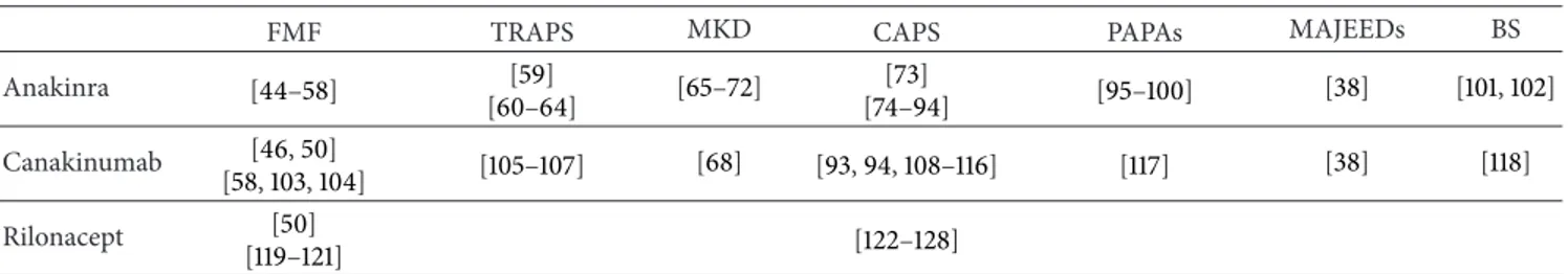
Table 2: Overview of the medical literature regarding anti-interleukin-1 therapies in the monogenic autoinflammatory disorders.
FMF TRAPS MKD CAPS PAPAs MAJEEDs BS
Anakinra [44–58] [59] [60–64] [65–72] [73] [74–94] [95–100] [38] [101,102] Canakinumab [46,50] [58,103,104] [105–107] [68] [93,94,108–116] [117] [38] [118] Rilonacept [50] [119–121] [122–128]
BS: Blau syndrome; CAPS: cryopyrin-associated periodic syndromes; FMF: familial Mediterranean fever; MAJEEDs: Majeed syndrome; MKD: mevalonate kinase deficiency syndrome; PAPAs: pyogenic arthritis-pyoderma gangrenosum-acne syndrome; TRAPS: tumor necrosis factor receptor-associated periodic syndrome.
improves beta-cell secretory function: moreover, the bene-ficial effects on insulin production and systemic proinflam-matory parameters were prolonged over time, even after anakinra cessation [182, 183]. Convincing evidence derives also from the positive effects on HbA1c levels of a single dose of canakinumab in T2DM [184–186]. A large phase III clinical trial CANTOS (Canakinumab Anti-inflammatory Thrombo-sis Outcomes Study) is currently recruiting participants to assess whether canakinumab may increase insulin secretion and insulin sensitivity in patients with T2DM (ClinicalTri-als.govNCT01327846). Gevokizumab has also proven to be useful in improving HbA1c [187–190]. Several clinical trials are in progress to determine whether gevokizumab might improve glycemic control in subjects with T2DM treated or not with metformin (ClinicalTrials.gov NCT01144975,
NCT01066715, NCT00513214). Despite these encouraging
results, further studies are needed to consider anti-IL1 agents as new therapeutic instruments for treating T2DM and probably a wider range of metabolic disturbances.
6. Uveitis
This sporadic disease has a baffling etiology and is the most frequent extra-articular sign of different systemic autoim-mune rheumatologic disorders, such as oligoarticular variant of juvenile idiopathic arthritis, seronegative spondyloarthri-tis, and Behc¸et’s disease [191]. The inflammatory process leading to uveitis is mainly driven by Th17 cells and sustained by intricate sceneries directed by many proinflammatory cytokines, chiefly TNF-𝛼 and IL-1𝛽 [192]. In this regard Kitamei et al. have suggested that NF-𝜅B, activated by IL-1𝛽, plays a pivotal role in an experimental murine model of auto-immune uveoretinitis: in fact, pyrrolidine dithiocarbamate administration, which inhibits NF-𝜅B signaling, has amelio-rated clinical symptoms and suppressed ocular IL-1𝛽 mRNA expression [193]. Lennikov et al. have shown that uveitis improves after inhibition of I𝜅B kinase 𝛽 in an animal model of endotoxin-induced uveitis, particularly when the disease is due to IL-1𝛽 and TNF-𝛼 oversecretion [194]. Several years before other authors had demonstrated that foot-pad injection of lipopolysaccharide in rats induced elevated mRNA expression of various cytokines, such as TNF-𝛼, IL-1𝛽, and IL-1 receptor antagonist (IL-1Ra) in the uvea and retina, suggesting that these mediators contribute to the develop-ment and recovery of ocular inflammation [195,196]. These
evidences have also been corroborated by the capability of anakinra to suppress immune-mediated ocular inflammation not only in animal models [197] but also in a patient with Blau syndrome and in a patient with CINCA syndrome [73, 198]. More recently a 4-year-old boy diagnosed with early onset sarcoidosis and presenting with refractory severe panuveitis has experienced a rapid remission of uveitis and normalization of most gene expression profiles following canakinumab administration [118]. In addition, after the results of the previous study conducted by G¨ul et al. on Behc¸et’s disease-resistant uveitis [199], three multicenter phase III clinical trials are ongoing to test safety and effi-cacy of gevokizumab in the treatment of active nonin-fectious uveitis (ClinicalTrials.gov NCT01684345), quies-cent noninfectious uveitis (ClinicalTrials.govNCT01747538), and Behc¸et’s disease-associated uveitis (ClinicalTrials.gov
NCT01965145). In the end, a phase II clinical trial is also being
conducted with gevokizumab in patients with active scleritis (ClinicalTrials.govNCT01835132).
7. Pericarditis
Acute pericarditis may be the first manifestation of an under-lying systemic disease [200–203]. Although a specific exact etiology may be identified, its cause remains obscure in up to 85% of patients. Idiopathic recurrent acute pericarditis (IRAP) is a troubling complication of acute pericarditis, occu-rring in approximately 30% of cases [204]. Recently, con-sidering clinical and laboratory features of IRAP (absence of autoantibodies or self-antigen-specific T lymphocytes) [205–208] and the growing evidence about IRAP favourable response to IL-1 inhibition, it has been hypothesized that this condition can be included in the group of AIDs [209–211]. However, IRAP may occur in the framework of two peculiar AIDs, familial Mediterranean fever (FMF) and TNF receptor-associated periodic syndrome (TRAPS), becoming a diagnos-tic clue for identifying these disorders [22,59,212–214]. IRAP may also be the only clinical TRAPS symptom in patients carrying low-penetrance TNFRSF1A mutations [215–217]. According to some authors this recurrent pericarditis can be considered an example of a multifactorial disorder with overlapping pathogenic mechanisms, both autoinflammatory and autoimmune [218]. On this basis, a multicentre study evaluating the incidence of TRAPS mutations in patients with IRAP has demonstrated that positive family history
T a ble 3: R ecen t and o n go in g clinical tr ials o n in te rleuk in-1 b lo ck ade in the mo nog enic au to infla m ma to ry dis o rd er s. Ph as e St atu s Stu dy Dis eas e C linicalT rials.g o v iden tifier Anakinra 3 R ecr u it in g K iner et (a nakinra) in ad ul t p at ien ts w it h co lc hicine-r esist an t fa m ilial M edi ter ra ne an fe ve r F MF NC T 0170 575 6 1 C o m p let ed The us e o f K iner et (a na kinra) in th e tr ea tmen t o f fa mi li al cold au to infl am ma to ry sy ndr o me FA CS NC T 0 02 14 851 C ana k in u ma b 3 R ecr u it in g Efficac y, sa fety ,a n d to lera b il it y o f A CZ88 5 in p edia tr ic p at ien ts wi th th e fo llo win g cr yo p yr in-ass o cia ted p er io dic syndr o mes: fa milial co ld au to infla m ma to ry syndr o me ,M uc k le-W ells syndr o m e, an d C IN CA sy ndr o me CAPS N C T0157 63 6 7 3C o m p le te d The sa fe ty an d efficac y o f ca n akin uma b in pa ti en ts ag ed 4 ye ar s o r o lder d ia gnos ed wi th cr yo p yr in-ass o cia ted p er io dic syndr o mes in C an ada CAPS N C T011 05 50 7 3C o m p le te d Efficac y an d sa fety st u d y o f ca nakin u ma b administ er ed fo r 6 mo n th s (2 4 w eeks) in Ja p an es e p at ien ts wi th cr yo p yr in-ass o cia ted p er io dic syndr o mes fo llo w ed by an ext en sio n p h as e CAPS N C T0 0 9 911 4 6 2C o m p le te d E va lua ti o n o f th e sa fety and efficac y o f ca na kin u ma b in p edia tr ic p at ien ts wi th co lc hicine-in to lera n t o r co lc hicine-r esist an t fa m il ia lM edi ter ra ne an fe ve r FMF N C T011 4 8797 2 C o m p let ed Efficac y an d sa fety o f ca n ak in uma b in p at ien ts wi th co lc hicine-r esist an t fa m il ial M edi ter ra ne an fe ve r F MF N C T01088880 3 Ac ti ve ,n o t re cr u it ing Efficac y, sa fety ,a n d to lera b il it y o f A CZ88 5 in p edia tr ic p at ien ts wi th th e fo llo win g cr yo p yr in-ass o cia ted p er io dic syndr o mes: fa milial co ld au to infla m ma to ry syndr o me ,M uc k le-W ells syndr o m e, an d C IN CA sy ndr o me CAPS N C T013 02 86 0 — R ecr u it in g C linical o u tc o m es an d sa fety :a regist ry st u d y o f Ila ris (ca n akin uma b ) p at ien ts (B-co nfiden t) C APS N C T01213 6 4 1 2 Ac ti ve ,n o t re cr u it ing C ana k in u ma b in p at ien ts w it h ac tiv e h yp er -I gD sy ndr o me MKD N C T013 03 38 0 2C o m p le te d Efficac y an d sa fety st u d y o f A C Z88 5 in p at ien ts wi th ac ti ve re cu rr en t o r chr o nic tu mo r n ecr o sis fac to r re cep to r-ass o cia ted p er io dic syndr o me TRAPS NC T 01 24 281 3 3 R ecr u it in g E fficac y and sa fe ty o f ca nakin u ma b in p at ien ts w it h h er edi ta ry p er io dic fe ve rs HPFs NC T 02 0 592 91 CAPS: cr yo p yrin-a sso cia ted perio d ic syn d ro m es; FA CS: fa m ilial co ld au to infla m m at o ry syn d ro m e; F MF : fa m ilial M edi terra n ea n fe ve r; HP Fs: h er edi ta ry p er io dic fe ver syndr o mes; MKD: m ev al o n at e k inas e deficien cy syn d ro m e; T RAPS: tum o r n ecr osis fac to r re cep to r-ass o cia te d p er io dic syn dr o m e.
of pericarditis, pericarditis recurrence, failure of treatment with colchicine, and need of immunosuppressive agents were key-elements for suspecting a clinical diagnosis of TRAPS [219,220]. Due to these findings, FMF and above all TRAPS should be taken into account in the differential diagnosis of IRAP [221]. Treatment with anakinra has been described as dramatically effective in the control of IRAP, even the corticosteroid-dependent forms of IRAP or those resistant to conventional therapies [210, 211, 222, 223]. Additional data derive from the study of 15 young patients with IRAP performed by Finetti et al., who evaluated long-term use of anakinra: after a median follow-up of 39 months a 95% reduction of flares was observed, and patients experienced a persistent disease control [209]. More recently Lazaros et al. have confirmed that anakinra is highly effective also in adults with IRAP [224]. A phase IV study designed to demonstrate the efficacy of anakinra in patients with IRAP is actually ongoing (ClinicalTrials.gov NCT02219828), but there are no data about treatment with rilonacept, canakinumab, and gevokizumab.
8. Behçet’s Disease
The pathogenesis of Behc¸et’s disease (BD) is still largely unknown, and continuous efforts are in progress to character-ize its biologic background, suggesting that the disease may lie at the crossroad between autoinflammatory and autoim-mune syndromes [1,225]. The central role of innate immunity in its pathogenesis has been suggested not only by the increased levels of IL-1 in serum [226] and synovial fluid [227] of BD patients but also by the beneficial effects obtained with IL-1 inhibition [228]. In addition, the time of disease onset has been correlated with IL-1 gene specific single nucleotide polymorphisms [229,230]. Recently Castrichini et al. have observed an increased expression of the P2X7 receptor, a nucleotide-gated ion channel, in BD monocytes, acting in the promotion of IL-1𝛽 release [231]. With regard to therapy, the efficacy of anakinra on various BD manifestations has been well documented in some surveys and several case reports, albeit with variable duration of response [232–236]. A pilot study is ongoing to test whether anakinra given at a daily dose of 100 mg with a dose escalation up to 200 mg/day might con-trol all BD manifestations (ClinicalTrials.govNCT01441076). The favorable response to anti-IL-1 agents has been also con-firmed in three adult BD patients who received canakinumab (150 mg every 6 weeks) as monotherapy [237] and in other two cases [228,238].Table 4shows a list of papers dealing with IL-1 blockade in BD. Additional convincing evidence for a possible role of IL-1𝛽 in BD emerges from a trial with the monoclonal anti-IL-1𝛽 antibody gevokizumab, which has proven to be effective in both uveitis and retinal vasculitis, leading to complete resolution of intraocular inflammation [199]. These findings have represented an important aspect in the management of BD, revealing that IL-1 inhibition can be indicated in the treatment of the most severe ocular man-ifestations, especially if unresponsive to immunosuppressant drugs and other biologicals [239–241].
9. Gout and Chondrocalcinosis
Proinflammatory cytokines have a nodal role in orchestrating the body reaction to monosodium urate (MSU) and calcium pyrophosphate dihydrate crystals: recent attention has been focused on the role of IL-1 [242]. Experimental models have suggested that crystals engage the caspase-1-activating
NLRP3 inflammasome, resulting in the production of
bioac-tive IL-1𝛽 [243]. Further evidence for the proposed role of IL-1𝛽 in the pathogenesis of gout is shown by the study of Chen et al., in which they demonstrated that IL-1𝛽 receptor-deficient mice were not susceptible to MSU-induced inflammation [244]. Other authors have also assessed that activation of the P2X7R-mediated signalling pathway by MSU crystals leads to enhanced NLRP3 inflammasome activity and IL-1𝛽 oversecretion at the onset of an acute gouty attack [245]. Interestingly, Owyang et al. have reported that gevokizumab was able to reduce acute inflammation in a mouse model, blocking MSU crystal-induced peritonitis [246]. Further proofs of the concept that IL-1 is clearly involved derive from the favorable results obtained with anakinra in an open-label study and several gout-related case reports [247–252]. In line with these findings, Vitale et al. have reported three patients with chronic tophaceous gout unresponsive to standard ther-apy, in whom anakinra led to remarkable amelioration of joint symptoms within 24 hours. Interestingly, patients were also affected by T2DM and, along with amelioration of joint symptoms, they also experienced a marked improvement in the glycemic control during anakinra treatment [253]. Also canakinumab and rilonacept have proven their efficacy on a broad sample of patients with crystal-induced arthritis [254–
261], showing a superior therapeutic effect in comparison with corticosteroids. A phase III study testing efficacy of canakinumab in preventing gout relapses is now ongoing for patients with colchicine intolerance (ClinicalTrials.gov
NCT01362608). As a result, anti-IL-1 agents should be taken
into consideration for both patients affected with unrespon-sive gouty arthritis and those presenting with dysmetabolic comorbidities.
10. Sjogren’s Syndrome
This autoimmune disorder is characterized by infiltration of mononuclear cells in the salivary and lacrimal glands, leading to dryness of both mouth and eyes. Although pathogenesis is not completely understood, several studies focusing on cyto-kine profile that may contribute to the pathological scenery of this disease [262,263] have found an increased concentration of IL-1 in the salivary fluid and peripheral blood of patients with Sjogren’s syndrome (SS), indicating that IL-1 works as a pivotal regulator in the development of local and systemic manifestations [264, 265]. Functional consequences of IL-1 and low levels of IL-IL-1Ra in the saliva remain unclear. In this regard Dubost et al. have suggested that the salivary IL-1/IL-Ra imbalance may promote inflammatory lesions in the mouth [266], while Solomon et al. have shown that patients with SS produce higher concentration of IL-1𝛼 and IL-1𝛽 in the tears [262]. In addition, IL-1 expression in ocular epithe-lial cells has been correlated with keratinizing squamous
T a ble 4 :O ve rv ie w fr o m the medical li te ra tu re de alin g w it h in terleukin-1 b lo ck ade in B ehc ¸e t’s d is ea se . Fir st au th o r [re fe re n ce ] Nu m b er o f pa tie n ts B rief summa ry o f clinical an d la b o ra to ry fe at u re s A n ti-in te rleukin-1 ag en ts O u tco me Bo ts io s [ 23 5 ]1 F ev er ,m u cos al in vo lv emen t, gu t is chemic p er fo ra tio n, p o si ti ve p at h er gy te st, in cr ea sed acu te -p h ase re ac ti o n Anakinra C o mp le te re m is si o n w it h imp ro ve m ent o f infl am ma to ry ma rk ers in 7–10 d ay s B ilginer [ 234 ]1 F ev er ,m u cos al in vo lv emen t, er yt hema no dosum, ar th ri ti s, se co nda ry am yl o idosis, incr ea se d ac u te -p has e re ac ti o n ,s kin p at her gy re ac tio n s, o ver la p w it h fa m ilial M edi terra n ea n fe ve r Anakinra C o mp le te re m is si o n w it h imp ro ve m ent o f infl am ma to ry ma rk ers in 6 mo n ths G ¨ul [ 19 9 ] 7 A cut e p o st er ior uve it is an d p an u ve it is ,r et in al va sc u lit is G evok iz u m ab C o m p let e re missio n o f ret inal vas culi tis in 4 – 21 d ay s an d m ar ke d re d u ct ion in in tr ao cu la r infla m ma ti o n U gurl u [ 238 ]1 M u cos al in vo lv emen t, er yt hema no dosum, b ila te ra lp an u vei ti s, re ti na l vas cu li tis, skin p at her gy re ac tio n s C ana k in u ma b C o m plete remissio n fo r 8 w eeks Emmi [ 236 ]1 M u cos al and gast ro in te st inal in vo lv emen t, ar th ri ti s, ps eudo fo llic uli tis, bi la te ra lr et in al va sc u lit is Anakinra C o m p let e re missio n aft er 12 m o n th s o f fol lo w -u p C an ta rini [ 23 2 ]9 F ev er ,m u cos al in vo lv emen t, he adac he ,s kin lesio n s, ret ina lvas cu li tis, lo w bac k pa in, incr ea se d ac u te -p has e re ac ti o n ,a rt hr it is, ab d o minal pa in, Anakinra C o m p let e/pa rt ial re missio n w it h a va ri ab le du ra ti on of re sp ons e Ca so [ 23 3 ]1 M u cos al and o cu la r in vo lv emen t, ps eudo fo ll ic u li tis, sacr o ilii tis incr ea se d ac u te-p h as e re ac tio n Anakinra C o m p let e remissio n in fe w d ay s C an ta rini [ 228 ]1 F ev er ,m u cos al in vo lv emen t, skin lesio n s, ar th ri ti s, ab do minal pa in, he adac he , incr ea se d ac u te -p has e re ac ti o n ,o ve rla p wi th gra n ulo m a ann ula re C ana k in u ma b C o m plete remissio n aft er fe w w ee ks Vi ta le [ 23 7 ]3 F ev er ,m u cos al and gast ro in te st ina lin vo lv emen t, he adac he ,a n ter io r u ve it is, ar th ralgia, p se udo fo llic uli tis, d eep veno u s thr o m b o sis, pa n u ve it is, h ea dac h e, ar th ri ti s, in cr ea sed acu te -p h ase re ac ti o n C ana k in u ma b C o m plete remissio n w it hin fe w w ee ks
metaplasia, a condition resulting from uncontrolled ocular inflammation [267]. Some authors have also found that IL-1𝛽 is involved in the destruction of salivary and lacrimal glands [268]: in fact, IL-1𝛽 may have a proteolytic activity,
leading to acinar and ductal structure disruption in salivary glands of patients with SS [263]. Since acinar cells, duct cells, and blood vessels of the lacrimal glands are innervated by parasympathetic and sympathetic nervous system, several reports have shown that exogenously added IL-1 might inhibit neurotransmitter release [269–271]. Additional data substantiating the crucial role of innate immunity in SS derive from studies on nonobese autoimmune-prone mice. Bulosan et al. have investigated the potential involvement of inflammatory caspases, revealing a concurrent upregulation of caspase-11 in macrophages [272]. Moreover, the presence of the purinergic P2X7 receptor, an ATP-gated ion channel, in the salivary glands, would be capable of determining
NLRP3 inflammasome activation, leading to the release of
mature IL-1𝛽 and IL-18 [273–275]. Since IL-1 seems to be directly involved in the pathogenesis of SS, there might be a rationale for using anti-IL-1 agents as a potential treatment [276]. In light of this evidence, a randomized double-blind placebo-controlled trial has indicated that IL-1 inhibition with anakinra is able to influence favorably fatigue in patients with SS [277]. More recently, data from a prospective double-blind randomized trial have also demonstrated that targeting IL-1 by topical application of anakinra is effective in reducing dry eye disease-related symptoms and corneal epitheliopathy [278].
11. Interstitial Lung Diseases
Chronic interstitial lung diseases (ILDs), characterized by diffuse lung interstitial wall inflammation, often result in severe pulmonary fibrosis and impaired gas exchange [279]. Alveolar macrophages are involved in various pulmonary inflammatory processes and can constitutively release IL-1, probably due to various exogenous and endogenous stimuli [280]. However, their activity is limited by the presence of IL-1 inhibitory factors secreted from the same macrophages [281]. On this basis, an imbalance between the release of IL-1 and its inhibitor may evoke an inflammatory state [282]. Several studies have revealed the presence of IL-1𝛽 in the chronically inflamed lung tissues undergoing fibrogenesis, suggesting a causative link between IL-1 and fibrosis [283–
285]. Through IL-1𝛽 overexpression, induced by intratracheal
administration of adenoviral genes, Kolb et al. have caused an acute pulmonary inflammation with severe progressive tissue fibrosis in an animal model [286]; they also found that IL-1𝛽 led to increased concentrations of growth factors in the bronchoalveolar lavage fluid [287]. Interestingly, IL-1 antago-nists have been successfully used to block fibrosis in murine models of ILDs [288,289]. A genetic variability in the IL1RN gene, encoding the physiological IL-1Ra, may contribute to the pathogenesis of idiopathic pulmonary fibrosis [290,291]. Other authors have suggested that NLRP3 inflammasome is involved in the early inflammatory process of pneumoconio-sis and systemic scleropneumoconio-sis [292,293]. In this regard a phase
I/II study is ongoing to test the effect of the IL-1 inhibitor rilonacept on skin gene expression of patients with systemic sclerosis (ClinicalTrials.govNCT01538719). Moreover, a case of antisynthetase syndrome clinically characterized by pro-gressive and diffuse interstitial lung disease and myositis responding to anakinra has been reported [294], while ano-ther one lost its efficacy on lung disease manifestations [295]. Although IL-1𝛽 might be considered a valid target for
treatment of ILDs, further studies are required to fully explore and define its exact role in the pathogenesis of ILDs.
12. Still’s Disease
This rare inflammatory disorder of undisclosed etiology is characterized by fever, rash, arthritis, and prominent neu-trophilia, accompanied by high C-reactive protein and fer-ritin levels [296]. Similarly to what we observe in the mono-genic AIDs, the main proinflammatory cytokine increased in adult onset Still’s disease (AOSD) is IL-1𝛽 [297, 298]. Macrophage activation syndrome has been rarely reported in the course of AOSD, but mortality in adults is far higher than that for children with systemic-onset juvenile idiopathic arthritis [299]. Convincing evidence about IL-1 involvement in this disorder derives from the study by Pascual et al., reporting that peripheral blood mononuclear cells of healthy subjects incubated with sera from patients with systemic-onset juvenile idiopathic arthritis secreted large amounts of IL-1𝛽 and led to increased expression of innate immunity genes [300]. Benefits obtained with IL-1 antagonists have also reinvigorated the concept that IL-1𝛽 is largely implicated in the pathological scenery of AOSD. Notably, anakinra as monotherapy has proven to be highly effective in patients refractory to conventional treatments (corticosteroids and methotrexate). These findings rely not only on single case reports and small case series [301–321] but also on large num-bers of subjects: a study of 25 patients with active multiresis-tant AOSD reported a complete resolution of clinical activity in 84% of cases and normalization of laboratory markers in 80% [322]. Moreover, an open randomized multicentre study has enrolled 22 patients with AOSD, demonstrating that anakinra brings about remission in the refractory forms of the disease [323]. A recent retrospective study carried out to assess long-term efficacy of anakinra in 28 patients has shown a complete remission in 57% of them after a mean follow-up time of 23 months [324]. Also canakinumab and rilonacept have proven the efficacy of IL-1 inhibition in AOSD. In this regard Henderson et al. have investigated treatment with rilonacept in a small sample of patients, observing a good clinical response [325]. More recently the successful use of rilonacept in the management of three patients with refractory AOSD [326] as well as the effective-ness of canakinumab in curbing AOSD manifestations has been reported [327,328]. A phase II study is now ongoing to assess whether canakinumab may confirm its promising effects in the decrease of disease activity (ClinicalTrials.gov
T a ble 5: M ai n scene p la ye rs in vo lv in g the ro le o f in te rleukin-1 in the au to infla m ma to ry an d au to imm une d is o rder s des cr ib ed in this re vie w . D is eas e [r efe re nc e] C el ls in volve d Bi ol o gi c pl at for m s in volve d T re at m en t [r efe re nc e] PF AP A sy n dr o m e [ 13 7 , 13 8 ] M o no n u cle ar cel ls, n eu tr o p hi ls, ly m p ho cy te s, Th1 cells NLRP3 infla m mas o me Anakinra [ 13 9 , 14 0 ] Rh eu m at o id ar th ri ti s [ 14 1 – 14 3 ] Ta n d Bl ym p h o cy te s,p la sm ac el ls , syno vio cyt es M etallo p ro te in as es (co lla ge n as e, ela st as e) Anakinra [ 167 , 16 8 ], ca na k in u ma b [ 17 1 , 17 2 ] Ty p e 2 d ia b et es m el li tu s [ 17 3 – 181 ] M acr o p hag es, adi p o cyt es, p an cr ea ti c 𝛽 -cells Olig o m er s o f islet am yl o id p o ly p ep tide , NLRP3 infla m mas o me Anakinra [ 182 , 18 3 , 25 3 ], ca na k in u ma b [ 18 4 – 18 6 ], ge vok iz u m ab [ 187 – 19 0 ] Uv ei ti s [ 192 – 19 4 ]Th 17 ce ll s NLRP3 infla m mas o me Anakinra [ 73 , 19 8 ], ca na k in u ma b [ 11 8 ], ge vok iz u m ab [ 19 9 ] Pe ri ca rd it is [ 22 , 59 , 212 – 21 8 ] D endr it ic cel ls, Th 1 and Th17 cel ls, macr o p hag es P yr in, p5 5 tumo r necr osis fac to r re cep to r typ e 1 Anakinra [ 209 , 22 4 ] Be h c¸e t’s d is ea se [ 226 – 23 1 ] M o no cy te s/macr o p ha ge s, Th1 an d Th 2 cells, neu tr o phi ls P2X7 re cep to r, NLRP3 infla m mas o me Anakinra [ 23 2 – 236 ], ca na k in u ma b [ 228 , 23 7 , 238 ] G o u t an d cho ndr o ca lcinosis [ 24 2 – 24 5 ] N eu tr o p hi ls, m acr o p hag es L yso so m al an d cy to p la sm ic en zy m es ,P 2X 7 re cep to r, NLRP3 infla m mas o me Anakinra [ 24 7 – 25 3 ], ca na k in u ma b [ 254 – 25 7 ], ri lo nacep t [ 25 8 – 26 1 ] Sj ¨ogr en syndr o m e [ 26 2 – 26 6 , 27 2 – 27 5 ] M o n o n uc le ar cells, Th1 an d Th 17 cells U p re gula tio n o f caspas e-11, ST A T -1 ac ti vi ty ,P 2X7 re cep to r, NLRP3 infla m mas o me ,I L -1/IL -R a im bala nce Anakinra [ 27 7 , 27 8 ] In te rs ti tial lu n g d isea se s [ 280 , 28 1 ] A lve ol ar m ac roph age s, ne ut roph il s, macr o p hag es NLRP3 infla m mas o me ,I L -1/IL -R a im b ala n ce , tr an sf o rmin g gr o wt h fac to r-𝛽1 Anakinra [ 294 , 295 ] St ill ’s di sea se [ 300 ] M acr o p ha ge s, neu tr o p h ils, na tu ral k iller cel ls, Th1 an d Th 17 cel ls, dendr it ic cel ls NLRP3 infla m mas o me Anakinra [ 30 1 – 324 ], ca na k in u ma b [ 32 7 , 32 8 ], ri lo nacep t [ 32 5 , 32 6 ]
13. Conclusions
From a mere pathogenic point of view most autoinflamma-tory and autoimmune diseases share the chronic aberrant activation of the immune system, which leads to tissue inflammation and/or tissue damage of varying magnitude and extent in genetically predisposed individuals. The specific effectors of inflammation and damage are different in the two groups of disorders, respectively, the innate and adaptive immunity branches, even if in the last decade we began to rec-ognize the involvement of autoinflammatory circuits in many different diseases and also those having an autoimmune basis. Certainly, the role of IL-1 on lymphocyte function, favouring the expansion of autoreactive Th1 and Th17 cells or down-regulating regulatory T cells, has not been yet completely elucidated and requires further research to change our way of categorizing an expanding group of inflammatory disorders, even autoimmune diseases. Table 5 recapitulates all cells and the different biologic platforms involved in the various clinical settings driven by IL-1 oversecretion. Unearthing the molecular pathways of autoinflammation and autoimmunity has enlightened our capacity of understanding the human disease, and recent technological breakthroughs have also generated large quantities of novel information specifically in both autoinflammatory and autoimmune diseases at the genetic, transcriptional, proteic, and metabolic levels. The acceleration of clinical trials over the past decades has also included rare diseases, such as AIDs. Studies of such complex disorders and their relationship with IL-1 need to address heterogeneity in the human population, interaction with the environment, and effects of treatments, while a multidisci-plinary approach should be ultimately fostered to provide a significant change in the knowledge of these diseases across the scientific community.
Conflict of Interests
The authors declare that there is no conflict of interests regarding the publication of this paper.
Authors’ Contribution
Giuseppe Lopalco and Luca Cantarini equally contributed to this work.
References
[1] D. McGonagle and M. F. McDermott, “A proposed classification of the immunological diseases,” PLoS Medicine, vol. 3, no. 8, article e297, 2006.
[2] I. Touitou and I. Kon´e-Paut, “Autoinflammatory diseases,” Best
Practice and Research: Clinical Rheumatology, vol. 22, no. 5, pp.
811–829, 2008.
[3] D. Rigante, “The fresco of autoinflammatory diseases from the pediatric perspective,” Autoimmunity Reviews, vol. 11, no. 5, pp. 348–356, 2012.
[4] L. Cantarini, O. M. Lucherini, F. Iacoponi et al., “Development and preliminary validation of a diagnostic score for identifying patients affected with adult-onset autoinflammatory disorders,”
International Journal of Immunopathology and Pharmacology,
vol. 23, no. 4, pp. 1133–1141, 2010.
[5] L. Cantarini, D. Rigante, M. G. Brizi et al., “Clinical and bio-chemical landmarks in systemic autoinflammatory diseases,”
Annals of Medicine, vol. 44, no. 7, pp. 664–673, 2012.
[6] L. Cantarini, F. Iacoponi, O. M. Lucherini et al., “Validation of a diagnostic score for the diagnosis of autoinflammatory diseases in adults,” International Journal of Immunopathology and
Phar-macology, vol. 24, no. 3, pp. 695–702, 2011.
[7] I. Muscari, F. Iacoponi, L. Cantarini et al., “The diagnostic evaluation of patients with potential adult-onset autoinflam-matory disorders: our experience and review of the literature,”
Autoimmunity Reviews, vol. 12, no. 1, pp. 10–13, 2012.
[8] L. Cantarini, A. Vitale, O. M. Lucherini et al., “Childhood versus adulthood-onset autoinflammatory disorders: myths and truths intertwined,” Reumatismo, vol. 65, no. 2, pp. 55–62, 2013. [9] D. Rigante, A. Vitale, O. M. Lucherini, and L. Cantarini, “The
hereditary autoinflammatory disorders uncovered,”
Autoimmu-nity Reviews, vol. 13, no. 9, pp. 892–900, 2014.
[10] D. Rigante, G. Lopalco, A. Vitale et al., “Untangling the web of systemic autoinflammatory diseases,” Mediators of
Inflamma-tion, vol. 2014, Article ID 948154, 15 pages, 2014.
[11] L. Cantarini, A. Vitale, O. M. Lucherini et al., “The labyrinth of autoinflammatory disorders: a snapshot on the activity of a third-level center in Italy,” Clinical Rheumatology, 2014. [12] F. Caso, D. Rigante, A. Vitale et al., “Monogenic
autoinflam-matory syndromes: state of the art on genetic, clinical, and therapeutic issues,” International Journal of Rheumatology, vol. 2013, Article ID 513782, 15 pages, 2013.
[13] F. Caso, L. Cantarini, O. M. Lucherini et al., “Working the end-less puzzle of hereditary autoinflammatory disorders,” Modern
Rheumatology, vol. 24, no. 3, pp. 381–389, 2014.
[14] A. Vitale, D. Rigante, M. C. Maggio et al., “Rare NLRP12 vari-ants associated with the NLRP12-autoinflammatory disorder phenotype: an Italian case series,” Clinical and Experimental
Rheumatology, vol. 31, no. 3, supplement 77, pp. 155–156, 2013.
[15] D. Rigante, I. la Torraca, V. Ansuini, A. Compagnone, A. Sall`ı, and A. Stabile, “The multi-face expression of familial mediterranean fever in the child,” European Review for Medical
and Pharmacological Sciences, vol. 10, no. 4, pp. 163–171, 2006.
[16] D. Rigante, B. Frediani, M. Galeazzi, and L. Cantarini, “From the mediterranean to the sea of Japan: the transcontinental odyssey of autoinflammatory diseases,” BioMed Research International, vol. 2013, Article ID 485103, 8 pages, 2013.
[17] T. Lane, J. M. Loeffler, D. M. Rowczenio et al., “AA amyloidosis complicating the hereditary periodic fever syndromes,”
Arthri-tis & RheumaArthri-tism, vol. 65, no. 4, pp. 1116–1121, 2013.
[18] D. Rigante, V. Ansuini, M. Caldarelli, B. Bertoni, I. la Torraca, and A. Stabile, “Hydrocephalus in CINCA syndrome treated with anakinra,” Child’s Nervous System, vol. 22, no. 4, pp. 334– 337, 2006.
[19] A. Vitale, D. Rigante, O. M. Lucherini et al., “Biological treatments: new weapons in the management of monogenic autoinflammatory disorders,” Mediators of Inflammation, vol. 2013, Article ID 939847, 16 pages, 2013.
[20] N. Ter Haar, H. Lachmann, S. ¨Ozen et al., “Treatment of autoin-flammatory diseases: results from the Eurofever Registry and a literature review,” Annals of the Rheumatic Diseases, vol. 72, no. 5, pp. 678–685, 2013.
[21] L. Cantarini, O. M. Lucherini, B. Frediani et al., “Bridg-ing the gap between the clinician and the patient with
cryopyrin-associated periodic syndromes,” International
Jour-nal of Immunopathology and Pharmacology, vol. 24, no. 4, pp.
827–836, 2011.
[22] S. Ozen, E. Demirkaya, G. Amaryan et al., “Results from a multicentre international registry of familial Mediterranean fever: impact of environment on the expression of a monogenic disease in children,” Annals of the Rheumatic Diseases, vol. 73, pp. 662–667, 2014.
[23] L. Cantarini, P. L. Capecchi, O. M. Lucherini, F. Laghi Pasini, and M. Galeazzi, “Familial Mediterranean fever diagnosed in an elderly patient,” Clinical and Experimental Rheumatology, vol. 28, no. 4, supplement 6, p. S91, 2010.
[24] D. Rigante, G. Lopalco, A. Vitale et al., “Key facts and hot spots on tumor necrosis factor receptor-associated periodic syndrome,” Clinical Rheumatology, vol. 33, no. 9, pp. 1197–1207, 2014.
[25] H. J. Lachmann, R. Papa, K. Gerhold et al., “The pheno-type of TNF receptor-associated autoinflammatory syndrome (TRAPS) at presentation: a series of 158 cases from the Euro-fever/EUROTRAPS international registry,” Annals of the
Rheu-matic Diseases, vol. 73, no. 12, pp. 2160–2167, 2014.
[26] F. Magnotti, A. Vitale, D. Rigante et al., “The most recent advances in pathophysiology and management of tumour ne-crosis factor receptor-associated periodic syndrome (TRAPS): personal experience and literature review,” Clinical and
Exper-imental Rheumatology, vol. 31, supplement 77, no. 3, pp. S141–
S149, 2013.
[27] L. Cantarini, D. Rigante, O. M. Lucherini et al., “Role of etanercept in the treatment of tumor necrosis factor receptor-associated periodic syndrome: personal experience and review of the literature,” International Journal of Immunopathology and
Pharmacology, vol. 23, no. 3, pp. 701–707, 2010.
[28] L. Cantarini, O. M. Lucherini, R. Cimaz, C. T. Baldari, F. Laghi Pasini, and M. Galeazzi, “Sacroileitis and pericarditis: Atypical presentation of tumour necrosis factor receptor-associated periodic syndrome and response to etanercept therapy,” Clinical
and Experimental Rheumatology, vol. 28, no. 2, pp. 290–291,
2010.
[29] L. Cantarini, O. M. Lucherini, R. Cimaz et al., “Typical and severe tumor necrosis factor receptor-associated periodic syn-drome in the absence of mutations in the TNFRSF1A gene: a case series,” Rheumatology International, vol. 32, no. 12, pp. 4015–4018, 2012.
[30] L. Cantarini, O. M. Lucherini, M. Galeazzi et al., “Tumour necrosis factor receptor-associated periodic syndrome caused by a rare mutation in the TNFRSF1A gene, and with excellent response to etanercept treatment,” Clinical and Experimental
Rheumatology, vol. 27, no. 5, pp. 890–891, 2009.
[31] L. Cantarini, A. Vitale, F. Magnotti et al., “Weekly oral alen-dronate in mevalonate kinase deficiency,” Orphanet Journal of
Rare Diseases, vol. 8, no. 1, article 196, 2013.
[32] R. van der Burgh, N. M. ter Haar, M. L. Boes, and J. Frenkel, “Mevalonate kinase deficiency, a metabolic autoinflammatory disease,” Clinical Immunology, vol. 147, no. 3, pp. 197–206, 2013. [33] R. Levy, L. G´erard, J. Kuemmerle-Deschner et al., “Phenotypic and genotypic characteristics of cryopyrin-associated periodic syndrome: a series of 136 patients from the Eurofever Registry,”
Annals of the Rheumatic Diseases, 2014.
[34] A. Vitale, O. M. Lucherini, M. Galeazzi, B. Frediani, and L. Cantarini, “Long-term clinical course of patients carrying the Q703K mutation in the NLRP3 gene: a case series,” Clinical and
Experimental Rheumatology, vol. 30, no. 6, pp. 943–946, 2012.
[35] E. J. Smith, F. Allantaz, L. Bennett et al., “Clinical, molecular, and genetic characteristics of PAPA syndrome: a review,” Current
Genomics, vol. 11, no. 7, pp. 519–527, 2010.
[36] A. P. Demidowich, A. F. Freeman, D. B. Kuhns et al., “Genotype, phenotype, and clinical course in five patients with PAPA syndrome (pyogenic sterile arthritis, pyoderma gangrenosum, and acne),” Arthritis and Rheumatism, vol. 64, no. 6, pp. 2022– 2027, 2012.
[37] B. Tallon and M. Corkill, “Peculiarities of PAPA syndrome,”
Rheumatology, vol. 45, no. 9, pp. 1140–1143, 2006.
[38] T. Herlin, B. Fiirgaard, M. Bjerre et al., “Efficacy of anti-IL-1 treatment in Majeed syndrome,” Annals of the Rheumatic
Diseases, vol. 72, no. 3, pp. 410–413, 2013.
[39] H. I. El-Shanti and P. J. Ferguson, “Chronic recurrent multifocal osteomyelitis: a concise review and genetic update,” Clinical
Orthopaedics and Related Research, vol. 462, pp. 11–19, 2007.
[40] P. J. Ferguson and H. I. El-Shanti, “Autoinflammatory bone disorders,” Current Opinion in Rheumatology, vol. 19, no. 5, pp. 492–498, 2007.
[41] F. Caso, L. Costa, D. Rigante et al., “Caveats and truths in genet-ic, clinical, autoimmune and autoinflammatory issues in Blau syndrome and early onset sarcoidosis,” Autoimmunity Reviews, vol. 13, no. 12, pp. 1220–1229, 2014.
[42] F. la Torre, G. Lapadula, L. Cantarini, O. M. Lucherini, and F. Iannone, “Early-onset sarcoidosis caused by a rare
CARD15/NOD2 de novo mutation and responsive to infliximab:
a case report with long-term follow-up and review of the literature,” Clinical Rheumatology, 2014.
[43] P. Sfriso, F. Caso, S. Tognon, P. Galozzi, A. Gava, and L. Punzi, “Blau syndrome, clinical and genetic aspects,” Autoimmunity
Reviews, vol. 12, no. 1, pp. 44–51, 2012.
[44] K. S. Stojanovic, Y. Delmas, P. U. Torres et al., “Dramatic bene-ficial effect of interleukin-1 inhibitor treatment in patients with familial Mediterranean fever complicated with amyloidosis and renal failure,” Nephrology Dialysis Transplantation, vol. 27, no. 5, pp. 1898–1901, 2012.
[45] C. Moser, G. Pohl, I. Haslinger et al., “Successful treatment of familial Mediterranean fever with anakinra and outcome after renal transplantation,” Nephrology Dialysis Transplantation, vol. 24, no. 2, pp. 676–678, 2009.
[46] U. Meinzer, P. Quartier, J.-F. Alexandra, V. Hentgen, F. Retornaz, and I. Kon´e-Paut, “Interleukin-1 targeting drugs in familial Mediterranean fever: a case series and a review of the literature,”
Seminars in Arthritis and Rheumatism, vol. 41, no. 2, pp. 265–
271, 2011.
[47] L. Calligaris, F. Marchetti, A. Tommasini, and A. Ventura, “The efficacy of anakinra in an adolescent with colchicine-resistant familial Mediterranean fever,” European Journal of Pediatrics, vol. 167, no. 6, pp. 695–696, 2008.
[48] I. Mitroulis, V. P. Papadopoulos, T. Konstantinidis, and K. Ritis, “Anakinra suppresses familial Mediterranean fever crises in a colchicine-resistant patient,” The Netherlands Journal of
Medicine, vol. 66, no. 11, pp. 489–491, 2008.
[49] A. D. Petropoulou, M. Robin, G. Soci´e, and L. Galicier, “Transmission of familial Mediterranean fever mutation after bone marrow transplantation and successful treatment with anakinra,” Transplantation, vol. 90, no. 1, pp. 102–103, 2010. [50] A. Soriano, E. Verecchia, A. Afeltra, R. Landolfi, and R. Manna,
“IL-1𝛽 biological treatment of familial mediterranean fever,”
Clinical Reviews in Allergy & Immunology, vol. 45, no. 1, pp. 117–
[51] C. Estublier, K. S. Stojanovic, J.-F. Bergerot, C. Broussolle, and P. S`eve, “Myositis in a patient with familial Mediterranean fever and spondyloarthritis successfully treated with anakinra,” Joint
Bone Spine, vol. 80, no. 6, pp. 645–649, 2013.
[52] N. Alpay, A. S¸umnu, Y. C¸ alI´skan, H. Yazıcı, A. T¨urkmen, and A. G¨ul, “Efficacy of anakinra treatment in a patient with colchicine-resistant familial Mediterranean fever,”
Rheumatol-ogy International, vol. 32, no. 10, pp. 3277–3279, 2012.
[53] R. Belkhir, L. Moulonguet-Doleris, E. Hachulla, J. Prinseau, A. Baglin, and T. Hanslik, “Treatment of familial mediterranean fever with anakinra,” Annals of Internal Medicine, vol. 146, no. 11, pp. 825–826, 2007.
[54] R. Gattringer, H. Lagler, K. B. Gattringer et al., “Anakinra in two adolescent female patients suffering from colchicine-resistant familial Mediterranean fever: effective but risky,” European
Journal of Clinical Investigation, vol. 37, no. 11, pp. 912–914, 2007.
[55] L. M. Kuijk, A. M. A. P. Govers, W. J. D. Hofhuis, and J. Frenkel, “Effective treatment of a colchicine-resistant familial Mediterranean fever patient with anakinra,” Annals of the
Rheumatic Diseases, vol. 66, no. 11, pp. 1545–1546, 2007.
[56] S. ¨Ozen, Y. Bilginer, N. A. Ayaz, and M. Calguneri, “Anti-interleukin 1 treatment for patients with familial Mediterranean fever resistant to colchicine,” The Journal of Rheumatology, vol. 38, no. 3, pp. 516–518, 2011.
[57] R. Roldan, A. M. Ruiz, M. D. Miranda, and E. Collantes, “Anakinra: new therapeutic approach in children with familial Mediterranean fever resistant to colchicine,” Joint Bone Spine, vol. 75, no. 4, pp. 504–505, 2008.
[58] O. Akgul, E. Kilic, G. Kilic, and S. Ozgocmen, “Efficacy and safety of biologic treatments in familial Mediterranean fever,”
The American Journal of the Medical Sciences, vol. 346, no. 2, pp.
137–141, 2013.
[59] L. Cantarini, O. M. Lucherini, R. Cimaz, and M. Galeazzi, “Recurrent pericarditis caused by a rare mutation in the
TNFRSF1A gene and with excellent response to anakinra
treat-ment,” Clinical and Experimental Rheumatology, vol. 28, no. 5, article 802, 2010.
[60] M. Gattorno, M. A. Pelagatti, A. Meini et al., “Persistent efficacy of anakinra in patients with tumor necrosis factor receptor-associated periodic syndrome,” Arthritis & Rheumatism, vol. 58, no. 5, pp. 1516–1520, 2008.
[61] A. Simon, E. J. Bodar, J. C. H. van der Hilst et al., “Beneficial response to interleukin-1 receptor antagonist in TRAPS,” The
American Journal of Medicine, vol. 117, no. 3, pp. 208–210, 2004.
[62] K. Sacr´e, B. Brihaye, O. Lidove et al., “Dramatic improvement following interleukin 1𝛽 blockade in tumor necrosis factor receptor-1-associated syndrome (TRAPS) resistant to anti-TNF-𝛼 therapy,” Journal of Rheumatology, vol. 35, no. 2, pp. 357–358, 2008.
[63] M. Andr´es and E. Pascual, “Anakinra for a refractory case of intermittent hydrarthrosis with a TRAPS-related gene muta-tion,” Annals of the Rheumatic Diseases, vol. 72, no. 1, p. 155, 2013. [64] O. M. Lucherini, L. Obici, M. Ferracin et al., “First report of circulating microRNAs in tumour necrosis factor receptor-associated periodic syndrome (TRAPS),” PLoS ONE, vol. 8, no. 9, Article ID e73443, 2013.
[65] M. Cailliez, F. Garaix, C. Rousset-Rouvi`ere et al., “Anakinra is safe and effective in controlling hyperimmunoglobulinaemia D syndrome-associated febrile crisis,” Journal of Inherited
Metabolic Disease, vol. 29, no. 6, p. 763, 2006.
[66] D. Rigante, V. Ansuini, B. Bertoni et al., “Treatment with ana-kinra in the hyperimmunoglobulinemia D/periodic fever syn-drome,” Rheumatology International, vol. 27, no. 1, pp. 97–100, 2006.
[67] T. Lequerr´e, O. Vittecoq, S. Pouplin et al., “Mevalonate kinase deficiency syndrome with structural damage responsive to anakinra,” Rheumatology, vol. 46, no. 12, pp. 1860–1862, 2007. [68] C. Galeotti, U. Meinzer, P. Quartier et al., “Efficacy of
inter-leukin-1-targeting drugs in mevalonate kinase deficiency,”
Rheumatology, vol. 51, no. 10, pp. 1855–1859, 2012.
[69] E. J. Bodar, L. M. Kuijk, J. P. H. Drenth, J. W. M. van der Meer, A. Simon, and J. Frenkel, “On-demand anakinra treatment is effective in mevalonate kinase deficiency,” Annals of the
Rheumatic Diseases, vol. 70, no. 12, pp. 2155–2158, 2011.
[70] E. J. Bodar, J. C. H. van der Hilst, J. P. H. Drenth, J. W. M. van der Meer, and A. Simon, “Effect of etanercept and anakinra on inflammatory attacks in the hyper-IgD syndrome: introducing a vaccination provocation model,” Netherlands
Journal of Medicine, vol. 63, no. 7, pp. 260–264, 2005.
[71] A. R. Gomez, M. L. Couce, J. Garcia-Villoria et al., “Clinical, genetic, and therapeutic diversity in 2 patients with severe mevalonate kinase deficiency,” Pediatrics, vol. 129, no. 2, pp. e535–e539, 2012.
[72] H. M. Shendi, D. Walsh, and J. D. M. Edgar, “Etanercept and anakinra can prolong febrile episodes in patients with hyperim-munoglobulin D and periodic fever syndrome,” Rheumatology
International, vol. 32, no. 1, pp. 249–251, 2012.
[73] S. C. B. Teoh, S. Sharma, A. Hogan, R. Lee, A. V. Ramanan, and A. D. Dick, “Tailoring biological treatment: anakinra treatment of posterior uveitis associated with the CINCA syndrome,”
British Journal of Ophthalmology, vol. 91, no. 2, pp. 263–264,
2007.
[74] C. M. Hedrich, N. Bruck, D. Paul, M. Gahr, A. R¨osen-Wolv, and G. Hahn, “‘Mutation negative’ familial cold autoinXammatory syndrome (FCAS) in an 8-year-old boy: clinical course and functional studies,” Rheumatology International, vol. 32, no. 9, pp. 2629–2636, 2012.
[75] R. Goldbach-Mansky, N. J. Dailey, S. W. Canna et al., “Neonatal-onset multisystem inflammatory disease responsive to inter-leukin-1𝛽 inhibition,” The New England Journal of Medicine, vol. 355, no. 6, pp. 581–592, 2006.
[76] M. Gattorno, S. Tassi, S. Carta et al., “Pattern of interleukin-1𝛽 secretion in response to lipopolysaccharide and ATP before and after interleukin-1 blockade in patients with CIAS1 mutations,”
Arthritis & Rheumatism, vol. 56, no. 9, pp. 3138–3148, 2007.
[77] B. Neven, I. Marvillet, C. Terrada et al., “Long-term efficacy of the interleukin-1 receptor antagonist anakinra in ten patients with neonatal-onset multisystem inflammatory disease/chronic infantile neurologic, cutaneous, articular syndrome,” Arthritis
and Rheumatism, vol. 62, no. 1, pp. 258–267, 2010.
[78] L. Lepore, G. Paloni, R. Caorsi et al., “Follow-up and quality of life of patients with cryopyrin-associated periodic syndromes treated with Anakinra,” The Journal of Pediatrics, vol. 157, no. 2, pp. 310.e1–315.e1, 2010.
[79] T. Miyamae, Y. Inaba, G. Nishimura et al., “Effect of anakinra on arthropathy in CINCA/NOMID syndrome,” Pediatric
Rheuma-tology, vol. 8, article 9, 2010.
[80] D. J. Lovell, S. L. Bowyer, and A. M. Solinger, “Interleukin-1 blockade by anakinra improves clinical symptoms in patients with neonatal-onset multisystem inflammatory disease,”
[81] P. Aubert, M. Su´arez-Fari˜nas, H. Mitsui et al., “Homeostatic tissue responses in skin biopsies from NOMID patients with constitutive overproduction of IL-1𝛽,” PLoS ONE, vol. 7, no. 11, Article ID e49408, 2012.
[82] J. E. Balow Jr., J. G. Ryan, J. J. Chae et al., “Microarray-based gene expression profiling in patients with cryopyrin-associated periodic syndromes defines a disease-related signature and IL-1-responsive transcripts,” Annals of the Rheumatic Diseases, vol. 72, no. 6, pp. 1064–1070, 2013.
[83] C. H. Sibley, N. Plass, J. Snow et al., “Sustained response and prevention of damage progression in patients with neonatal-onset multisystem inflammatory disease treated with anakinra: a cohort study to determine three- and five-year outcomes,”
Arthritis & Rheumatism, vol. 64, no. 7, pp. 2375–2386, 2012.
[84] P. N. Hawkins, H. J. Lachmann, and M. F. McDermott, “Interleukin-1-receptor antagonist in the Muckle-Wells syn-drome,” The New England Journal of Medicine, vol. 348, no. 25, pp. 2583–2584, 2003.
[85] M. Rynne, C. Maclean, A. Bybee, M. F. McDermott, and P. Emery, “Hearing improvement in a patient with variant Muckle-Wells syndrome in response to interleukin 1 receptor antagonism,” Annals of the Rheumatic Diseases, vol. 65, no. 4, pp. 533–534, 2006.
[86] B. T. Stew, S. J. C. Fishpool, D. Owens, and S. Quine, “Muckle-Wells syndrome: a treatable cause of congenital sensorineural hearing loss,” B-ENT, vol. 9, no. 2, pp. 161–163, 2013.
[87] R. A. Sabroe, C. A. Stokes, L. C. Parker, K. Higgins, L. R. Prince, and I. Sabroe, “Muckle-Wells syndrome without mutation in exon 3 of the NLRP3 gene, identified by evidence of excessive monocyte production of functional interleukin 1𝛽 and rapid response to anakinra,” Clinical and Experimental Dermatology, vol. 38, no. 8, pp. 874–877, 2013.
[88] R. Enr´ıquez, A. E. Sirvent, S. Padilla et al., “Nephrotic syndrome and AA amyloidosis revealing adult-onset cryopyrin-associated periodic syndrome,” Renal Failure, vol. 35, no. 5, pp. 738–741, 2013.
[89] J. B. Kuemmerle-Deschner, P. N. Tyrrell, I. Koetter et al., “Efficacy and safety of anakinra therapy in pediatric and adult patients with the autoinflammatory Muckle-Wells syndrome,”
Arthritis & Rheumatism, vol. 63, no. 3, pp. 840–849, 2011.
[90] P. N. Hawkins, H. J. Lachmann, E. Aganna, and M. F. McDer-mott, “Spectrum of clinical Features in Muckle-Wells syndrome and response to anakinra,” Arthritis and Rheumatism, vol. 50, no. 2, pp. 607–612, 2004.
[91] S. K. Metyas and H. M. Hoffman, “Anakinra prevents symptoms of familial cold autoinflammatory syndrome and Raynaud’s disease,” Journal of Rheumatology, vol. 33, no. 10, pp. 2085–2087, 2006.
[92] H. M. Hoffman, S. Rosengren, D. L. Boyle et al., “Prevention of cold-associated acute inflammation in familial cold autoinflam-matory syndrome by interleukin-1 receptor antagonist,” The
Lancet, vol. 364, no. 9447, pp. 1779–1785, 2004.
[93] J. B. Kuemmerle-Deschner, H. Wittkowski, P. N. Tyrrell et al., “Treatment of Muckle-Wells syndrome: analysis of two IL-1-blocking regimens,” Arthritis Research and Therapy, vol. 15, no. 3, article R64, 2013.
[94] J. B. Kuemmerle-Deschner, P. Lohse, I. Koetter et al., “NLRP3 E311K mutation in a large family with Muckle-Wells syn-drome—description of a heterogeneous phenotype and respo-nse to treatment,” Arthritis Research and Therapy, vol. 13, no. 6, article R196, 2011.
[95] N. Pazyar, A. Feily, and R. Yaghoobi, “An overview of inter-leukin-1 receptor antagonist, anakinra, in the treatment of cuta-neous diseases,” Current Clinical Pharmacology, vol. 7, no. 4, pp. 271–275, 2012.
[96] M. Braun-Falco, O. Kovnerystyy, P. Lohse, and T. Ruzicka, “Pyoderma gangrenosum, acne, and suppurative hidradenitis (PASH)—a new autoinflammatory syndrome distinct from PAPA syndrome,” Journal of the American Academy of
Derma-tology, vol. 66, no. 3, pp. 409–415, 2012.
[97] M. Brenner, T. Ruzicka, G. Plewig, P. Thomas, and P. Herzer, “Targeted treatment of pyoderma gangrenosum in PAPA (pyo-genic arthritis, pyoderma gangrenosum and acne) syndrome with the recombinant human interleukin-1 receptor antagonist anakinra,” British Journal of Dermatology, vol. 161, no. 5, pp. 1199–1201, 2009.
[98] M. P. Dierselhuis, J. Frenkel, N. M. Wulffraat, and J. J. Boelens, “Anakinra for flares of pyogenic arthritis in PAPA syndrome,”
Rheumatology, vol. 44, no. 3, pp. 406–408, 2005.
[99] M. A. Schellevis, M. Stoffels, E. P. A. H. Hoppenreijs, E. Bodar, A. Simon, and J. W. M. van der Meer, “Variable expression and treatment of PAPA syndrome,” Annals of the Rheumatic
Diseases, vol. 70, no. 6, pp. 1168–1170, 2011.
[100] N. G. Shoham, M. Centola, E. Mansfield et al., “Pyrin binds the PSTPIP1/CD2BP1 protein, defining familial Mediterranean fever and PAPA syndrome as disorders in the same pathway,”
Proceedings of the National Academy of Sciences of the United States of America, vol. 100, no. 23, pp. 13501–13506, 2003.
[101] L. Punzi, A. Gava, P. Galozzi, and P. Sfriso, “Miscellaneous non-inflammatory musculoskeletal conditions. Blau syndrome.,”
Best Practice and Research: Clinical Rheumatology, vol. 25, no.
5, pp. 703–714, 2011.
[102] T. M. Martin, Z. Zhang, P. Kurz et al., “The NOD2 defect in Blau syndrome does not result in excess interleukin-1 activity,”
Arthritis and Rheumatism, vol. 60, no. 2, pp. 611–618, 2009.
[103] D. O. Hacihamdioglu and S. Ozen, “Canakinumab induces remission in a patient with resistant familial Mediterranean fever,” Rheumatology, vol. 51, no. 6, Article ID kes021, p. 1041, 2012.
[104] I. Mitroulis, P. Skendros, A. Oikonomou, A. G. Tzioufas, and K. Ritis, “The efficacy of canakinumab in the treatment of a patient with familial Mediterranean fever and longstanding destructive arthritis,” Annals of the Rheumatic Diseases, vol. 70, no. 7, pp. 1347–1348, 2011.
[105] M. G. Brizi, M. Galeazzi, O. M. Lucherini, L. Cantarini, and R. Cimaz, “Successful treatment of tumor necrosis factor receptor-associated periodic syndrome with canakinumab,” Annals of
Internal Medicine, vol. 156, no. 12, pp. 907–908, 2012.
[106] M. Gattorno, L. Obici, and A. Meini, “Efficacy and safety of canakinumab in patients with TNF receptor-associated peri-odic syndrome,” Arthritis & Rheumatism, vol. 64, article 749, 2012.
[107] G. Lopalco, D. Rigante, A. Vitale, B. Frediani, F. Iannone, and L. Cantarini, “Tumor necrosis factor receptor-associated periodic syndrome managed with the couple canakinumab-alendronate,” Clinical Rheumatology, 2014.
[108] C. H. Sibley, A. Chioato, S. Felix et al., “A 24-month open-label study of canakinumab in neonatal-onset multisystem inflammatory disease,” Annals of the Rheumatic Diseases, 2014. [109] R. Caorsi, L. Lepore, F. Zulian et al., “The schedule of admin-istration of canakinumab in cryopyrin associated periodic syndrome is driven by the phenotype severity rather than the age,” Arthritis Research and Therapy, vol. 15, article R33, 2013.