Phytopathologia Mediterranea 59(2): 269-284, 2020
Mediterranean Phytopathological Union
ISSN 0031-9465 (print) | ISSN 1593-2095 (online) | DOI: 10.14601/Phyto-11225
Citation: F. Aloi, S. Giambra, L.
Sche-na, G. Surico, A. Pane, G. Gusella, C. Stracquadanio, S. Burruano, S.O. Cac-ciola (2020) New insights into scabby canker of Opuntia ficus-indica, caused by Neofusicoccum batangarum. Phy-topathologia Mediterranea 59(2): 269-284. DOI: 10.14601/Phyto-11225
Accepted: May 26, 2020 Published: August 31, 2020
Copyright: © 2020 F. Aloi, S.
Giam-bra, L. Schena, G. Surico, A. Pane, G. Gusella, C. Stracquadanio, S. Bur-ruano, S.O. Cacciola. This is an open access, peer-reviewed article published by Firenze University Press (http:// www.fupress.com/pm) and distributed under the terms of the Creative Com-mons Attribution License, which per-mits unrestricted use, distribution, and reproduction in any medium, provided the original author and source are credited.
Data Availability Statement: All
rel-evant data are within the paper and its Supporting Information files.
Competing Interests: The Author(s)
declare(s) no conflict of interest.
Editor: Alan J.L. Phillips, University of
Lisbon, Portugal.
Research Papers
New insights into scabby canker of Opuntia
ficus-indica, caused by Neofusicoccum
batangarum
Francesco ALOI1,2,§, Selene GIAMBRA2,§, Leonardo SCHENA3, Giuseppe SURICO4, Antonella PANE1, Giorgio GUSELLA2, Claudia STRAC-QUADANIO1,3, Santella BURRUANO2, Santa Olga CACCIOLA1,*
1 Department of Agriculture, Food and Environment (Di3A), University of Catania, Via Santa Sofia 100, I-95123, Catania, Italy
2 Department of Agricultural, Food and Forestry Sciences, University of Palermo, Viale delle Scienze 4, 90128 Palermo, Italy
3 Department of Agriculture, University Mediterranea of Reggio Calabria, Feo di Vito, I89122, Reggio Calabria, Italy
4 Department of Agrifood production and Environmental Sciences, University of Florence, P.le delle Cascine 18, I-50144, Firenze, Italy
§ These two authors contributed equally to the study *Corresponding author: [email protected]
Summary. This study characterizes a fungal disease of cactus pear (Opuntia
ficus-indi-ca, Cactaceae), reported from the minor islands of Sicily. The disease, originally named
‘gummy canker’, was first reported in 1973 from Linosa, a small island of the Pelagian archipelago, south of Sicily. The causal agent was identified as Dothiorella ribis (cur-rently Neofusicoccum ribis, Botryosphaeriaceae). In a recent survey the disease has been found to be widespread in minor islands around Sicily, including Lampedusa, Linosa, Favignana and Ustica. The causal agent was identified in Botryosphaeriaceae as
Neofu-sicoccum batangarum on the basis of the phylogenetic analysis of the DNA sequences
from ITS, tef1 and tub2 sequences, and the disease was renamed ‘scabby canker’, which describes the typical symptoms on cactus pear cladodes. In artificial inoculations, N.
batangarum induced symptoms on cactus pear cladodes identical to those observed in
naturally infected plants. The fungus also induced cankers on artificially wound-inoc-ulated stems of several common Mediterranean plants including Aleppo pine (Pinus
halepensis), almond (Prunus dulcis), sweet orange (Citrus × sinensis), citrange (Citrus sinensis × Poncirus trifoliata) and holm oak (Quercus ilex), indicating that the
patho-gen has a wide potential host range. Isolates of N. batangarum from cactus pear from several small islands around Sicily were genetically uniform, as inferred from micro-satellite primed (MSP)-PCR electrophoretic profiles, suggesting the pathogen popula-tions in these islands have a common origin. A preliminary report of the identity of the causal agent of this disease has been published as the first record of N. batangarum in Europe and on cactus pear worldwide.
Keywords. Botryosphaeriaceae, cactus pear, phylogenetic analysis, marker genes, host
INTRODUCTION
Cactus pear [Opuntia ficus-indica (L.) Mill.] is prob-ably native to Mexico (Kiesling and Metzing, 2017), and, after the discovery of America, this plant was intro-duced into the Mediterranean basin where it is natural-ized (Ochoa and Barbera, 2017). In Sicily, cactus pear has become an economically important fruit crop and is a characteristic feature of the landscape. It is also cultivated in the Sardinia, Apulia, Calabria, and Basili-cata regions of Italy, and this country is the second cac-tus pear fruit producer, after Mexico. Sicily produces approx. 90% of cactus pear fruit in the European Union. Cactus pear was introduced into the small islands around Sicily, where it is mainly grown as productive living fences. According to Pretto et al. (2012), O.
ficus-indica was introduced into the small Mediterranean
islands in the nineteenth century, as fodder or to fence fields.
Somma et al. (1973) reported a severe and unusual disease of O. ficus-indica on Linosa, a small island of the Pelagian archipelago, south of Sicily. The disease was named ‘gummy canker’, referring to the typical symp-toms on cladodes, and the causal agent was identified as
Dothiorella ribis, currently Neofusicoccum ribis (Botry-osphaeriaceae). In a subsequent review of cactus pear
dis-eases (Granata et al., 2017), the ‘gummy canker’ described by Somma et al. (1973) was equated to a disease named ‘cladode and fruit rot’, and the causal agent was consid-ered to be the cosmopolitan fungus Lasiodiplodia
theo-bromae (Pat.) Giff. & Maubl. This fungus is in the same
family as N. ribis. Recently, ‘gummy canker’ was seen to be destroying the cactus pear stands in Lampedusa, the southernmost island of the Pelagian archipelago, and the disease was also present in other minor islands near Sic-ily, including Favignana of the Aegadian archipelago, and Ustica, a small island around 67 km northwest of Palermo in the Tyrrhenian sea (Schena et al., 2018). The name of the disease was changed to ‘scabby canker’, as this was more appropriate to describe the characteristic symp-toms on cladodes. The fungus responsible for the disease was identified as Neofusicoccum batangarum Begoude, Jol. Roux & Slippers (Schena et al., 2018), not previously reported in Europe (Phillips et al., 2013; Dissanayake et
al., 2016a). This was the first report of N. batangarum on
cactus pear, but there are records of this fungus in Bra-zil as a pathogen of cochineal cactus [Nopalea
cochenilli-fera (L.) Salm-Dyck, syn. Opuntia cochenillicochenilli-fera (L.) Mill.],
which is a close relative of cactus pear (Conforto et al., 2016; Garcete-Gómez et al., 2017).
The disease of cochineal cactus, named ‘cladode brown spot’, has some traits in common with ‘scabby
canker’ occurring in the minor Sicilian islands. Howev-er, other fungi, besides N. batangarum, are responsible for ‘cladode brown spot’ in Brazil (Conforto et al., 2019; Feijo et al., 2019). Neofusicoccum batangarum was first described as an endophyte of Indian almond
(Termina-lia catappa L., Combretaceae) in Africa (Begoude et al.,
2010, 2011). This fungus was also reported in Florida (USA) as a contaminant of seeds of Schinus
terebinthi-folius, the Brazilian pepper tree (Shetty et al., 2011),
and more recently as an aggressive pathogen causing stem cankers of fruit trees in the tropics (Netto et al., 2017; Serrato-Diaz et al., 2020). The distribution of N.
batangarum includes Africa, Brazil, Puerto Rico and the
United States of America (Phillips et al., 2013; Dissan-ayake et al., 2016a; Conforto et al., 2019; Serrato-Diaz et
al., 2020).
The present study aimed to gain insights into the aetiology and epidemiology of ‘scabby canker’ that is destroying cactus pear on the minor islands of Sicily, and also poses a serious threat to cactus pear crops in Sicily. Specific objectives included: i) to determine the present distribution of ‘scabby canker’; ii) to characterize
N. batangarum isolates from different minor islands of
Sicily; and iii) to investigate if the potential host range of this fungus includes other Mediterranean plants which could act as alternative hosts or inoculum reservoirs for this pathogen.
MATERIALS AND METHODS
Fungus isolates, distribution and incidence of the disease
Samples were collected from 2013 to 2018, from the minor islands of Sicily, and a survey was carried out in the islands of Favignana, Lampedusa, Linosa and Ustica (Figure 1) to determine the distribution and the inci-dence of the disease. Since the extent of these islands is limited, all prickly pear hedges and plantations in each island were examined systematically. Isolations were made from the margins of active cankers developing on cladodes of the host plants. Pieces (5 mm) of dis-eased tissue were plated onto potato dextrose agar (PDA, Oxoid Limited) supplemented with 1 mg mL-1 of strep-tomycin, and were incubated at 22°C. Isolates were also obtained as single conidium isolations from conidiomata emerging from cankers of diseased plants, as described by Phillips et al. (2013). A total of 26 representative iso-lates of N. batangarum from cactus pear were character-ized in this study. Table 1 lists these isolates and their origins. Cultures were routinely grown and maintained on PDA in the collection of the Molecular Plant Pathol-ogy laboratory of the Di3A, University of Catania.
Morphological characteristics and cardinal temperatures for growth of the isolates
The isolates were induced to sporulate by plat-ing them on PDA containplat-ing sterilized pine needles (Smith et al., 1996), and incubating at room temperature (approx. 20 to 25ºC) under diffused day light or near-UV light, until pycnidia developed. For microscopy, pyc-nidia and copyc-nidia were mounted in sterile distilled water or 100% lactic acid and observed microscopically at ×40 and ×100 magnifications, with an Axioskop (Zeiss) microscope. Images were captured with an AxioCam MRc5 camera (Zeiss), and measurements were made with the software AxioVision. For each isolate, 50 conid-ia were randomly selected and their lengths, widths and shape were recorded. For pycnidium dimensions, 20 measurements were made. Colony characters and pig-ment production were noted after 4 to 6 d of growth on PDA or malt extract agar (MEA) at 25°C, in the dark. Colony colours (upper and lower surfaces) were rated according to Rayner (1970).
Four isolates, one from each island, were deposited at Westerdijk Fungal Biodiversity Institute, with strain code numbers CBS 143023, CBS 143024, CBS 143025, and CBS 143026 (Schena et al., 2018).
Radial growth rate and cardinal temperatures for radial growth were determined by growing the isolates on PDA in Petri dishes (9 cm diam.), and incubating at 5, 10, 15, 20, 25, 30 35°C, in the dark. Means of radial growth at the different temperatures were adjusted to a regression curve using Statgraphics Plus 5.1 software (Manugistics Inc.), and the best polynomial model was chosen based on parameter significance (P < 0.05) and coefficient of determination (R2) to estimate the opti-mum growth temperature for each isolate. Four repli-cates of each isolate were evaluated and each experiment was repeated twice.
Amplification and sequencing of target genes
Genomic DNA was isolated from 1-week-old cultures grown on PDA at 25°C in the dark using the procedure of Schena and Cooke (2006). The internal transcribed spacer (ITS) region of the ribosomal DNA was ampli-fied and sequenced with primers ITS5/ITS4 (White et al. 1990), part of the translation elongation factor 1 alpha gene (tef1) was sequenced and amplified with primers EF1-728F/EF1-986R (Carbone and Kohn, 1999), and the β-tubulin gene (tub2) was sequenced and amplified with Bt2a and Bt2b (Glass and Donaldson, 1995).
Amplified products with both forward and reverse primers were sequenced by Macrogen Europe. CHRO-MASPRO v. 1.5 (http://www.technelysium.com.au/) was used to evaluate reliability of sequences and to cate consensus sequences. Unreliable sequences were re-sequenced.
Molecular identification and phylogenetic analyses
The preliminary identification of isolates and their association to N. batangarum were carried by BLAST analyses. For the accurate identification, sequences of ITS, tef1 and tub2 loci from the isolates obtained in the present study were phylogenetically analyzed along with validated sequences representative of N. batangarum and closely related species as defined by comprehensive phy-logenetic studies (Slippers et al., 2013; Lopes et al., 2017; Yang et al., 2017). Two additional isolates of N.
batangar-um for which ITS, tef1 and tub2 sequences were available
in GenBank were included in the analysis. Table 2 lists the analyzed isolates. For all isolates, ITS, tef1 and tub2 sequences were trimmed to a common length and con-catenated using the Sequence Matrix software (Vaidya
et al., 2011). Concatenated sequences were aligned with
MUSCLE (Edgar, 2004) as implemented in Mega Version 7.0 (Kumar et al., 2016), and edited manually for check-Figure 1. Sicily and the minor surrounding islands.
ing indels and single nucleotide polymorphisms. Phylo-genetic analyses were performed in Mega with the maxi-mum likelihood method using the Tamura–Nei model and 1000 bootstrap replications (Tamura and Nei, 1993; Tamura et al., 2013).
Analysis of genetic variability of isolates
Six representative isolates (FIU 1B, OP6, FIF D, FILA 4, OB43, and FILI F1) collected from four different islands were characterized according to their microsatel-lite-primed PCR (MSP-PCR) profiles using primer M13 (Meyer et al., 1993; Santos and Phillips, 2009), (CAG)5 (Freeman and Shabi, 1996), and (GGA)5 (Uddin et al., 1997). Each amplification was carried out in a total vol-ume of 25 μL, containing 1 μL (50 ng) of fungal DNA, 1 μM of primer, 2 mM [(primer (CAG)5 and (GGA)5] or 4 mM (primer M13) of MgCl2 and 1U of GoTaq DNA Pol-ymerase (Promega Corporation). Reactions were incu-bated for 2 min at 95°C, followed by 35 cycles of 30s at
95°C, 30 s at 49°C [primers (GGA5)5 and M13] or 52°C [primer CAG)5] and 1 min at 72°C. All reactions ended with a final extension of 5 min at 72°C. PCR profiles were visualized on 2% agarose electrophoresis gels (Mer-ck) in 1× TBE buffer stained with SYBR Safe DNA Gel Stain (Thermo Fisher Scientific).
Pathogenicity tests
Four N. batangarum isolates obtained from cactus pear cankers (isolates FIF D, FIU 1B, OP6 and OB43 from, respectively, Favignana, Ustica, Lampedusa and Linosa) were used in pathogenicity tests on cac-tus pear plants. These isolates were used to inoculate mature cladodes (cladodes of the previous year) and the stems of field-grown cactus pear plants (three plants per isolate and two cladodes per plant). On each clad-ode, two holes (5 mm diam.) were made 20 cm apart with a cork-borer, while only one hole was made on the stem. An agar plug from a 5-d-old colony grow-Table 1. Identity of the Neofusicoccum batangarum isolates studied, and GenBank accession numbers, for isolates recovered from the small
islands of Sicily.
Isolate Species Island origin ITS β -tubulin EF1-α
FIF G N. batangarum Favignana MF414731 MF414750 MF414769
FIF A N. batangarum Favignana MF414732 MF414751 MF414770
FIF D N. batangarum Favignana MF414730 MF414749 MF414768
FIF F1 N. batangarum Favignana MF414733 MF414752 MF414771
FIF E N. batangarum Favignana MF414734 MF414753 MF414772
FIF F N. batangarum Favignana MF414735 MF414754 MF414773
FIF I N. batangarum Favignana MF414736 MF414755 MF414774
FIF H N. batangarum Favignana MF414737 MF414756 MF414775
FILI F1 N. batangarum Linosa MF414747 MF414766 MF414785
OB43 N. batangarum Linosa MG609040 MG609057 MG609074
OB44 N. batangarum Linosa MG609041 MG609058 MG609075
OB46 N. batangarum Linosa MG609043 MG609060 MG609077
FIU 1 N. batangarum Ustica MF414739 MF414758 MF414777
FIU 1B N. batangarum Ustica MF414738 MF414757 MF414776
FIU 2 N. batangarum Ustica MF414740 MF414759 MF414778
FIU 3 N. batangarum Ustica MF414741 MF414760 MF414779
FIU 3A N. batangarum Ustica MF414742 MF414761 MF414780
FIU 3B N. batangarum Ustica MF414743 MF414762 MF414781
FIU 4 N. batangarum Ustica MF414744 MF414763 MF414782
FIU 5 N. batangarum Ustica MF414745 MF414764 MF414783
FIU 6 N. batangarum Ustica MF414746 MF414765 MF414784
FILA 4 N. batangarum Lampedusa MF414748 MF414767 MF414786
OP5 N. batangarum Lampedusa MG609050 MG609067 MG609084
OP6 N. batangarum Lampedusa MG609051 MG609068 MG609085
OP9 N. batangarum Lampedusa MG609052 MG609069 MG609086
Ta bl e 2. G enB an k acces sio n n um ber s o f s eq uen ces o f t he Ne of us ico cc um sp p. i so la tes o f diff er en t co un tr y a nd h os t o rig in s u se d a s r ef er en ces in p hy log en et ic a na lys es Sp ecies Is ol at e C ou nt ry Ho st So ur ce G enB an k acces sio n n um ber IT S tef1 β-t ub ulin N. a lger iens e CAA322 Po rt uga l M al us d om est ica Lo pes et al ., 2017 KX505906 KX505894 KX505916 N. a lger iens e CBS137504 A lg er ia Vi tis v in ifer a Lo pes et al ., 2017 KJ657702 KX505893 KX505915 N. b at an ga ru m CBS124922 Ca m er oo n Te rm in ali a c at ap pa Ya ng et al ., 2017 FJ900606 FJ900652 FJ900633 N. b at an ga ru m CBS127348 USA: Flo rid a Sc hin us te re bin th ifo liu s Ya ng et al ., 2017 HM357636 KX464674 KX464952 N. b at an ga ru m CMM4553 Bra sil An ac ar dium sp. U np ub lishe d KT728917 KT728921 KT728913 N. b at an ga ru m CBS124924 (ex-t yp e) Ca m er oo n Ter m in al ia c at ap pa Lo pes et al ., 2016 FJ900607 FJ900653 FJ900634 N. b at an ga ru m CBS124923 Ca m er oo n Ter m in al ia c at ap pa Lo pes et al ., 2016 FJ900608 FJ900654 FJ900635 N. b ras ili ens e CMM1285 Brazi l M ang ife ra in di ca Lo pes et al ., 2016 JX513628 JX513608 KC794030 N. b ras ili ens e CMM1338 Brazi l M ang ife ra in di ca Lo pes et al ., 2016 JX513630 JX513610 KC794031 N . c or da tic ola CBS123634 So ut h A fr ic a Syzy gi um c or da tu m Lo pes et al ., 2016 EU821898 EU821868 EU821838 N . c or da tic ola CBS123635 So ut h A fr ic a Syzy gi um c or da tu m Lo pes et al ., 2016 EU821903 EU821873 EU821843 N. k wa m bo na m bi ens e CBS123639 So ut h A fr ic a Syzy gi um c or da tu m Lo pes et al ., 2016 EU821900 EU821870 EU821840 N. k wa m bo na m bi ens e CBS123641 So ut h A fr ic a Syzy gi um c or da tu m Lo pes et al ., 2016 EU821919 EU821889 EU821859 N. m acr oc la va tu m CBS118223 Au stra lia Eu ca ly pt us gl ob ul us Lo pes et al ., 2016 D Q093196 D Q093217 D Q093206 N. m acr oc la va tu m W AC12445 Au stra lia Eu ca ly pt us gl ob ul us Lo pes et al ., 2016 D Q093197 D Q093218 D Q093208 N. o ccu la tu m CBS128008 A us tra lia Eu ca ly pt us g ran di s h yb rid Lo pes et al ., 2016 EU301030 EU339509 EU339472 N. o ccu la tu m MUC C286 A us tra lia Eu ca ly pt us p ell ita Lo pes et al ., 2016 EU736947 EU339511 EU339474 N. p ar vu m CBS110301 P or tuga l Vi tis v in ifer a Lo pes et al ., 2017 AY259098 AY573221 EU673095 N. p ar vu m CMW 9081 Ne w Z ea la nd Po pu lu s n ig ra Lo pes et al ., 2017 AY236943 AY236888 AY236917 N. r ib is CBS115475 USA Rib es sp. Lo pes et al ., 2016 AY236935 AY236877 AY236906 N . u m do nic ola CBS123645 So ut h A fr ic a Syzy gi um c or da tu m Lo pes et al ., 2016 EU821904 EU821874 EU821844 N . u m do nic ola CBS123646 So ut h A fr ic a Syzy gi um c or da tu m Lo pes et al ., 2016 EU821905 EU821875 EU821845 Ne of us ico cc um sp. 5 CBS157.26 Sr i L an ka Ca m ell ia s in ens is Ya ng et al ., 2017 KX464214 KX464744 KX465034
ing on PDA was inserted into each hole. Three plants inoculated with sterile agar served as controls. Wounds were sealed with the excised tissues and inspected daily for 20 d after inoculation (a.i.). Lesion diameters was recorded 30 d a.i., and the size of the cankers on cladodes was calculated as the circle area. Inoculations were first performed in June 2014 in an experimen-tal field at the University of Catania (Sicily), and these were repeated each year in June for three consecutive years on different healthy cactus pear plants. Com-mencing from 20 d a.i., inoculated plants were inspect-ed each month to observe development of symptoms. Trials were also repeated in an experimental field at the University of Palermo (Sicily).
Isolates FIFD and FIU1B were also used to inocu-late twigs and stems of 2-year-old trees of sweet orange ‘Navelina’ (Citrus × sinensis) grafted on citrange ‘Car-rizo’ (Citrus sinensis × Poncirus trifoliata) rootstock, grown in a greenhouse maintained at 20 to 26°C. These isolates were also used to inoculate the stems of field-grown trees of woody plants typical of the Mediterra-nean region, in an experimental field at the University of Catania. These included 3-year-old trees of almond [Prunus dulcis (Mill.) D.A. Webb], 5-year-old trees of holm oak (Quercus ilex L.) and 6-year old trees of Aleppo pine (Pinus halepensis Mill.). Inoculations were performed in June 2016. On sweet orange (two twigs per tree) were inoculated (four trees per isolate). A hole in each twig was made with a 3 mm cork-borer. A 3 mm diam. mycelium plug from 5-d-old PDA culture was placed on the freshly wounded surface, the wound was covered with the excised bark disk and sealed with Parafilm®. The stem of each tree (the ‘Carrizo’ citrange rootstock) was inoculated 10 cm above soil level (a single hole per stem) using the same method. Four trees inoculated with sterile agar served as con-trols. The length and breadth of each resulting lesion were recorded 30 d a.i., and the outer surface areas of the bark cankers on twigs and stems were calculated as ellipses. Almond, holm oak and Aleppo pine trees were wound inoculated on the stems using a cork borer (5 mm diam.). Four trees per host species were inoculat-ed (three holes per tree 60 cm apart), and the wounds were each covered with the excised bark disk. Four trees inoculated with sterile agar served as controls. The lengths and breadths of the lesions were recorded at 30 and 70 d a.i.
In all pathogenicity tests, re-isolations were made from lesions, and resulting fungal colonies were con-firmed morphologically and by sequencing part of the ITS, tef1 and tub2 genes, as described above, to fulfill Koch’s postulates.
Statistical analyses of data
Data from pathogenicity tests were analyzed using RStudio v.1.2.5 (R). When comparing the means of mul-tiple groups, a one-way ANOVA followed by Tukey’s HSD post hoc test was performed. Significant differences between groups (P < 0.05) were denoted with different letters. When comparing independent groups, Student’s t-test was used. The significance level was reported as follows: * = P < 0.05, ** = P < 0.01, or *** = P < 0.001.
RESULTS
Symptoms, distribution and incidence of the disease
Symptoms were visible on cladodes and included radially expanding, crusty, concentric, silvery, peren-nial cankers, each with a leathery, brown halo (Fig-ure 2A–C). Pycnidia were erumpent from the host epidermis, visible to the naked eye as minute, black dots, formed on the silver-coloured internal area of each canker (Figure 2C) The cankers had radial and tangential cracks (Figure 2B–C). A milky to buff-col-oured abundant and viscous exudate of polysaccharide nature, caking on contact with air, oozed from active cankers and formed strips or cerebriform masses. The exudate, being water soluble, was partially washed away by rain, while masses of exudates that remained on the cankers became black due to the growth of sooty molds, giving the cankers an appearance of carbona-ceous crusts. The cankers ceased to expand in the cold-est season of each year. An individual canker rarely reached a maximum diameter of more than 25 cm, but cankers often coalesced and formed larger lesions extending to the whole cladode or up to its edge, caus-ing wiltcaus-ing. The cladodes collapsed when the bases were girdled by cankers. Infections on thicker clad-odes and stems gave rise to very prominent cankers and eruptions of solid exudates at scattered points far from the lesions. Cladodes and stems of heavily infect-ed plants became senescent and the whole plants col-lapsed, appearing gray and ghostly. In a systematic sur-vey of prickly pear hedges and plantations, symptoms were observed in all hedges and plantings with 80% of plants symptomatic in the island of Lampedusa (20.2 km2) and 40% of plants symptomatic plants on Linosa (5.43 km2). Conversely, on Favignana (19.8 km2), symp-toms were observed at two sites three km apart, with incidences of 40% and 100% of plants with symptoms. On the island of Ustica (8.24 km2), symptoms were observed only at one site on the northern coast, over-looking the sea (Figure 2A). However, this disease
out-break was severe, with approx. 400 m of hedges con-taining 100% of plants with symptoms.
Morphological and molecular identification of the pathogen
A fungus with white aerial mycelium that turned gray with age was consistently recovered from canker tissues, with 100% of positive isolations. The same fun-gus was obtained by plating single conidia taken from conidiomata emerging individually or in groups from
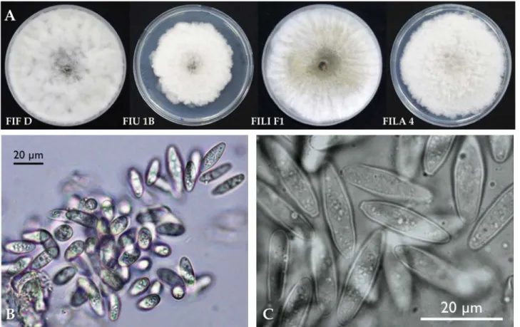
cankers (Figure 2D). Colonies on MEA formed concen-tric rings. On PDA mycelium was white and became smoky gray to gray-olivaceous after 5 d (Figure 3A). The mycelium was fast-growing (Table 3) and covered the 9 cm diam. Petri dishes after 5 d incubation at 25° C in the dark. Optimum temperature for radial colony growth was between 25 and 30°C for all the isolates test-ed. Little growth was observed at 10 or 35°C. Stromat-ic conidiomata were produced in pine needle cultures within 14 d. The condidiomata were solitary, covered by mycelium, obpyriform to ampulliform, and each had Figure 2. A. Cankers on cladodes incited by Neofusicoccum batangarum in a cactus pear hedge on the Island of Ustica, May 2014. B.
Coalesc-ing, concentric cankers incited by Neofusicoccum batangarum on a cactus pear cladode. C. Concentric expanding canker incited by Neofusico-ccum batangarum on a cactus pear cladode. Note pycnidia, as small dark spots, and cracking on the silvery, intermediate area of the canker, the dark colour and the sooty appearance of the exudate after rain and the tan colour of the edge, indicating that the canker is still active. D. Mycelium emerging from conidiomata of Neofusicoccum batangarum formed on cankers (photograph taken using a stereomicroscope).
a central and circular unilocular ostiole, and measured 250-300 μm in diameter. Conidia were non-septate (bi-cellular conidia were observed only very occasionally), hyaline, smooth, fusoid to ovoid, thin-walled, and meas-ured 17.1-21.8 × 4.6-8.9 μm, with a mean length to width ratio = 2.9 (Figure 3B–C).
The isolates obtained had identical ITS, tef1 and
tub2 sequences. Preliminary BLAST analyses of these
three genes yielded several identical sequences of
Neo-fusicoccum spp., deposited with different taxa names.
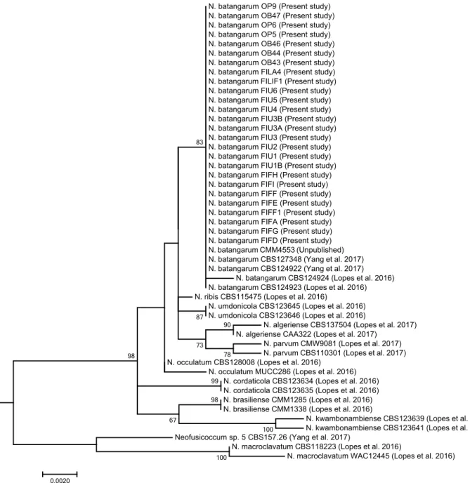
Consequently, this analysis enabled the identification at the genus level, but did not provide reliable infor-mation on the species. The phylogenetic analysis of the combined data set of sequences from ITS, tef1 and
tub2 sequences (Figure 4) produced trees with a high
concordance with those reported by Lopes et al. (2017) and Yang et al. (2017). According to this analysis, iso-lates from cactus pear were identified as N.
batangar-um, since they clearly clustered with the ex-type (CBS
124924 from Terminalia catappa; Lopes et al., 2016) and other reference isolates of this species, and were differ-entiated from other Neofusicoccum species, including Figure 3. A. Four representative isolates of Neofusicoccum batangarum after 5 d of incubation on PDA at 25°C; CBS143023 (FIF D),
CBS143024 (FIU 1B), CBS143025 (FILI F1), and CBS143026 (FILA 4). B and C. Unicellular, fusiform, thin-walled hyaline conidia of Neofu-sicoccum batangarum.
Table 3. Mean radial growth rates of colonies of Neofusicoccum batangarum isolates on PDA at three different temperatures, as determined after 3 d of incubation.
Isolates of N.
batangarum Island origin
15°C (mm d-1) mean ± S.D.a 25°C (mm d-1) mean ± S.D. 30°C (mm d-1) mean ± S.D. FILI-F1 Linosa 3.83 ± 0.35 7.50 ± 0.00 5.90 ± 0.26 FILA 4 Lampedusa 3.97 ± 0.34 7.50 ± 0.00 7.50 ± 0.00 FIU-1B Ustica 3.00 ± 0.58 6.53 ± 0.63 6.26 ± 0.42 FIF-D Favignana 3.40 ± 0.41 7.50 ± 0.00 7.50 ± 0.00 OP5 Lampedusa 4.32 ± 0.05 9.77 ± 0.09 9.83 ± 0.06 OP6 Lampedusa 4.20 ± 0.21 12.94 ± 0.23 9.75 ± 0.13 OP9 Lampedusa 4.31 ± 0.09 13.00 ± 0.19 9.33 ± 0.24 OB44 Linosa 3.73 ± 0.81 12.83 ± 0.10 9.69 ± 0.06 OB46 Linosa 3.44 ± 0.44 13.03 ± 0.18 9.23 ± 0.42 OB47 Lampedusa 4.16 ± 0.10 12.94 ± 0.16 9.77 ± 0.11 OB43 Linosa 2.40 ± 0.22 7.82 ± 0.07 6.47 ± 0.06
the closely related species N. ribis, N. umdonicola, and
N. occulatum (Annex 1).
Analysis of the genetic variability of isolates
The MSP-PCR characterization of the six representa-tive isolates of N. batangarum revealed high genetic
uni-formity, since isolates showed identical banding patterns with the three tested primers (Annex 2).
Pathogenicity tests
Four isolates, one from each island, were deposited at Westerdijk Fungal Biodiversity Institute [CBS 143023
N. batangarum OP9 (Present study) N. batangarum OB47 (Present study) N. batangarum OP6 (Present study) N. batangarum OP5 (Present study) N. batangarum OB46 (Present study) N. batangarum OB44 (Present study) N. batangarum OB43 (Present study) N. batangarum FILA4 (Present study) N. batangarum FILIF1 (Present study) N. batangarum FIU6 (Present study) N. batangarum FIU5 (Present study) N. batangarum FIU4 (Present study) N. batangarum FIU3B (Present study) N. batangarum FIU3A (Present study) N. batangarum FIU3 (Present study) N. batangarum FIU2 (Present study) N. batangarum FIU1 (Present study) N. batangarum FIU1B (Present study) N. batangarum FIFH (Present study) N. batangarum FIFI (Present study) N. batangarum FIFF (Present study) N. batangarum FIFE (Present study) N. batangarum FIFF1 (Present study) N. batangarum FIFA (Present study) N. batangarum FIFG (Present study) N. batangarum FIFD (Present study) N. batangarum CMM4553 (Unpublished) N. batangarum CBS127348 (Yang et al. 2017) N. batangarum CBS124922 (Yang et al. 2017)
N. batangarum CBS124924 (Lopes et al. 2016) N. batangarum CBS124923 (Lopes et al. 2016) N. ribis CBS115475 (Lopes et al. 2016)
N. umdonicola CBS123645 (Lopes et al. 2016) N. umdonicola CBS123646 (Lopes et al. 2016)
N. algeriense CBS137504 (Lopes et al. 2017) N. algeriense CAA322 (Lopes et al. 2017)
N. parvum CMW9081 (Lopes et al. 2017) N. parvum CBS110301 (Lopes et al. 2017) N. occulatum CBS128008 (Lopes et al. 2016)
N. occulatum MUCC286 (Lopes et al. 2016) N. cordaticola CBS123634 (Lopes et al. 2016) N. cordaticola CBS123635 (Lopes et al. 2016) N. brasiliense CMM1285 (Lopes et al. 2016) N. brasiliense CMM1338 (Lopes et al. 2016)
N. kwambonambiense CBS123639 (Lopes et al. 2016) N. kwambonambiense CBS123641 (Lopes et al. 2016) Neofusicoccum sp. 5 CBS157.26 (Yang et al. 2017)
N. macroclavatum CBS118223 (Lopes et al. 2016)
N. macroclavatum WAC12445 (Lopes et al. 2016)
83 87 90 78 73 99 98 100 67 98 100 0.0020
Figure 4. Phylogenetic tree of isolates of Neofusicoccum collected in the present study from Opuntia ficus-indica, and representative isolates
of Neofusicoccum batangarum and closely related species as defined by in comprehensive phylogenetic studies (Tables 1 and 2). The tree was built using concatenated sequences of ITS-5.8S-ITS2 region, tef1-α gene and β-tubulin gene. Numbers on nodes indicate the posterior prob-abilities from the maximum likelihood method.
(FIF D), CBS 143024 (FIU 1B), CBS 143025 (FILI F1), and CBS 143026 (FILA 4)], and these were tested for their pathogenicity on cactus pear and other species of plants. Additional isolates from Lampedusa and Linosa were also included in the pathogenicity tests. Since the results of four inoculation series performed, respectively, in 2014, 2015, 2016 and 2017 were very similar, only the results of inoculations performed in 2014 are reported here in detail. All isolates were pathogenic on the inoc-ulated plant species (Tables 4, 5, 6 and 7). The isolates induced cankers in all inoculated plants while no symp-toms were observed on the controls.
On cactus pear plants, symptoms appeared after 4 d, as brown circular halos around the inoculation wounds with viscous exudates oozing from the lesions that con-solidated in contact with the air to form long strips
(Figure 5A–B). Cankers expanded progressively and concentrically (Figure 5D). They were roughly circular, with irregular margins, identical or very similar to those observed on plants with natural infections, and the can-ker expansion reduced during the coldest months (late December to early February). Some cankers stopped growing permanently and healed, but most cankers resumed growth and the production of exudate when temperatures increased after winter. Table 4 presents the mean lesion areas induced on cactus pear cladodes by the four tested isolates 30 d after wound inoculation. Analysis of variance revealed no statistically significant differences in pathogenicity between the isolates. Five years after the first inoculation, most cankers were still active, and they continued to expand and produce abun-dant exudates (Figure 5D). In some cases, the cankers expanded more rapidly in one direction and became asymmetrical and irregular (Figure 5D). After rain, the cankers became dark, with a carbonaceous appearance. Table 4. Mean lesion areas on cladodes of cactus pear (Opuntia
ficus-indica) 30 d after wound inoculation with representative iso-lates of Neofusicoccum batangarum obtained from cactus pear from four minor islands of Sicily.
Isolate Island origin Mean lesion area (cm2) ± S.D. a,b
FIF D Favignana 4.4 ± 1.2
OP6 Lampedusa 4.4 ± 1.8
OB43 Linosa 4.5 ± 2.3
FIU 1B Ustica 4.6 ± 0.6
a Means of 12 replicate values. b ANOVA, F (3,44) = 0.372, P > 0.05
Table 5. Mean lesion areas on stems of sweet orange ‘Navelina’ (Cit-rus × sinensis) trees grafted on ‘Carrizo’ Citrange (C. sinensis × Pon-cirus trifoliata) rootstock, 30 d after wound inoculation with rep-resentative isolates of Neofusicoccum batangarum from four minor islands of Sicily
Isolate f Island origin Mean lesion area (cm 2) ± S.D.a
rootstock b,d scionc,e
FIF D*** Favignana 1.4 ± 0.18 1.8 ± 0.1
FIU 1B** Ustica 1.4 ± 0.12 1.8 ± 0.2
FILI F1* Linosa 1.4 ± 0.15 1.7 ± 0.2
FILA 4** Lampedusa 1.5 ± 0.26 1.9 ± 0.4
a Rootstock: means of four replicate values. Scion: means of eight
replicate values.
b Symptoms included barely noticeable gummy exudate. c Symptoms included abundant gummosis.
d ANOVA rootstock, F
(3,12) = 0, P > 0.05. e ANOVA scion, F
(3,28) = 0, P > 0.05.
f Comparing rootstock and scion according to Student’s t-test.
(* = P < 0.05, ** = P < 0.01, and *** = P < 0.001).
Table 6. Mean lesion areas on stems of holm oak (Quercus ilex)
trees 30 d and 70 d after wound inoculation with representative iso-lates of Neofusicoccum batangarum from two minor islands of Sicily. Isolate Island origin Mean lesion area (cm
2) ± S.D.a
30 db 70 dc
FIF D Favignana 5.2 ± 1.3 8.2 ± 1.1
FIU 1B Ustica 5.8 ± 2.4 10.0 ± 3.0
a Means of six replicate values. b ANOVA 30 d, F(1,10) = 0.274, P > 0.05. c ANOVA 70 d, F
(1,10) = 1.517, P >0 .05.
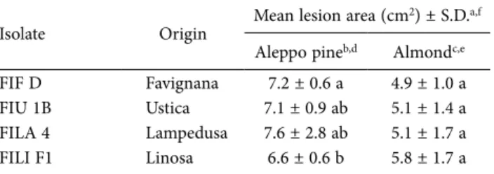
Table 7. Mean lesion areas on stems of Aleppo pine (Pinus halepen-sis) or almond (Prunus dulcis) trees 70 d after wound inoculations with representative isolates of Neofusicoccum batangarum from four minor islands of Sicily.
Isolate Origin Mean lesion area (cm
2) ± S.D.a,f
Aleppo pineb,d Almondc,e
FIF D Favignana 7.2 ± 0.6 a 4.9 ± 1.0 a FIU 1B Ustica 7.1 ± 0.9 ab 5.1 ± 1.4 a FILA 4 Lampedusa 7.6 ± 2.8 ab 5.1 ± 1.7 a FILI F1 Linosa 6.6 ± 0.6 b 5.8 ± 1.7 a
a Means of six replicates.
b Symptoms included resinous exudates.
c Symptoms included abundant gummous exudates. d ANOVA Aleppo pine, F(3,20) = 6.015, P = 0.004. e ANOVA Almond, F
(3,20) = 2.783, P = 0.06.
f Means accompanied by the same letters are not statistically
Pycnidia were visible on cankers 14-20 d a.i. From six months to 5 years a.i., small cankers and scattered erup-tions of exudate, similar to runny wax, appeared on arti-ficially inoculated cladodes approx. 10-20 cm from the inoculation points.
All four tested N. batangarum isolates induced necrotic lesions on the citrange rootstock and the sweet orange scion at 7 d a.i. Symptoms were more severe on
the sweet orange scions (Table 5) and included abun-dant gummosis (Figure 5G), while gummosis was much less abundant on citrange. Pycnidia emerged from the necrotic lesions on sweet orange stems from 10 to 14 d a.i. (Figure 5H). Differences in mean lesion size between the sweet orange scion and the citrange rootstock were significant (P < 0.05), according to Student’s t-test. How-ever, no statistically significant differences in patho-Figure 5. A. Brown, circular lesions and waxy exudate oozing from lesions on a cactus pear cladode wound-inoculated with Neofusicoc-cum batangarum, 7 d after inoculation (a.i.) B. Brown lesion and exudate oozing from a lesion on a cactus pear cladode wound-inoculated with N. batangarum, 4 d a.i. C. A fusiform dry canker induced by wound-inoculation of N. batangarum on the stem of a holm oak tree 70 d a.i. D. A still active canker on a cactus pear cladode artificially inoculated with Neofusicoccum batangarum in the field, September 2019, 5 years a.i. E. A resinous canker induced by wound-inoculation with N. batangarum on the stem of an Aleppo pine tree, 70 d a.i. F. A gummy canker induced by wound-inoculation with N. batangarum on the stem of a young almond tree, 14 d a.i. G. A gummy canker induced by wound-inoculation with N. batangarum on the stem of a young ‘Navelina’ sweet orange tree, 14 d a.i. H. Pycnidia on a canker induced by wound-inoculation with N. batangarum on the stem of a young ‘Navelina’ sweet orange tree, 14 d a.i.
genicity (P > 0.05) were observed among the fungal isolates on both citrus symbionts. No symptoms were observed on the controls.
On holm oak (Table 6), no gummy reaction was observed (Figure 5 C), but cankers expanded progres-sively along the stems and were still active 3 years after inoculation. No symptoms were observed on the con-trols. There were no significant differences (P > 0.05) among isolates in the pathogenicity test on holm oak at 30 d a.i. or 70 d a.i.
On almond and Aleppo pine (Table 7), N.
batan-garum isolates incited necrotic bark lesions with
gum-my exudates on almond (Figure 5F) and resinous exu-dates on Aleppo pine (Figure 5E). No symptoms were observed on the controls. Also on almond and Aleppo pine, cankers were still active 3 years a.i.
In the pathogenicity tests on almond and Aleppo pine there were small but statistically significant differ-ences between the isolates on these two hosts (almond,
P = 0.06; Aleppo pine, P = 0.004). All the N. batangarum
isolates were re-isolated from inoculated plants, while no fungal pathogens were isolated from control plants, thus fulfilling Koch’s postulates for the N. batangarum iso-lates.
DISCUSSION
Identification of the Botryosphaeriaceae prior to the application of DNA sequencing and phylogenetic infer-ence should be considered with caution. The classification and nomenclature of these fungi have evolved rapidly and have been substantially revised, as previous classifi-cation based on morphological characters was confus-ing and most species were actually complexes of differ-ent taxa (Slippers et al., 2004, 2013; Crous et al., 2006; Phillips et al., 2008, 2013; Crous et al., 2017). According to the molecular taxonomy of the Botryosphaeraceae,
N. batangarum is the appropriate name of the fungus
responsible for the chronic disease observed on the clad-odes of cactus pear in the minor islands of Sicily.
Neofu-sicoccum batangarum was confirmed to be the sole causal
agent of the ‘scabby canker’ disease. The fungus was con-sistently associated with symptomatic cactus pear plants and, when artificially inoculated onto this host, induced the same type of cankers as natural infections. From an etiological and ecological perspective, N. batangarum was the only fungus in the family Botryosphaeriaceae recov-ered from infected cactus pear in these small islands belonging to different archipelagos.
Several species of this family frequently occur together on the same host (Jami et al., 2017), although
not all are able to cause disease (Lawrence et al., 2017). In Brazil, N. batangarum, alone or in association with other fungi including several species of
Botryospha-eriaceae, was reported to be responsible for ‘brown spot’
of cladodes, a severe disease of cochineal cactus that is grown as fodder for livestock in the semi-arid region of the north-east of that country (Conforto et al., 2016, 2019). Although the syndromes of ‘cladode brown spot’ in north-eastern Brazil and ‘scabby canker’ in minor islands of Sicily have some traits in common, they are distinct. Differences between the symptoms of these dis-eases include the presence of crusty, silvery, perennial cankers, and exudates oozing from the cankers in the ‘scabby canker’ disease. However, differences might be due to environmental conditions, host plant and/or cul-tivation systems. In Brazil, cochineal cactus is pruned repeatedly for the production of fresh forage. The cactus pear fences in the minor islands near Sicily are pruned only occasionally, thus allowing the disease to become chronic on mature cladodes. The cladode brown spot in Brazil is also a complex disease and the causal agent may vary according to the season and the geographical region (Santana et al., 2020).
For many years, Botryosphaeriaceae, which are wide-spread in tropical and temperate regions, were con-sidered to be opportunistic pathogens infecting hosts exclusively through wounds or natural openings in their periderms. Since the late 1980s, however, these fungi have been recognized as endophytes that remain latent in woody host plants for long periods. With the onset of abiotic stress conditions (drought, physical damage, water-logging, frost and unsuitable environments for the growth) the latent pathogens cause disease (Slippers and Wingfield, 2007; Pavlic-Zupanc et al., 2015; Marsberg et
al., 2017). The prolonged latent infection or endophytic
phase implies that these fungi can easily pass undetected through phytosanitary controls or during the selection of propagation material.
The genus Neofusicoccum comprises species with widespread geographical and host distributions, and these fungi are typically endophytes, which in stress-ful environments can cause symptoms such as dieback, cankers and gummosis (Crous et al., 2006; Lopes et al., 2017; Zhang et al., 2017; Burgess et al., 2018). Wounds caused by hailstorms may have been the factor trigger-ing the epidemic outbreak of N. batangarum on cactus pear in the small islands of Sicily. The climate of these islands is affected by the proximity to the sea, and this may have favoured the development of the disease and the survival of the inoculum. In in vitro tests, N.
batan-garum showed an optimum temperature for growth
maxi-mum of approx. 35°C. On artificially inoculated clad-odes, the fungus formed pycnidia between 14 and 20 d a.i. In winter, conidia collected from pycnidia formed on cladodes artificially inoculated in the spring or autumn of the previous year were viable.
Most species of Botryosphaeriaceae have broad host ranges, and only very few have been described from a limited number of host species or are host specific (Slip-pers and Wingfield, 2007; Marsberg et al., 2017). The ability to infect multiple hosts and to move among unre-lated hosts facilitates the establishment and spread of species and genotypes of this family into new areas.
Neofusicoccum batangarum has been reported as an
endophyte as well as a pathogen of several host plants in the tropics (Begoude et al., 2010, 2011; Shetty et al., 2011; Conforto et al., 2016; Netto et al., 2017). A very recent report has further expanded the known hosts (Serrato-Diaz et al., 2020). Pathogenicity tests in the present study showed that this fungus has an even wider poten-tial host range, encompassing woody forest and culti-vated plants typical of the Mediterranean macro-region. These hosts include Aleppo pine, almond, citrus and holm oak. Like other Botryosphaeriaceae, N.
batanga-rum can be regarded as a generalist pathogen, although
in natural conditions the host affinity of polyphagous
Botryosphaeriaceae species is strongly influenced by the
environment (Slippers and Wingfield, 2007). On arti-ficially inoculated sweet orange stems, N. batangarum induced the typical symptoms of ‘gummy cankers’ or ‘bot gummosis’, already known as ‘Dothiorella gum-mosis’. These are minor, but widespread, diseases caused by diverse species of Botryosphaeriaceae, and they com-monly occur in citrus groves in California and in the Mediterranean basin (Adesemoye et al., 2014; Guar-naccia and Crous, 2017). Consistently with the typical symptoms of ‘gummy canker’ of citrus, in artificially inoculated symbiont citrus plant, symptoms were more severe on the sweet orange scion than on rootstock.
The polyphagy of N. batangarum may be related to its ability to produce non-host specific phytotoxins (Masi
et al., 2020). These toxins may have roles in
pathogenic-ity as virulence factors and may also enhance the eco-logical fitness of the fungus by inhibiting other micro-organisms competing in plant biospheres. Production of diffusible phytotoxins could also explain the systemic spread of symptoms on cactus pear cladodes and their appearance far from inoculation points (Masi et al. 2020).
Reports of Botryosphaeriaceae associated with vari-ous hosts have increased worldwide in recent years. In Italy, this family is common and widespread on a broad range of hosts, and is an increasing concern for
agri-cultural crops and urban and natural forest ecosystems (Burruano et al., 2008; Linaldeddu et al., 2014, 2016; Dissanayake et al., 2016b;). The disease of cactus pear caused by N. batangarum was noticed for the first time in Linosa more than 45 years ago (Somma et al., 1973), and at that time it was widespread and had been estab-lished for many years. Similarly, the chronic nature of symptoms observed recently in Favignana, Lampedusa and Ustica and the widespread occurrence of the dis-ease in Lampedusa, clearly indicate it has not emerged recently. During the last 50 years the disease has prob-ably been favoured by the low frequency of cactus pear pruning as a consequence of the reduced importance of this plant as a crop, and the drastic reduction in the use of cladodes as fodder. Although Sicily is the main cactus pear fruit producer in Italy, with more than 3,500 ha of specialized culture (Ochoa and Barbera, 2017), this dis-ease has not been reported in cactus pear cultivations in Sicily. The present study has shown that N. batangarum populations from Favignana, Lampedusa, Linosa and Ustica were genetically uniform, despite their geographi-cal isolation.
It can be assumed that conidia and ascospores of
Botryosphaeriaceae are dispersed by wind and rain only
over short distances. The occurrence of N. batangarum only on cactus pear plantations of the minor islands, and the genetic uniformity of the fungus populations, may indicate that these populations have a common ori-gin, and that the widespread distribution of a single gen-otype of the pathogen has resulted from anthropogenic activity. Neofusicoccum batangarum, as an endophytic or latent pathogen, may have been introduced with cactus pear cladodes collected in other geographical areas and used as propagation material. This hypothesis is consist-ent with cactus pear being introduced on a large scale into small islands of the Mediterranean Sea as fodder for livestock, for field fences or for edible fruit produc-tion, at one recent time (Pretto et al., 2012). This was when more intensive colonization and exploitation of agriculture in these islands were promoted by the public authorities.
From an ecological perspective, the emergence of this disease in such an aggressive form, which has become a limiting factor for the cultivation of cactus pear in these small islands, may be partly due to the failure of the acclimatization of a non-native plant species. However, the occurrence of a serious disease of a crop of economic and landscaping relevance for Sicily in a restricted geo-graphical area, but very close and frequently connected to the main island by tourist traffic, may have phytosani-tary implications. Appropriate actions should be taken to prevent further spread and introduction of N.
batan-garum into areas where the non-native cactus pear host
is naturalized and intensively cultivated. LITERATURE CITED
Adesemoye A. O., Mayorquin J. S., Wang D. H., Twizeyi-mana M., Lynch S. C., Eskalen A. 2014. Identification of species of Botryosphaeriaceae causing bot gum-mosis in citrus in California. Plant Disease 98:55–61. Begoude B.A.D., Slippers B., Wingfield M.J., Roux J. 2010.
Botryosphaeriaceae associated with Terminalia
cat-appa in Cameroon, South Africa and Madagascar. Mycological Progress 9: 101–123.
Begoude B.A.D., Slippers B., Wingfield M.J., Roux J. 2011. The pathogenic potential of endophytic Botryospha-eriaceous fungi on Terminalia species in Cameroon.
Forest Pathology 41: 281–292
Burgess T.I., Tan Y.P., Garnas J., Edwards J., Scarlett K.A., …, Jami F. 2018. Current status of the Botryospha-eriaceae in Australia. Australasian Plant Pathology DOI: 10.1007/s13313–018-0577-5
Burruano S., Mondello V., Conigliaro G., Alfonzo A., Spagnolo A., Mugnai L. 2008. Grapevine decline in Italy caused by Lasiodiplodia theobromae.
Phytopath-ologia Mediterranea 47: 132–136.
Carbone I., Kohn L.M. 1999. A method for design-ing primer sets for speciation studies in filamentous ascomycetes. Mycologia 91: 553–556.
Conforto C., Lima N.B., Garcete-Gómez J.M., Câmara M.P.S., Michereff S.J. 2016. First Report of Cladode Brown Spot in Cactus Prickly Pear Caused by
Neo-fusicoccum batangarum in Brazil. Plant Disease 100:
1238.
Conforto C., Lima N.B., Silva F.J.A., Câmara M.P.S., Maharachchikumbura S., Michereff S.J. 2019. Char-acterization of fungal species associated with cladode brown spot on Nopalea cochenillifera in Brazil.
Euro-pean Journal of Plant Pathology 155: 1179–1194.
Crous P.W., Slippers B., Wingfield M.J., Rheeder J., Mara-sas W.F.O., …, Groenewald J.Z. 2006. Phylogenetic lineages in the Botryosphaeriaceae. Studies in
Mycol-ogy 55: 235–253.
Crous P.W., Slippers B., Groenewald J.Z., Wingfield M.J. 2017. Botryosphaeriaceae: systematics, pathology, and genetics. Fungal Biology 121: 305–306.
Dissanayake A.J., Phillips A.J.L., Li X.H., Hyde K.D. 2016a. Botryosphaeriaceae: Current status of genera and species. Mycosphere 7: 1001–1073.
Dissanayake A.J., Camporesi E. Hyde K.D., Phillips A.J.L., Fu C.Y., …, Li X.H.2016b. Dothiorella species associ-ated with woody hosts in Italy. Mycosphere 7: 51–63.
Feijo F.M., Silva M.J.S., Nascimento A.D., Infante N.B., Ramos-Sobrinho R., …, Lima G.S.A. 2019. Botry-osphaeriaceae species associated with the prickly pear cactus, Nopalea cochinellifera. Tropical Plant
Pathology 44: 452–459.
Freeman S., Shabi, E.E. 1996 Cross-infection of subtropi-cal and temperate fruits by Colletotrichum species from various hosts. Physiological Molecular Plant
Pathology 49:395–404.
Garcete-Gómez J.M., Conforto C., Domínguez-Monge S., Flores-Sánchez J.L.F., Mora-Aguilera G., Micher-eff S.J. 2017. Sample size for assessment of cladode brown spot in prickly pear cactus. European Journal
of Plant Pathology 149: 759–763.
Edgar R.C. 2004. Muscle: multiple sequence alignment with high accuracy and high throughput. Nucleic
Acids Research 32: 1792–1797.
Glass N.L., Donaldson G.C. 1995. Development of primer sets designed for use with the PCR to amplify con-served genes from filamentous ascomycetes. Applied
and Environmental Microbiology 61: 1323–1330.
Granata G., Faedda R., and Ochoa M.J. 2017. Diseases of cactus pear. In: Crop Ecology, Cultivation and user of Cactus pear (P. Inglese, C. Mondragon, A. Nefzaoui, C. Saenz, eds.), FAO and ICARDA, Rome, Italy, 115– 126.
Guarnaccia V., Crous P.W. 2017. Emerging citrus diseases in Europe caused by species of Diaporthe. IMA
Fun-gus 8: 317–334.
Jami F., Wingfield M.J., Gryenhout M., Slippers B. 2017. Diversity of tree-infecting Botryosphaeriales on native and non-native trees in South Africa and Namibia. Australasian Plant Pathology 46: 529–545. Kiesling R., Metzing D. 2017. Origin and Taxonomy of
Opuntia ficus-indica. In: Crop Ecology, Cultivation
and user of Cactus pear (P. Inglese, C. Mondrag-on, A. Nefzaoui, C. Saenz eds.), FAO and ICARDA, Rome, Italy, 14–19
Kumar S., Stecher G., Tamura K. 2016. MEGA7: Molecu-lar Evolutionary Genetics Analysis Version 7.0 for Bigger Datasets. Molecular Biology and Evolution 33: 1870–1874
Lawrence P.D., Peduto Hand F., Gubler W.D., Trouillas F.T. 2017. Botryosphaeriaceae species associated with dieback and canker disease of bay laurel in northern California with the description of Dothiorella
califor-nica sp. nov. Fungal Biology 121: 347–360.
Linaldeddu B.T., Scanu B., Maddau L., Franceschini A. 2014. Diplodia corticola and Phytophthora
cinnamo-mi: the main pathogens involved in holm oak decline
on Caprera island (Italy). Forest Pathology 44: 191– 200.
Linaldeddu B.T., Alves A., Phillips A.J.L. 2016. Sardiniella
urbana gen. et sp. nov., a new member of the Botry-osphaeriaceae isolated from declining Celtis australis
trees in Sardinian streets capes. Mycosphere 7: 893– 905.
Lopes A., Barradas C., Phillips A.J.L., Alves A. 2016. Diversity and phylogeny of Neofusicoccum species occurring in forest and urban environments in Por-tugal. Mycosphere 7: 906–920.
Lopes A., Phillips A.J.L., Alves A. 2017. Mating type genes in the genus Neofusicoccum: Mating strategies and usefulness in species delimitation. Fungal Biology 121, 394–404.
Marsberg A., Kemler M., Jami F., Nagel J.H., Postma-Smidt A., …, Slippers B., 2017. Botryosphaeria
doth-idea: a latent pathogen of global importance to
woody plant health. Molecular Plant Pathology 18: 477–488.
Masi M., Aloi F., Nocera P., Cacciola S.O., Surico G., Evi-dente A. 2020. Phytotoxic metabolites isolated from
Neofusicoccum batangarum, the causal agent of the
scabby canker of cactus pear (Opuntia ficus-indica L.). Toxins 12: 126; DOI: 10.3390/toxins12020126 Meyer W., Mitchell T.G., Freedman E.Z., Vilgalys R. 1993.
Hybridization probes for conventional DNA finger-printing used as single primers in the polymerase chain reaction to distinguish strains of
Cryptococ-cus neoformans. Journal of Clinical Microbiology 31:
2274–2280.
Netto M.S.B., Lima W.G., Correia K.G., Da Silva C.F.B., Thon M., … Camâra M.P.F. 2017. Analysis of phy-logeny, distribution and pathogenicity of
Botryospha-eriaceae species associated with gummosis of Ana-cardium in Brasil, with a new species of Lasiodiplo-dia. Fungal Biology 121: 437–451.
Ochoa M.J., Barbera G., 2017. History and economic and agro-ecological importance. In: Crop Ecology, Culti-vation and user of Cactus pear (P. Inglese, C. Mon-dragon, A. Nefzaoui, C. Saenz eds.), FAO and ICAR-DA, Rome, Italy, 1–12
Pavlic-Zupanc D., Wingfield M.J., Boissin, E., Slippers, B. 2015. The distribution of genetic diversity in
the Neo-fusicoccum parvum/N. ribis complex suggests
struc-ture correlated with level of disturbance. Fungal
Ecol-ogy 13: 93–102.
Phillips A.J.L., Alves A., Pennycook S. R., Johnston P.R., Ramaley A., ..., Crous P.W. 2008. Resolving the phy-logenetic and taxonomic status of dark-spored tele-omorph genera in the Botryosphaeriaceae. Persoonia 21: 29–55
Phillips A.J.L., Alves A., Abdollahzadeh J., Slippers B., Wingfield M.J., …, Crous P.W. 2013. The
Botryospha-eriaceae: Genera and species known from culture.
Studies in Mycology 76: 51–167
Pretto F., Celesti-Grapow L., Carli E., Brundu G., Blasi C. 2012. Determinants of non-native plant species rich-ness and composition across small Mediterranean islands. Biological Invasions 14: 2559–2572.
Rayner R.W. 1970. A Mycological Colour Chart. Com-monwealth Mycological Institute, Kew, Surrey, UK. Santana M.D., de Lima Leite I.C.H., da Silva Santos I.C.,
Michereff S.J., Freitas-Lopes R.do L., Lopes U.P. 2020. Reduction of cladode brown spot in cactus pear in semiarid growing areas and yield increase using fun-gicides. Journal of Plant Pathology DOI: 10.1007/ s42161–019-00462-9
Santos J.M., Phillips A.J.L. 2009. Resolving the com-plex of Diaporthe (Phomopsis) species occurring on
Foeniculum vulgare in Portugal. Fungal Diversity 34:
111–125.
Schena L., Cooke D.E.L., 2006. Assessing the potential of regions of the nuclear and mitochondrial genome to develop a “molecular tool box” for the detection and characterization of Phytophthora species. Journal of
Microbiological Methods 67: 70–85.
Schena L., Surico G., Burruano S., Giambra S., Pane A., …, Cacciola S.O. 2018. First report of Neofusicoccum
batangarum as causal agent of scabby cankers of
cac-tus pear (Opuntia ficus-indica) in minor islands of Sicily. Plant Disease 102: 445.
Serrato-Diaz L.M., Aviles-Noriega A., Soto-Bauzó A., Rivera-Vargas L.I., Goenaga R., Bayman P. 2020. Bot-ryosphaeriaceae fungi as causal agents of dieback and corky bark in rambutan and longan. Plant Disease 104: 105–115.
Shetty K.G., Minnis A.M., Rossman A.Y., Jayachandran K. 2011. The Brazilian peppertree seedborne patho-gen, Neofusicoccum batangarum, a potential biocon-trol agent. Biological Conbiocon-trol 56: 91–97.
Slippers B., Wingfield M.J. 2007.
Botryosphaeriace-ae as endophytes and latent pathogens of woody
plants: diversity, ecology and impact. Fungal Biology
Reviews21: 90–106.
Slippers B., Crous P.W., Denman S., Coutinho T.A., Wingfield B.D., Wingfield M.J. 2004. Combined mul-tiple gene genealogies and phenotypic characters dif-ferentiate several species previously identified as
Bo-tryosphaeria dothidea. Mycologia 96: 83–101.
Slippers B., Boissin E., Phillips A.J.L., Groenewald J.Z., Lombard L., …, Crous P.W. 2013. Phylogenetic line-ages in the Botryosphaeriales: a systematic and evolu-tionary framework. Studies in Mycology 76: 31–49. Smith H., Wingfield M.J., Crous P.W., Coutinho T.A.,
doth-idea endophytic in Pinus spp. and Eucalyptus spp.
in South Africa. South African Journal of Botany 62: 86–88.
Somma V., Rosciglione B., Martelli G.P. 1973. Preliminary observations on gummous canker, a new disease of prickly pear. Tecnica Agricola 25: 437–443.
Tamura K., Nei M. 1993. Estimation of the number of nucleotide substitutions in the control region of mitochondrial DNA in humans and chimpan-zees. Molecular Biology and Evolution 10: 512–526. Tamura K., Stecher G., Peterson D., Filipski A., Kumar
S. 2013. MEGA6: Molecular Evolutionary Genetics Analysis Version 6.0. Molecular Biology and Evolution 30: 2725–2729.
Uddin W., Stevenson, K.L., Pardo-Schultheiss R.A. 1997. Pathogenicity of a species of Phomopsis causing a shoot blight on peach in Georgia and evaluation of possible infection courts. Plant Disease 81: 983–998. Yang T., Groenewald J.Z., Cheewangkoon R., Jami F.,
Abdollahzadeh J., … , Crous P.W. 2017. Families, genera, and species of Botryosphaeriales. Fungal
Biol-ogy 121: 322–346.
Vaidya N.H., Hadjicostis C.N., Dominguez-Garcia A.D. 2011. Distributed algorithms for consensus and coor-dination in the presence of packet-dropping commu-nication links-part II: Coefficients of ergodicity anal-ysis approach. arXiv preprint arXiv:1109.6392.
White T. J., Bruns T. D., Lee S. B., Taylor J. W. 1990. Amplification and direct sequencing of fungal ribo-somal RNA Genes for phylogenetics. In: PCR -
Pro-tocols and Applications - A Laboratory Manual (M.A.
Innis, D.H. Gelfand, J.J. Sninsky, T.J White eds.), Aca-demic Press Inc., New York, USA, 315–322.
Zhang M., Lin S., He W., Zhang Y. 2017. Three species of Neofusicoccum (Botryosphaeriaceae, Botryospha-eriales) associated with woody plants from southern China. Mycosphere 8: 797–808.