CHRONIC OBSTRUCTIVE PULMONARY DISEASE
COPD increases the risk of squamous histological subtype in
smokers who develop non-small cell lung carcinoma
A Papi, G Casoni, G Caramori, I Guzzinati, P Boschetto, F Ravenna, N Calia, S Petruzzelli,
L Corbetta, G Cavallesco, E Forini, M Saetta, A Ciaccia, L M Fabbri
. . . .
See end of article for authors’ affiliations . . . . Correspondence to: Dr A Papi, Research Centre on Asthma and COPD, University of Ferrara, Via Savonarola 9, 44100 Ferrara, Italy; [email protected] Received3November2003 Accepted 7 April 2004 . . . . Thorax 2004;59:679–681. doi: 10.1136/thx.2003.018291
Background:Squamous cell carcinoma has a stronger association with tobacco smoking than other non-small cell lung cancers (NSCLC). A study was undertaken to determine whether chronic obstructive pulmonary disease (COPD) is a risk factor for the squamous cell carcinoma histological subtype in smokers with surgically resectable NSCLC.
Methods:Using a case-control design, subjects with a surgically confirmed diagnosis of squamous cell carcinoma were enrolled from smokers undergoing lung resection for NSCLC in the District Hospital of Ferrara, Italy. Control subjects were smokers who underwent lung resection for NSCLC in the same hospital and had a surgically confirmed diagnosis of NSCLC of any histological type other than squamous cell.
Results:Eighty six cases and 54 controls (mainly adenocarcinoma, n = 50) were enrolled. The presence of COPD was found to increase the risk for the squamous cell histological subtype by more than four times. Conversely, the presence of chronic bronchitis was found to decrease the risk for this histological subtype by more than four times. Among patients with chronic bronchitis (n = 77), those with COPD had a 3.5 times higher risk of having the squamous cell histological subtype.
Conclusions:These data suggest that, among smokers with surgically resectable NSCLC, COPD is a risk factor for the squamous cell histological subtype and chronic bronchitis, particularly when not associated with COPD, is a risk factor for the adenocarcinoma histological subtype.
L
ung cancer is the leading cause of death from cancer worldwide, and tobacco smoking is associated with 90% of cases of lung cancer.1Cigarette smoking is also the most important risk factor for chronic bronchitis and/or chronic obstructive pulmonary disease (COPD).2
Lung cancer, COPD, and chronic bronchitis therefore share a common risk factor—tobacco smoking—through which they may also share similar pathogenetic mechanisms.
Squamous cell carcinoma has a stronger association with active tobacco smoking than other non-small cell lung cancers (NSCLC), while smokers with chronic bronchitis are at a higher risk of developing lung adenocarcinoma.3
It is not known whether smokers with COPD have a higher risk of developing a specific histological subtype of NSCLC, particu-larly squamous cell carcinoma.
Without pathological confirmation on surgically resected samples it is not always possible to perform the differential diagnosis between NSCLC and small cell lung cancer.4 We
therefore selected a population of subjects undergoing lung resection for lung cancer and determined whether COPD increases the risk of developing the squamous cell histolo-gical subtype in non-selected smokers with a surhistolo-gically confirmed diagnosis of NSCLC.
METHODS
Subjects
Using a case-control design, smokers undergoing thoracot-omy for lung cancer who had a preoperative diagnosis of NSCLC were enrolled in the study. Initial work-up included complete physical examination, chest radiography, broncho-scopy, computed tomographic (CT) scan of the chest, abdo-men and brain, radionuclide bone scan, electrocardiography, routine blood tests, and pulmonary function tests including vital capacity (VC), forced expiratory volume in 1 second
(FEV1), mid expiratory forced expiratory flow (FEF25-75),
forced vital capacity (FVC), total lung capacity (TLC), residual volume (RV), expiratory reserve volume (ERV), and transfer coefficient for carbon monoxide (KCO). Staging was done
according to current TNM classification.5
Surgical resect-ability of the lung cancer was established on the basis of current guidelines.5
Subjects with clinical stages I–IIIA were considered for lung resection. The histological subtype was diagnosed on surgical specimens at the Department of Pathology of the University of Ferrara according to the World Health Organization classification.6
Bronchoalveolar carci-noma was included in the adenocarcicarci-noma group.
On the basis of the histological diagnosis of surgical specimens, cases were identified as smokers with surgically resectable squamous cell lung carcinoma and controls were smokers with surgically resectable NSCLC of non-squamous cell histological subtype. Localisation was established on the basis of surgical/pathological findings. When more than one area (for example, segmental and subsegmental bronchi) was involved, the more central localisation was chosen for the analysis.
COPD was defined according to the most recent guide-lines.2 Chronic airflow limitation was defined as
post-bronchodilator FEV1/FVC ,70% with FEV1 reversibility of
,12% after inhalation of 400 mg salbutamol. According to international guidelines, chronic bronchitis was defined as a cough and sputum production occurring on most days of the month for at least 3 months in a year during the 2 years before the study.2
Tobacco smoke exposure was evaluated as current smoking status, number of cigarettes smoked per day, years of tobacco smoking, and (in ex-smokers) number of years since quitting. Ex-smokers were defined as those who had quit smoking at least 6 months before surgery.
679
The study conformed to the Declaration of Helsinki and informed consent was obtained from each subject under-going surgery.
Statistical analysis
Group data were expressed as mean (SE) values. Statistically significant differences between patients and control subjects and differences between groups were evaluated by either the x2or Student’s t test. Logistic regression analysis was used to
evaluate the risk factors for the squamous cell histological subtype. Univariate analysis was performed initially, intro-ducing as independent variables age, sex, pack years, smok-ing status (current or ex), presence of chronic bronchitis, FEV1, presence of COPD, and localisation of the tumour
(main bronchi, lobar bronchi, segmental bronchi, subseg-mental bronchi, more peripheral location). These variables were subsequently introduced into a multivariate model to eliminate the possibility of mutual confounding. A p value of ,0.05 was considered statistically significant.
RESULTS
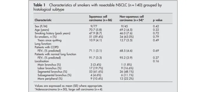
Between January 2001 and December 2002 140 smokers with surgically resectable NSCLC were recruited (86 with squamous cell carcinoma and 54 with a different NSCLC histological subtype, almost exclusively adenocarcinoma (controls, n = 50)). The two groups of patients were matched for age, sex, smoking history, and tumour localisation (table 1).
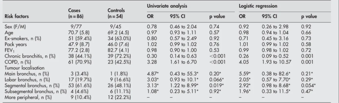
Logistic regression analysis revealed that the presence of COPD indicated an increased risk of more than fourfold for having squamous cell carcinoma in patients with resectable NSCLC (table 2). Conversely, chronic bronchitis was found to increase by more than fourfold the risk for histological subtypes other than squamous cell in patients with resectable NSCLC.
By univariate analysis we found that, when the most peripheral localisation was compared with the more central localisations, some of the latter had a significantly higher risk (or a trend towards a significantly higher risk) for squamous cell carcinoma (table 2). When this parameter was tested in the logistic regression model, no significant difference was observed between different localisations and only a trend persisted for an increased risk for squamous cell carcinoma between segmental localisation and the more peripheral localisation (p = 0.054, table 2).
Separate univariate and logistic regression analyses (including all the parameters evaluated in the previous logistic regression) were performed to determine whether the effects of chronic bronchitis were the same in subjects with and without COPD. In the subgroup of patients with chronic bronchitis (n = 77), both analyses showed that the presence of COPD indicated a more than threefold increase in the risk of having squamous cell carcinoma in subjects with chronic bronchitis undergoing lung resection for resectable NSCLC (78% of cases had COPD compared with 41% of controls; odds ratio 3.49 (95% CI 1.63 to 18.5); p = 0.006).
Conversely, a specific logistic regression analysis on the subgroup of subjects with COPD (n = 84) found no sig-nificant difference in terms of the risk for squamous cell carcinoma between those with and those without chronic bronchitis.
DISCUSSION
This study was designed to investigate whether there is a relationship between COPD and different lung cancer histological subtypes. A pilot study was conducted on a population with a surgically confirmed histological subtype since the proportion of the histological cell types of lung cancer is affected by the method by which the pathological material is obtained.4Indeed, when the cytological cell type is
compared with subsequent histopathological diagnosis made on a resection specimen, only about 80% of cytologically diagnosed lung cancers are correctly typed.4
By selecting patients with a surgically confirmed diagnosis of NSCLC we avoided a possible bias due to inaccurate histological characterisation.
To our knowledge, this is the first study to report that COPD increases the risk of squamous cell carcinoma in patients with surgically resectable NSCLC. A well defined progression of smoking associated pathological changes occurs in the bronchial epithelium before the development of squamous cell carcinoma.7
It is conceivable that such a progression could be facilitated in smokers who develop COPD because of their impaired clearance of carcinogenic substances resulting from chronic airflow limitation. Increased deposition of particulate matter in the major bronchi has recently been described in a computer model of the lungs of patients with COPD, 8
and an association between particle deposition and the onset of lung carcinoma has been reported.
Table 1 Characteristics of smokers with resectable NSCLC (n = 140) grouped by histological subtype Characteristic Squamous cell carcinoma (n = 86) Non-squamous cell carcinoma (n = 54)* p value Sex (F/M) 9/77 9/45 0.42 Age (years) 70.7 (5.8) 69.2 (4.5) 0.22 Smoking history (pack years) 47.9 (8.7) 46.0 (7.6) 0.72 Ex-smokers, n (%) 51 (59.4%) 34 (63.0%) 0.79 Years since quitting 10.9 (4.1) 12.7 (3.5) 0.49 Lung function:
Patients with COPD
FEV1(% predicted) 71.1 (3.1) 68.5 (4.6) 0.69
Patients with normal lung function
FEV1(% predicted) 91.7 (3.3) 95.2 (3.9) 0.27 Localisation 0.16 Main bronchus (%) 3 (3.4%) 1 (1.8%) Lobar bronchus (%) 17 (19.7%) 9 (16.6%) Segmental bronchus (%) 53 (61.6%) 26 (48.1%) Subsegmental bronchus (%) 4 (4.6%) 6 (11.1%) More peripheral (%) 9 (10.4%) 12 (22.2%) Values are expressed as mean (SE) where appropriate.
*Adenocarcinoma (n = 50), large cell carcinoma (n = 4).
680 Papi, Casoni, Caramori, et al
Previous studies have shown an association between chronic bronchitis and lung adenocarcinoma.3 We confirmed those
observations and further extended the analysis by taking into account pulmonary function. Indeed, by analysing a subgroup of subjects with chronic bronchitis, we found that the presence of COPD significantly increases the risk for squamous cell carcinoma, indicating the importance of COPD in influencing the histological subtype even in the presence of chronic bron-chitis. A possible explanation for the increased risk of lung adenocarcinoma in subjects with chronic bronchitis without COPD is that, in the absence of chronic airflow limitation, a more peripheral distribution of tobacco smoke can cause wide-spread activation of cell signalling pathways—for example, the epidermal growth factor/epidermal growth factor receptor loop—which is potentially able to cause both increased mucus production (and therefore the symptoms of chronic bron-chitis) and adenocarcinoma differentiation of the preneoplastic lesions of the bronchial/bronchiolar epithelium.
In agreement with the literature, we found that a more peripheral localisation is associated with a lower prevalence of squamous cell carcinoma among the NSCLC histological subtypes, although the statistical significance of the result tended to disappear in the logistic regression analysis (possibly due to the limited number of cases analysed). Nevertheless, one could argue that a more central localisation of squamous cell carcinoma could itself directly cause bronchial obstruction. Against this hypothesis is the fact that COPD represents a significant risk factor for squamous cell carcinoma even when the logistic regression had been adjusted for the localisation of the tumour. Furthermore, to exclude such a possibility we performed a univariate analysis to evaluate whether the localisation of the tumour is a risk factor for airflow limitation in the population studied. The results indicated that a central localisation did not represent an increased risk factor for airflow limitation in our population (data not shown). Nevertheless, we recognise that a potential bias of our results is that the central localisation of lung cancer might be associated with an increased risk of airflow limitation because of central airflow limitation. Indeed, a central mass large enough to markedly reduce the lumen of the central bronchi may cause fixed airflow limitation, but the few studies that have addressed the relationship between cancer cell type and degree of airflow limitation found no correlation between them.9
We are aware that the small number of subjects in our study is a limitation. However, it has to be emphasised that the epidemiological characteristics, prevalence of histological subtypes, and age/sex distribution of the population are in agreement with recently published data on the epidemiology
of lung cancer.10 Further studies are required to evaluate
whether our findings can be extended to non-surgically resectable NSCLC.
ACKNOWLEDGEMENTS
The authors thank Ms Mary McKenney for her careful editorial review of the manuscript and Dr Elisa Veratelli for scientific secretarial assistance.
Authors’ affiliations
. . . .
A Papi, G Casoni, G Caramori, I Guzzinati, F Ravenna, N Calia, A Ciaccia, L M Fabbri,Research Centre on Asthma and COPD, University of Ferrara, Ferrara, Italy
P Boschetto,Department of Clinical and Experimental Medicine, Section of Occupational Medicine, University of Ferrara, Ferrara, Italy S Petruzzelli, L Corbetta, L M Fabbri,Department of Medical and Surgical Specialties, Section of Respiratory Diseases, University of Modena and Reggio Emilia, Modena, Italy
G Cavallesco,Thoracic Surgery Department, University of Ferrara, Ferrara, Italy
E Forini,Department of Medical Statistics, University Hospital of Ferrara, Ferrara, Italy
M Saetta,Department of Clinical and Experimental Medicine, Section of Respiratory Medicine, University of Padova, Padova, Italy
Supported in part by a grant from the Fondazione Cassa di Risparmio di Ferrara, Ferrara, Italy and from the Associazione Studio Tumori and Malattie Polmonari, Padova, Italy.
REFERENCES
1 Bach P, Ginsberg RJ. Epidemiology of lung cancer. In: Ginsberg RJ, eds. Lung cancer. Hamilton: BC Decker, 2002:1–10.
2 Pauwels RA, Buist AS, Calverley PM, et al. Global strategy for the diagnosis, management, and prevention of chronic obstructive pulmonary disease. NHLBI/WHO Global Initiative for Chronic Obstructive Lung Disease (GOLD) Workshop summary. Am J Respir Crit Care Med 2001;163:1256–76. 3 Wang SY, Hu YL, Wu YL, et al. A comparative study of the risk factors for lung
cancer in Guangdong, China. Lung Cancer 1996;14:S99–105.
4 Corrin B. Classification and pathology of bronchopulmonary carcinoma and of metastases in the lung. In: Gibbson GJ, Geddes DM, Costabel U, et al. Respiratory medicine.3rd ed. London: Saunders, 2003:1789–802. 5 Mountain CF. Revisions in the International System for Staging Lung Cancer.
Chest 1997;111:1710–7.
6 Travis W, Colby T, Corrin T, et al. International histological classification of tumors, 3rd ed. Berlin: Springer, 1999.
7 Auerbach O, Hammond EC, Garfinkel L. Changes in bronchial epithelium in relation to cigarette smoking, 1955–1960 vs. 1970–1977. N Engl J Med 1979;300:381–5.
8 Segal RA, Martonen TB, Kim CS, et al. Computer simulations of particle deposition in the lungs of chronic obstructive pulmonary disease patients. Inhal Toxicol 2002;14:705–20.
9 Congleton J, Muers MF. The incidence of airflow obstruction in bronchial carcinoma, its relation to breathlessness, and response to bronchodilator therapy. Respir Med 1995;89:291–6.
10 Janssen-Heijnen ML, Coebergh JW. Trends in incidence and prognosis of the histological subtypes of lung cancer in North America, Australia, New Zealand and Europe. Lung Cancer 2001;31:123–37.
Table 2 Risk factors for squamous cell carcinoma
Risk factors
Cases Controls Univariate analysis Logistic regression
(n = 86) (n = 54) OR 95% CI p value OR 95% CI p value Sex (F/M) 9/77 9/45 0.78 0.46 to 2.04 0.74 0.92 0.26 to 2.98 0.92 Age 70.7 (5.8) 69.2 (4.5) 0.97 0.93 to 1.11 0.57 0.98 0.94 to 1.04 0.66 Ex-smokers, n (%) 51 (59.4%) 34 (63.0%) 0.80 0.57 to 2.49 0.92 0.71 0.45 to 3.16 0.73 Pack years 47.9 (8.7) 46.0 (7.6) 1.02 0.99 to 1.02 0.76 1.01 0.99 to 1.02 0.58 FEV1 77.2 (2.8) 82.7 (4.1) 0.98 0.90 to 1.00 0.53 0.99 0.98 to 1.02 0.72 Chronic bronchitis, n (%) 38 (44.1%) 39 (72.2%) 0.30 0.14 to 0.63 ,0.001 0.26 0.09 to 0.52 0.001 COPD, n (%) 61 (70.9%) 23 (42.5%) 3.28 1.61 to 6.70 ,0.001 4.05 1.93 to 10.57 0.001 Tumour localisation Main bronchus, n (%) 3 (3.4%) 1 (1.8%) 4.87* 0.43 to 55.3* 0.20* 5.59* 0.38 to 82.6* 0.21* Lobar bronchus, n (%) 17 (19.7%) 9 (16.6%) 3.03* 0.93 to 10.1* 0.066* 2.05* 0.57 to 7.70* 0.29* Segmental bronchus, n (%) 53 (61.6%) 26 (48.1%) 3.13* 1.22 to 8.99* 0.019* 2.92* 0.98 to 8.68* 0.054* Subsegmental bronchus, n (%) 4 (4.6%) 6 (11.1%) 1.08* 0.23 to 5.11* 0.92* 1.96* 0.33 to 11.5* 0.47* More peripheral, n (%) 9 (10.4%) 12 (22.2%) – – – – – – *Compared with more peripheral localisation.
FEV1= forced expiratory volume in 1 second; OR = odds ratio; CI = confidence intervals.
COPD and squamous cell carcinoma 681