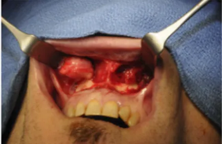
The surgery was realized through a vestibular route shifting the incision in the lip to avoid penetrating the cysts directly. A careful dissection of the horizontal then vertical fibers of the orbicularis oris lets the 2 cysts appear, showing the typical pearl-white aspect of dermoid cysts. Their thick envelopes were totally independent from each other. The adhesion area was found on the anterior surface, in contact with the muscle, toward the deep layer of the dermis (Fig. 4). The histologic examination revealed a typical aspect of dermoid cyst in both tumors. The surgical suites were simple with no re-currence at 1 year after surgery.
DISCUSSION
Dermoid cysts are slow-growing benign tumoral formations. They have 3 possible origins:
- Traumatic implantation of skin cells into deeper layers: they are similar to epidermal cysts. They represent 10% of dermoid cysts.2 - Congenital teratoma are composed of mesoderm, entoderm, and ectoderm derivates3Y5; they are principally located in the testes and ovaries but have been reported in the tongue.6,7
- Congenital inclusion dermoid cysts form along the lines of embryologic closure. They contain both dermal and epidermal derivatives.
At the neck and the face level, New and Erich1distinguish 4 groups of possible location. The most frequent ones being the lateral ends of eyebrows (47%Y70%) then the nasal dorsum (8%Y12%)8Y10 with a possible intracranial extension.11,12 The possibility of this intracranial location imposes the realization of a computed tomo-graphic or MRI scan. The sublingual and neck regions are the 2 last locations described.1Bauer13reported a case of dermoid cyst located on the base of the columella in relation with the maxillary sinus. In our patient, the attachment points were situated in the deep dermis.
The human face derives from 5 prominences of ectomesen-chyme covered by surface epithelia. Union of the facial prominences occurs between the sixth and the eighth week of development.14 Fusion of the medial nasal and maxillary prominences provides for continuity of the upper jaw and lip and for separation of the nasal pits from the stomodeum. Medial nasal prominences form the columella, the tip of the nose, the philtrum of the upper lip, and the primary palate. This fusion requires the disintegration of con-tacting surface epithelia of prominences, allowing the contact of underlying mesenchymal cells. At this time, dermal inclusions may arise between frontonasal and medial nasal prominences, which manifest themselves more than 40 years later.
According to literature data, we present the first described case of bilateral dermoid cysts of the upper lip.
REFERENCES
1. New GB, Erich JB. Dermoid cysts of the head and neck. Surg Gynecol Obstet 1937;65:48Y55
2. Gold BD, Sheinkopf DE, Levy B. Dermoid, epidermoid, and teratomatous cysts of the tongue and the floor of the mouth. J Oral Surg 1974;32:107Y111
3. Flom GS, Donovan TJ, Landgraf JR. Congenital dermoid cyst of the anterior tongue. Otolaryngol Head Neck Surg 1989;101:388Y391
4. Towne BM, Hurst R. Firm mass on the posterior tongue. J Oral Maxillofac Surg 1997;55:987Y991
5. Meyer I. Dermoid cysts (dermoids) of the floor of the mouth. Oral Surg Oral Med Oral Pathol 1955;8:1149Y1164
6. Obiechina AE, Arotiba JT, Ogunbiyi JO. Coexisting congenital sublingual dermoid and bronchogenic cyst. Br J Oral Maxillofac Surg 1999;37:58Y60
7. Gleizal A, Abouchebel N, Lebreton F, et al. Dermoid cyst of the tongue: an association of dermoid cyst with bronchogenic epithelium. J Craniomaxillofac Surg 2006;34:113Y116
8. Toriyama K, Kamei Y, Morishita T, et al. Congenital dermoid cyst of the upper lip: a case report. J Plast Reconstr Aesthet Surg 2008;61:970Y971
9. McAvoy JM, Zuckerbraun L. Dermoid cysts of the head and neck in children. Arch Otolaryngol 1976;102:529Y531
10. Taylor BW, Erich JB, Dockerty MB. Dermoids of the head and neck. Minn Med 1966;49:1535Y1540
11. Matson DD, Ingraham FD. Intracranial complications of congenital dermal sinuses. Pediatrics 1951;8:463Y474
12. Pratt LW. Midline cysts of the nasal dorsum: embryologic origin and treatment. Laryngoscope 1965;75:968Y980
13. Bauer BS. Benign tumors and conditions of the head and neck. In: Achauer BM, Ericksson E, Guyuron B, et al, eds. Plastic Surgery: Indications, Operations, Outcomes. St Louis, MO: Mosby; 2000:1133Y1138
14. Sperberg G. Craniofacial Development. Hamilton, Ontario: Decker, 2001
Surgical Technique of the
Transoral Approach to Remove a
Lipoma of the Buccal Fat Pad
Matteo Brucoli, MD,* Francesco Arcuri, MD,* Giovanni Borello, MD,Þ Arnaldo Benech, MD, PhD*
Background: In 1727, Heister (Compendium anatomicum. Altdorf, Guill, Koleshii: editio tertia 1727: 134, table VIII and figs. 36Y37) described the buccal fat pad (BFP) as an independent anatomic structure of the face; in 1801, Bichat (Anatomie generale appliquee a la physiologie et a la medecine. Paris, France: Brosson, Gabon et Cie Libraires, 1801:60) reported his fatty histologic finding. Ac-cording to the literature, several pathologic tumorous conditions can
From the Departments of *Maxillo-Facial Surgery and †Otorhinolaryngol-ogy, Azienda Ospedaliera Maggiore della Carita`, University of Piemonte OrientaleBAmedeo Avogadro,[ Novara, Italy.
Received March 28, 2011.
Accepted for publication July 26, 2011.
Address correspondence and reprint requests to Francesco Arcuri, MD, SCDU di Chirurgia Maxillo-Facciale, Ospedale Maggiore della Carita`, Corso Mazzini 18, 28100 Novara, Italy; E-mail: [email protected] The authors report no conflicts of interest.
Copyright* 2011 by Mutaz B. Habal, MD ISSN: 1049-2275
DOI: 10.1097/SCS.0b013e318231fe2b
FIGURE 4. Intraoperative view showing the adhesion of cysts.
The Journal of Craniofacial Surgery
&
Volume 22, Number 6, November 2011 Brief Clinical Studies* 2011 Mutaz B. Habal, MD
2415
arise from BFP, such as lipoma, lipoblastomatosis, liposarcoma, hemangioma, arteriovenous malformation, and nodular fasciitis; all of which are rare. After a revision of the English literature performed through PubMed between 1948 and 2008, we found 10 cases of lipomas arising from the BFP (7 cases are simple subtype, 2 are spindle cell lipoma, and 1 is fibrolipoma). The aims of this study were to introduce our clinical report of this rare pathologic entity, describe the surgical technique of the transoral approach, and dis-cuss the potential pitfalls regarding the preoperative diagnosis and the close interrelation among the BFP, the facial buccal branches (FBBs), and the parotid duct (PD).
Clinical Report: A 43-year-old man was referred to the Maxillo-facial Unit of the Novara Major Hospital with a 6-month history of a painless swelling in the right cheek. Clinical examination revealed a clearly visible, tender, slightly fluctuant mass, situated anterior to the masseter muscle and extended to the submandibular region. The patient underwent an ultrasound, a computed tomography, and a magnetic resonance imaging. Under general anesthesia with na-sotracheal intubation, the patient underwent intraoral resection of BFP lipoma.
Discussion: The 2 major areas of discussion are the potential pitfall regarding the preoperative diagnosis and the close anatomic inter-relation among the BFP, the FBB, and the PD. First, the spindle cell lipoma, one of the most common BFP lipoma variant, can be histologically and clinically similar to a well-differentiated lipo-sarcoma, which can be recurrent and metastatic. This issue warrants that a careful workup of the tumorous mass of the buccal space and a BFP origin must be considered in every situation. Finally, ac-cording to the recent literature, the anatomic variations of the in-terrelation between the FBB and the BFP are classified into 2
groups: (1) FBB passing lateral to the BFP and (2) branches crossing inside the BFP. The anatomic variations of the interrelation between the PD and the BFP are classified into 3 groups: (1) PD passing lateral to the BFP, (2) PD crossing deep to the BFP, and (3) PD running along the superior border of the BFP.
Key Words: Buccal fat pad, lipoma, liposarcoma, transoral approach, facial nerve, parotid duct
I
n 1727, Heister1described the buccal fat pad (BFP) as an inde-pendent anatomic structure of the face; in 1801, Bichat2reported his fatty histologic finding. According to the literature, several pathologic tumorous conditions can arise from BFP, such as li-poma,3lipoblastomatosis,4liposarcoma,5 hemangioma,6 arteriove-nous malformation,7and nodular fasciitis8; all of which are rare.Moreover, pseudoherniation of the BFP can result in the for-mation of a mass. This condition is present either subcutaneously or intraorally. The former occurs in older individuals, and it is fre-quently associated with corticosteroids, trauma, or surgery due to loss of the ligamentous support of the fascia; the latter is usually seen in children, and it is associated with trauma.9
Although most of the BFP lipomas are of the simple subtype, other histologic variants, such as spindle cell lipoma and fibroli-poma, are also reported. After a revision of the English literature performed through PubMed between 1948 and 2008, we found 10 cases of lipomas arising from the BFP (7 cases are simple sub-type, 2 are spindle cell lipoma, and 1 is fibrolipoma).10Y14
The aims of this study were to introduce our clinical report of this rare pathologic entity, describe the surgical technique of FIGURE 1. A photograph demonstrating the
echotomography.
FIGURE 2. A computed tomographic scan showing the tumor.
FIGURE 3. T2-weighed MRI scan demonstrating the hyperintensity of the lesion.
FIGURE 4. The mass clearly visible in the right cheek and extended to the submandibular region.
Brief Clinical Studies The Journal of Craniofacial Surgery
&
Volume 22, Number 6, November 20112416
* 2011 Mutaz B. Habal, MDthe transoral approach, and discuss the potential pitfalls regard-ing the preoperative diagnosis and the close interrelation among the BFP, the facial buccal branches (FBBs), and the parotid duct (PD).
CLINICAL REPORT
A 43-year-old man was referred by his general physician to the Maxillofacial Unit of the Novara Major Hospital with a 6-month history of a painless swelling in the right cheek, around the body of the mandible; his medical history was unremarkable. He denied having other complaints, and there was no history of trauma. Clin-ical examination revealed a clearly visible, tender, slightly fluctuant mass, situated anterior to the masseter muscle and extended to the submandibular region.
The patient underwent an ultrasound, a computed tomogra-phy, and a magnetic resonance imaging (MRI; Figs. 1Y3). These examinations showed a round, finely encapsulated fatty mass de-formity with fibrotic changes anterior in the right cheek, lateral to the buccinator muscle, without infiltration of the surrounding struc-tures (Fig. 4). This location led to the suspicion of a BFP lipoma, and a transoral approach to remove this tumor was planned (Fig. 5). Further preoperative investigations included a complete blood count, blood chemistry, electrocardiogram, and chest x-ray. Under general anes-thesia with nasotracheal intubation, the patient underwent intraoral resection of BFP lipoma.
A bite block is inserted on the opposite side to operate on an adequately open mouth; infiltration of anesthetic and vasoconstric-tor is performed to avoid bleeding, and steroid ointment is placed on the lips to avoid damage during the surgery. The incision is made with a no. 15 blade through the buccal mucosa and the buccinator muscle from just lingual to the external oblique ridge halfway up the mandibular ramus superiorly, to mesial of the second molar in-feriorly (Fig. 6).
The cut is performed in a straight line with 5 mm of non-keratinized mucosa left buccally at the lower end of the incision for ease of suturing later; insufficient nonkeratinized soft tissue on the alveolar aspect can cause difficulty with suturing later and may lead to wound dehiscence. An incision too far lingually may damage the lingual nerve, causing lingual nerve anesthesia of the tongue; an incision too far buccally may cause excessive hemor-rhage from the vessels in the masseter muscle. An incision too high may cause excessive hemorrhage from the vessels in the temporalis and/or masseter muscle.
The masseter muscle is left attached to the bone because tearing of periosteum and/or excessive stripping of the masseter muscle may cause hemorrhage, hematoma formation, and excessive postop-erative swelling. Blunt dissection is performed to gain direct access to the anterior masticatory space. The tumor was extirpated entirely, and the wound was closed with slowly resorbing interrupted sutures FIGURE 5. The planned incision.
FIGURE 6. The cut through the buccal mucosa revealing the buccinator muscle.
FIGURE 8. The closure of the wound.
FIGURE 7. The intraoral removal of the tumor.
FIGURE 9. The surgical specimen.
The Journal of Craniofacial Surgery
&
Volume 22, Number 6, November 2011 Brief Clinical Studies* 2011 Mutaz B. Habal, MD
2417
(Figs. 7Y9). The lesion was sent for pathologic analysis that con-firmed the diagnosis of BFP lipoma: microscopically mature fatty tissue was seen separated by a fine network of connective tissue. No rich cellularity or lipoblasts were seen, and thus, the diagnosis of lipoma was confirmed. The postoperative course was uneventful; the patient was discharged 4 days after the surgical procedure, and a good mucosal healing was observed after 20 days, without any inju-ries of the FBB and the PD.
DISCUSSION
Despite the fact that several scientific speculations could arise from this case, we focus on 2 major areas of discussion: the potential pitfall regarding the preoperative diagnosis and the close anatomic interrelation among the BFP, the FBB, and the PD.
Lipoma of the BFP is a rare and underestimated pathologic condition; BFP lipomas show characteristics that can lead to con-fusion with other diseases. Extensive preoperative imagings are essential to understand the tumor extension and the potential infil-tration. The spindle cell lipoma, one of the most common BFP lipoma variant, can be histologically and clinically similar to a well-differentiated liposarcoma, which can be recurrent and metastatic. This issue warrants that a careful workup of the tumorous mass of the buccal space and a BFP origin must be considered in every situation.5,15
Liposarcoma of the BFP has been reported, constituting 10% to 15% of all the sarcomas; liposarcoma is the second most common soft-tissue sarcoma in adults, very rare in the head and neck region. Enzinger and Weiss16 described an incidence of 5.6%, and Cawson et al17reported that the BFP is the most fre-quent head and neck location for liposarcoma. Moreover, if the clinical and histologic image of a well-differentiated liposarcoma can be similar to a lipoma, then it seems likely that more lipo-sarcomas arise from the BFP than it is reported in the scientific literature.18
We think that a preoperative ultrasound and an MRI must be always performed to determine the potential origin from the BFP, visualize the extent in the various extensions, and exclude a lipo-sarcoma; Gaskin and Helms19stated that an MRI is 100% specific in the diagnosis of simple lipoma and has a 100% sensitivity with 83% specificity in distinguishing a liposarcoma. Moreover, MRI could be used preoperatively to perform an MRI sialography leading to a better visualization of any potential duct obstruction before the surgical resection.20
These statements guide the surgical approach; whether an in-traoral access could be sufficient in suspected simple lipoma or if a more radical cutaneous cervicofacial approach must be evaluated. Every resected BFP lipoma should be sent for pathologic analysis to determine the histologic feature because the clinical image is not always reliable.
Usually, a lipoma of the BFP is present as a mass in the cheek, underneath the superficial musculoaponeurotic system and lateral to the buccinator muscle. The clinical history is that of a slow-growing lesion with no history of trauma. On palpation, the over-lying skin and oral mucosa are intact, and the tumor is tender. It does not adhere to the subcutis, and the mucosa can be moved freely over it.21
Finally, according to the recent literature, the anatomic variations of the interrelation between the FBB and the BFP are classified into 2 groups: (1) FBB passing lateral to the BFP and (2) branches cross-ing inside the BFP. The anatomic variations of the interrelation be-tween the PD and the BFP are classified into 3 groups: (1) PD passing lateral to the BFP, (2) PD crossing deep to the BFP, and (3) PD running along the superior border of the BFP.22
The results of these findings are that, during the procedure of removal of BFP by an intraoral approach, even with careful manipulation, some injuries to the PD can be inevitable and that some functions of facial muscles can be hampered; therefore, we recommend the use of a facial nerve monitoring during the surgical dissection, reducing the risk of any injuries.
REFERENCES
1. Heister L. Compendium anatomicum. Altdorf, Guill, Koleshii: editio tertia 1727: 134, table VIII and figs. 36Y37
2. Bichat X. Anatomie generale appliquee a la physiologie et a la medecine. Paris, France: Brosson, Gabon et Cie Libraires, 1801:60
3. Manara E. Case of lipoma of the fatty ball of Bichat. G Clin Med 1952;33:166Y171
4. Greenwood RE, Hook PC. Lipoblastomatosis of the cheek: a case report. Br J Oral Surg 1982;20:135Y141
5. Sxenyuva C, Yu¨cel A, Okur I, et al. A well-differentiated giant liposarcoma originating from the buccal fat pad. Ann Plast Surg 1996;36:439Y443
6. Tanaka A, Hatoko M, Tada H, et al. A case of hemangioma of the buccal fat pad. Ann Plast Surg 2000;44:346Y347
7. Dubin B, Jackson IT, Halim A, et al. Anatomy of the buccal fat pad and its clinical significance. Plast Reconstr Surg 1989;83:257Y262
8. Smith JF. Nodular fasciitis of the buccal pad. Arch Otolaryngol 1967;86:217Y218
9. Matarasso A. Pseudoherniation of the buccal fat pad: a new clinical syndrome. Plast Reconstr Surg 1997;100:723Y730
10. Birnmeyer G. Kasuistischer beitrag zur seltenen lokalisation eines lipoms in der wange. Monatsschr Ohrenheilkd Laryngorhinol 1958;92:182Y185
11. Colombo E, Tosi C. Sui lipomi del corpo adiposo di Bichat. Arch Ital Otol Rinol Laringol 1963;74:733Y745
12. Calhoun NR. Lipoma of the buccal space. Oral Surg Oral Med Oral Pathol 1963;16:246Y249
13. Rodgers GK, Myers EN. Surgical management of the mass in the buccal space. Laryngoscope 1988;98:749Y753
14. De Wijn RS, Van der Heijden EP, Kon M. On lipoma of the buccal fat pad: report of two cases and review of the literature. J Plast Reconstr Aesthet Surg 2009;62:28Y35
15. Eidinger G, Katsikeris N, Gullane P. Liposarcoma: report of a case and review of the literature. J Oral Maxillofac Surg 1990;48:984Y988
16. Enzinger FM, Weiss SW. Soft Tissue Tumors. 3rd ed. St Louis, MO: Mosby, 1995
17. Cawson RA, Speight P, Binnie WH. Tumors of adipose tissue. In: Lucas RB, ed. Pathology of Tumours of the Oral Tissues. 5th ed. London: Churchill Livingstone, 1998:324
18. Charnock D, Jett T, Heise G, et al. Liposarcoma arising in the cheek: report of a case and review of the literature. J Oral Maxillofac Surg 1991;49:298Y300
19. Gaskin CM, Helms CA. Lipomas, lipoma variants and well-differentiated liposarcomas (atypical lipomas): results of MRI evaluations of 126 consecutive fatty masses. AJR Am J Roentgenol 2002;182:733Y739
20. Schro¨der U, Jungehu¨lsing M, Fischbach R, et al. Magnetic resonance sialography. A new diagnostic method for imaging salivary duct patency. HNO 1998;46:38Y43
21. Furlong AM, Fanburg-Smith JC, Childers ELB. Lipoma of the oral and maxillofacial region: site and subclassification of 125 cases. Oral Surg Oral Med Oral Pathol 2004; 98:441Y450
22. Hwang K, Cho HJ, Battuvshin D, et al. Interrelated buccal fat pad with facial buccal branches and parotid duct. J Craniofac Surg 2005;16:658Y660
Brief Clinical Studies The Journal of Craniofacial Surgery