Università degli Studi di Pisa
Dottorato di Ricerca in Fisiopatologia e Clinica dell’Apparato
Cardiovascolare e Respiratorio
Presidente Prof. A. Mussi
A.A. 2007-2008
Tesi di Diploma
Randomized Comparison of Sirolimus and Paclitaxel Drug-Eluting
Stents for Long Lesions in the Left Anterior Descending Artery: an
Intravascular Ultrasound Study
Candidata: Dr.ssa Giulia Branchitta
Index
Abstract
3
Abbreviations
4
Introduction
5
Methods
16
Results
22
Discussion
25
Conclusions
31
Appendix
32
References
42
Abstract
The objective of this prospective randomized study was to verify whether the superiority of the sirolimus-eluting stent (SES) in inhibiting neointimal hyperplasia could be demonstrated in complex coronary lesions. The SES was compared with the paclitaxel-eluting stent (PES) in long lesions of the left anterior descending artery with intravascular ultrasound (IVUS) and quantitative angiography. Late luminal loss and mean neointimal hyperplasia were significantly lower in the SES group compared to the PES group and peri-stent plaque area was significantly reduced in the SES group only. Both groups had excellent IVUS and angiographic results at 9-month follow up.
Abbreviations
1. SES=sirolimus-eluting stent 2. PES=paclitaxel-eluting stent 3. IVUS=intravascular ultrasound
4. NIHA=mean neointimal hyperplasia area 5. PSPA=mean peri-stent plaque area
6. PCI=percutaneous coronary intervention 7. MLA=minimal lumen area
8. RVD=reference vessel diameter 9. MLD=minimal luminal diameter 10. LL=late luminal loss
Introduction
Coronary stenting, by reducing both the acute risk of major complications and the incidence of restenosis, has gradually replaced conventional balloon angioplasty as the standard technique to accomplish percutaneous coronary interventions (PCIs) 1,2. However, in-stent restenosis still occurs in 10 to 50% of the patients, depending upon a number of clinical, angiographic, and procedural variables 3-5.
In April 2002, the first drug-eluting stent (DES) became commercially available in Europe, beginning what has been called the third revolution in PCI following the introduction of balloon angioplasty and stents. The DES combines the advantages of a stainless steel scaffold with controlled release of an antiproliferative agent to prevent restenosis 6. Local drug delivery allows to achieve appropriate drug concentration at the treatment site while avoiding systemic toxic effects.
A DES has three basic components: the stent, the coating, and the biological agent. A number of DESs have been developed using different carrier stents, different kinds of coating, and different drugs 6,7. To ensure uniform drug delivery, the ideal DES should have a large surface area, minimal gaps between cells, and minimal strut deformation after deployment. Furthermore, these stents would need to maintain a good deliverability even in more complex lesions, through a low profile, good conformability and radial support, and appropriate flexibility.
A variety of different formulations have been developed that provide appropriate stent coating for clinical use, including direct drug binding, coatings with phosphorylcholine, non-erodible or bioabsorbable polymers, or ceramic layers. The coating should not induce an excessive vascular reaction 8, should be suitable for sterilization, it must follow the geometric change of configuration during stent
expansion, be resistant to mechanical abrasion during stent implantation, and it should be able to release the drug in a controlled way. A potential universal coating is unlikely, and different pharmacological agents may require different delivery vehicles.
The biological agent carried by the stent should interfere with one or more steps involved in the restenosis process, yet preserving vascular healing. A number of antiproliferative, immunosuppressive, anti-inflammatory, antithrombotic, and prohealing drugs have been tested or are under investigation. To date, only two DESs have received CE (Conformité Européenne) and FDA (Food and Drug Administration) approval for clinical use in Europe and in the United States, respectively: the Cypher™ sirolimus eluting stent (SES) (Cordis, Johnson & Johnson, Miami Lake, FL, USA), and the Taxus™ paclitaxel-eluting stent (PES) (Boston Scientific, Natick, MA, USA). In randomized trials, both these stents have been shown to dramatically reduce late luminal loss, binary restenosis, and the need for repeat revascularization when compared to bare metal stents (BMSs) 9-13. Sirolimus (rapamycin) is a macrocyclic lactone produced by Streptomyces hygroscopicus, which inhibits cellular proliferation by blocking cell cycle progression at the G1 to S transition 14-16, and inhibits vascular smooth muscle cell migration 14. In the Cypher™ stent, a combination of two non-erodible polymers (polyethylene-co-vinyl acetate, and poly n-butyl methacrylate [PBMA]) mixed with sirolimus (67%/33%) makes up the basecoat formulation which is applied to a parylene C-treated stent. A drug-free topcoat of PBMA polymer is applied to the stent surface to control the release kinetics of sirolimus (> 28 days) 6.
Paclitaxel (taxol) is an antineoplastic agent that shifts the microtubule equilibrium toward assembly. This enhances the assembly of extraordinarily stable microtubules, interrupting proliferation, migration, and signal transduction 17-19. In the
Taxus™ stent, 1 μg/mm2 paclitaxel is incorporated in a poly (lactide-co-Σ-caprolactone) copolymer attached to a conventional stent. Two different release kinetics were evaluated: slow release (continuous drug release throughout the first 15-20 days), and moderate release (the drug is released within the first 2 days after stent implantation).
To date, many randomized controlled trials on DESs have been completed. However, while studies on SESs represent a homogeneous group utilizing one single device, studies on PESs comprised a variety of devices, both non-polymer-based and polymer-based, which therefore need to be evaluated separately. In all these trials chronic total occlusion, ostial lesion, thrombus-containing lesion, unprotected left main PCI, acute myocardial infarction, low left ventricular ejection fraction, and multivessel stenting were exclusion criteria.
The sirolimus-eluting stent
Six randomized trials comparing the outcomes of patients treated with SESs and conventional BMSs have been concluded to date 9, 10, 12, 20-24. In these studies, SESs were evaluated in the treatment of de novo coronary lesions, < 33 mm long, in native coronary arteries 2.5 to 3.5 mm in diameter.
The landmark Randomized Study with Sirolimus-Eluting Velocity Balloon-Expandable Stent in the Treatment of Patients with de Novo Native Coronary Artery Lesions (RAVEL) trial included 238 patients with single non-complex de novo lesions. Remarkably, the 6-month angiographic restenosis rate of the SES group was zero, as well as the late loss. Intravascular ultrasound examination at follow-up further confirmed the marked neointimal inhibition after SES implantation 25. The clinical outcomes were significantly better among patients treated with the sirolimus stents, with
94% of patients being free of any major cardiac events at 1 year compared to 71% in the BMS group (p < 0.01) 9-13, 20-24. The Sirolimus-Eluting Bx VelocityTM Balloon-Expandable Stent (SIRIUS) trial randomized 1101 patients with de novo lesions to sirolimus or bare stents 10. In-stent binary restenosis (within the margins of the stent) was reduced by 91% (3.2 vs 35.4%, p < 0.01) and in-segment restenosis (including the stented portion and the 5-mm segments proximal and distal to the stent) was reduced by 75% (8.9 vs 36.3%, p < 0.01) 10. At 9 months, the incidence of major adverse cardiac events was significantly lower in the sirolimus group (7.1 vs 18.9%, p < 0.01), mainly due to a decrease in the need of target lesion revascularization (TLR) (4.1 vs 16.6%, p < 0.01). At 12 months, the absolute difference in TLR continued to increase (4.9 vs 20%, p < 0.001) 26. The E-SIRIUS trial enrolled 352 patients with longer lesions and smaller vessels than the RAVEL and SIRIUS trials 12. Nevertheless, the 8-month in-stent restenosis rate was 3.9% in the sirolimus and 41.7% in the bare stent group (p < 0.01). Similarly, the incidence of in-segment restenosis (5-mm edges included) was significantly reduced (5.9 vs 42.3%, p < 0.01). The 9-month incidence of major adverse cardiac events was 8 vs 22.6% in the sirolimus and bare stent groups (p < 0.01). In the C-SIRIUS trial, which randomized 100 patients to sirolimus or conventional stenting, in-stent restenosis was not detected in any patient after SES implantation 20. In-segment restenosis occurred in 2.3% of SES patients and 52.3% of BMS patients (p < 0.001). At 270 days, clinically-driven TLR was 4% in the SES group and 18% in the BMS group (p = 0.05). The Kaplan-Meier estimate of freedom from major adverse cardiac events at 270 days was 96.0% for SES patients and 81.7% for BMS patients (p = 0.029).
The Sirolimus-Eluting Stent in Small Arteries (SES-SMART) study was specifically designed to evaluate SESs in small vessels 23. The mean vessel size in the
patients was 2.2 mm. At 8 months, in-segment binary restenosis was 9.8% in the SES group and 53.1% in the control group (p < 0.001). Acute myocardial infarction and overall clinical events were also significantly reduced in patients who received the DES. All findings appeared to be independent of gender, diabetic status, and stent size 23.
The Diabetes and Sirolimus-Eluting Stent Trial (DIABETES) assessed the efficacy of SESs in 160 diabetic patients with de novo coronary stenoses, half of whom received BMSs 24. Abciximab was recommended for all patients. There was an 88% reduction in late lumen loss in the SES group compared to the BMS group (from 0.44 to 0.08 mm, p < 0.0001). Binary restenosis was reduced from 33% in the BMS group to 7.7% in the SES group. At 9-month follow-up, there was a significant difference in TLR (7.5 vs 31.3%, p < 0.0001) and major adverse cardiac events (11.3 vs 36.3%, p < 0.0001) in favor of the SES group.
The Paclitaxel (polymer-coated) eluting stent
The large variety of PESs should lead us to consider that variations in drug dosing, release kinetics, stent design, and stent coating technologies may result in different vascular reactions and efficacy. Four clinical trials utilizing polymer-coated PESs have been reported 11, 13, 27. Overall, more than 2300 patients with de novo lesions have been enrolled in the TAXUS I 27 , II 11, IV 13, and VI trials, and randomized to paclitaxel or bare stents. Different stent platforms with different release kinetics were used in these studies. The Taxus NIRx stent coated with paclitaxel (1 µg/mm2 paclitaxel per unit of stent surface area) in a slow-release formulation was employed in the TAXUS I, the same stent with both a slow-release and a moderate-release formulation was used for the TAXUS II. In TAXUS IV and VI, the drug was loaded onto the
Express stent, in the slow-release formulation in the former, and the moderate-release formulation in the latter. A marked reduction in neointimal proliferation and binary restenosis was observed in the active groups of all the trials, leading to a significant reduction in revascularization procedures and overall adverse cardiac events compared to controls 11, 13, 27. Interestingly, the TAXUS VI is the only trial specifically designed for long lesions (lesion length for inclusion 18-42 mm). In this trial, PESs reduced restenosis from 35.7 to 12.4% and repeat revascularizations from 19.4 to 9.1% (Grube E., personal communication). In all these studies, DESs and BMSs showed similar rates of subacute stent thrombosis.
The Paclitaxel (direct dip-coating) eluting stent
Paclitaxel elution from a metal stent without a polymer coating is an attractive choice because some polymers were associated with an exaggerated inflammatory response and increased neointimal hyperplasia in animal studies. However, contradictory clinical results have been obtained with devices utilizing direct dip-coating of paclitaxel stents 28, 29, 30. Some of the studies evaluating paclitaxel or paclitaxel derivatives never came to full completion due to an unacceptable adverse event rate (9.4% stent thrombosis in the active arm of the Study to Compare Restenosis Rate between QueST and QuaDS-QP2 [SCORE] trial) 31 or did not yield sufficient clinical benefit to justify commercialization of the device. The European Evaluation of Paclitaxel Eluting Stent (ELUTES) trial 30 and the Asian Paclitaxel-Eluting Stent Clinical Trial (ASPECT) 29 have shown a significant dose-dependent reduction in restenosis with paclitaxel stents, whereas the larger RX AchieveTM Drug-Eluting Coronary Stent System in the Treatment of Patients with de Novo Native Coronary
Lesions (DELIVER-I) study failed to demonstrate the beneficial effect of these devices 28. In the ASPECT, two different types of antiplatelet therapy were given to patients treated with PESs 29. This choice contributed to the relative increase in adverse events in the group treated with aspirin and cilostazol compared to those treated with aspirin and clopidogrel.
The relative efficacy of the more successful DES cannot be easily assessed because they are most often compared with BMS. Randomized trials such as the Prospective, Randomized, Multi-Center Comparison of the Cypher Sirolimus-Eluting and the Taxus Paclitaxel-Eluting Stent Systems (REALITY), Paclitaxel and Sirolimus Stents in the Real World of Interventional Cardiology (TAXI) study, Drug-Eluting Stent for Complex Lesions: Cordoba–Las Palmas Study (CORPAL), Sirolimus-Eluting Stent Compared with Paclitaxel-Eluting Stent for Coronary Revascularization (SIRTAX) study, Intracoronary Stenting and Angiographic Results—Drug-Eluting Stents for In-Stent Restenosis (ISAR-DESIRE) trial, and the ISAR-Diabetes trial directly compared SES and PES in a wide variety of patient and lesion types.32-37 Both the SIRTAX and ISAR-DESIRE showed target lesion revascularisation rates that favored SES. 35,36. SIRTAX randomised 1012 patients to PCI of de novo coronary lesions with SES/PES showing at 9 months a TLR of 4,8% in SES and 8,3% in PES group (p 0,03) 35. ISAR-DESIRE enrolled only patients with in-stent restenosis 36. However, the TAXI, REALITY, ISAR-DIABETES, and CORPAL studies did not show a significant difference between both devices 32-34, 37. TAXI trial randomised 202 patients and was essentially a clinical study with angiographic follow up performed only in few cases 33. REALITY involved 1386 patients and did not find differences in the rates of binary
restenosis or MACE between SES and PES 32. ISAR-DIABETES (250 patients with diabetes and coronary artery disease randomized to SES/PES) showed a significantly greater late luminal loss in PES group but not an increased rate of revascularisation for restenosis 37. CORPAL enrolled 515 patients with coronary lesions at high risk for restenosis and obtained an angiographic and IVUS follow-up in a subgroup (respectively 154 and 51 pts), showing a significantly lower angiographic late loss (0,30±0,4 mm in SES vs 0,59±0,8 mm in PES, p<0,01) and neointimal area (0,41±0,4 mm2 in SES vs 1,37±1,4 mm2, p<0,01) 34. Two recent meta-analyses showed that the use of SES was associated with lower angiographic restenosis rates and a lower incidence of target vessel revascularization as compared with PES 38,39 . Of note, no significant differences were found in the incidence of death or myocardial infarction between both groups. Among the PES registries, the TAXUS Stent Evaluated at Rotterdam Cardiology Hospital (T-SEARCH) registry is worth mention because it was one of the first studies to reflect real-world clinical practice. The recently published 2-year results did not show a significant difference between PES and SES in any of the clinical end points.40 The much larger Strategic Transcatheter Evaluation of New Therapies (STENT) registry also did not show a significant difference between both devices at 6 months in 6659 patients. 41
IVUS and DES
Intravascular ultrasound (IVUS) is unique in its ability to assess stent implantation. Specifically, serial (postintervention and follow-up) IVUS can measure intimal hyperplasia (IH), assess acute and late acquired incomplete stent apposition, detect the presence and persistence of edge dissections, assess vascular responses such
as remodeling, study edge effects, compare overlapping with nonoverlapping segments, and look for causes of restenosis and thrombosis. In particular, IVUS determined percentage IH volume (IH volume/stent volume) has been a useful indicator of the ultimate clinical success or failure of a particular DES. In BMS, %IH volume averaged 30% of stent volume; %IH volume was consistently greater in diabetics versus nondiabetics; and in BMS that did not restenose, IH remained stable or regressed slightly after 6 months.
The sirolimus-eluting stent
In the RAVEL (Randomized Comparison of a Sirolimus-Eluting Stent with a Standard Stent for Coronary Revascularization) trial, 6-month %IH volume (1 ± 3%) was lower with Cypher stents compared with control subjects (29 ± 20%; p < 0.001) 25. In the subset of diabetic patients, Cypher stent %IH volume measured 0.82 ± 1.38% (vs. 30.2 ± 22.9% in control subjects; p = 0.008). Furthermore, Cypher stents compared favorably in diabetics versus nondiabetics in whom %IH volume measured 1.14 ± 2.68% 42.
In the SIRIUS (Sirolimus-Eluting Stent in Coronary Lesions) trial, Cypher stents reduced 9-month %IH volumes from 33.4% to 3.1% (p < 0.001) 10. There was a tendency for greater neointima suppression in the middle of the stent compared with the edges, the opposite of BMS 43. There was: 1) no difference in IH between lesions with moderate/severe calcium (arc _120°) versus no/mild calcium; 2) no difference between lesions with positive versus negative pre-intervention vessel remodeling; 3) no correlation between IH and pre-interventional plaque burden (plaque and media [P&M]/external elastic membrane [EEM]); 4) no effect from stent asymmetry; and 5) no difference between overlap versus nonoverlapped segments 44-46. Many weak
predictors of BMS restenosis (i.e., intervention positive remodeling and pre-intervention or residual plaque burden) were not predictive after Cypher implantation, because marked IH suppression masked any effect from weaker predictors.
In the São Paulo registry, IH within Cypher stents was stable or increased slightly beyond the time points reported in the RAVEL (6 months) and SIRIUS (9 months) trials. The %IH volume in 14 patients treated with the fast-release formulation was 2.3% at 1 year, 9.2% at 2 years, and 9.1% at 4 years; %IH volume in 14 patients treated with the slow-release formulation was 2.2% at 1 year, 3.3% at 2 years, and 5.7% at 4 years 47-49.
The paclitaxel-eluting stent
In the TAXUS-II trial, 6-month %IH volume measured 7.8 ± 9.9% in slow-release Taxus stents (vs. 23.2 ± 18.2% in control subjects) and 7.8 ± 9.7% in moderate-release Taxus stents (vs. 20.5 ± 16.7% in control subjects) 11. The distribution of IH over the length of the stent was neither increased (as in BMS) nor more suppressed in the center (as in Cypher) 50.
The findings of the TAXUS-II trial were supported by the TAXUS-IV trial 51; 9-month %IH measured 12.2 ± 12.4% in slow-release Taxus stents versus 29.4 ± 14.0% in control subjects (p < 0.0001). As in the TAXUS-II trial, IH was flat over the length of the stent. The TAXUS-V trial randomized more complex lesions than the TAXUS-IV trial; %IH volume measured 13.2 ± 12.0% with Taxus stents versus 31.8 ± 15.1% with control subjects (p < 0.0001) 52. The TAXUS-VI trial studied moderate-release Taxus stents in longer lesions; %IH volume measured 10.7 ± 10.8% versus 33.0 ± 15.1% in control subjects (p < 0.0001) (N. J. Weissman, unpublished data, 2005).
A subset meta-analysis of 566 patients from the TAXUSIV, V, and VI trials showed: 1) %IH measured 9.8 ± 12.0% in overlapping Taxus segments versus 11.3 ± 10.9% in nonoverlapping segments; and 2) in lesions > 26 mm in length, %IH measured 13.4 ± 9.2% (N. J. Weissman, unpublished data, 2005) In that meta-analysis, %IH in 87 diabetics treated with Taxus stents measured 13.7 ± 12.4%, i.e., less than the 34.6 ± 16.7% in 75 diabetics treated with BMS (p < 0.0001) but no different from the 217 nondiabetics treated with Taxus stents (11.6 ± 11.6%) 53.
Two-year TAXUS-II IVUS trial data has been reported in a highly selected group of 32% of Taxus-treated patients who had baseline and 6 month follow-up IVUS. The IH continued to be significantly suppressed compared with BMS. However, IH increased in both the slow-release and moderate-release Taxus stents (0.64 ± 0.81 mm2 to 0.94 ± 0.76 mm2 [p = 0.01] and 0.66 ± 0.83 mm2 to 1.06 ± 0.90 mm2 [p = 0.009], respectively) 54.
Even if almost all studies on DES have included some IVUS analyses, few data are available on IVUS assessment of long, complex coronary lesions treated with the SES or the PES, and to date, no randomized comparison of IVUS findings has been reported in these subjects. Available data on lesions treated with the SES or the PES shows that the SES appears to exert a more powerful inhibition of neointimal growth. The objective of this study was to verify whether the superiority of the SES in inhibiting neointimal hyperplasia could be demonstrated in complex coronary lesions.
Methods
Patient Selection
Patients with stable angina, non-ST-segment elevation acute coronary syndrome, or documented silent ischemia undergoing coronary angiography at Pisa University Hospital were screened for enrollment. Inclusion criteria were: (a) presence of an angiographically significant stenosis in the proximal and/or mid portion of the left anterior descending artery; (b) American College of Cardiology/American Heart Association (ACC/AHA) lesion class B2 or C, requiring a stent ≥ 16 mm in length; (c) vessel reference diameter between 2.5 and 3.7 mm at lesion site; and (d) consent to undergo a follow-up angiography at nine months. Exclusion criteria were: (a) ST-segment elevation acute myocardial infarction; (b) intolerance to aspirin or clopidogrel; (c) severe comorbidity; and (d) participation in another clinical study. The study complied with the Declaration of Helsinki and was approved by the local ethics committee. All patients gave written informed consent.
Randomization and Treatment
Randomization was performed after diagnostic angiography and before angioplasty. Sequentially numbered, sealed randomization envelopes were used, with a computer-generated random allocation sequence. Patients were randomized on a 1:1 basis to treatment with a SES (Cypher, Cordis, Miami Lakes, Florida, USA) or a PES (Taxus, Boston Scientific, Natick, Massachusetts, USA). Stent diameter ranged from 2.75 to 3.50 mm; minimum stent length was 18 mm for the SES and 16 mm for the PES. Shorter stents could be used as a second stent for complete lesion coverage, or to treat edge dissection. In case of inability to deliver a single stent to cover the whole length of
the lesion, the use of two shorter stents with overlapping edges was allowed. If a mixture of drug-eluting stents could not be avoided, the patient was excluded from the study.
Before percutaneous coronary intervention (PCI), all patients received ≥ 100 mg of aspirin and a 300 mg loading dose of clopidogrel was administered before or at the time of PCI. PCI was performed by radial approach in more than 90% of patients. In our center this is first choice vascular access because of less bleeding complications and best patient’s comfort than femoral approach, with similar procedural success. After crossing the stenosis with a guidewire, if needed, the lesion was pre-dilated with a balloon catheter; then, according to randomization, SES or PES was implanted. At the beginning of the procedure unfractionated heparin (70 U/kg of body weight) was administered and ACT (activated clotting time) was periodically checked to be between 250-300 sec. Use of glycoprotein IIb/IIIa inhibitors was at the physician’s discretion. Successful intervention was defined as a patent vessel with antegrade Thrombolysis In Myocardial Infarction (TIMI) flow grade 3 and angiographic residual stenosis<50%. Patients were discharged on aspirin 100 mg daily indefinitely, and clopidogrel 75 mg daily for ≥ 6 months after PCI. In addition, all patients were discharged on simvastatin indefinitely, at a daily dose of 20 mg or 40 mg, as necessary to obtain LDL-cholesterol levels of<110 mg/dL.
IVUS Analysis
A commercially available IVUS system (Boston Scientific, Natick, Massachusetts, USA) was used. All IVUS studies were performed at the end of the procedure, after the intracoronary administration of nitroglycerin 200 μg. The IVUS catheter was advanced
over the same guidewire used for PCI at least 10 mm distal to the stent, and imaging was performed to at least 10 mm proximal to the stent, with a motorized transducer pullback speed of 0.5 mm/s. IVUS images were recorded onto S-VHS tape and analyzed by two expert readers blinded to the treatment arm, with intra-observer and inter-observer agreements of r=0.96 and r=0.91, respectively. The inter-observer variability was assessed comparing the measurement of lumen volume in 15 recordings. Intra-observer variability was assessed by reanalyzing 15 recordings 1 month after the initial analysis. IVUS characteristics were identified according to the criteria of the ACC Clinical Expert Consensus document on IVUS 55, using computerized planimetry (Tapemeasure, Indec Inc., Mountain View, California).
The external elastic membrane, stent, and lumen contours were identified, both at baseline and follow-up, every millimeter within the stented segment by semiautomatic detection contour mode. When the external elastic membrane could not be identified (due to acoustic shadowing) over a>90° arc, it was not measured. If the external elastic membrane could not be measured in>75% of the stent length, the patient was excluded from the study. The total vessel volume, stent volume, and lumen volume were measured, and the mean vessel area, mean stent area, and mean lumen area, were calculated as the ratio of the corresponding volume to the analyzed length. The minimal lumen area (MLA) was also measured. The length of each stent that was free of
IVUS-detectable neointimal hyperplasia was determined. The following parameters were calculated: mean peri-stent plaque area (PSPA, defined as [(vessel area)-(stent area)], PSPA% [defined as PSPA/(vessel area) x 100], mean neointimal hyperplasia area [NIHA, defined as (stent area)-(lumen area) at follow-up], and NIHA% [defined as
NIHA/(stent area) x 100]. Minimal in-stent lumen area after implantation was considered optimal when>6 mm256.
Incomplete stent apposition was defined as a separation of at least one stent strut from the intimal surface of the arterial wall. Incomplete apposition at nine months was considered “persistent” if already present post-procedure, and “late-acquired” if it was not present after implantation 57.
Quantitative Coronary Angiography
Coronary angiograms at baseline, immediately after PCI, and at follow-up were performed in at least two orthogonal views after intracoronary administration of nitroglycerin 0.2 mg. Care was taken to avoid vessel overlapping and to obtain the same views at baseline and follow-up. Angiograms were analyzed by two experienced readers, unaware of treatment allocation, with the use of an automated edge-detection system (Quantcor Siemens System, Siemens AG, Erlangen, Germany). The intra-observer and inter-observer agreements were r=0.95 and r=0.88, respectively. The inter-observer variability was assessed comparing the measurement of minimal lumen diameter in 15 angiograms. Intra-observer variability was assessed by reanalyzing 15 angiograms 1 month after the initial analysis. Parameters measured were: the reference vessel diameter (RVD), the minimal luminal diameter (MLD), the percent stenosis [defined as (RVD-MLD)/RVD x 100], and the late luminal loss [LL, defined as (MLD after PCI)-(MLD at follow-up)]. All angiographic measurements of the target lesion were obtained within the stent and within its proximal and distal 5 mm margins.
Study End Points
The primary pre-specified end point was the comparison of NIHA% at follow-up between SES and PES use. Secondary end points included the comparison of: (1) change in PSPA% from baseline to follow-up; and (2) angiographic LL at follow-up. Major adverse cardiac events were assessed at nine months, but were not an end point. They included death from cardiac causes, myocardial infarction, and target-lesion revascularization. Myocardial infarction was defined as an increase in creatine kinase to more than twice the upper limit of the normal range, with elevation of troponin I. Target-lesion revascularization was defined as revascularization for a stenosis within the stent or its 5 mm borders.
Statistical Analysis
At the time of study design, IVUS data on the SES and the PES in the literature involved short coronary lesions in small populations, with a NIHA% of 6.6±5.5% for the SES 48, and of 7.8±9.9% (0.7±0.9 mm2) for the PES 11. Since total stented length increases the risk of in-stent restenosis also for drug eluting stents 58, we anticipated a 60% increase in NIHA% in long, complex lesions. Thus a NIHA% of 9±5% and of 13±7% with the SES and the PES, respectively, was anticipated. Consequently, a total of 74 patients provided 81% power to detect a 4% absolute difference in NIHA%, with a significance level of 0.05. Allowing for 25% attrition due to lack of follow-up angiography and insufficient IVUS quality, we calculated that 100 patients would need to be enrolled.
Data are presented as frequencies or mean ± SD. Comparisons between the SES and the PES were performed with the 2-tailed, unpaired t-test or the Mann-Whitney test
for continuous parameters, the paired t-test for change from post-procedure to follow-up, and the Fisher exact test for categorical variables. Correlations between NIHA and PSPA% post-procedure and between angiographic LL and NIHA% were performed by linear regression analysis. Significance was set at alpha of 0.05.
Results
Between May 2004 and April 2005, 101 patients were enrolled in the study. Figure 1 depicts the flow of participants throughout the study. Complete IVUS analysis was obtained in 42 patients receiving the SES and 43 receiving the PES.
No significant differences in baseline clinical and angiographic characteristics between the two groups were found (Tables I and II). The degree of coronary atherosclerosis was indicated by the high prevalence of multivessel disease (75.3%), previous myocardial infarction (30.6%), and diabetes mellitus (24.7%), and by lesion length (mean 20.6±9.4 mm, range 12-49 mm). A trend towards a larger reference vessel diameter in the PES group was found (p=0.09).
Procedural characteristics, including the number of stents per lesion, stent diameter and length, and the rate of direct stenting, were also similar between the two groups (Table II). Delivery of the single long stent initially chosen failed in two SES and one PES patients (p>0.2).
IVUS Results
Mean vessel, stent, and lumen areas were similar in the SES and PES groups, both after stent implantation and at 9-month follow-up (Table III). The changes in mean vessel, plaque, and lumen areas at follow-up are represented in Figure 2. Mean lumen area showed a significant reduction in both groups (p=0.004 and p<0.001, for the SES and PES groups, respectively), related to the development of neointimal hyperplasia. The reduction in lumen area at follow-up was significantly higher in the PES group (p=0.002). Conversely, NIHA and NIHA%, the primary end point of the study, were significantly smaller in the SES group (p<0.001).
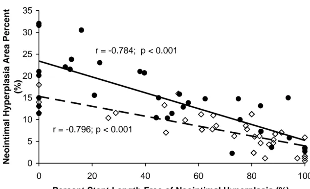
The stent length free of IVUS-detectable neointimal hyperplasia was significantly shorter in the PES group (p<0.001). More than 75% of stent length was neointimal hyperplasia-free in 59.5% of SES vs. 25.6% of PES (p=0.002), while less than 25% of stent length was neointimal hyperplasia-free in 9.5% of SES vs. 41.9% of PES (p=0.001). Neointimal hyperplasia-free stent length was inversely correlated with NIHA% in both SES and PES (p<0.001) (Fig. 3).
Mean vessel area and PSPA in the SES group showed a trend towards a reduction at follow-up (p=0.14 and p=0.09, respectively), while in the PES group they showed no significant change (Table III) (Fig. 2). In terms of PSPA%, the reduction in the SES group from post-procedure to follow-up reached statistical significance (p=0.008), making the mean change in PSPA% from post-procedure to follow-up significantly different between the SES and PES groups (-2.1±4.8 vs 0.1±3.4%, respectively; p=0.01). No correlation was found between NIHA at follow-up and PSPA% post-procedure (r=0.062, p>0.2).
An in-stent MLA of>6 mm2 after implantation (72.6 vs 74.4%, in SES and PES groups, respectively; p>0.2) was associated with no target-lesion revascularization during follow-up (0 vs 9.2%; p=0.06), and with minimal binary in-stent restenosis (1.6 vs 13.6%; p=0.05), compared with an in-stent MLA ≤6 mm2.
Incomplete stent apposition was comparable in the SES and PES groups, both after implantation (7.1 vs 9.3%, respectively; p>0.2) and at nine months (14.3 vs 11.7%; p>0.2), being persistent in 4.8 vs 4.7% of patients in the SES and PES groups, respectively (p>0.2), and late-acquired in 9.5 vs 7.0% (p>0.2). Incomplete apposition was never associated with a major adverse cardiac event.
Subgroup analysis for patients with and without acute coronary syndrome (ACS) confirmed general results with a significant lower NIHA% for SES treated patients in both subgroups (ACS: NIHA% 7,9 ± 4,5 in SES vs 13,9 ± 8,8 in PES, p = 0,003. No ACS: NIHA% 6,7 ± 3,9 in SES vs 17,8 ± 6,7 in PES, p < 0,0001).
A typical example of IVUS findings for both the SES and the PES is depicted in Figure 4.
Angiographic Results
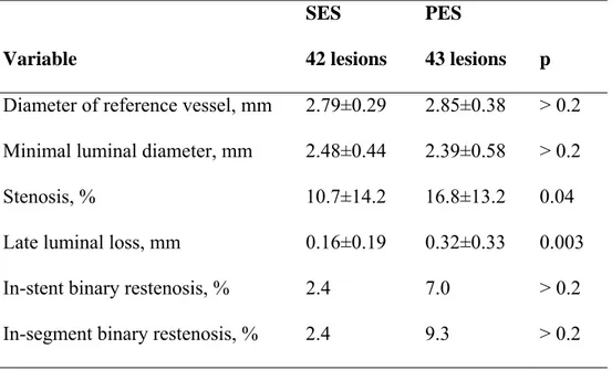
The mean in-stent LL at follow-up was 0.16±0.19 mm in the SES group and 0.32±0.33 mm in the PES group (p=0.003) (Table IV). The mean percent stenosis was quite low in both groups (10.7% vs 16.8% for the SES and PES groups, respectively; p=0.04), with a rate of in-stent binary restenosis of only 2.4% vs 7.0% (p>0.2). With 5 mm proximal and distal stent margins, the rate of in-segment restenosis was 2.4% vs 9.3%, for the SES and PES groups, respectively (p>0.2). A highly significant linear correlation between angiographic LL and NIHA% was observed in the entire population (r=0.567, p<0.001).
Major Adverse Cardiac Events
The overall rate of major adverse cardiac events was very low and not statistically different in both groups (4.8 vs 6.8%, for the SES vs the PES, respectively; p>0.2), with one cardiac death in the PES group (0 vs 2.3%, for the SES vs the PES, respectively; p>0.2), and one myocardial infarction and one target-lesion revascularization in each group (2.4 vs 2.3%; p>0.2).
Discussion
Not surprisingly, in light of previous angiographic and clinical studies in short lesions, the present study demonstrates that the SES reduces neointimal tissue proliferation significantly more than the PES in long, complex coronary lesions. However, the NIHA% was remarkably low for both types of drug-eluting stents, compared with previous data on bare-metal stents (30-40%) 51,60, and in agreement with previously reported IVUS data on the SES 10, 47-49, 25 and the PES. 11, 50, 51, 54, 61
In particular, in our SES population, with a mean lesion length 21.2 mm, the NIHA% was 7.4%, which is higher than that reported with SESs for shorter lesions at 8-12 months follow-up: 2.3% was reported in the first series from São Paulo, Brazil (mean lesion length 12.9 mm) 47, 1% in the RAVEL trial (mean lesion length of 14.4 mm) 25, 1.2% in the E-SIRIUS trial (mean lesion length of 15.5 mm) 62, and 3.1% in the SIRIUS trial (mean lesion length of 14.4 mm) 10. Interestingly, in the first series by São Paulo, NIHA% appeared to increase to 6.6% at two years follow-up 48, and to 7.9% at four years 49.
Regarding the PES, in our population with mean lesion length 20.1 mm, the NIHA% (15.4%) was higher than the 7.8% reported in the TAXUS II trial with a mean lesion length 10.6 mm 11, but similar to the 12.2% of the TAXUS IV trial (mean lesion length 12.5 mm) 51, and the 13.1% of the TAXUS V trial (mean lesion length 17.3 mm) 61.
The present study describes, for the first time in the literature, a direct comparison of IVUS results after SES and PES implantation for long lesions. Most published IVUS reports involve short lesions treated with a single SES 47-49, 25 or PES 11, 50, 51 . Fewer IVUS data involve more complex lesions, with use of multiple SESs 10,62 or PESs 61.
The only randomized comparison of the SES and the PES in complex lesions, the CORPAL trial, has only been presented orally 34.
The length of stent free of IVUS-evident neointimal hyperplasia has been reported to be greater in a non-polymeric paclitaxel-eluting stent than in the corresponding bare-metal stent, and to correlate inversely with NIHA% 63. In the present study, the inverse correlation with NIHA% was confirmed both for Cypher and Taxus stents (p<0.001). Interestingly, the mean neointimal hyperplasia-free stent length in SES was almost double than in PES (p<0.001), and IVUS-detectable neointimal hyperplasia was observed in over 75% of stent length in 41.9% of PES vs only 9.5% of SES (p=0.001). Thus, neointimal hyperplasia inside PES is often represented along the entire stent length, while SES appear neointimal hyperplasia-free for most of their length. However, since the assessment of stent endothelization is below the resolution of IVUS 63, the absence of IVUS-detectable hyperplasia does not imply the absence of re-endothelization.
There was no correlation between neointimal hyperplasia and post-procedural peri-stent plaque burden in our study. This finding is in agreement with previous reports showing that suppression of neointimal proliferation occurs irrespective of residual plaque burden after procedures with both the SES 45 and the PES 50. Late-acquired incomplete stent apposition was infrequent in both the SES and PES groups (9.5 vs 7.0%, p>0.2) in the current study, which is consistent with a recent large, retrospective study by Hong et al. 64, indicating a prevalence of 13.2% with the SES and 8.4% with the PES in a real-world population. The number of patients with late-acquired incomplete stent apposition in our study was too small to draw any conclusions about its possible mechanism or clinical relevance.
Although the absolute changes in vessel and plaque area from baseline to follow-up in the current study were small and did not reach statistical significance, the reduction in PSPA% in the SES group was significant. This finding is in contrast to previous reports showing no such variations at 6-month follow-up 25. With regard to PES, we did not observe significant changes in PSPA%, which is in agreement with the TAXUS IV trial 51
In our opinion, factors other than the stent-eluted drug may play a major role in peri-stent plaque changes. We recently demonstrated that treatment with simvastatin 20 mg/day in normocholesterolemic patients undergoing bare-metal stent implantation significantly reduces PSPA% compared with placebo (-14% vs +6%) 60. This observation, together with the apparent clinical benefits of statin treatment, underlies the aggressive lipid-lowering approach (aiming for an LDL-cholesterol level<110 mg/dL) we routinely use with all patients undergoing coronary interventions. At follow-up, 93% of SES patients and 95% of PES patients were on statins. Statin treatment may play a role in the observed reduction in PSPA% in the SES group in our study, and may mask the positive remodeling tendency described with the PES in the TAXUS II trial 1
We observed a trend for a reduction of vessel area in SES group and not in PES group; the effects of DES on the vessel wall have not been fully investigated with few, discordant findings in literature. In an analysis of 30 patients in the Sao Paulo registry, vessel area did not change in the first 2 year after Cypher stent implantation but decreased between 2 and 4 year 65. In an analysis of the TAXUS II trial patients, there was a greater 6-month increase in vessel area in the moderate-release group compared with control subjects but not in the slow- release group, suggesting a dose dependent
drug effect 50; at 2 year the 6-month vessel area increase regressed completely in the slow-release group but only incompletely in the moderate-release group 54.
An MLA >6 mm2 has been reported to reduce the risk of target-lesion revascularization and qualifies an “optimal IVUS result” 56. In our SES and PES populations, an MLA >6 mm2 post-procedure was associated with a very low rate of angiographic binary in-stent restenosis (1.6% vs 13.6%; p=0.05), and a with no need for target-lesion revascularization during follow-up.
In agreement with the IVUS findings, quantitative angiography in our study showed a significantly lower LL with the SES than the PES (0.16±0.19 vs 0.32±0.33 mm). In fact, a tight linear correlation between NIHA% and LL was observed (P<0.001). In-stent LL values are comparable to those reported in other randomized trials comparing the SES and the PES, such as the SIRTAX trial (0.12±0.36 vs 0.25±0.49 mm with the SES and the PES, respectively; p<0.001) 35, the ISAR-DIABETES trial (0.19±0.44 vs 0.46±0.64 mm; p<0.001) 37, and the REALITY trial (0.09±0.43 vs 0.31±0.44 mm; p<0.001) 32. Angiographic restenosis rates in the present study were also similar to those reported in the literature, but the population size was too small to allow meaningful considerations.
The present study was not powered to detect differences in clinical end points; however, the rate of major adverse cardiac events was low and comparable between the two groups. Our data confirm the results of subgroup analyses of patients with a stenosis in the left anterior descending artery in the SIRIUS 66 and TAXUS IV 67 trials, showing a 1-year binary in-stent restenosis rate of 2.0% and 8.7%, respectively, and a target-lesion revascularization rate of 6.0% and 5.8%, respectively. In a recent meta-analysis of six randomized trials comparing the clinical and angiographic outcome of
SES and PES implantation, Kastrati et al. concluded that patients receiving the SES have a significantly lower risk of restenosis and target-vessel revascularization 38. However, until results of a multicenter study with an adequately powered clinical end point are available, the SES and the PES should be considered clinically equivalent.
It is important to keep in mind that Cypher stent and Taxus stent differ not only for eluted drug but even for polymer and stent geometry. Different performance of two devices could so depend at least in part on difference of polymer and stent design. However it’s almost impossible to analyze separately the effects of three components; that’s why we think it’s more correct to ascribe results to the whole device rather than to the eluted-drug.
This study is limited by the absence of an IVUS core lab. However, we perform an average of 150 IVUS examinations per year, thus providing reliable expertise in the evaluation of IVUS images. Secondly, the identification of the external elastic membrane beyond the stent struts can be difficult or even impossible due to acoustic shadowing and presence of side branches, making the measurement of vessel volumes unreliable. In order to improve the quality of our data, we excluded those patients in whom the external elastic membrane could not be identified in>75% of stent length, and calculated the mean vessel area as the ratio of vessel volume to the actually analyzed length. We also acknowledge that the inclusion of patients with acute coronary syndromes and the discretional administration of glycoprotein IIb/IIIa inhibitors may have had confounding effects on neointimal proliferation. Finally, the sample size was too small to compare the clinical outcome, which was not an end point of the study, but it was adequate for the prespecified comparison of neointimal hyperplasia.
Whether or not the use of the SES (with the lower neointimal hyperplasia formation) provide superior clinical outcomes in the prognostically important left anterior descending artery cannot be answered in this study but is certainly an area deserving further investigation in future studies.
Conclusions
The present study demonstrates that both the SES and the PES cause limited neointimal hyperplasia in complex lesions, with a significant difference in favor of the SES. Comparison with IVUS data obtained in previous studies involving shorter lesions shows a higher neointimal net volume obstruction for both the SES and PES in more complex lesions and this was particularly evident for the PES. The difference in neointimal hyperplasia observed between the SES and the PES in the present study, however, does not translate into higher rates of angiographic in-stent and in-segment restenosis.
Appendix
Table I. Baseline Clinical Characteristics
Characteristic SES 42 patients PES 43 patients p Age (yr) 61±11 64±10 0.12 Male sex, n (%) 36 (85.7) 34 (79.1) > 0.2 Current smoker, n (%) 20 (47.6) 18 (41.9) > 0.2 Diabetes mellitus, n (%) 13 (31.0) 8 (18.6) 0.19 Hypertension, n (%) 28 (66.7) 26 (60.5) > 0.2 Hyperlipidemia, n (%) 30 (71.4) 26 (60.5) > 0.2 Dialysis, n (%) 3 (7.1) 0 (0) 0.12
Previous myocardial infarction, n (%) 15 (35.7) 11 (25.6) > 0.2 Acute coronary syndrome, n (%) 19 (45.2) 22 (51.2) > 0.2 Multivessel disease, n (%) 31 (73.8) 33 (76.7) > 0.2 Left ventricular ejection fraction (%) 50±8 51±8 > 0.2
SES=sirolimus-eluting stent PES=paclitaxel-elutingstent
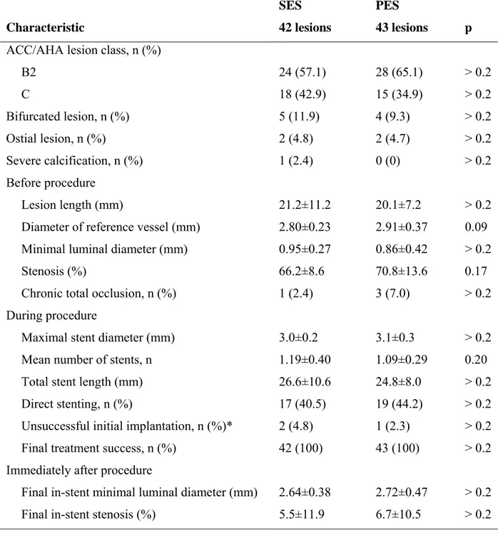
Table II. Baseline Angiographic Characteristics and Procedural Results Characteristic SES 42 lesions PES 43 lesions p
ACC/AHA lesion class, n (%)
B2 24 (57.1) 28 (65.1) > 0.2 C 18 (42.9) 15 (34.9) > 0.2 Bifurcated lesion, n (%) 5 (11.9) 4 (9.3) > 0.2 Ostial lesion, n (%) 2 (4.8) 2 (4.7) > 0.2 Severe calcification, n (%) 1 (2.4) 0 (0) > 0.2 Before procedure Lesion length (mm) 21.2±11.2 20.1±7.2 > 0.2
Diameter of reference vessel (mm) 2.80±0.23 2.91±0.37 0.09 Minimal luminal diameter (mm) 0.95±0.27 0.86±0.42 > 0.2
Stenosis (%) 66.2±8.6 70.8±13.6 0.17
Chronic total occlusion, n (%) 1 (2.4) 3 (7.0) > 0.2 During procedure
Maximal stent diameter (mm) 3.0±0.2 3.1±0.3 > 0.2
Mean number of stents, n 1.19±0.40 1.09±0.29 0.20
Total stent length (mm) 26.6±10.6 24.8±8.0 > 0.2
Direct stenting, n (%) 17 (40.5) 19 (44.2) > 0.2
Unsuccessful initial implantation, n (%)* 2 (4.8) 1 (2.3) > 0.2 Final treatment success, n (%) 42 (100) 43 (100) > 0.2 Immediately after procedure
Final in-stent minimal luminal diameter (mm) 2.64±0.38 2.72±0.47 > 0.2 Final in-stent stenosis (%) 5.5±11.9 6.7±10.5 > 0.2
* defined as unsuccessful implantation of the first single stent chosen to cover the whole lesion.
Table III. Intravascular Ultrasound Measurements In-Stent SES 42 lesions PES 43 lesions p Vessel area (mm2) Post-procedure 16.08±5.55 15.69±3.61 > 0.2 9-month follow-up 15.30±4.04 15.73±3.70 > 0.2 P 0.14 > 0.20 Stent area (mm2) Post-procedure 8.13±2.46 8.23±1.86 > 0.2 9-month follow-up 8.07±1.80 8.21±1.78 > 0.2 P > 0.20 > 0.20 Lumen area (mm2) Post-procedure 8.13±2.46 8.23±1.86 > 0.2 9-month follow-up 7.46±1.60 7.01±1.94 > 0.2 P 0.004 < 0.001
Peri-stent plaque area (mm2)
Post-procedure 7.95±3.62 7.46±2.30 > 0.2
9-month follow-up 7.23±2.79 7.52±2.49 > 0.2
P 0.09 > 0.20
Peri-stent plaque area percent (%)
Post-procedure 48.1±8.0 46.9±6.9 > 0.2
9-month follow-up 46.0±7.9 47.0±7.1 > 0.2
P 0.008 > 0.20
Neointimal hyperplasia area (mm2)
9-month follow-up 0.61±0.41 1.20±0.56 < 0.001 Neointimal hyperplasia area percent, %
9-month follow-up 7.4±4.2 15.4±8.1 < 0.001
Neointimal hyperplasia-free stent length, %
Table IV. Angiographic Results at Follow-up Variable SES 42 lesions PES 43 lesions p
Diameter of reference vessel, mm 2.79±0.29 2.85±0.38 > 0.2 Minimal luminal diameter, mm 2.48±0.44 2.39±0.58 > 0.2
Stenosis, % 10.7±14.2 16.8±13.2 0.04
Late luminal loss, mm 0.16±0.19 0.32±0.33 0.003 In-stent binary restenosis, % 2.4 7.0 > 0.2 In-segment binary restenosis, % 2.4 9.3 > 0.2
Figure 2. p=0.14 p=0.15 p=0.002 -1.4 -1.2 -1.0 -0.8 -0.6 -0.4 -0.2 0.0 0.2
Vessel Plaque Lumen
A re a C h a n ge a t Fol lo w -up ( m m 2 ) Cypher Taxus
Figure 3. r = -0.784; p < 0.001 r = -0.796; p < 0.001 0 5 10 15 20 25 30 35 0 20 40 60 80 100
Percent Stent Length Free of Neointimal Hyperplasia (%)
Neo in tim al Hyp e rp lasi a Area P e rcen t (%)
Figure Legends
Figure 1. Flow of participants through the study. IVUS=intravascular ultrasound.
Figure 2. Averaged changes (follow-up minus post-procedure) in vessel, plaque, and
lumen areas in the stented segment.
Figure 3. Correlation of percent neointimal hyperplasia-free stent length and percent
neointimal hyperplasia area in paclitaxel (solid circles – solid regression line) and sirolimus-eluting stents (open diamonds – dashed regression line).
Figure 4. Example of IVUS semiautomatic detection of vessel area (red line), stent area
(green line) and lumen area (yellow line).
Figure 5. IVUS evaluation of SES (A) and PES (C) immediately post stent deployment
and at follow-up (respectively B and D). PES shows greater intra-stent neointimal hyperplasia (seven o’clock).
References
1. Fischman DL, Leon MB, Baim DS, et al. A randomized comparison of coronary-stent placement and balloon angioplasty in the treatment of coronary artery disease. Stent Restenosis Study Investigators. N Engl J Med 1994; 331: 496-501.
2. Serruys PW, de Jaegere P, Kiemeneij F, et al. A comparison of balloon –expandable-stent implantation with ballon angioplasty in patients with coronary artery disease. Benestent Study Group. N Engl J Med 1994; 331: 489-95
3. Serruys PW, Kay IP, Disco C, Deshpande NV, de Feyter PJ. Periprocedural quantitative coronary angiography after Palmaz-Schatz stent implantation predicts the restenosis rate at six month: results of a meta-analysis of the Belgian Netherlands Stent study (BENESTENT) I, BENESTENT II Pilot, BENESTENT II and MUSIC trials. Multicenter Ultrasound Stent in Coronaries. J Am Coll Cardiol 1999; 34: 1067-74.
4. de Feyter PJ, Kay IP, Disco C, Serruys PW. Reference chart derived from post-stent-implantation intravascular ultrasound preictors of 6-months expected restenosis on quantitative coronary angiography. Circulation 19999; 100: 1777-83.
5. Mercado N, Boersma E, Wijns W, et al. Clinical and quantitative coronary angiographic predictors of coronary restenosis: a comparative analysis from the balloon-to-stent era. J Am Coll Cardiol 2001; 38: 645-52.
6. Sousa JE, Serruys PW, Costa MA. New frontiers in cardiology: drug eluting stents. Part I. Circulation 2003; 107: 2274-9
7. Sousa JE, Serruys PW, Costa MA. New frontiers in cardiology: drug eluting stents. Part II. Circulation 2003; 107: 2383-9
8. van der Giessen WJ, Lincoff AM, Schwartz RS, et al. Marked inflammatory sequelae to implantation of biodegradable and nonbiodegradable polymers in porcine coronary arteries. Circulation 1996; 94:1690-7
9. Morice MC, Serruys PW, Sousa JE, et al, for the RAVEL Study Group. A randomized comparison of a sirolimus eluting stent with a standard stent for coronary revascularization. N Engl J Med 2002; 346: 1773-80.
10. Moses JW, Leon MB, Popma JJ, et al, for the SIRIUS Investigators. Sirolimus-eluting stents versus standard stents in patients with stenosis in a native coronary artery. N Engl J Med 2003; 349: 1315-23.
11. Colombo A, Drzewiecki J, Banning A, et al, for the TAXUS II Study Group. Randomized study to assess the effectiveness of slow- and moderate-release polymer-based paclitaxel-eluting stents for coronary artery lesions. Circulation 2003; 108: 788-94.
12. Schofer J, Schluter M, Gershlick AH, et al, for the E-SIRIUS Investigators. Sirolimus-eluting stents for treatment of patients with long atherosclerotic lesions in small coronary arteries: double-blind, randomised controlled trial (E-SIRIUS). Lancet 2003; 362: 1093-9.
13. Stone GW, Ellis SG, Cox DA, et al, for the TAXUS IV Investigators. A polymer-based, paclitaxel-eluting stent in patients with coronary artery disease. N Engl J Med 2004; 350:221-31. 14. Marx SO, Marks AR. Bench to bedside: the development of rapamycin and its application to
stent restenosis. Circulation 2001; 104: 852-5.
15. Sun J, Marx SO, Chen HJ, Poon M, Marks AR, Rabbani LE. Role for p27(Kip1) in vascular smooth muscle cell migration. Circulation 2001; 103: 2967-72.
16. Serruys PW, Regar E, Carter AJ. Rapamycin eluting stent: the onset of a new era in interventional cardiology. Heart 2002; 87: 305-7.
17. Rowinsky EK, Donehower RC. Paclitaxel (taxol). N Engl J Med 1995; 332: 1004-14.
18. Schiff PB, Horwitz SB. Taxol stabilizes microtubules in mouse fibroblast cells. Proc Natl Acad Sci USA 1980; 77: 1561-5.
19. Sollott SJ, Cheng L, Pauly RR, et al. Taxol inhibits neointimal smooth muscle cell accumulation after angioplasty in the rat. J Clin Invest 1995; 95: 1869-76.
20. Schampaert E, Cohen EA, Schluter M, et al, for the C-SIRIUS Investigators. The Canadian study of the sirolimus eluting stent in the treatment of patients with long de novo lesions in small native coronary arteries (C-SIRIUS). J Am Coll Cardiol 2004; 43: 1110-5.
21. Morice M, Serruys PW, Costantini, C, et al. Two-year follow-up of the RAVEL study: a Randomized Study With the Sirolimus-Eluting Bx VelocityTM Stent in the Treatment of Patients with de-Novo Native Coronary Artery Lesions. (abstr) J Am Coll Cardiol 2003; 41: 32A
22. Regar E, Serruys PW, Bode C, et al. Angiographic findings of the multicenter Randomized Study With the Sirolimus-Eluting Bx Velocity Balloon-Expandable Stent (RAVEL): sirolimus-eluting stents inhibit restenosis irrespective of the vessel size. Circulation 2002; 106: 1949-56. 23. Ardissino D, Cavallini C, Bramucci E, et al, for the SES-SMART Investigators.
Sirolimus-eluting vs uncoated stents for prevention of restenosis in small coronary arteries: a randomized trial. JAMA 2004; 292: 2727-34.
24. Sabate M, Jimenez-Quevedo P, Angiolillo DJ et al. Randomized comparison of sirolimus-eluting stent versus standard stent for percutaneous coronary revascularisation in diabetic patients: the diabetes and sirolimus-eluting stent (DIABETES) trial. Circulation 2005; 112: 2175-83.
25. Serruys PW, Degertekin M, Tanabe K, et al, for the RAVEL Study Group. Intravascular ultrasound findings in the multicenter, randomized, double-blind RAVEL (Randomized Study With the Sirolimus-Eluting Velocity Balloon-Expandable Stent in the Treatment of Patients with de Novo Native Coronary Artery Lesions) trial. Circulation 2002; 106:798-803.
26. Holmes DR Jr, Leon MB, Moses JW, et al. Analysis of 1-year clinical outcomes in the SIRIUS trial: a randomized trial of a sirolimus-eluting stent versus a standard stent in patients at high risk for coronary restenosis. Circulation 2004;109: 634-40.
27. Grube E, Silber S, Hauptmann KE, et al. TAXUS I: six- and twelve-month results from a randomized, double-blind trial on a slow-release paclitaxel-eluting stent for de novo coronary
28. Lansky AJ, Costa RA, Mintz GS, et al, for the DELIVER Clinical Trial Investigators. Non-polymer-based paclitaxel coated coronary stents for the treatment of patients with de novo coronary lesions: angiographic follow-up of the DELIVER clinical trial. Circulation 2004; 109: 1948-54.
29. Park SJ, Shim WH, Ho DS, et al. A paclitaxel-eluting stent for the prevention of coronary restenosis. N Engl J Med 2003; 348: 1537-45.
30. Gershlick A, De Scheerder I, Chevalier B, et al. Inhibition of restenosis with a paclitaxel-eluting, polymer-free coronary stent: the European Evaluation of Paclitaxel Eluting Stent (ELUTES) trial. Circulation 2004; 109: 487-93.
31. Kataoka T, Grube E, Honda Y, et al. 7-Hexanoyltaxol-eluting stent for prevention of neointimal growth: an intravascular ultrasound analysis from the Study to Compare Restenosis Rate between QueST and QuaDS-QP2 (SCORE). Circulation 2002; 106: 1788-93.
32. Morice MC, Colombo A, Meier B, Serruys P, Tamburino C, Guagliumi G, Sousa E, Stoll HP. Sirolimus- vs paclitaxel-eluting stents in de novo coronary artery lesions: the REALITY trial: a randomized controlled trial. JAMA. 2006;295:895–904.
33. Goy JJ, Stauffer JC, Siegenthaler M, Benoit A, Seydoux C. A prospective randomized
comparison between paclitaxel and sirolimus stents in the real world of interventional cardiology: the TAXI trial. J Am Coll Cardiol. 2005;45:308 –311
34. de Lezo JMA, Pan M. Drug-eluting stents for complex lesions: randomized rapamycin versus paclitaxel CORPAL study. J Am Coll Cardiol. 2005;45(Suppl):75a. Abstract.
35. Windecker S, Remondino A, Eberli FR, Juni P, Raber L, Wenaweser P, Togni M, Billinger M, Tuller D, Seiler C, Roffi M, Corti R, Sutsch G, Maier W, Luscher T, Hess OM, Egger M, Meier B. Sirolimus-eluting and paclitaxel-eluting stents for coronary revascularization. N Engl J Med. 2005;353:653– 662.
36. Kastrati A, Mehilli J, von Beckerath N, Dibra A, Hausleiter J, Pache J, Schuhlen H, Schmitt C, Dirschinger J, Schomig A. Sirolimus-eluting stent or paclitaxel-eluting stent vs balloon angioplasty for prevention of recurrences in patients with coronary in-stent restenosis: a randomized controlled trial. JAMA. 2005;293:165–171.
37. Dibra A, Kastrati A, Mehilli J, Pache J, Schuhlen H, von Beckerath N, Ulm K, Wessely R, Dirschinger J, Schomig A. Paclitaxel-eluting or sirolimus-eluting stents to prevent restenosis in diabetic patients. N Engl J Med. 2005;353:663– 670.
38. Kastrati A, Dibra A, Eberle S, Mehilli J, Suarez de Lezo J, Goy JJ, Ulm K, Schomig A. Sirolimus-eluting stents vs paclitaxel-eluting stents in patients with coronary artery disease: meta-analysis of randomized trials. JAMA. 2005;294:819–825.
39. Windecker S. CYPHER vs TAXUS: an independent patient-based metaanalysis including the currently completed randomized controlled trials. Paper presented at: Transcatheter Cardiovascular Therapeutics Symposium; October 17–21, 2005; Washington, DC.
compared to the sirolimuseluting stent as part of the Taxus-stent evaluated at Rotterdam Cardiology Hospital (T-SEARCH) Registry. Euro Intervention. 2006;6:330 –337.
41. Simonton CA. Nonrandomized Comparison of Late Clinical Outcomes With Paclitaxel-Eluting and Sirolimus-Eluting Stents From a Large Multicenter Registry. Paper presented at: Transcatheter Cardiovascular Therapeutics Symposium; October 17–21, 2005; Washington, DC. 42. Abizaid A, Costa MA, Blanchard D, et al. Sirolimus-eluting stents inhibit neointimal hyperplasia
in diabetic patients: insights from the RAVEL trial. Eur Heart J 2004;25:107–12.
43. Morino Y, Ako J, Sonoda S, et al. Neointimal distribution within sirolimus-eluting stents: a 3-D IVUS interim analysis from the SIRIUS trial (abstr). Circulation 2002;106:II391.
44. Kaneda H, Koizumi T, Ako J, et al. Impact of intravascular ultrasound lesion characteristics on neointimal hyperplasia following sirolimus eluting stent implantation. Am J Cardiol 2005;96:1237– 41.
45. Kaneda H, Ako J, Honda Y, et al. Impact of asymmetric stent expansion on neointimal hyperplasia following sirolimus-eluting stent implantation. Am J Cardiol 2005;96:1404 –7. 46. Miyazawa A, Shimada Y, Waseda K, et al. Vascular responses after overlapping
sirolimus-eluting stent implantation: serial intravascular ultrasound analysis (abstr). Circulation 2005;112:II770.
47. Sousa JE, Costa MA, Abizaid AC, et al. Sustained suppression of neointimal proliferation by sirolimus-eluting stents: one-year angiographic and intravascular ultrasound follow-up. Circulation 2001; 104:2007–11.
48. Sousa JE, Costa MA, Sousa AG, et al. Two-year angiographic and intravascular ultrasound follow-up after implantation of sirolimus eluting stents in human coronary arteries. Circulation 2003;107:381–3.
49. Sousa JE, Costa MA, Abizaid A, et al. Four-year angiographic and intravascular ultrasound follow-up of patients treated with sirolimuseluting stents. Circulation 2005;111:2326 –9.
50. Tanabe K, Serruys PW, Degertekin M, et al. Chronic arterial responses to polymer-controlled paclitaxel-eluting stents: comparison with bare metal stents by serial intravascular ultrasound analyses: data from the randomized TAXUS-II trial. Circulation 2004;109:196 –200.
51. Weissman NJ, Koglin J, Cox DA, et al. Polymer-based paclitaxeleluting stents reduce in-stent neointimal tissue proliferation: a serial volumetric intravascular ultrasound analysis from the TAXUS-IV trial. J Am Coll Cardiol 2005;45:1201–5.
52. Weissman NJ, Ellis SE, Mintz GS, et al. Insights on the effect of polymer-based paclitaxel-eluting stents in long lesions and overlapping stents: final results from the TAXUS-V IVUS substudy (abstr). Circulation 2005;112:II769.
53. Weissman NJ, Ellis SG, Grube E et al. Effect of the polymer-based, paclitaxel-eluting TAXUS Express stent on vascular tissue responses: a volumetric intravascular ultrasound integrated analysis from the TAXUS IV, V and VI trial. Eur Heart J 2007; 28: 1574-82.
54. Aoki J, Colombo A, Dudek D, et al. Peristent remodeling and neointimal suppression two years after polymer-based, paclitaxeleluting stent implantation. Insights from serial intravascular ultrasound analysis in the TAXUS-II study. Circulation 2005;112:3876 – 83.
55. Mintz GS, Nissen SE, Anderson WD, et al. American College of Cardiology Clinical Expert Consensus Document on Standards for Acquisition, Measurement and Reporting of Intravascular Ultrasound Studies (IVUS). A report of the American College of Cardiology Task Force on Clinical Expert Consensus Documents. J Am Coll Cardiol 2001;37:1478-92.
56. Cheneau E, Pichard AD, Satler LF, Suddath WO, Weissman NJ, Waksman R. Intravascular ultrasound stent area of sirolimus-eluting stents and its impact on late outcome. Am J Cardiol 2005;95:1240-2.
57. Mintz GS, Shah VM, Weissman NJ. Regional remodeling as the cause of late stent malapposition. Circulation 2003;107:2660-3.
58. Lemos PA, Hoye A, Goedhart D, et al. Clinical, angiographic, and procedural predictors of angiographic restenosis after sirolimus-eluting stent implantation in complex patients: an evaluation from the Rapamycin-Eluting Stent Evaluated At Rotterdam Cardiology Hospital (RESEARCH) study. Circulation 2004;109:1366-70.
59. Mintz GS, Nissen SE, Anderson WD, et al. American College of Cardiology Clinical Expert Consensus Document on Standards for Acquisition, Measurement and Reporting of Intravascular Ultrasound Studies (IVUS). A report of the American College of Cardiology Task Force on Clinical Expert Consensus Documents. J Am Coll Cardiol 2001;37:1478-92.
60. Petronio AS, Amoroso G, Limbruno U, et al. Simvastatin does not inhibit intimal hyperplasia and restenosis but promotes plaque regression in normocholesterolemic patients undergoing coronary stenting: a randomized study with intravascular ultrasound. Am Heart J
2005;149:520-6.
61. Stone GW, Ellis SG, Cannon L et al. Comparison of a polymer based paclitaxel- eluting stent in patients with complex coronary artery disease: a randomised controlled trial. JAMA 2005; 294: 1215-23.
62. Hoffmann R, Guagliumi G, Musumeci G, et al. Vascular response to sirolimus-eluting stents delivered with a nonaggressive implantation technique: Comparison of intravascular ultrasound results from the multicenter, randomized E-SIRIUS, and SIRIUS trials. Catheter Cardiovasc Interv 2005; 66: 499-506.
63. Mintz GS, Hong MK, Raizner AE, et al. Intravascular ultrasound assessment of neointima distribution and the length of stent that was free of intravascular ultrasound-detectable intimal hyperplasia in paclitaxel-eluting stents. Am J Cardiol 2005;95:107-9.
64. Hong MK, Mintz GS, Lee CW, et al. Late stent malapposition after drug-eluting stent implantation: an intravascular ultrasound analysis with long-term follow-up. Circulation 2006;113:414-9.
computer-assisted grayscale value analysis for plaque composition in event-free patients. J Am Coll Cardiol 2005;46:1670-6.
66. Sawhney N, Moses JW, Leon MB, et al. Treatment of left anterior descending coronary artery disease with sirolimus-eluting stents. Circulation 2004;110:374-9.
67. Dangas G, Ellis SG, Shlofmitz R, et al. Outcomes of paclitaxel-eluting stent implantation in patients with stenosis of the left anterior descending coronary artery. J Am Coll Cardiol 2005;45:1186-92.