Abstract— Rehabilitation procedures require an objective evaluation of motor recovery to optimize their functional outcomes. Here, we propose an innovative framework for the identification of neural biomarkers of motor recovery in the forearm muscles of tetraplegic and stroke patients. The system incorporates a custom-made ergometer and high-density surface EMG recordings on the flexor and extensor finger/wrist muscles of the forearm. Blind source separation was used to identify motor unit activity during isometric maximal voluntary contractions. Preliminary results on two patients show a significant increase in the average discharge rate of the identified motor units after some weeks of rehabilitation training. Moreover, the relative change of this parameter matched with the motor recovery exhibited by the patients. This study demonstrates that the identification of individual motor units from the forearm muscles can provide a reliable neural biomarker of motor recovery.
I. INTRODUCTION
OTOR system injuries are the most important causes of disability worldwide. For example, according to the World Health Organization [1], several million people suffer injuries at the level of the motor system (e.g. occlusion/ hemorrhagic stroke, spinal cord lesion, cervical compression, etc…) each year. In most cases, the motor deficits of upper and lower limb may be substantial.
In general, early rehabilitation interventions are essential to minimize long-term motor impairment and improve functional recovery of the patients. Specifically, rehabilitation of the hand is crucial because impaired hand function can lead to the development of “learned non-use” of the entire upper limb [2].
After an injury to the motor system, the success of the
F. Negro was funded by the European Union's Horizon 2020 research and innovation programme under the Marie Skłodowska‐Curie grant agreement No 702491 (NeuralCon).
F. Negro is with the Department of Clinical and Experimental Sciences, Università degli Studi di Brescia, Brescia, Italy (corresponding author e-mail: [email protected]).
M. Cogliati is with the Department of Clinical and Experimental Sciences, Università degli Studi di Brescia, Brescia, Italy (e-mail: [email protected]).
A. Cudicio is with the Department of Clinical and Experimental Sciences, Università degli Studi di Brescia, Brescia, Italy (e-mail: [email protected]).
L. Bissolotti is with the Rehabilitation Service, Fondazione Teresa Camplani-Casa di Cura Domus Salutis, Brescia, Italy (e-mail: [email protected]).
C. Orizio is with the Department of Clinical and Experimental Sciences, Università degli Studi di Brescia, Brescia, Italy (e-mail: [email protected]).
rehabilitation procedure relies on the evaluation of the motor deficits and the level of spasticity during the whole recovery phase. For this monitoring, several clinical scales exist, but they often rely on the experience and the subjective judgment of the clinician. Objective measurements such as magnetic resonance imaging can provide only indirect information not well correlated with the actual impairment of the motor function of the patient. Therefore, the neurorehabilitation requires the development of innovative techniques to measure the degree of functional recovery at the level of alpha motor neurons. In this study, we propose an innovative framework to estimate neural biomarkers of motor recovery in the forearm of patients with injured motor system and provide preliminary evidence that such markers are a reliable indicator of motor recovery.
II. MATERIAL AND METHODS
Two patients participated in the experimental procedure. One patient (56 years) was tetraplegic due to a spinal cord injury and the other patient (43 years) had a cerebral stroke. At the time of the hospital admission, the two patients had substantial impairment of the forearm muscles. All subjects were given a verbal and written description of the study protocols and gave written informed consent prior the participation in the experiment.
Each patient participated in two experimental sessions: the first session was recorded when the patient was able to perform a detectable finger flexion. The second session was performed after some weeks of standard rehabilitation training. The tetraplegic patient improved from an average of 2/3 mobility score (hand-mouth, reaching and pronation/supination) for the upper limb to 3/3 in the last session. The stroke patient improved from 1/3 to 2/3.
In the experiment, the most affected forearm of the patients was fixed on a custom-made ergometer (Fig. 1) in order to measure the isometric flexion force of the fingers. Three high-density surface EMG matrices (64 channels) with different inter-electrode distances (ied) were placed on the extrinsic flexor muscles of the forearm (ied = 8 mm) and on the intrinsic flexor pollicis brevis of the hand (ied = 4 mm). Force signals were recorded using two force transducers (Interface, Arizona USA), one measuring the flexor force of the thumb and the second of the other four fingers. All fingers were secured with Velcro straps. The subjects performed five maximal voluntary contractions (MVCs) of the finger flexion for ~5 s with a rest of 3 min in between.
Neural Biomarkers of Functional Recovery in Patients with Injured
Motor System
Negro Francesco, Marta Cogliati, Alessandro Cudicio, Luciano Bissolotti, Claudio Orizio
The monopolar surface EMG signals were amplified (EMG-USB2+, OT Bioelettronica, Italy), band-pass filtered (20 Hz to 500 Hz), and sampled at 2048 Hz together with the force signals. The EMG signals were decomposed into series of motor unit (MU) discharges using a convolutive blind source separation method [3]. Briefly, the algorithm compensates for the action potential shapes first by the whitening of the extended measurement matrix and subsequently by an iterative approach aimed to maximized specific statistical properties of the extracted sources (non-gaussianity/sparsity). This algorithm has been previously validated and guarantees high accuracy in the identification of MU discharge times [3]. The decomposition accuracy was estimated with the silhouette measure (SIL), with an acceptable threshold of 0.90 [3]. From the decomposed discharge times, the average discharge rate of the individual motor units was computed from the signals recorded in the two sessions.
Fig. 1. Example of a recording session. The load cells are positioned to record the flexion force of the five fingers. High-density recordings are placed on the main extrinsic flexor muscles of the forearm and on the flexor pollicis brevis of the hand. The visual feedback of the two forces is provided on a computer screen in real time.
III. RESULTS
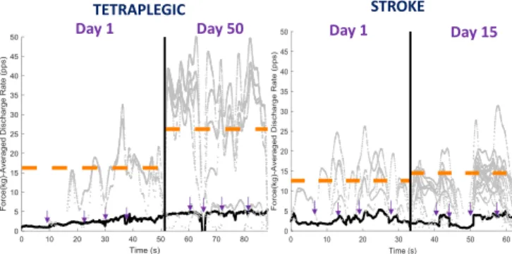
For each session, the five MVC recordings were cut in the plateau part, concatenated and decomposed. Only results for the four finger flexions are shown (no thumb). A total of 41 motor units were identified in the two subjects across the two recordings sessions (Averaged SIL = 0.92 ± 0.09). In patient I (tetraplegic), the averaged discharge rates of the identified motor units increased from 16.1 ± 5.5 pps to 25.5 ± 7.5 pps after approximately six weeks of rehabilitation training. The number of identified motor units was equal in the two sessions (8 vs 8). After approximately two weeks of rehabilitation procedure, the patient II (stroke) showed an increase in the number of identified motor units (from 8 to 17 MUs), but more moderate neural improvement with an increase of only 1.5 pps, and no detectable change in force. The amplitude of the surface EMG signal averaged across all channels increased similarly in both subjects.
IV. DISCUSSION
In this study, we developed a new framework for the
assessment of the neural recovery of patients with injured motor system after a rehabilitation period. In general, the designed ergometer has proved to be intuitive and relatively easy to use for both patients. Separated handles provide the possibility to modify the distance and the angle of the force cells in respect to the hand of the patient. The high-density matrices cover most flexor or extensor muscles of the forearm and can provide a spatial information of their activation. Moreover, blind source separations techniques were able to identify several motor units during the task and show changes in the neural drive to the muscles. Future investigations will focus on increasing the number of subjects and match motor units in multiple recording sessions [4].
Fig. 2. Smoothed discharge rates of the identified motor units (gray) are plotted for the two recording sessions in the two patients. Mean discharge rates for all units identified in the two trials is shown in orange. Force signal is in black. In each session, signals are the combination/concatenation of five MVC recordings. Purple arrows indicate the MVC segments.
V. CONCLUSIONS
We demonstrated that the proposed framework can provide neural information in patients with injured motor system. We showed that some weeks of rehabilitation training may induce significant neural modifications at the level of the flexor forearm motor pools. A larger patient population will be recruited to perform a correlation between the neural biomarkers and the standard disability scales. Future work will focus on the analysis of the motor unit action potential characteristics and the intra-muscular coherence between motor unit spike trains.
REFERENCES
[1] World Health Organization. The world health report 2002: reducing
risks, promoting healthy life. World Health Organization. (2002)
[2] Wolf SL. Revisiting constraint-induced movement therapy: are we too smitten with the mitten? Is all nonuse "learned"? and other quandaries.
Phys Ther. 2007;87:1212-23.
[3] Negro, Francesco, Silvia Muceli, Anna Margherita Castronovo, Ales Holobar, and Dario Farina. "Multi-channel intramuscular and surface EMG decomposition by convolutive blind source separation." Journal
of neural engineering 13, no. 2 (2016): 026027.
[4] Martinez‐Valdes, E., F. Negro, C. M. Laine, D. Falla, F. Mayer, and
D. Farina. "Tracking motor units longitudinally across experimental sessions with high‐density surface electromyography." The Journal of
physiology 595, no. 5 (2017): 1479-1496. Fingers %MVC Th u mb %M V C VISUAL FEEDBACK FLEXOR DIGITORUM PROXIMAL-MATRIX FLEXOR DIGITORUM DISTAL-MATRIX 64-CHANNELS MATRIX 500 N LOAD CELL 100 N LOAD CELL Day 1 Day 50 TETRAPLEGIC Day 1 Day 15 STROKE