Cardiac Involvement in a Patient With Coronavirus Disease
2019 (COVID-19)
Riccardo M. Inciardi, MD; Laura Lupi, MD; Gregorio Zaccone, MD; Leonardo Italia, MD; Michela Raffo, MD; Daniela Tomasoni, MD; Dario S. Cani, MD; Manuel Cerini, MD; Davide Farina, MD; Emanuele Gavazzi, MD; Roberto Maroldi, MD; Marianna Adamo, MD; Enrico Ammirati, MD, PhD; Gianfranco Sinagra, MD; Carlo M. Lombardi, MD; Marco Metra, MD
IMPORTANCEVirus infection has been widely described as one of the most common causes of myocarditis. However, less is known about the cardiac involvement as a complication of severe acute respiratory syndrome coronavirus 2 (SARS-CoV-2) infection.
OBJECTIVETo describe the presentation of acute myocardial inflammation in a patient with coronavirus disease 2019 (COVID-19) who recovered from the influenzalike syndrome and developed fatigue and signs and symptoms of heart failure a week after upper respiratory tract symptoms.
DESIGN, SETTING, AND PARTICIPANTThis case report describes an otherwise healthy 53-year-old woman who tested positive for COVID-19 and was admitted to the cardiac care unit in March 2020 for acute myopericarditis with systolic dysfunction, confirmed on cardiac magnetic resonance imaging, the week after onset of fever and dry cough due to COVID-19. The patient did not show any respiratory involvement during the clinical course.
EXPOSURECardiac involvement with COVID-19.
MAIN OUTCOMES AND MEASURESDetection of cardiac involvement with an increase in levels of N-terminal pro–brain natriuretic peptide (NT-proBNP) and high-sensitivity troponin T, echocardiography changes, and diffuse biventricular myocardial edema and late gadolinium enhancement on cardiac magnetic resonance imaging.
RESULTSAn otherwise healthy 53-year-old white woman presented to the emergency department with severe fatigue. She described fever and dry cough the week before. She was afebrile but hypotensive; electrocardiography showed diffuse ST elevation, and elevated high-sensitivity troponin T and NT-proBNP levels were detected. Findings on chest radiography were normal. There was no evidence of obstructive coronary disease on coronary angiography. Based on the COVID-19 outbreak, a nasopharyngeal swab was performed, with a positive result for SARS-CoV-2 on real-time reverse transcriptase– polymerase chain reaction assay. Cardiac magnetic resonance imaging showed increased wall thickness with diffuse biventricular hypokinesis, especially in the apical segments, and severe left ventricular dysfunction (left ventricular ejection fraction of 35%). Short tau inversion recovery and T2-mapping sequences showed marked biventricular myocardial interstitial edema, and there was also diffuse late gadolinium enhancement involving the entire biventricular wall. There was a circumferential pericardial effusion that was most notable around the right cardiac chambers. These findings were all consistent with acute myopericarditis. She was treated with dobutamine, antiviral drugs (lopinavir/ritonavir), steroids, chloroquine, and medical treatment for heart failure, with progressive clinical and instrumental stabilization.
CONCLUSIONS AND RELEVANCEThis case highlights cardiac involvement as a complication associated with COVID-19, even without symptoms and signs of interstitial pneumonia.
JAMA Cardiol. 2020;5(7):819-824. doi:10.1001/jamacardio.2020.1096 Published online March 27, 2020.
Viewpointpage 743and Editorialpage 751
Related articlespages 811, 802, and831
Video
Author Affiliations: Institute of Cardiology, Department of Medical and Surgical Specialties, Radiological Sciences, and Public Health, University of Brescia, Brescia, Italy (Inciardi, Lupi, Zaccone, Italia, Raffo, Tomasoni, Cani, Cerini, Adamo, Lombardi, Metra); Institute of Radiology, Department of Medical and Surgical Specialties, Radiological Sciences, and Public Health, University of Brescia, Brescia, Italy (Farina, Gavazzi, Maroldi); “De Gasperis” Cardio Center and Transplant Center, Niguarda Hospital, Milan, Italy (Ammirati);
Cardiovascular Department, “Ospedali Riuniti” and University of Trieste, Trieste, Italy (Sinagra). Corresponding Author: Marco Metra, MD, Institute of Cardiology, c/o Spedali Civili, Piazzale Spedali Civili 1, Brescia BS 25123, Italy ([email protected]).
JAMA Cardiology |
Brief Report
T
he first cases of coronavirus disease 2019 (COVID-19) were reported in December 2019, originating in Wu-han, China,1with rapid spread worldwide, and COVID-19 became a public health emergency of international concern.2 The pathogen has been identified as a novel enveloped RNA beta-coronavirus and has been named severe acute respira-tory syndrome coronavirus 2 (SARS-CoV-2).3
The clinical course of SARS-CoV-2 infection is mostly characterized by respira-tory tract symptoms, including fever, cough, pharyngodynia, fatigue, and complications related to pneumonia and acute respiratory distress syndrome.4
Data regarding cardiovascular involvement due to SARS-CoV-2 infection are less described. Previous severe acute re-spiratory syndrome (SARS) beta-coronavirus infections could be associated with tachyarrhythmias and signs and symp-toms of heart failure.5The present report describes a case of cardiac involvement in a patient affected by COVID-19. The pa-tient provided written informed consent, and the diagnostic procedures were conducted in accordance with institutional guidelines about the protection of human subjects.
Report of a Case
An otherwise healthy 53-year-old white woman without pre-vious history of cardiovascular disease presented to the emer-gency department with severe fatigue for 2 previous days. She denied chest pain, dyspnea, and further symptoms. She re-ported having fever and cough the week before.
On arrival to the emergency department, physical exami-nation revealed blood pressure of 90/50 mm Hg, heart rate of 100 beats per minute, oxygen saturation of 98% while breath-ing ambient air, and body temperature of 36.6 °C. (She re-mained afebrile during the subsequent clinical course.) Arte-rial gas analysis showed a pH of 7.46, oxygen partial pressure of 82 mm Hg, carbon dioxide partial pressure of 32 mm Hg, and
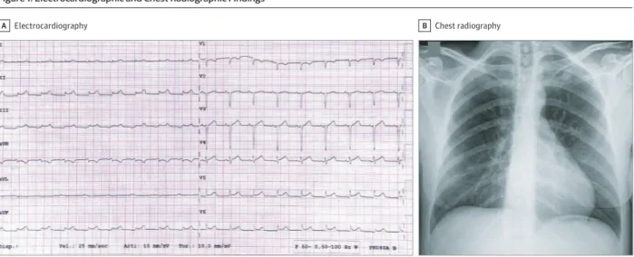
lactate level of 17.1 mg/dL (to convert to millimoles per liter, multiply by 0.111). A 12-lead electrocardiogram (ECG) showed low voltage in the limb leads, minimal diffuse ST-segment el-evation (more prominent in the inferior and lateral leads), and an ST-segment depression with T-wave inversion in lead V1 and aVR (Figure 1A).
Findings on chest radiography were unremarkable (Figure 1B). Blood tests revealed elevated levels of markers of myocyte necrosis (high-sensitivity troponin T level of 0.24 ng/mL [to convert to micrograms per liter, multiply by 1] and creatine kinase–MB level of 20.3 ng/mL [to convert to micro-grams per liter, multiply by 1]), elevated N-terminal pro– brain natriuretic peptide (NT-proBNP) levels (5647 pg/mL [to convert to nanograms per liter, multiply by 1]), slight increase in C-reactive protein levels (1.3 mg/dL [to convert to milli-grams per liter, multiply by 10), and normal blood cell counts (Table). Blood sample tests also revealed hyperkalemia, hy-ponatremia, and hypochloremia. These abnormalities were treated with kayexalate, glucose and insulin solution, and so-dium bicarbonate. Given the echocardiography changes, re-gional wall motion abnormalities, and elevated markers of
Key Points
QuestionWhat are the cardiac complications associated with the emerging outbreak of coronavirus disease 2019 (COVID-19)?
FindingsIn this case report, an otherwise healthy 53-year-old patient developed acute myopericarditis with systolic dysfunction confirmed on cardiac magnetic resonance imaging a week after onset of fever and dry cough due to COVID-19. The patient was treated with inotropic support, antiviral drugs, corticosteroids, and chloroquine, with progressive stabilization of the clinical course.
MeaningThe emerging outbreak of COVID-19 can be associated with cardiac involvement, even after the resolution of the upper respiratory tract infection.
Figure 1. Electrocardiographic and Chest Radiographic Findings
Electrocardiography
A B Chest radiography
A, Electrocardiography showing sinus rhythm with low voltage in the limb leads, diffuse ST-segment elevation (especially in the inferior and lateral leads), and ST-segment depression with T-wave inversion in leads V1 and aVR. B,
Posteroanterior chest radiography at presentation. No thoracic abnormalities were noted.
myocardial necrosis, urgent coronary angiography was per-formed, which showed no evidence of obstructive coronary disease.
The patient was admitted to the intensive care unit with a diagnosis of suspected myopericarditis. Based on the clini-cal history and the COVID-19 outbreak, COVID-19 was deemed as likely. A nasopharyngeal swab was performed with a posi-tive result for SARS-CoV-2 on real-time reverse transcriptase– polymerase chain reaction assay. Search for common cardio-tropic infectious agents yielded negative results.
Transthoracic echocardiography revealed normal left ventricular (LV) dimensions with an increased wall thickness (interventricular septum, 14 mm, posterior wall, 14 mm) and a diffuse echo-bright appearance of the myocardium. There was diffuse hypokinesis, with an estimated LV ejection fraction (LVEF) of 40%. There was no evidence of heart valve disease. Left ventricular diastolic function was mildly impaired with mitral inflow patterns, with an E/A ratio of 0.7 and an average E/e′ ratio of 12. There was a circumferential pericardial effu-sion that was most notable around the right cardiac cham-bers (maximum, 11 mm) without signs of tamponade. Cardiac magnetic resonance imaging (MRI) confirmed the increased wall thickness with diffuse biventricular hypokinesis, espe-cially in the apical segments, and severe LV dysfunction (LVEF of 35%) (Video 1 and Video 2). Short tau inversion recovery and T2-mapping sequences showed marked biventricular myo-cardial interstitial edema. Phase-sensitive inversion recovery sequences showed diffuse late gadolinium enhancement
extended to the entire biventricular wall (Figure 2). The myo-cardial edema and pattern of late gadolinium enhancement ful-filled all the Lake Louise criteria for the diagnosis of acute myocarditis.6The circumferential pericardial effusion was con-firmed, especially around the right cardiac chambers (maxi-mum, 12 mm).
During the first days of her hospitalization, the patient re-mained hypotensive (systolic blood pressure less than 90 mm Hg) and required inotropic support (dobutamine) in the first 48 hours, during which there was a further increase in levels of NT-proBNP (8465 pg/mL), high-sensitivity troponin T (0.59 ng/mL), and creatine kinase–MB (39.9 ng/mL), with a progres-sive stabilization and reduction during the following days (Table). Blood pressure progressively stabilized, although sys-tolic pressure remained less than 100 mm Hg, and dobuta-mine treatment was weaned on day 4. Heart failure–directed medical treatment was started with daily doses of 50 mg of kan-renone, 25 to 50 mg of furosemide, and 2.5 mg of bisoprolol, then reduced and finally withdrawn on day 5 owing to sinus bradycardia. The patient was treated on admission with intra-venous aspirin (500 mg twice daily), and given the cardiac MRI findings, hydroxychloroquine (200 mg twice daily), lopinavir/ ritonavir (2 tablets of 200/50 mg twice daily), and intrave-nous methylprednisolone (1 mg/kg daily for 3 days)7,8
were ad-ministrated. Chest radiography was repeated on day 4 and showed no thoracic abnormalities. Transthoracic echocardi-ography, performed on day 6, revealed a significant reduc-tion of LV wall thickness (interventricular septum, 11 mm; pos-Table. Clinical Laboratory Results
Measure Reference range
Result
Day 1 Day 2 Day 3 Day 4 Day 5 Day 6 Day 7
Red blood cell count, ×106/μL 4.0-5.2 5.5a 4.6 4.0b 3.9b 3.8b 3.6b 3.7b
Hemoglobin, g/dL 12.0-16.0 17.1a 14.5 12.4 11.9b 12.0 11.4b 11.2b
Hematocrit, % 37.0-47.0 49.3a 42.1 36.0b 34.9b 35.1b 33.9b 33.6b
White blood cell count, per μL 4000-10 800 8900 12 090a 9920 10 900 13 470a 13 730a 13 500a
Lymphocyte count Relative, % 20.0-40.0 10.6b NA NA NA NA NA 7.7b Absolute, per μL 900-4000 950 NA NA NA NA NA 1040 Platelet count, ×103/μL 130-400 152 168 164 213 317 317 360 Sodium, mEq/L 136-145 129b 133b 129b 136 132b 134b 137 Potassium, mEq/L 3.4-4.5 5.7a 6.3a 3.9 3.7 3.5 3.6 3.6 Chloride, mEq/L 98-107 89b 96b 92b 92b NA 92b 94b Calcium, mg/dL 8.60-10.20 8.63 NA 7.84b 8.15b NA NA NA Creatinine, mg/dL 0.60-1.00 0.75 0.76 0.53b 0.88 0.99 0.96 0.80 C-reactive protein, mg/dL <0.5 1.3a 0.7a 1.0a 1.1a 0.6 0.4 0.3 Creatine kinase–MB, ng/mL <4.9 20.3a 39.9a 30.7a 13.3 5.2 3.3 2.8 High-sensitivity troponin T, ng/mL <0.01 0.24 0.59 0.78 0.89 0.76 0.65a 0.63a NT-proBNP, pg/mL <300c 5647 8465 8133 5113 2827 NA NA
Abbreviations: NA, not applicable; NT-proBNP, N-terminal pro–brain natriuretic peptide.
SI conversion factors: To convert red blood cell count to ×1012per liter, multiply by 1; hemoglobin to grams per liter, multiply by 10; white blood cell count to ×109per liter, multiply by 0.001; lymphocyte count to ×109per liter, multiply by 0.001; platelet count to ×109
per liter, multiply by 1; sodium to millimoles per liter, multiply by 1; potassium to millimoles per liter, multiply by 1; chloride to millimoles per liter, multiply by 1; C-reactive protein to milligrams per liter,
multiply by 10; creatine kinase–MB to micrograms per liter, multiply by 1; high-sensitivity troponin T to micrograms per liter, multiply by 1; and NT-proBNP to nanograms per liter, multiply by 1.
a
The value in the patient was greater than normal. bThe value in the patient was less than normal.
cLess than 300 pg/mL was a rule-out for acute heart failure; greater than 900 pg/mL was a rule-in for acute heart failure.
terior wall, 10 mm), an improvement of LVEF to 44%, and a slight decrease of pericardial effusion (maximum, 8-9 mm). At the time of submission, the patient was hospitalized with progressive clinical and hemodynamic improvement.
Discussion
Herein, we describe a patient without a history of cardiovas-cular disease admitted to the hospital with COVID-19 and severe LV dysfunction and acute myopericarditis. Our main findings are that cardiac involvement may occur with
COVID-19 even without respiratory tract signs and symp-toms of infection.
After the first cases describing pneumonia cases of un-known origin in Wuhan, China, SARS-CoV-2 rapidly spread worldwide with critical challenges for the public health and medical communities.1,2
The World Health Organization has declared SARS-CoV-2 a public health emergency of interna-tional concern, with a global estimate of 98 192 laboratory-confirmed cases and 3380 deaths as of March 6, 2020.
A 2020 report by the China Medical Treatment Expert Group for COVID-199
showed the spectrum of clinical and di-agnostic features associated with SARS-CoV-2 infection among Figure 2. 1.5-T Cardiac Magnetic Resonance Imaging
STIR sequence in short-axis view
A B STIR sequence in 4-chamber view
T2-mapping sequence in short-axis view
C D T2-mapping sequence in 4-chamber view
PSIR sequence in short-axis view
E F PSIR sequence in 4-chamber view
Short tau inversion recovery (STIR) sequences in short-axis view (A) and 4-chamber view (B) showed diffuse myocardial signal hyperintensity of the biventricular wall, suggesting interstitial edema. Results were confirmed on the T2-mapping sequences in short-axis view (C) and 4-chamber view (D). Phase-sensitive inversion recovery (PSIR) sequences in short-axis view (E) and 4-chamber view (F) showed diffuse biventricular late gadolinium enhancement. All images demonstrated a
circumferential pericardial effusion, especially around the right ventricle.
Chinese patients. The most common symptoms were fever (in up to 88.7% of patients during hospitalization) and cough (in 67.8% of patients), followed by dry cough, headache, fatigue, or shortness of breath. Complications were mostly related to physician-diagnosed pneumonia (91.1%) and acute respiratory distress syndrome.3,4While the spectrum of clinical manifesta-tion is highly related to the inflammamanifesta-tion process of the respi-ratory tract, this case provides evidence of cardiac involve-ment as a possible late phenomenon of the viral respiratory infection. This process can be subclinical with few interstitial inflammatory cells, as reported by an autopsy study,10or can present with overt manifestations even without respiratory symptoms, as in the present case.
Virus infection has been widely described as one of the most common infectious causes of myocarditis, especially associated with influenza and parvovirus B-19 infection.11 However, less is known about the cardiac involvement as a complication of SARS-CoV-2 infection.
Myocarditis results in focal or global myocardial inflam-mation, necrosis, and eventually ventricular dysfunction. Focal myocarditis is often suspected in patients presenting with chest pain after an influenzalike syndrome, with clinical evidence suggesting an acute coronary syndrome on electro-cardiography or laboratory testing or with evidence of wall mo-tion abnormalities without evidence of obstructive coronary artery disease on coronary angiography.12
The pathogenesis of cardiac involvement associated with SARS-CoV-2 may reflect a process of replication and dissemi-nation of the virus through the blood or the lymphatic sys-tem from the respiratory tract. However, to our knowledge, there are no reports of influenza virus or coronavirus RNA in the heart, to date. Alternatively, SARS-CoV-2 could trigger an exaggerated inflammatory response that can cause myocar-dial injury, and this could justify the use of corticosteroids to attenuate inflammation, as in the present case. Evidence of a significant inflammatory cell infiltration has been reported in the alveoli of patients with acute respiratory distress syn-drome associated with SARS-CoV-2 infection,10and this could explain the use of corticosteroids in patients with COVID-19 (up to 58% in a series of critically ill patients13
). Although ul-trastructural mechanisms are not certain, a potential binding to a viral receptor of the myocyte can favor the
internaliza-tion and subsequent replicainternaliza-tion of the capsid proteins and the viral genome.14,15In this patient, increases of cardiac tropo-nin levels as a sensitive marker of myocardial injury, the cardiac MRI findings showing diffuse edema, and the slow gadolinium washout are in line with an acute myocarditis. In addition, the onset of symptoms several days after the influ-enzalike syndrome may reflect these proposed mechanisms with a potential myocyte dissemination of the virus, the acti-vation of the immune system, and, ultimately, the clinical onset of heart failure.
Limitations
As endomyocardial biopsy was not performed, limitations of this report are the lack of the histological demonstration of myocarditis and the absence of viral genome search in the heart. Except for the first 48 hours during which she required inotropic support, the patient was mainly treated with heart failure–directed medical treatment. However, as described in the literature, viral myocarditis has a wide spectrum of clini-cal presentations, ranging from life-threating arrhythmias to advanced heart failure requiring invasive support.10
Conclusions
We believe that recognition by the scientific community of acute myocarditis as a possible complication associated with COVID-19 may be helpful for strict monitoring of affected patients and also for furthering knowledge of such complica-tions for public health officials. This report highlights the importance of clinical surveillance and laboratory testing, including troponin levels, in individuals with recent symp-toms of an acute illness to guarantee appropriate identifica-tion and prompt isolaidentifica-tion of patients at risk of COVID-19 and eventually to reduce further transmission. Further evidence is needed to determine whether corticosteroids are useful in reducing the myocardial inflammatory response. We cannot exclude that a spontaneous resolution occurred or that antivi-ral drugs or chloroquine contributed to the improvement of this patient. Finally, awareness of atypical presentations such as this one is important to prompt patient isolation and pre-vent interhuman transmission.
ARTICLE INFORMATION
Accepted for Publication: March 13, 2020. Published Online: March 27, 2020. doi:10.1001/jamacardio.2020.1096
Author Contributions: Drs Inciardi and Metra had full access to all of the data in the study and take responsibility for the integrity of the data and the accuracy of the data analysis.
Study concept and design: Inciardi, Lupi, Zaccone, Italia, Raffo, Tomasoni, Cani, Maroldi, Sinagra, Lombardi, Metra.
Acquisition, analysis, or interpretation of data: Inciardi, Lupi, Zaccone, Raffo, Cerini, Farina, Gavazzi, Adamo, Ammirati, Metra.
Drafting of the manuscript: Inciardi, Lupi, Zaccone, Italia, Raffo, Tomasoni, Cani, Farina, Metra. Critical revision of the manuscript for important
intellectual content: Inciardi, Lupi, Cerini, Gavazzi, Maroldi, Adamo, Ammirati, Sinagra, Lombardi, Metra.
Administrative, technical, or material support: Lupi, Cerini, Metra.
Study supervision: Inciardi, Lupi, Farina, Maroldi, Adamo, Ammirati, Sinagra, Metra.
Conflict of Interest Disclosures: Dr Farina has received personal fees from Bayer and Bracco Group. Dr Metra has received personal fees from Abbott Vascular, Amgen, Bayer, Edwards Therapeutics, and Vifor Pharma. No other disclosures were reported.
Additional Contributions: We thank the patient for granting permission to publish this information.
REFERENCES
1. World Health Organization. Pneumonia of unknown cause—China. Accessed January 5, 2020. https://www.who.int/csr/don/05-january-2020-pneumonia-of-unkown-cause-china/en/ 2. World Health Organization. Novel coronavirus—China. Accessed January 12, 2020. https://www.who.int/csr/don/12-january-2020-novel-coronavirus-china/en/
3. Lu R, Zhao X, Li J, et al. Genomic characterisation and epidemiology of 2019 novel coronavirus: implications for virus origins and receptor binding. Lancet. 2020;395(10224):565-574. doi: 10.1016/S0140-6736(20)30251-8
4. Huang C, Wang Y, Li X, et al. Clinical features of patients infected with 2019 novel coronavirus in
Wuhan, China. Lancet. 2020;395(10223):497-506. doi:10.1016/S0140-6736(20)30183-5
5. Yu CM, Wong RS, Wu EB, et al. Cardiovascular complications of severe acute respiratory syndrome. Postgrad Med J. 2006;82(964):140-144. doi:10.1136/pgmj.2005.037515
6. Friedrich MG, Sechtem U, Schulz-Menger J, et al; International Consensus Group on Cardiovascular Magnetic Resonance in Myocarditis. Cardiovascular magnetic resonance in myocarditis: a JACC White Paper. J Am Coll Cardiol. 2009;53(17):1475-1487. doi:10.1016/j.jacc.2009.02.007
7. Young BE, Ong SWX, Kalimuddin S, et al; Singapore 2019 Novel Coronavirus Outbreak Research Team. Epidemiologic features and clinical course of patients infected with SARS-CoV-2 in Singapore. JAMA. 2020. Published online March 3, 2020. doi:10.1001/jama.2020.3204
8. Gao J, Tian Z, Yang X. Breakthrough: chloroquine phosphate has shown apparent efficacy in treatment of COVID-19 associated pneumonia in clinical studies. Biosci Trends. 2020;14(1):72-73. doi: 10.5582/bst.2020.01047
9. Guan WJ, Ni ZY, Hu Y, et al; China Medical Treatment Expert Group for Covid-19. Clinical characteristics of coronavirus disease 2019 in China. N Engl J Med. Published online February 28, 2020. doi:10.1056/NEJMoa2002032
10. Xu Z, Shi L, Wang Y, et al. Pathological findings of COVID-19 associated with acute respiratory distress syndrome. Lancet Respir Med. Published online February 18, 2020. doi:10.1016/S2213-2600 (20)30076-X
11. Fung G, Luo H, Qiu Y, Yang D, McManus B. Myocarditis. Circ Res. 2016;118(3):496-514. doi:10. 1161/CIRCRESAHA.115.306573
12. Esfandiarei M, McManus BM. Molecular biology and pathogenesis of viral myocarditis. Annu Rev Pathol. 2008;3:127-155. doi:10.1146/annurev. pathmechdis.3.121806.151534
13. Yang X, Yu Y, Xu J, et al. Clinical course and outcomes of critically ill patients with SARS-CoV-2 pneumonia in Wuhan, China: a single-centered, retrospective, observational study. Lancet Respir Med. Published online February 24, 2020. doi:10.1016/ S2213-2600(20)30079-5
14. Rahman JE, Helou EF, Gelzer-Bell R, et al. Noninvasive diagnosis of biopsy-proven cardiac amyloidosis. J Am Coll Cardiol. 2004;43(3):410-415. doi:10.1016/j.jacc.2003.08.043
15. Liu PP, Mason JW. Advances in the
understanding of myocarditis. Circulation. 2001;104 (9):1076-1082. doi:10.1161/hc3401.095198