This article was published in the above mentioned Springer issue. The material, including all portions thereof, is protected by copyright;
all rights are held exclusively by Springer Science + Business Media. The material is for personal use only;
commercial use is not permitted. Unauthorized reproduction, transfer and/or use may be a violation of criminal as well as civil law.
REGULAR ARTICLE
The shape modulation of osteoblast
–osteocyte
transformation and its correlation
with the fibrillar organization in secondary osteons
A SEM study employing the graded osmic maceration technique
Ugo E. Pazzaglia&Terenzio Congiu&
Marcella Marchese&Carlo Dell’Orbo
Received: 13 December 2009 / Accepted: 29 March 2010 / Published online: 28 April 2010 # Springer-Verlag 2010
Abstract Cortex fractured surface and graded osmic maceration techniques were used to study the secretory activity of osteoblasts, the transformation of osteoblast to osteocytes, and the structural organization of the matrix around the cells with scanning electron microscopy (SEM). A specialized membrane differentiation at the base of the cell was observed with finger-like, flattened processes which formed a diffuse meshwork. These findings suggested that this membrane differentiation below the cells had not only functioned in transporting collagen through the membrane but also in orienting the fibrils once assembled. Thin ramifications arose from the large and flat membrane foldings oriented perpendicular to the plane of the osteo-blasts. This meshwork of fine filaments could not be visualized with SEM because they were obscured within the matrix substance. Their 3-D structure, however, should be similar to the canalicular system. The meshwork of large, flattened processes was no more evident in the cells which had completed their transformation into osteocytes.
Keywords Secondary osteon . Osteoblast . Osteocyte . Collagen fibrillogenesis . Fibripositor .
Rabbit (New Zealand white)
Introduction
The concept that osteoblasts are cells committed to producing bone matrix is well established (Baud1968). However, other related aspects like the osteoblast–osteocyte transformation, the formation and extrusion of collagen fibrils, and the control of their spatial orientation remain unanswered questions and are the subject of conflicting theories (Rouiller et al. 1952; Ruth 1953; Smith 1960; Boyde and Hordell
1969; Giraud-Guille1988; Marotti1993).
The asymmetry of the osteoblast structure observed in transmission electron-microscopy (TEM) studies suggested a polarity of the collagen precursor assembly line within the cellular membrane and also the recognition of a specialized secretory territory on the bone-facing surface of the cell (Palumbo et al.1990). The definition of secretory territories of osteoblasts was introduced by Jones (1974) in a scanning electron-microscopy (SEM) study of rat parietal bones where the latter parameter was correlated to the rate of matrix production. However, due to the particular topography of osteoblasts on the bone growing surface, the most interesting zone of the cell was not accessible with SEM. In this area, the specialized functions related to the extrusion of the collagen fibril precursors, their aggregation and the spatial organization of the extracellular matrix were supposed to occur. To overcome this problem, Jones and colleagues (1975) removed single osteoblasts from the sheet of cells lining the parietal bones of rats and rhesus monkeys with a microneedle. This gave a direct visualization of the collagen fibrils laid down beneath the cell.
Osmium tetroxide is currently used as post-fixative in TEM and SEM because of its ability to fix and to enhance the contrast of lipids and phospholipids. A prolonged osmic treatment, however, is capable of denaturating the mem-U. E. Pazzaglia (*)
:
M. MarcheseClinica Ortopedica dell’Università di Brescia, Spedali Civili di Brescia,
25123 Brescia, Italy
e-mail: [email protected] T. Congiu
:
C. Dell’OrboDipartimento di Morfologia Umana, Università dell’Insubria, 21100 Varese, Italy
T. Congiu
e-mail: [email protected]
brane proteins but not collagen and elastin. This technique has also been applied to evaluate the plasmalemma and the intracellular organelles with SEM (Congiu et al.2004; Riva et al. 1993; Tanaka and Mitsushima 1984; Tanaka et al.
1986).
In this study, the bone surface of the osteoblasts lining the central canal of forming osteons were exposed through longitudinal cortical fractures (Pazzaglia et al.2009), and the graded osmic maceration technique was applied. By removing most of the osteoblast cellular body, it was possible to expose the membrane processes at the base of the cells. Bone lining cells present on the endosteal surface of the diaphysis could also be evaluated by the longitudinal cortical fractures and could be compared with the active osteoblasts of the osteons.
In light of these new findings, it has been possible to reconsider several aspects related to the collagen organization in the lamellar bone of secondary osteons and the shape modulation of the osteoblast–osteocyte transformation.
Materials and methods
The study was carried out on the tibia of six male New Zealand white rabbits (Charles River, Calco, BG, Italy) of around 8 months of age, with a body weight between 3.0 and 3.5 kg. The care and use of the experimental animals was consistent with procedures and regulations of the Italian Health Ministery.
The rabbits were anaesthetized with ketamine hydro-chloride (Imagel) and xylazine (Rompum), the aorta and the vena cava were exposed through a midline abdominal incision and a 1.5-mm catheter was inserted into the aorta between the diaphragm and the renal arteries in a proximal to distal direction. The artery was then tightly ligated with two ligatures around the catheter, and the rabbit was euthanased with a further dose of the anaesthetic just prior to commencing the perfusion of the vascular tree. The rabbits were injected with 300 ml of formaldehyde solution (2%) with a hand syringe at a pressure of 150–200 mm Hg until the lower limbs were completely perfused. This took about 5 s. It was necessary to clamp the vena cava prior to infusion to balance the difference in resistence to perfusion between the extra-cortical and intra-cortical system. By doing so, a complete perfusion of the intra-cortical sector vessels was obtained (Pazzaglia et al.1997).
Following infusion, the tibias were immediately dissect-ed from the soft tissues and stordissect-ed in formaldehyde solution (2%). The central part of the diaphysis (including the distal tibio-fibular junction) was cut with a hand saw in a plane perpendicular to the major axis of the bone, and the proximal and distal parts were discarded. The central cylinder of each right tibia was split in a longitudinal direction after creating three or four fractures initiated by a notch made with a chisel on the transversally cut surface. The marrow was then gently washed out and the specimens were then submitted to a graded osmic maceration. They were washed in phosphate-buffered saline (PBS, pH 7.2) Fig. 1 SEM (osmic maceration,
6 h). Bone linig cells covering the endosteal surface of the diaphysis. Small processes are present on the peripheral mar-gin, but the upper face of the cell membrane is flat and the small roughness is due to osmic corrosion. The layer of cells continues within the vascular canals which open in the bone marrow cavity (arrows)
and then post-fixed in a solution of 1% osmium tetroxide and 1.25% potassium ferrocyanide for 2 h. They were then washed in PBS, immersed in 0.1% osmium tetroxide in PBS for 6, 12, 48, and 72 h at room temperature and again washed in PBS. All the specimens were dehydrated in ascending grades of ethanol, subjected to critical point drying in CO2, coated with 10 nm of gold palladium in a vacuum sputter Emitech K550 (Edax, Mahwah, NJ, USA) and studied in the direct mode with a Philips XL 30 SEM-FEG scanning electron microscope (Philips, Eindhoven, Nederlands).
Results
The endosteal surface of the diaphysis was lined by a sheet of flattened, polygonal cells which formed an extensive and almost complete layer covering the bone (bone lining cells). This layer appeared in continuity with the surface of the vascular canals which opened within the bone marrow cavity (Fig. 1). The cells had a flat, smooth surface and small processes at the periphery, forming a finely indented margin. The thin gap between each cell and its neighbor suggested a shrinkage artefact of processing.
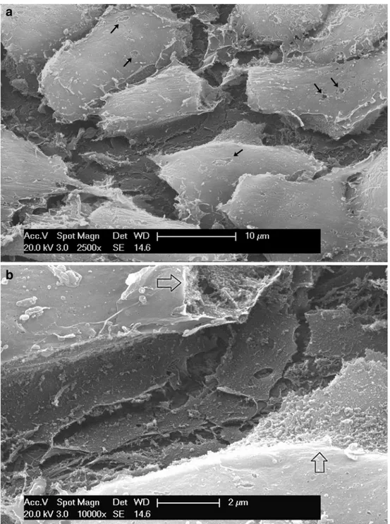
Fig. 2 a SEM (osmic macera-tion, 12 h). Sheet of osteoblasts lining the central canal of a structuring osteon. Flat process-es spreading from beneath the cells form a meshwork on the bone surface. The small defects on the cellular membrane have been produced by osmic macer-ation (arrows). b SEM (osmic maceration, 12 h). Detail of the meshwork of flat processes at the base of an osteoblast. Where the cellular membrane has been corroded by osmic acid the ergastoplasmatic reticulum in-side the cells has been exposed (arrows)
No specialized membrane differentiation on the cell surface could be evidenced either in cells which had been occasionally torn off mechanically or macerated by osmium.
The osteoblasts lining the central canal of the active osteons had a similar polygonal shape. Their surface was slightly domed with a flat base adjacent to the bone surface. There was no evidence of any cytoplasmic processes on the
dome corresponding to the vascular face. The base of the cell (bone face) was not freely accessible to direct observation; however, shrinkage artefacts associated with osmic acid action opened a gap between the cells showing a network of flat, finger-like processes and crests derived from the basal cellular membrane (Fig.2a, b). By prolonging osmic maceration and removing most of the cellular body, it was possible to get a full view of the basal surface. The cellular membrane of each Fig. 3 SEM (osmic maceration,
48 h). Basal processes of differ-ent osteoblasts forming a mesh-work beneath the cellular bodies which have been removed by maceration. Processes are large and flat and show ramifications (arrows). The internal aspect of the cellular membrane was ex-posed by osmic maceration (*). In the deeper layers, more com-pact bundles of collagen fibrils are evident (#). The numbers correspond to processes of dif-ferent cells
Fig. 4 SEM (osmic maceration, 48 h). Basal processes with exocytosis vescicles (arrows) whose content is assembled to form fibrils (arrowheads) on the external surface of the cell membrane
osteoblast presented flattened, finger-like processes, wider at their base, and becoming thin as they projected away from their origin. Ramifications existed through frequent bi-furcations which prevailed in a horizontal plane forming an intertwined meshwork beneath each cell (Fig. 3). The processes of the adjoining cells contributed to the formation of this meshwork; therefore, a single osteoblast had largely expanded its ramifications outside the perimeter of its cellular body. Exocytosis vescicles could be observed in large numbers at these sites, and their content aggregated to form filaments adherent to the outer aspect the cellular
membrane, but initially without a periodic pattern (Fig. 4). Within the membrane meshwork, collagen fibrils were aggregated into bundles with a definite orientation in the horizontal plane beneath the cell (Fig.4). A second order of ramifications was observed beneath underlying matrix orientated perpendicular to the plane of the meshwork (Fig.5a, b).
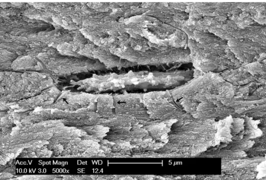
There was no such organization of the membrane processes around the osteocytes exposed in the plane of fracture; they looked like thin filaments which were observed to seep straight into the canaliculi (Fig.6). Fig. 5 a SEM (osmic
macera-tion, 72 h). Basal processes and oriented bundles of collagen fibrils. Some ramifications of processes turn perpendicularly to the horizontal plane and deepen between collagen bun-dles (arrows). b SEM (osmic maceration, 72-h). Detail of box in (a) showing the thin branches of processes with a direction perpendicular to the plane of the meshwork. The parallel packing of fibril bundles is evident
Discussion
A functional polarization of the secretory activity of the osteoblasts has been suggested by the ultrastructural organization and by the morphological changes undergone by these cells during their differentiation into osteocytes (Marotti1976; Palumbo1986; Palumbo et al.1990). These observations were assumed as a base for models of the entrapment of the pre-osteocyte within the bone matrix (Boyde 1972; Marotti et al. 1992) and the matrix organization of the secondary osteons.
The present application of cortex-fractured surfaces associated with graded osmic maceration to SEM observa-tion allowed an original approach to the study of the osteoblast secretory activity. This has enabled a novel perspective on the transformation from osteoblast to osteocyte to be achieved as well as a new appreciation of the relationship with the fibrillar organization of the matrix around the cells.
The collagen is synthetized within the RER of osteo-blasts and it is extruded from the cell within exocytotic vesicles, but the fibrils are assembled outside the cellular membrane as documented with cultured fibroblasts (Church et al.1971; Canty et al.2004; Iwasaki et al.2008).
We were able to document that the osteoblasts of forming osteons develop a network of flat processes on that part of the cellular membrane lying on the growing bone surface. The concept of secretory territory proposed by Boyde (1969) and by Jones (1974) and later developed by others (Palumbo et al. 1990) refers to this zone, where the extension of the cellular membrane surface is augment-ed by the folding and the finger-like processes. Our findings confirmed this view, but further suggested that these processes on the bone face of the osteoblasts formed a meshwork; therefore, it was not possible to define an individual secretory territory beneath each cell (Fig. 7). This structural asset can explain the regular polarization of collagen fibrils in the lamellae of the osteon, because it is Fig. 6 SEM (direct mode).
Os-teocyte within lacuna exposed by cortical fracture. No mesh-work or flat processes are pres-ent on either the upper or lower cell surfaces. The processes appear as thin filaments which seep straight into the canaliculi (arrows)
Fig. 7 Scheme of the basal processes beneath the osteoblasts showing the meshwork formed by the flat processes orienting the collagen fibrils and a second order of thinner processes turning in the depth of the fibrillar matrix
compatible with a temporal and spatial synchronism of all the osteoblasts of the sheet when they assemble the fibrils into more compact bundles. The morphological observations of this study suggest that the membrane differentiation beneath the base of the osteoblasts not only functions to transport collagen through the membrane and to assemble it into chains but it also functions to orientate the collagen fibrils and the bundles.
Evidence concerning how the cells can perform the assembly, parallelism, and packing of collagen fibrils has previously been reported in tendons. It has been seen that in tendons, where plasma membrane projections termed “fibripositors” were documented, they played a role in the extracellular collagen fibril orientation (Birk and Trelstad
1986; Birk and Zycband1994; Canty et al.2004; Kapacee et al. 2008). Similar aspects were also reported in teeth cementogenesis (Bosshardt and Schroeder1991; Yamamoto et al.1996). The cell membrane configuration documented at the base of the osteoblasts suggested a similar mechanism in the osteonal lamella.
The thin ramifications originating from the meshwork beneath the osteoblast could not be explored with SEM because they were obscured within the matrix substance. Their course and architecture, however, can be extrapolated by other methods of study, for example histological analysis of not-demineralized ground sections, as these cellular processes correspond to the canalicular network.
Despite continuity between the two types of processes, they differ in size, shape, and spatial arrangement. This suggests a different function existing for each; namely, collagen fibril orientation for the large and flat foldings of the membrane and interconnections between osteocytes for the thin filaments (Weinger and Holtrop 1974; Pawlicki
1975; Stanka 1975; Doty 1981; Shapiro 1988). It is these thin filaments which are oriented in a radial direction with respect to the vascular axis of the osteon. A consequence of this structural asset is that the osteoblasts, in the course of bone apposition, have a very limited ability to change their position within the organizing osteon. The only free direction is radially allowed by the lengthening of the processes.
The basal, horizontal meshwork was observed ex-clusively beneath active osteoblasts, while the processes originated from the osteocytes with a cone from the cell body and then seeped straight inside the canaliculi. The transformation from osteoblast to osteocyte implies the entrapment of the cell within the matrix and has been correlated to the slowing down of the rate of bone apposition of a single osteoblast with respect to the neighboring cells (Marotti et al. 1992); the loss of below-the-cell membrane differentiation appeared to be correlated with the synthetic activity reduction.
If the regular polarization of the individual lamellar fibrils can be explained by the synchronous activity of a
sheet of osteoblasts connected through the network of flat and horizontal processes, no explanation has been presented as yet for the change of polarization in the sequence of lamellae. A possible hypothesis may be that there is a modulation in the activity of the osteoblast–osteocyte transformation resulting in phasic activity of the osteoblast. During the active phase, there is apposition of a single lamella and remodulation of a new network. This is followed by a period of inactivity and then a new active phase where apposition and remodulation takes place but with a different orientation of the lamella.
Aknowledgements The study was carried out using a scanning electron microscope from the “Centre Great Instruments” at the University of Insubria and was supported by research funds from Brescia University. The authors thank Mr. Livio Di Muscio, registrar in orthopaedics at the RNOH (Stanmore, UK), for revision of the English text.
References
Baud CA (1968) Submicroscopic structure and functional aspects of the osteocyte. Clin Orthop Relat Res 56:227–236
Birk DE, Trelstad RL (1986) Extracellular compartments in tendon morphogenesis: collagen fibril, bundle, and macroaggregate formation. J Cell Biol 103(1):231–240
Birk DE, Zycband E (1994) Assembly of the tendon extracellular matrix during development. J Anat 184(Pt 3):457–463
Bosshardt DD, Schroeder HE (1991) Establishment of acellular extrinsic fiber cementum on human teeth. A light- and electron-microscopic study. Cell Tissue Res 263(2):325–336
Boyde A (1969) Correlation of ameloblast size with enamel prism pattern: use of scanning electron microscope to make surface area measurements. Z Zellforsch Mikrosk Anat 93(4):583–593 Boyde A (1972) Scanning electron microscope studies of bone. In:
Bourne GH (ed) The biochemistry and physiology of bone, vol 1. Academic, New York, pp 259–310
Boyde A, Hordell MH (1969) Scanning electron microscopy of lamellar bone. Z Zellforsch Mikrosk Anat 93(2):213–231 Canty EG, Lu Y, Meadows RS, Shaw MK, Holmes DF, Kadler KE
(2004) Coalignment of plasma membrane channels and protru-sions (fibripositors) specifies the parallelism of tendon. J Cell Biol 165(4):553–563
Church RL, Pfeiffer SE, Tanzer ML (1971) Collagen biosynthesis: synthesis and secretion of a high molecular weight collagen precursor (procollagen). Proc Natl Acad Sci USA 68(11):2638– 2642
Congiu T, Radice R, Raspanti M, Reguzzoni M (2004) The 3D structure of the human urinary bladder mucosa: a scanning electron microscopy study. J Submicrosc Cytol Pathol 36(1):45– 53
Doty SB (1981) Morphological evidence of gap junctions between bone cells. Calcif Tissue Int 33(5):509–512
Giraud-Guille MM (1988) Twisted plywood architecture of collagen fibrils in human compact bone osteons. Calcif Tissue Int 42 (3):167–180
Iwasaki S, Hosaka Y, Iwasaki T, Yamamoto K, Nagayasu A, Ueda H, Kokai Y, Takehana K (2008) The modulation of collagen fibril assembly and its structure by decorin: an electron microscopic study. Arch Histol Cytol 71(1):37–44
Jones SJ (1974) Secretory territories and rate of matrix production of osteoblasts. Calcif Tissue Res 14(4):309–315
Jones SJ, Boyde A, Pawley JB (1975) Osteoblasts and collagen orientation. Cell Tissue Res 159(1):73–80
Kapacee Z, Richardson SH, Lu Y, Starborg T, Holmes DF, Baar K, Kadler KE (2008) Tension is required for fibripositor formation. Matrix Biol 27(4):371–375
Marotti G (1976) Decrement in volume of osteoblasts during osteon formation and its effect on the size of the corresponding osteocytes. In: Meunier PJ (ed) Bone histomorphometry. Armour Montagu, Levallois, pp 385–397
Marotti G (1993) A new theory of bone lamellation. Calcif Tissue Int 53(Suppl 1):S47–S55
Marotti G, Ferretti M, Muglia MA, Palumbo C, Palazzini S (1992) A quantitative evaluation of osteoblast-osteocyte relationships on growing endosteal surface of rabbit tibiae. Bone 13(5):363–368 Palumbo C (1986) A three-dimensional ultrastructural study of
osteoid-osteocytes in the tibia of chick embryos. Cell Tissue Res 246(1):125–131
Palumbo C, Palazzini S, Zaffe D, Marotti G (1990) Osteocyte differentiation in the tibia of newborn rabbit: an ultrastructural study of the formation of cytoplasmic processes. Acta Anat (Basel) 137(4):350–358
Pawlicki R (1975) Bone canaliculus endings in the area of the osteocyte lacuna, Electron-microscopic studies. Acta Anat (Basel) 91(2):292–304
Pazzaglia UE, Andrini L, Di Nucci A (1997) The reaction to nailing or cementing of the femur in rats. A microangiographic and fluorescence study. Int Orthop 21(4):267–273
Pazzaglia UE, Congiu T, Raspanti M, Ranchetti F, Quacci D (2009) Anatomy of the intracortical canal system: scanning electron
microscopy study in rabbit femur. Clin Orthop Relat Res 467:2446–2456
Riva A, Congiu T, Faa G (1993) The application of the OsO4
maceration method to the study of human bioptic material. A procedure avoiding freeze-fracture. Microsc Res Technique 26:526–527
Rouiller C, Huber L, Kellenberger E, Rutishauser E (1952) The lamellar structure of the osteon. Acta Anat (Basel) 14(1–2):9–22 Ruth EB (1953) Bone studies. II. An experimental study of the
Haversian-type vascular channels. Am J Anat 93(3):429–455 Shapiro F (1988) Cortical bone repair. The relationship of the
lacunar-canalicular system and intercellular gap junctions to the repair process. J Bone Jt Surg Am 70(7):1067–1081
Smith JW (1960) The arrangement of collagen fibres in human secondary osteones. J Bone Jt Surg Br 42-B:588–605
Stanka P (1975) Occurrence of cell junctions and microfilaments in osteoblasts. Cell Tissue Res 159(3):413–422
Tanaka K, Mitsushima A (1984) A preparation method for observing intracellular structures by scanning electron microscopy. J Microsc 113:213–222
Tanaka K, Mitsushima A, Fukudome H, Kashima Y (1986) Three-dimensional architecture of the Golgi complex observed by high resolution scanning electron microscopy. J Submicrosc Cytol 18:1–9
Weinger JM, Holtrop ME (1974) An ultrastructural study of bone cells: the occurrence of microtubules, microfilaments and tight junctions. Calcif Tissue Res 14(1):15–29
Yamamoto T, Domon T, Takahashi S, Wakita M (1996) Cellular cementogenesis in rat molars: the role of cementoblasts in the deposition of intrinsic matrix fibers of cementum proper. Anat Embryol (Berl) 193(5):495–500