ANNO ACCADEMICO 2016/2017
UNIVERSITÀ DEGLI STUDI DI SALERNO
DIPARTIMENTO DI MEDICINA E CHIRURGIA
DOTTORATO DI RICERCA IN MEDICINA TRASLAZIONALE E
DELL’INVECCHIAMENTO ATTIVO
Curriculum: Marcatori molecolari, radiologici, clinici e
cognitivo-comportamentali dello sviluppo e del declino funzionale
Ciclo: XXIX
Coordinatore: Prof. Corrado Rubino
TESI DI DOTTORATO IN:
Unraveling biomarkers in Parkinson’s disease: the role of Insulin-like
growth factor-1 (IGF-1) and DAT imaging
! !
!
RELATORE
Chiar.ma Prof.
Maria Teresa Pellecchia
CANDIDATA
Table of contents
List of abbreviations 2 Part I – Introduction 4 Parte II - Scope of the thesis 10 Part III - Exploring the association between serum IGF-1 and cognitive functions
in early, drug-naïve Parkinson’s disease
Part IV - Investigating the association between dopaminergic dysfunction and
anxiety in de novo Parkinson's disease
Part V - Unraveling the relationship between cognition and dopaminergic
dysfunction in de novo Parkinson's disease: a longitudinal prospective study Appendix Acknowledgments 69 ! ! ! ! ! 12 33 49
List of abbreviations
BJLOT: Benton Judgment of Line Orientation test DAT: dopamine transporter
GDS-15: 15-item Geriatric Depression Scale
HC: healthy controls
HVLT-DLRY: Hopkins Verbal Learning Test Delayed Recall
HVLT-DR: Hopkins Verbal Learning Test discrimination recognition HVLT-IR: Hopkins Verbal Learning Test immediate recall
HVLT-R: Hopkins Verbal Learning Test – Revised IGF-1: insulin-like growth factor-1
JOLO: Benton Judgment of Line Orientation LNS: Letter Number Sequencing
MCI: mild cognitive impairment
MDS-UPDRS: Movement Disorder Society-Unified Parkinson’s Disease Rating Scale
MDS-UPDRS-III: Movement Disorders Society version of the Unified Parkinson’s disease rating scale part III
MoCA: Montreal Cognitive Assessment PD: Parkinson’ s disease
PPMI: Parkinson's Progression Markers initiative SBRs: striatal binding ratios
SDMT: Symbol Digit Modalities test SF: Semantic fluency
SPECT: single positron emission computerized tomography STAI: State-Trait Anxiety Inventory
! ! ! ! ! ! ! ! ! ! ! ! ! ! !
Introduction
Parkinson's disease (PD) is the second most common neurodegenerative disease after Alzheimer's disease, affecting up to 10 million individuals worldwide [1]. Although symptomatic treatment ameliorates motor symptoms, currently there are no disease-modifying treatments.
A biomarker is defined by the National Institutes of Health as “a characteristic that is objectively measured and evaluated as an indicator of normal biological processes, pathogenic processes or pharmacological responses to a therapeutic intervention” [2]. Thus, biomarkers include clinical data, measurements of biological samples (e.g., plasma, serum, cerebrospinal fluid) and application of brain imaging techniques to detect changes in brain structure and function.
As for PD, biomarkers represent tools potentially suitable for either clinical or research settings and useful in predicting onset, confirming diagnosis, detecting progression and evaluating the response to disease-modifying treatments. In addition, biomarkers’ trends in different stages of disease may reflect the widespread neurochemical and neuroanatomical changes that occur throughout the course of PD and, thus, possibly suggest new insights in the pathophysiological mechanisms underlying disease progression[3,4] (Figure 1). The range of available biomarkers in PD is fast expanding and includes an increasing number of laboratory, clinical and imaging data [5]. Indeed, the latter two represent the cornerstones of the diagnostic criteria for PD recently proposed by the International Parkinson and Movement Disorders Society (MDS) task force on the definition of PD [5-7]. As for imaging biomarkers, the largest amount of evidence is available for SPECT DAT Scan which is currently used in both clinical and research settings [8,9]. On the contrary, scant of data are
available for laboratory biomarkers. As such, although serum biomarkers would represent a fascinating, cost-effective and easy to collect source of information in PD, no valid and reliable serum biomarkers have been identified so far.
References
1) Wright-Willis A, Evanoff BA, Lian M, et al. Geographic and ethnic variation in Parkinson disease: a population-based study of US Medicare beneficiaries. Neuroepidemiology 2010;34(3):143–151.
2) Biomarkers Definitions Working Group, Biomarkers and surrogate endpoints: preferred definitions and conceptual framework. Clin Pharmacol Ther 2001;69:89–95.
3) O'Sullivan SS, Williams DR, Gallagher DA, et al. Non motor symptoms as presenting complaints in Parkinson’s disease: a clinicopathological study. Mov Disord 2008;23(1):101-106.
4) Jellinger KA, Neuropathobiology of non-motor symptoms in Parkinson disease. J Neural Transm 2015;122:1429–1440.
5) Picillo M, Barone P, Pellecchia MT. Merging clinical and imaging biomarkers to tackle Parkinson’s disease. Mov Disord Clinical Practice 2017;4(5):652-662.
6) Berg D, Postuma RB, Adler CH, et al. MDS research criteria for prodromal Parkinson's disease. Mov Disord 2015;30(12):1600-1611.
7) Postuma RB, Berg D, Stern M, et al. MDS clinical diagnostic criteria for Parkinson's disease. Mov Disord 2015;30(12):1591-1601.
8) Arena JE, Stoessl AJ. Optimizing diagnosis in Parkinson's disease: Radionuclide imaging. Parkinsonism Relat Disord. 2016;22 Suppl 1:S47-51.
9) Suwijn SR, de Bruin K, de Bie RM, Booij J. The role of SPECT imaging of the dopaminergic system in translational research on Parkinson's disease. Parkinsonism Relat Disord. 2014 Jan;20 Suppl 1:S184-6.
Figure 1. Clinical and imaging biomarkers tackling pathological changes in Parkinson’s
disease from prodromal to middle/advanced phase.
Figure 1 legend
AR:! akinetic4rigid;! EDS:! Excessive! daytime! sleepiness;! MIBG:! iodine41234meta4 iodobenzylguanidine;!MRI:!magnetic!resonance!imaging;!MSA:!multiple!system!atrophy,!NMS:!non! motor!symptoms;!PD:!Parkinson's!disease;!PDD:!Parkinson’s!disease!with!dementia;!PET:!positron! emission! tomography;! PIGD:! postural! instability! and! gait! disorders;! ! RBD:!REM4Sleep! Behavior!
Disorders;! SN:! substantia! nigra;! SPECT:! single! photon! emission! computed! tomography;! TCS:!
correlates)!has!been!taken!from:!Olanow!CW,!Stern!MB,!Sethi!K.!The!scientific!and!clinical!basis!for! the!treatment!of!Parkinson!disease!(2009).!Neurology!2009;72!(Suppl!4):S14S136.! ! ! ! ! ! ! ! !
Scope of the thesis
The scope of this thesis is to explore the relationship between specific laboratory and imaging biomarkers and cognitive as well as behavioral features in de novo, drug-naïve PD patients.
In Part III of the thesis, we explored the role of serum Insulin-Growth Factor 1 (IGF-1) at diagnosis as a marker of worse cognitive performances.
In Part IV and V, we focused on the SPECT DAT Scan and aimed at demonstrating a direct relationship between nigro-striatal denervation and specific cognitive and behavioral features. Indeed, we already reached such results in our small, single-center de novo PD population [1-4]. Thus, the main goal of the studies proposed in the present thesis were to replicate our previous findings in the largest cohort of de novo, drug-naïve PD patients existing worldwide, the Parkinson’s Progression Markers Initiative (PPMI) study promoted by the Michael J Fox Foundation for Parkinson’s research and following prospectically 424 de novo PD patients and 196 Healthy Controls (http://www.ppmi-info.org). Indeed, we took advantage of being an active part of the PPMI study and obtained the approval from the Steering Committee for dosing the IGF-1 serum levels of all the PPMI participants. The remaining data used for this thesis have been downloaded from the PPMI website (http://www.ppmi-info.org/access-data-specimens/download-data). The whole statistical design as well the execution for the three studies proposed have been performed entirely by the PhD candidate.
References
1) Pellecchia MT, Santangelo G, Picillo M, Pivonello R, Longo K, Pivonello C, Vitale C, Amboni M, De Rosa A, Moccia M, Erro R, De Michele G, Santoro L, Colao A, Barone P. Insulin-like growth factor-1 predicts cognitive functions at 2-year follow-up in early, drug-naïve Parkinson's disease. Eur J Neurol 2014;21(5):802-7.
2) Erro R, Pappatà S, Amboni M, Vicidomini C, Longo K, Santangelo G, Picillo M, Vitale C, Moccia M, Giordano F, Brunetti A, Pellecchia MT, Salvatore M, Barone P. Anxiety is associated with striatal dopamine transporter availability in newly diagnosed untreated Parkinson's disease patients. Parkinsonism Relat Disord 2012 ;18(9):1034-8.
3) Santangelo G, Vitale C, Picillo M, Cuoco S, Moccia M, Pezzella D, Erro R, Longo K, Vicidomini C, Pellecchia MT, Amboni M, Brunetti A, Salvatore M, Barone P, Pappatà S. Apathy and striatal dopamine transporter levels in de-novo, untreated Parkinson's disease patients. Parkinsonism Relat Disord 2015;21(5):489-93.
4) Pellecchia MT, Picillo M, Santangelo G, Longo K, Moccia M, Erro R, Amboni M, Vitale C, Vicidomini C, Salvatore M, Barone P, Pappatà S. Cognitive performances and DAT imaging in early Parkinson's disease with mild cognitive impairment: a preliminary study. Acta Neurol Scand. 2015 May;131(5):275-81.
Exploring the association between serum IGF-1 and cognitive functions in
early, drug-naïve Parkinson’s disease
Published in Plos One 2017;12(10):e0186508. !
Abstract
Objective: Cognitive deficits are common in Parkinson’s disease (PD) since the early stages and
many patients eventually develop dementia. Yet, occurrence of dementia in PD is unpredictable. Evidence supports the hypothesis that insulin-like growth factor-1 (IGF-1) is involved in cognitive deficits. Our aim was to evaluate the relationship between serum IGF-1 levels and neuropsychological scores in a large cohort of drug-naïve PD patients during the earliest stages of the disease.
Methods: Serum IGF-1 levels were determined in 405 early, drug-naïve PD patients and 191
healthy controls (HC) enrolled in the Parkinson’s Progression Markers Initiative (PPMI). The association between serum IGF-1 levels and neuropsychological scores was evaluated with linear regression analysis.
Results: IGF-1 levels were similar in PD and HC. In PD patients the lowest IGF-1 quartile was a
predictor of lower performances at the Semantic Fluency task (β=-3.46, 95%CI: -5.87 to -1.01, p=0.005), the Symbol Digit Modalities Score (β=-2.09, 95%CI: -4.02 to -0.15, p=0.034), and Hopkins Verbal Learning Test Retention (β=-0.05, 95%CI: -0.09 to -0.009, p=0.019).
Conclusions: Lower serum IGF-1 levels are associated to poor performances in cognitive tasks
assessing executive function, attention and verbal memory in a large cohort of early PD patients. Follow-up studies are warranted to assess if IGF-1 is related to the development of dementia in PD.
Introduction
Cognitive disturbances are common findings in Parkinson’s disease (PD) since the earliest stages, manifesting with executive as well as with visuospatial deficits [1]. Cognitive function typically decline over time, with many patients eventually developing dementia [2]. Evolution to dementia is a key milestone in PD progression as denotes a subgroup of patients with worse prognosis and represents a heavy burden for both patients and caregivers. Among all, age, male gender and presence of subtle cognitive impairment have been identified as the most consistent predictors of dementia [2]. Despite considerable efforts of the research community, valid biomarkers of cognitive impairment for PD have yet to come. As minimally invasive and economically affordable, serum biomarkers for neurodegenerative diseases gained increasing attention in recent years.
Both preclinical and clinical evidence support the hypothesis that Insulin-like growth factor 1 (IGF-1) is involved in neuroprotection and plays a role in determining cognitive deficits: a) animal and in vitro studies show that IGF-1 is critical for neuronal cell functioning, enhancing neuronal survival and inhibiting apoptosis and Β-amyloid deposition [3-5]; b) studies disclose cognitive impairment in children with growth hormone (GH) deficit [6]; c) cross-sectional epidemiologic studies report an association between low IGF-1 levels and reduced cognitive performance in healthy, elderly subjects [7,8], with particular regard to executive function, working memory and attention tasks [9];
d) prospective studies demonstrate that higher serum IGF-1 levels are associated with less cognitive decline in the elderly [10-13].
In keeping with this evidence, we reported a significant association between low serum IGF-1 levels and poor performances on executive tasks at PD diagnosis and on attention/executive and verbal memory tasks after two-years since diagnosis in a cohort of 65 drug-naïve, early patients, suggesting a link between IGF-1 and early cognitive impairment in PD [14].
In the present study, we sought to analyze the relationship between serum IGF-1 levels and cognitive scores at diagnosis in a larger cohort of drug-naïve PD patients. Based on previous evidence, lower serum IGF-1 levels were expected to be associated with worse cognitive performances. Furthermore, the association between IGF-1 levels and cognition was explored in a large cohort of healthy controls (HC).
Methods
Data used for this study were obtained from the Parkinson’s Progression Markers Initiative (PPMI) database (www.ppmi-info.org/data). PPMI-a public-private partnership-is funded by the Michael J.
Fox Foundation for Parkinson’s Research and funding partners
(info.org/fundingpartners). The aims and methodology of the study are available at www.ppmi-info.org/study-design. The study was approved by the institutional review board at each site, and participants provided written informed consent. For up-to-date information on the study visit www.ppmi-info.org.
PD patients were enrolled in the PPMI study provided they were drug-naïve, had less than 2-years disease duration and evidence of dopamine transporter (DAT) deficit on DAT Scan imaging.
Enrolled subjects underwent scheduled assessments to collect clinical data and biospecimens every
3 months for the first year and then every 6 months up to 5 years. Disease severity has been
assessed by means of the Movement Disorders Society version of the Unified Parkinson’s disease rating scale part III (MDS-UPDRS-III).
Baseline serum samples for the IGF-1 analysis were obtained from the PPMI (Cognition biomarkers, 2013; Grant ID 8800; www.ppmi-info.org/access-data-specimens/request-specimens/). Demographic, clinical and neuropsychological data were gathered from the PPMI database according to guidelines for data access and use (http://www.ppmi-info.org/data).
According to PPMI Laboratory Manual, venous blood samples from PD patients and HC were drawn at baseline visit in the morning after an overnight fast. Blood samples were centrifuged and serum was frozen (-80°C). Serum samples from 405 PD patients and 191 HC collected at the baseline assessment of the PPMI were shipped on dry ice and analyzed at the Department of Clinical Medicine and Surgery, Federico II University, Naples.
IGF-1 Measurement
We performed the IGF-1 measurement using the Quantikine Human IGF-1 Immunoassay (R&D Systems, Minneapolis, MN, USA), that is a solid-phase ELISA designed to measure human IGF-1 in serum and plasma. The immunoassay is calibrated against a highly purified Escherichia coli-expressed recombinant human IGF-1. To reduce analytical variance, samples were batch analyzed
using the same assay lot. Serum was pretreated to release IGF-1 from IGF-binding proteins with acid-ethanol extraction. Each sample was tested twice in the same assay and the mean of these two values was considered for statistical analysis. The NIBSC/WHO IGF-1 International Reference Reagent 02/254 was evaluated in this assay. The minimal detectable dose (MDD) of IGF-1 in this assay ranged from 0.007 to 0.056 ng/ml and the mean MDD was 0.026 ng/ml. The intra-assay coefficients of variation (CVs) were 3.5%, 4.3% and 4.3%, and the inter-assay CVs were 8.1%, 8.3% and 7.5% for low, medium and high points of the standard curve, respectively.
Cognitive evaluation
Global cognition was screened with the Montreal Cognitive Assessment (MoCA) [15]. An extensive battery of neuropsychological tests was administered to evaluate different cognitive domains. In detail, memory was tested with the Hopkins Verbal Learning Test-Revised (HVLT-R) [16]; visuospatial function with the Benton Judgment of Line Orientation (JOLO) 15-item version [17]; processing speed-attention with the Symbol-Digit Modalities Test (SDMT) [18]; and executive function and working memory with Letter-Number Sequencing (LNS) [19] and semantic fluency (SF) [20]. With regard to HVLT-R, Immediate Recall (IR), Delayed Recall (DLRY), Retention and Discrimination recognition (DR) were analyzed. Language domain was not assessed.
Statistical analysis
Differences in the distribution of categorical variables among groups were assessed by the chi-square test. After Kolmogorov-Smirnov testing for normal distribution, parametric testing was performed for group comparisons (independent sample t-test or one-way ANOVA with post-hoc
Bonferroni test, as applicable). Descriptive statistics are given as means and standard deviation (SD) with range.
Those variables of interest assessing specific cognitive domains (ie, all except the MoCA score) reaching a significance threshold≤0.1 further underwent linear regression analysis to evaluate the association between serum IGF-1 levels and cognitive scores. Based on the observation that only the lowest quartile of IGF-1 tended to present worse cognitive outcomes compared to the other three quartiles (Figure 1 and Table 1), further analysis were performed comparing the lowest IGF-1 quartile to all other quartiles combined. Visual inspection of graphs also confirmed the lowest
IGF-1 quartile tended to present worse scores. Linear regression analysis included age, education,
gender as covariates with no interaction terms. Education years were divided into 2 groups based on the median (16 years); group 0: ≤16 years, group 1: >16 years. Results were considered statistically significant at p<0.05.
Results
Demographic and clinical data of PD patients are reported in Table 1. Serum IGF-1 and age were not different between PD and HC (136.6±56.1 vs 134.45±56.13 ng/ml; p=0.64). Serum IGF-1 levels were divided in quartiles as follows: lowest quartile ≤ 97.9 ng/ml; second quartile=97.9001-124 ng/ml; third quartile=124.0001-162.200 ng/ml; highest quartile ≥ 162.2001 ng/ml.
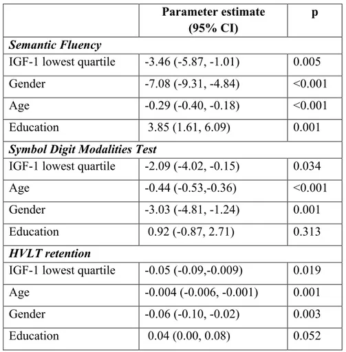
Linear regression analysis showed that in PD patients the lowest IGF-1 quartile was associated to worse performances in several cognitive tasks compared to all the others quartiles (Table 2). In detail, the lowest IGF-1 quartile was a predictor of lower scores at the SF task (p=0.005), along
with age (p<0.001), male gender (p<0.001) and education (p=0.001) (Figure 1A). Lower performances at the SDMT were associated to the lowest IGF-1 quartile (p=0.034), age (p<0.001) and male gender (p=0.001) (Figure 1B). Finally, the lowest IGF-1 quartile was a predictor of lower performances at the HVLT Retention (p=0.019), along with age (p=0.001) and male gender (p=0.003) (Figure 1C).
No significant associations were found between IGF-1 and any of the neuropsychological tasks in HC.
Discussion
Confirming our hypothesis, we demonstrated that lower serum IGF-1 levels are associated with lower performances on cognitive tasks in a large cohort of early, drug-naïve PD patients. In detail, lower serum IGF-1 levels are linked to poor performances in cognitive tasks assessing executive function, attention and verbal memory. Deficits in such tasks are driven by a dysfunction in the prefrontal and temporal cortex. In detail, neuroimaging studies suggest that while the capability of holding information and sustaining attention (evaluated with the retention and delayed recall of words list and the SDMT) are both ensured by the prefrontal cortex [21], the ability to access the conceptual knowledge stores (evaluated by the Semantic fluency task) is provided by the temporal cortex [22]. Therefore, our data suggest that low IGF-1 levels are linked to impaired cognitive functions (i.e. cognitive flexibility, attention, devising a search strategy) mediated by prefrontal and temporal cortex.
In spite of using different neuropsychological tests, these results are consistent with our previous study in a cohort of 65 drug-naïve, early PD patients showing a significant correlation between low serum IGF-I levels and poor performance on both executive tasks at baseline and attention/executive and verbal memory tasks at 2-year follow-up [14]. Therefore, taken together the findings from the current and previous study support the link between IGF-1 and cognitive impairment in PD patients even in the earliest stages of disease.
Although explored in a spoonful of studies [14,23], the relation between IGF-1 and cognitive performance in PD is not surprising. Indeed, a relevant number of IGF-1 receptors have been reported in several brain areas essential for cognitive performance, with highest concentrations in the hippocampus and the frontal cortex [3,24,25].Preclinical evidence also suggest,IGF-1 plays out an active defence mechanism against degeneration of the brain [26]. As a matter of fact, executive functioning, memory, attention and verbal fluency present specific improvement in GH-deficient populations treated with hormone replacement therapy [27].
Several preclinical and clinical studies highlighted a link between IGF-1 and neurodegeneration and demonstrated that it may play an important neuroprotective role by various mechanisms [28]. Indeed, animal and in vitro studies showed that IGF-1 enhances neuronal survival and inhibits apoptosis. In both Alzheimer’s disease (AD) models and patients, IGF-1 has been suggested to increase Β-amyloid clearance and protect neurons against Β-amyloid toxicity [4,5]. Interestingly, a recent study including more than three thousands subjects from the Framingham community showed that healthy subjects with IGF-1 in the lowest quartile had a 51% greater risk of developing AD and, among persons without dementia, higher levels of IGF-1 were associated with greater
brain volumes, strongly suggesting that higher levels of IGF-1 may protect against subclinical and clinical neurodegeneration [29]. As regards relationship between IGF-1 and α-synuclein pathology, IGF-1 can rescue α-synuclein toxicity and suppress α-synuclein aggregation through the activation of Akt pathway in cultured cells [30,31].
Indeed, the pathophysiology of cognitive impairment in PD is complex. It involves multiple neurotransmitter systems and diffuse neurodegeneration, and significant gaps still remain in our knowledge of cognitively impaired, non-demented PD patients [32,33].We acknowledge the range of neuropsychological findings in patients attributed to different IGF-1 quartiles is remarkable with a great overlap. Although our analysis took into account several additional factors possibly influencing cognitive performances in PD patients as age, gender and education, still several aspects were omitted. However, as there is a dearth of consistent serum biomarkers in PD, our intent was to focus on the role of serum IGF-1 as possible marker of cognitive impairment in a large cohort of early stage PD. Future studies should clarify the strength of the relationship between IGF-1 and cognitive performances when considering other clinical, genetic and neuroimaging markers.
In contrast with previous findings [34,35], we found IGF-1 levels similar in PD and HC, implying this is not a valid biomarker for PD diagnosis. Godau et al first detected higher IGF-1 levels in PD compared to healthy subjects and proposed IGF-1 as a marker for PD diagnosis [36,37]. Then, we replicated their findings in our small cohort of de novo PD patients [14,38]. On the other hand, Numao et al already reported no difference when comparing IGF-1 levels between Japanese PD patients and healthy subjects [39]. Indeed, the present study is the largest conducted so far and the sample size may in part account for the discrepancy with previous evidence.
Several confounding factors may affect IGF-1 levels including body mass index (BMI), diabetes, cancer, thyroid dysfunction, inflammatory diseases and medications as corticosteroids [14,35,38]. We identified such subjects in the PD PPMI cohort (diabetes: 17; cancer: 11; thyroid dysfunction: 59; on corticosteroids: 4) and repeated all the analysis after their exclusion. However, results were largely confirmed. In addition, such subjects did not show different IGF-1 values. Finally, the different IGF-1 quartiles did not display significant differences in terms of BMI (Table 1).
Our study has some limitations. First, our analysis focused only on the baseline assessments of the PPMI study and, thus, included patients with relatively preserved cognitive performances. Being the cognitive performances of recruited patients relatively stable over the first 2-year follow up, it is challenging nowadays to establish the real significance of any biomarker of cognitive impairment. However, the PPMI cohort is currently being followed and the next step is to verify if baseline biomarkers levels can predict the onset of PD with dementia on the long-term follow up. Second, in spite of being the largest existing cohort of de novo PD patients, subjects enrolled in the PPMI study may not completely represent the respective counterparts in standard clinical settings. Differently from a previous study analyzing preliminary data from the PPMI cohort (63 PD and 39 HC), no major differences in cognitive performances have been observed between PD and HC [40,41]. It should be noted that the PPMI PD cohort comprises predominantly high-educated volunteers committed to clinical examinations, neuroimaging and longitudinal follow-ups. As such, PD patients are generally more educated and present better cognitive performances as compared to previous, smaller cohorts of PD patients. Thus, our results may be not unhesitatingly generalizable to all PD patients. On the other hand, PPMI database ensures high data quality collection. Finally,
future studies evaluating the impact of a combined set of biomarkers (eg, IGF-1 and ApoE4 carrier status) are warranted.
In conclusion, this is the first large study to indicate a relationship between IGF-1 and specific cognitive functions in early PD patients. Long-term follow up of the same cohort will clarify if serum biomarkers represent useful tools for the early detection of dementia in PD.
References
1) Williams-Gray CH, Foltynie T, Brayne CE, Robbins TW, Barker RA. Evolution of cognitive dysfunction in an incident Parkinson’s disease cohort. Brain 2007;130:1787-1798. 2) Aarsland D, Andersen K, Larsen JP, Lolk A, Nielsen H, Kragh-Sørensen P. Risk of
dementia in Parkinson’s disease: a community-based, prospective study. Neurology 2001; 56:730-736.
3) Van Dam PS, Aleman A. Insulin-like growth factor I, cognition and brain aging. Eur J Pharmacol 2004; 490:87-95.
4) Carro E, Torres-Aleman I. The role of insulin and insulin-like growth factor I in the molecular and cellular mechanisms underlying the pathology of Alzheimer’s disease. Eur J Pharmacol 2004; 490:127-133.
5) Dore S, Kar S, Quition R. Insulin-like growth factor I protects and rescues hippocampal neurons against a-amyloid and human amylin-induced toxicity. Proc Natl Acad Sci USA 1997; 94:4772-4777.
6) Burman P, Deijen JB. Quality of life and cognitive function in patients with pituitary insufficiency. Psychother Psychosomat 1998; 67:154-167.
7) Aleman A, Verhaar HJ, De Haan EH, De Vries WR, Samson MM, Drent ML, et al. Insulin-like growth factor-I and cognitive function in healthy older men. J Clin Endocrinol Metab 1999; 84:471-475.
8) Rollero A, Murialdo G, Fonzi S, Garrone S, Gianelli MV, Gazzerro E, et al. Relationship between cognitive function, growth hormone and insuline-like growth factor I plasma levels in aged subjects. Neuropsychobiology 1998; 38:73-79.
9) Bellar D, Glickman EL, Juvancic-Heltzel J, Gunstad J. Serum insulin like growth factor-1 is associated with working memory, executive function and selective attention in a sample of healthy, fit older adults. Neuroscience 2011; 178:133-137.
10) Kalmijn A, Janssen JA, Pols HA, Lamberts SW, Breteler MM. A prospective study on circulating insulin-like growth factor I (IGF-1), IGF-binding proteins, and cognitive function in the elderly. J Clin Endocrinol Metab 2000; 85:4551-4555.
11) Dik MG, Pluijm SM, Jonker C, Deeg DJ, Lomecky MZ, Lips P. Insulin-like growth factor I (IGF-1) and cognitive decline in older persons. Neurobiol Aging 2003; 24:573-581.
12) Okereke OI, Kang JH, Ma J, Gaziano JM, Grodstein F. Midlife plasma insulin-like growth factor I and cognitive function in older men. J Clin Endocrinol Metab 2006;91:4306-4312. 13) Okereke OI, Kang JH, Ma J, Hankinson SE, Pollak MN, Grodstein F. Plasma IGF-1 levels
and cognitive performance in older women. Neurobiol Aging 2007; 28:135-142.
14) Pellecchia MT, Santangelo G, Picillo M, Pivonello R, Longo K, Pivonello C, et al. Insulin-like growth factor-1 predicts cognitive functions at 2-year follow-up in early, drug-naïve Parkinson's disease. Eur J Neurol 2014;21:802-807.
15) Nasreddine ZS, Phillips NA, Bédirian V, Charbonneau S, Whitehead V, Collin I, et al. The Montreal Cognitive Assessment, MoCA: a brief screening tool for mild cognitive impairment. J Am Geriatr Soc 2005;53:695-699.
16) Brandt J, Benedict RHB. The Hopkins Verbal Learning Test-Revised. Odessa, FL: Psychological Assessment Resources; 2001.
17) Benton AL, Varney NR, Hamsher KS. Visuospatial judgment: a clinical test. Arch Neurol 1978;35:364-367.
18) Smith A. Symbol digit modalities test: Manual. Los Angeles: Western Psychological Services; 1982.
19) Wechsler D. Wechsler Adult Intelligence Scale, 4th ed. San Antonio: Psychological Corporation; 2008.
20) Gladsjo JA, Shuman CC, Evans JD, Peavy GM, Miller SW, Heaton RK. Norms for letter and category fluency: demographic corrections for age, education, and ethnicity. Assessment 1999;6:147-178.
21) Wager TD, Smith EE. Neuroimaging studies of working memory: a meta-analysis. Cogn. Affect. Behav. Neurosci 2003;3:255–274.
22) Perani D, Cappa SF, Tettamanti M, et al. A fMRI study of word retrieval in aphasia. Brain Lang 2003;85:357-368.
23) Ma J, Jiang Q, Xu J, Sun Q, Qiao Y, Chen W, et al. Plasma insulin-like growth factor 1 is associated with cognitive impairment in Parkinson's disease. Dement Geriatr Cogn Disord. 2015;39(5-6):251-6.
24) Al-Delaimy WK, von Muhlen D, Barrett-Connor E. Insulin-like growth factor binding protein-1, and cognitive function in older men and women. J Am Geriatr Soc 2009;57:1441-1446.
25) De Kayser J, Wilczak N, De Backer JP, Herroelen L, Vauquelin G. Insulin-like growth factor-I receptors in human brain and pituitary gland: an autoradiographic study. Synapse 1994;17:196-202.
26) Torres-Aleman I. Toward a comprehensive neurobiology of IGF-1. Dev Neurobiol 2010;70:384-396.
27) Sathiavageeswaran M, Burman P, Lawrence D, et al. Effects of GH on cognitive function in elderly patients with adult-onset GH deficiency: a placebo-controlled 12-month study. Eur J Endocrinol 2007;156:439-447.
28) Bassil F, Fernagut PO, Bezard E, Meissner WG. Insulin, IGF-1 and GLP-1 signaling in neurodegenerative disorders: Targets for disease modification? Prog Neurobiol 2014;118:1-18.
29) Westwood AJ, Beiser A, Decarli C, Harris TB, Chen TC, He XM, et al. Insulin-like growth factor-1 and risk of Alzheimer dementia and brain atrophy. Neurology 2014; 82:1613-1619. 30) Kao SY. Rescue of alpha-synuclein cytotoxicity by insulin-like growth factors. Biochem
Biophys Res Commun 2009;385:434-438.
31) Fernandez AM, Torres-Aleman I. The many faces of insulin-like peptide signaling in the brain. Nat Rev Neurosci 2012;13:225-239.
32) Barone P, Aarsland D, Burn D, Emre M, Kulisevsky J, Weintraub D. Cognitive impairment in nondemented Parkinson’s disease. Mov Disord 2011;26:2483-2495.
33) Pappatà S, Santangelo G, Aarsland D, Vicidomini C, Longo K, Bronnick K, et al. Mild cognitive impairment in drug-naïve patients with PD is associated with cerebral hypometabolism. Neurology 2011;77:1357-1362.
34) Li DH, He YC, Quinn TJ, Liu J. Serum Insulin-Like Growth Factor-1 in Patients with De
Novo, Drug Naïve Parkinson's Disease: A Meta-Analysis. PLoS One.
2015;10(12):e0144755. 10.1371/journal.pone.0144755.
35) Bernhard FP, Heinzel S, Binder G, Weber K, Apel A, Roeben B, et al. Insulin-Like Growth Factor 1 (IGF-1) in Parkinson's Disease: Potential as Trait-, Progression- and Prediction Marker and Confounding Factors. PLoS One. 2016;11(3):e0150552.
36) Godau J, Herfurth M, Kattner B, Gasser T, Berg D. Increased serum insulin-like growth factor I in early idiopathic Parkinson’s disease. J Neurol Neurosurg Psychiatry 2010; 81:536-538.
37) Godau J, Knauel K, Weber K, Brockmann K, Maetzler W, Binder G, et al. Serum insulin like growth factor-1 as possible marker for risk and early diagnosis of Parkinson disease. Archives of neurology. 2011;68(7):925–31.
38) Picillo M, Erro R, Santangelo G, Pivonello R, Longo K, Pivonello C, et al. Insulin-like growth factor-1 and progression of motor symptoms in early, drug-naive Parkinson's disease. Journal of neurology. 2013; 260(7):1724–30.
39) Numao A, Suzuki K, Miyamoto M, Miyamoto T, Hirata K. Clinical correlates of serum insulin-like growth factor-1 in patients with Parkinson's disease, multiple system atrophy
and progressive supranuclear palsy. Parkinsonism and related disorders. 2014;20(2):212– 216.
40) Liu R, Umbach DM, Pedadda S, Xu Z, Troster AI, Huang X, Chen H. Potential sex differences in non motor symptoms in early drug-naive Parkinson disease. Neurology 2015;84:2107-2115.
41) Kang JH, Irwin DJ, Chen-Plotkin AS, Siderowf A, Caspell C, Coffey CS, et al. Association of cerebrospinal fluid β-amyloid 1-42, T-tau, P-tau181, and α-synuclein levels with clinical features of drug-naive patients with early Parkinson disease. JAMA Neurol 2013;70(10):1277-1287. ! ! ! ! ! ! ! ! !
Table 1. Characterization of the PD cohort according to IGF-1 quartiles Total cohort (405) The lowest quartile (110) The second quartile (86) The third quartile (100) The highest quartile (109) p IGF-1, ng/ml 33.800-412.200 ≤ 97.9 97.9001-124 124.0001-162.200 ≥ 162.2001 NA
Demographic and motor variables
Age, years 61.20 (9.76) (33, 84) 63.33 (9.04) (33, 85) 60.62 (11.63) (33, 82) 62.14 (11.91) (33, 83) 61.07 (10.44) (33, 82) 0.278 Gender (M/F) 264/141 58/52 (52.70/47.30) 62/24 (72.10/27.90) 65/35 (65/35) 79/30 (72.50/27.50) <0.05* BMI 27.20 (5.10) (16.80, 45) 26.92 (6.62) (18.18, 45) 28.01 (4.64) (19.65, 41.57) 27.21 (4.61) (17.72,41.05) 26.95 (3.91) (16.80, 36.85) 0.437 Education, years 15.56 (2.98) (5, 26) 15.25 (3.35) (5, 22) 15.84 (3.37) (8, 26) 15.39 (2.67) (8, 24) 15.76 (2.53) (11, 23) 0.443 Disease duration, months 6.33 (6) (0, 35) 5.8 (4.31) (0, 28) 5.92 (5.71) (4, 35) 6.21 (5.81) (6, 30) 6.51 (5.72) (8, 28) 0.499 MDS- UPDRS-III 20.25 (8.93) (4, 51) 21.03 (9.38) (6, 51) 20.87 (7.42) (6, 39) 18.44 (8.52) (6, 45) 20.55 (9.81) (4, 49) 0.140 Cognitive variables MoCA, total score 27.10 (2.34) (17, 30) 27 (2.37) (19, 30) 27.02 (2.32) (17, 30) 27.62 (2.08) (21, 30) 26.86 (2.51) (17, 30) 0.078 JOLO 12.69 (2.23) (5, 15) 12.81 (2.41) (5,15) 12.76 (2.28) (5,15) 12.96 (2.08) (7,15) 12.3 (2.13) (6, 15) 0.164 HVLT IR 24.45 (4.99) (9, 36) 24.11 (4.88) (11, 32) 24.52 (5.15) (14, 34) 25.49 (4.89) (9, 35) 24.86 (5.02) (13, 36) 0.097 § HVLT DRLY 8.36 (2.52) (0, 12) 7.95 (2.72) (0, 12) 8.56 (2.14) (3, 12) 8.79 (2.57) (0, 12) 8.22 (2.51) (0, 12) 0.084 ^ HVLT retention 0.85 (0.20) (0, 1.29) 0.81 (0.22) (0, 1.22) 0.89 (0.16) (0.44, 1.29) 0.86 (0.19) (0, 1.22) 0.85 (0.21) (0, 1.29) 0.074 ° HVLT DR 9.65 (2.54) (-2, 12) 9.51 (2.52) (-1, 12) 9.73 (2.52) (-1, 12) 9.89 (2.54) (0, 12) 9.55 (2.61) (-2, 12) 0.683 LNS 10.56 (2.66) (2-20) 10.15 (2.92) (4, 20) 10.41 (2.56) (2, 16) 10.68 (2.71) (3, 20) 10.98 (2.38) (5, 17) 0.120 Semantic fluency 48.52 (11.72) (20, 103) 46.31 (10.73) (20, 75) 49.15 (12.19) (26, 83) 50.13 (11.37) (25, 91) 48.77 (12.36) (24, 103) 0.100 $ SDMT 41.17 (9.78) (7, 82) 38.89 (9.88) (7, 61) 41.76 (10.74) (20, 82) 41.77 (8.9) (16, 70) 42.54 (9.35) (17, 66) 0.036&
Table 1 legend
Data are in mean (SD) (range), unless otherwise specified. Significance level≤0.1 are shown in italics.
* p=0.005 for the lowest versus the second quartile; p= 0.002 for the lowest versus the fourth
quartile.
§ -0.403, p=0.937 for the lowest versus the second quartile; -1.370, p=0.286 for the lowest versus
the third quartile; -0.256, p=0.989 for the lowest versus the highest quartile.
^ -0.615, p=0.543 for the lowest versus the second quartile; -0.835, p=0.101 for the lowest versus
the third quartile; -0.266, p=0.787 for the lowest versus the highest quartile.
° -0.075, p=0.060 for the lowest versus the second quartile; -0.045, p=0.608 for the lowest versus
the third quartile; -0.040, p=0.811 for the lowest versus the highest quartile
$ -2.848, p=0.547 for the lowest versus the second quartile; -3.827, p=0.109 for the lowest versus
the third quartile; -2.468, p=0.714 for the lowest versus the highest quartile
& -2.866, p=0.247 for the lowest versus the second quartile; -2.88, p=0.196 for the lowest versus the
third quartile; -3.560, p=0.042 for the lowest versus the highest quartile
BMI: Body Mass Index; HVLT DLRY: Hopkins Verbal Learning Test Delayed Recall; HVLT DR: Hopkins Verbal Learning Test Discrimination Recognition; HVLT IR: Hopkins Verbal Learning Test Immediate Recall; JOLO: Benton Judgment of Line Orientation (15-item version); LNS: Letter Number Sequencing; MDS-UPDRS-III: Movement Disorders Society version of the Unified Parkinson’s disease rating scale part III; MoCA: Montreal Cognitive Assessment; SDMT: Symbol Digit Modalities Test.
Table 2. Significant predictors of cognitive performances in PD patients at baseline.
Table 2 legend
CI: confidence interval; HVLT: Hopkins Verbal Learning Test
Parameter estimate (95% CI)
p
Semantic Fluency
IGF-1 lowest quartile -3.46 (-5.87, -1.01) 0.005
Gender -7.08 (-9.31, -4.84) <0.001
Age -0.29 (-0.40, -0.18) <0.001
Education 3.85 (1.61, 6.09) 0.001
Symbol Digit Modalities Test
IGF-1 lowest quartile -2.09 (-4.02, -0.15) 0.034
Age -0.44 (-0.53,-0.36) <0.001
Gender -3.03 (-4.81, -1.24) 0.001
Education 0.92 (-0.87, 2.71) 0.313
HVLT retention
IGF-1 lowest quartile -0.05 (-0.09,-0.009) 0.019
Age -0.004 (-0.006, -0.001) 0.001
Gender -0.06 (-0.10, -0.02) 0.003
Figure 1: Box plots showing differences in cognitive performances in different IGF-1 quartiles
(1A= Semantic fluency; 1B= Symbol-Digit Modalities Test; 1C= Hopkins Verbal Learning Test-Retention).
! !
Figure 1 legend
Y axis shows cognitive scores adjusted for age, gender and education according to the regression analysis. According to one-way ANOVA, the lowest quartile presents worse performances as compared to the other quartiles. *: p<0.001. Outliers are showed as circles.
Exploring the association between dopaminergic dysfunction and anxiety
in de novo Parkinson's disease
Published in Parkinsonism and related disorders 2017; 37:106-110.
Abstract
Objectives: To explore the relationships between nigrostriatal dysfunction and neuropsychiatric
symptoms (including anxiety, depression and apathy) in a large cohort of newly diagnosed, drug-naïve Parkinson disease (PD) patients compared to a cohort of healthy controls (HC).
Methods: This is a cross-sectional analysis of the Parkinson's Progression Markers Initiative
(PPMI) cohort at baseline, including 405 PD patients and 187 HC. Nigrostriatal degeneration was evaluated by means of SPECT DAT scan. Relationships between neuropsychiatric symptoms and DAT uptakes were analysed by means of stepwise multiple regression analysis.
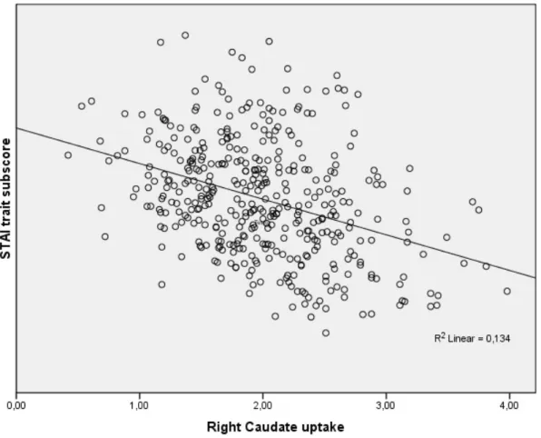
Results: In the PD group, lower DAT uptake in the right caudate was associated with higher STAI trait subscore (β=-2.939, 95%CI: -4.634 to -1.254, p=0.001). Depression and apathy scores were not related with DAT uptakes. No associations were found in the HC group.
Conclusions: Our cross-sectional analysis of the PPMI data shows that lower caudate DAT uptake
is associated with higher level of anxiety. The data strengthens the relationship between dopaminergic dysfunction and neuropsychiatric symptoms in early PD.
Introduction
Neuropsychiatric symptoms are frequent complaints in patients with Parkinson disease (PD) with a negative impact on quality of life and associated caregiver burden [1].Anxiety, depression and apathy are among the most common non motor symptoms, being present since the earliest stages of the disease [1].Nigrostriatal dopamine depletion in PD contributes directly to motor symptoms, however there is increasing evidence that dopaminergic dysfunction spreading to the anteroventral part of the striatum has a major role in determining neuropsychiatric disturbance in PD patients, especially in the earliest stage of the disease [2-5].Data from our single-center cohort of patients with newly diagnosed PD suggested an inverse correlation between both anxiety and apathy and dopamine transporter (DAT) availability in the right caudate [4,5]. However, previous studies have found conflicting results [2,3], likely due to the small sample size.
The aim of the present study is to investigate the relationships between nigrostriatal denervation and neuropsychiatric symptoms (including anxiety, depression and apathy) in a large cohort of newly diagnosed, drug-naïve PD patients compared to a cohort of healthy controls (HC). According to our initial hypothesis DAT availability would present an inverse correlation with neuropsychiatric symptoms.
Methods
Study Participants
The data used in this study was downloaded from the PPMI database, according to guidelines (http://www.ppmi-info.org/data, Grant ID 8800). PPMI is an ongoing, international multicenter study designed to identify biomarkers of PD progression.
PD patients were enrolled in the study if they met the following criteria: a) an asymmetric resting tremor or asymmetric bradykinesia; or evidence of either bradykinesia or resting tremor and rigidity; b) diagnosed within two years; c) a Hoehn and Yahr stage of I or II; d) age 30 years or older at diagnosis; e) evidence of dopamine transporter deficit on SPECT DATscan imaging; and f) drug-naïve for PD. Demographically comparable healthy subjects were also recruited into the study if they had no current or active neurological disorder, no first-degree relative with PD and no detectable evidence of dopamine transporter deficit on the SPECT DAT scan imaging. All participants underwent a comprehensive clinical, neuropsychiatric, neuropsychological and imaging assessment at enrollment.
The current analysis includes 405 newly diagnosed, drug-naïve PD patients and 187 HC and their clinical, neuropsychiatric and SPECT DAT scan imaging data collected at enrollment (either at screening or baseline visit).
Clinical and neuropsychiatric evaluations
Clinical examination included the Movement Disorder Society-sponsored revision of the Unified Parkinson’s Disease Rating Scale part III (MDS-UPDRS-III) to evaluate motor dysfunction and disease severity [6].
Severity of anxiety was assessed by means of the State-Trait Anxiety Inventory (STAI) [7], while depressive symptoms were evaluated using the 15- item Geriatric Depression Scale (GDS-15) [8],
with a cut off score of 5 or more indicating clinically significant symptoms [9].STAI sub-scores
can differentiate between the temporary condition of state anxiety (i.e., the temporary fear and nervousness due to a certain situation) and the more general and long-lasting trait anxiety (i.e., the stress, worry and discomfort perceived in typical situations on a daily basis) [9].Apathy was assessed with the single item from the MDS-UPDRS part I (MDS-UPDRS-I) [6]. Any non-zero score was considered as an indicator of presence of apathy.
SPECT studies
SPECT studies were performed during the screening visit. All subjects received an intravenous injection of 185 MBq of [123I]FP-CIT (DAT scan). The acquisition started between 3.30 and 4.30 h after the radiotracer injection. This time window between 3 and 6 h allows stable measurement of specific-to-non-displaceable ratio of [123I]FP-CIT.Specific acquisition parameters were selected for each participating center during a preceding technical visit. Images were acquired with a 128 x 128 matrix in a step and shoot mode. Subsequently, the raw projection data was reconstructed using iterative reconstruction algorithm at a central SPECT Core lab in New Haven, Connecticut, USA.
Site-specific attenuation correction was applied to the reconstructed data by an automated ellipse drawing technique and corrected for attenuation using Chang’s algorithm (l = 0.06/cm). The site-specific attenuation co-efficient, l, was based on an anthropomorphic distributed source phantom acquired during the preceding technical site visit. Hermes (Hermes Medical Solutions, Stockholm, Sweden) and Pmod (PMOD Technologies Ltd., Zurich, Switzerland) brain softwares for quantification were used. Spatial normalization was performed in Pmod software using a template image derived from the Hermes FP-CIT template based on a European multicenter database of healthy controls for [123I]FP-CIT SPECT (ENC-DAT).
Regions of interest (ROI) were then placed on the left and right caudate, the left and right putamen, and the occipital cortex (reference tissue). Count densities for each region were extracted and used to calculate striatal binding ratios (SBRs) for each of the 4 striatal regions. SBR was calculated as (target region/reference region)-1.
Statistic analysis
T tests and chi-squared tests were used for comparisons of demographic, clinical, neuropsychiatric and imaging variables between PD and HC. Descriptive statistics are given as means and standard
deviation (SD). Stepwise multiple linear regression analysis evaluating the association between
DAT scan uptakes (right and left caudate and putamen uptakes set as independent variables) and neuropsychiatric scores (GDS-15 and STAI sub-scores set as dependent variables) was performed both in PD and HC. As for apathy, multiple logistic regression analysis was used since it was evaluated with a dichotomous assessment. All the analyses included age, education, gender and
disease severity as assessed by the MDS-UPDRS-III as covariates with no interaction terms.
Results were considered statistically significant at p<0.05 (two-tailed). Analyses were performed
with the Statistical Package for the Social Sciences (SPSS) statistics (version 19;SPSS, Inc., Chicago, IL).
Standard protocol approvals, registrations, and patient consents
Each participating PPMI site obtained written informed consent from all participants, and received approval from an ethical standards committee on human experimentation.
Results
Demographic, clinical, imaging and neuropsychiatric features of PD and HC are displayed in Table 1.
In the PD group, lower DAT uptake in the right caudate was associated with higher STAI trait subscore (β=2.939, 95%CI: 4.634 to 1.254, p=0.001) along with younger age (β=0.187, 95%CI: -0.282 to -0.092, p<0.001) and female gender (β=-2.139, 95%CI: -4.051 to -0.226, p=0.029) (Figure 1). STAI state sub-score and 15-GDS showed no relationship with DAT uptakes. Presence of apathy was predicted by MDS-UPDRS-III (β=0.047, p=0.001, OR=1.048, 95%CI=1.020 to -1.078) and younger age (β=-0.033, p=0.015, OR=0.968, 95%CI=0.943 to -0.994), but not by any DAT uptakes. Given the well-known comorbidity between anxiety, depression and apathy [10-12], the regression analysis for anxiety was run again controlling also for the presence of apathy and severity of depression and the results were largely confirmed (lower right caudate uptake predicted higher STAI trait sub-score, β=-1.536, 95%CI: -2.687 to -0.384, p=0.009).
When comparing DAT uptakes between PD patients with and without anxiety (according to the STAI total score cut off of 39 [9]), anxious patients (88 out of 405) presented a tendency towards significance for lower right caudate uptake (1.918 vs 2.01, p=0.1).
Finally, we did not detect any difference in executive and attentive functions between anxious and non-anxious PD patients (data not shown), further supporting the specificity of the relationship between anxiety and caudate uptakes.
No correlation between neuropsychiatric symptoms and DAT uptakes was found in the HC group.
Discussion
Confirming previous findings, a recent paper on PPMI data showed that neuropsychiatric symptoms are more common in early, drug-naïve PD compared to the general population [9]. Here, we sought to detect associations between striatal dopaminergic dysfunction and anxiety, depression and apathy in PPMI cohort. After controlling for possible confounding factors, anxiety severity was found to relate to lower right caudate uptake in PD. No such correlation was found in the control group. The relationship between dopaminergic dysfunction and anxiety disorders in PD is complex and still only partially understood. Neuropsychiatric symptoms in PD are not a mere reaction to motor symptoms and chronic disability, but are likely due to the combination of psycho-social factors and the underlying cortical-limbic-striatal dysfunction [1]. Regarding the association between nigrostriatal DAT availability deficits and anxiety symptoms in PD, previous studies have found conflicting results. Weintraub et al., by TRODAT-1 imaging, found left anterior putamen DAT availability to be negatively correlated with STAI scores [2]. Conversely, Moriyama et al. found an
increased DAT density in left striatum and right putamen in PD patients with social anxiety disorder [3]. Different methodologies and definition of anxiety may account for such discrepancies. Moreover, both studies enrolled smaller cohorts of PD patients with longer disease duration and under dopaminergic treatment [2,3]. Indeed, the results of the present study largely confirm our previous data on a smaller cohort of early drug-naïve PD patients suggesting that the association between anxiety and right caudate may be specific for drug-naïve patients in the earliest stages of the disease [5]. This relationship is not surprising and can be explained in light of the crucial non motor role played by the caudate in PD [13]. Indeed, this association suggests a more pronounced dysruption of dopaminergic-frontal circuits in PD patients with anxiety. According to the dopaminergic theory, the neurodegeneration of mesolimbic and mesocortical dopaminergic projections observed as a primary neuroanatomical alteration in PD is strongly associated with
anxiety [13].Furthermore, the exclusive association between STAI trait sub-score and DAT binding
suggests that dopaminergic dysfunction is specifically related to anxiety-related personality traits rather than the temporary condition of state anxiety. Although the role of other neurotransmitters (i.e. serotonin and noradrenalin) cannot be overlooked, the dopaminergic involvement in anxiety of PD is supported by clinical evidence, as the common relationship between anxiety and motor fluctuations and the occurrence of anxiety symptoms following the withdrawal of dopaminergic
medications [14].
Indeed, anxiety is also recognized as part of the pre-motor symptoms of PD. A recent population-based cohort study found that the likelihood of developing PD was greater amongst patients with
anxiety than patients without anxiety, and the severity of anxiety correlated with the risk of PD [15].
From a practical standpoint, these results may have treatment implications. For instance, dopamine agonists, which help compensate for nigrostriatal dopamine deficiencies in PD [14], may be of particular use in PD patients with anxiety. Although ad hoc studies are lacking, we previously described a reduction in anxiety prevalence in de novo PD patients after the addiction of the dopaminergic treatment [16,17].
In contrast to previous findings [2,4], we failed to detect any associations between either depression or apathy and DAT availability in PD patients enrolled in PPMI.This discrepancy may reflect the different methodologies used to evaluate depressive symptoms and apathy. As for depressive symptoms, Weintraub et al. [2] used the depression subscale of Profile of Mood States, whereas PPMI study used GDS-15, which disclosed only mild depressive symptoms in PD patients, with only 13.9% presenting with significant depression [9]. As for apathy, in contrast to our previous study on a smaller cohort of early, drug-naïve patients, where we found an association between apathy assessed by Apathy Evaluation Scale and right caudate uptake [4], here the occurrence of apathy was evaluated by one single item of the MDS-UPDRS-I which is not considered to be an adequate screening measure. Thus, we suggest that both the GDS-15 "floor effect" reflecting mild depressive symptoms and the lack of specific scales assessing apathy may in part account for the lack of associations with DAT availability in the present study.
Limitations of the study include the cross-sectional design, which precludes firm conclusions of
assessment of meso-limbic and meso-cortical dopaminergic dysfunction with appropriate imaging techniques. We acknowledge our data describe only in part the complex scenario underlying neuropsychiatric disturbances in PD patients. The mild severity of depressive symptoms and the low number of subjects defined depressed according to the GDS-15 cut-off may in part account for the lack of association between dopaminergic dysfunction and depression in the PD cohort. As such, our results on depression and apathy should be interpreted with caution and deserve further investigations. Furthermore, the DAT scan was performed during the screening visit while behavioral assessments during the baseline visit (within 45 days from the screening). We cannot exclude this timeframe has a role in the lack of association with depression and STAI-state anxiety. Despite these drawbacks, this is the largest controlled-study to date assessing the relationship between dopaminergic dysfunction and anxiety in PD.
In conclusion, our cross-sectional analysis of the PPMI data shows that high level of anxiety is associated with reduced DAT uptake in the right caudate.
References
1) Weintraub D, Burn DJ. Parkinson’s disease: the quintessential neuropsychiatric disorder. Mov Disord 2011;26(6):1022-31.
2) Weintraub D, Newberg AB, Cary MS, Siderowf AD, Moberg PJ, Kleiner-Fisman G, Duda JE, Stern MB, Mozley D, Katz IR. Striatal dopamine transporter imaging correlates with anxiety and depression symptoms in Parkinson’s disease. J Nucl Med 2005;46(2):227-232. 3) Moriyama TS, Felicio AC, Chagas MHN, et al. Increased dopamine transporter density in
Parkinson’s disease patients with social anxiety disorder. J Neurol Sci 2011;310(1-2):53-57. 4) Santangelo G, Vitale C, Picillo M, Tardelli VS, Ferraz HB, Tumas V, Amaro-Junior E,
Andrade LA, Crippa JA, Bressan RA. Apathy and striatal dopamine transporter levels in de-novo, untreated Parkinson's disease patients. Parkinsonism Relat Disord 2015;21(5):489-493.
5) Erro R, Pappatà S, Amboni M, Caterina Vicidomini, Katia Longo, Gabriella Santangelo, Marina Picillo, Carmine Vitale, Marcello Moccia, Flavio Giordano, Arturo Brunetti, Maria Teresa Pellecchia, Marco Salvatore, Paolo Barone. Anxiety is associated with striatal dopamine transporter availability in newly diagnosed untreated Parkinson's disease patients. Parkinsonism Relat Disord 2012;18(9):1034-1038.
6) Goetz CG, Fahn S, Martinez-Martin P, Poewe W, Sampaio C, Stebbins GT, Stern MB, Tilley BC, Dodel R, Dubois B, Holloway R, Jankovic J, Kulisevsky J, Lang AE, Lees A, Leurgans S, LeWitt PA, Nyenhuis D, Olanow CW, Rascol O, Schrag A, Teresi JA, Van
Hilten JJ, LaPelle N. Movement Disorder Society-sponsored revision of the Unified Parkinson’s Disease RatingScale (MDS-UPDRS): Process, format, and clinimetric testing plan. Mov Disord 2007;22(1):41-47.
7) Spielberger CD, Gorsuch RL, Lushene RE. Manual for the State Trait Inventory. Palo Alto, CA: Consulting Psychologists Press, 1970.
8) Sheikh JI, Yesavage JA. Geriatric Depression Scale GDS: recent evidence and development of a shorter version. In: Brink TL, ed.Clinical Gerontology: A Guide to Assessment and Intervention. New York: The Haworth Press, 1986:165-173.
9) Weintraub D, Simuni T, Caspell-Garcia C, Coffey C, Lasch S, Siderowf A, Aarsland D, Barone P, Burn D, Chahine LM, Eberling J, Espay AJ, Foster ED, Leverenz JB, Litvan I, Richard I, Troyer MD, Hawkins KA; Parkinson's Progression Markers Initiative. Cognitive performance and neuropsychiatric symptoms in early, untreated Parkinson's disease. Mov Disord 2015;30(7):919-927.
10) Ceravolo R, Frosini D, Poletti M, Kiferle L, Pagni C, Mazzucchi S, Volterrani D, Bonuccelli U. Mild affective symptoms in de novo Parkinson's disease patients: relationship with dopaminergic dysfunction. Eur J Neurol 2013;20(3):480-485.
11) Guttman M, Boileau I, Warsh J, Saint-Cyr JA, Ginovart N, McCluskey T, Houle S, Wilson A, Mundo E, Rusjan P, Meyer J, Kish SJ. Brain serotonin transporter binding in non-depressed patients with Parkinson's disease. Eur J Neurol. 2007;14(5):523-528.
12) Remy P, Doder M, Lees A, Turjanski N, Brooks D. Depression in Parkinson's disease: loss of dopamine and noradrenaline innervation in the limbic system. Brain. 2005;128(Pt
6):1314-1322.
13) Peron J, Dondaine T, Le Jeune F, et al. Emotional processing in Parkinson’s disease: a systematic review. Mov Disord 2012;27(2):186-199.
14) Leentjens AF, Dujardin K, Marsh L, Martinez-Martin P, Richard IH, Starkstein SE. Anxiety and motor fluctuations in Parkinson’s disease: a cross-sectional observational study. Parkinsonism Relat Disord 2012;18(10):1084-1088.
15) Lin CH, Lin JW, Liu YC, Chang CH, Wu RM. Risk of Parkinson's disease following anxiety disorders: a nationwide population-based cohort study. Eur J Neurol 2015;22(9):1280-1287.
16) Erro R, Picillo M, Vitale C, Amboni M, Moccia M, Longo K, Cozzolino A, Giordano F, De Rosa A, De Michele G, Pellecchia MT, Barone P. Non-motor symptoms in early Parkinson's disease: a 2-year follow-up study on previously untreated patients. J Neurol Neurosurg Psychiatry. 2013;84(1):14-17.
17) Picillo M, Erro R, Amboni M, Longo K, Vitale C, Moccia M, Pierro A, Scannapieco S, Santangelo G, Spina E, Orefice G, Barone P, Pellecchia MT. Gender differences in non-motor symptoms in early Parkinson's disease: a 2-years follow-up study on previously untreated patients. Parkinsonism Relat Disord. 2014;20(8):850-854.
Table 1. Demographic, clinical, imaging and neuropsychiatric data of PD and HC. PD (n=405) HC (n=187) p Age, years 61.20 (9.76) 60.24 (11.21) 0.31 Gender (M/W, %) 264/140 (65.3/34.7) 121/66 (64.7/35.3) 0.41 Education, years 15.56 (2.98) 16.12 (2.9) 0.032 MDS-UPDRS-III 20.25 (8.93) NA NA
Most affected side (R/L/B, %) 224/173/7 (55.6/42.7/1.7) a NA NA Right Caudate uptake 1.98 (0.59) 2.95 (0.63) <0.001
Left Caudate uptake 1.99 (0.59) 3.0 (0.65) <0.001
Right Putamen
uptake 0.84 (0.36) 2.15 (0.58) <0.001 Left Putamen uptake 0.81 (0.35) 2.14 (0.56) <0.001
STAI total score 65.35 (18.47)b 57.03 (14.33) <0.001 STAI state sub-score 32.93 (10.22)b 28.03 (8.14) <0.001
STAI trait sub-score 32.39 (9.56)b 29.01 (7.5) <0.001
GDS-15 score 2.29 (2.37)b 1.28 (2.08) <0.001
GDS-15 cut off (not depressed/depressed) 347/56 (85.9/13.9) 174/12 (93/6.4) 0.005 MDS-UPDRS-I Apathy score (negative/any positive, %) 80/324 (19.8/80.2) 178/9 (95.1/4.9) <0.001 Table 1 Legend
Data are expressed in mean (standard deviation) unless otherwise specified. Significant differences are in italic.
a Data available for 404 subjects; b Data available for 400 subjects; B: bilateral; GDS-15: 15-item
Geriatric Depression Scale; HC: healthy controls; L: left; M: men; MDS-UPDRS-I: the Movement
Disorder Society-sponsored revision of the Unified Parkinson’s Disease Rating Scale part I; MDS-UPDRS-III: Movement Disorder Society-sponsored revision of the Unified Parkinson’s Disease Rating Scale part III; NA: not applicable; PD: Parkinson disease; R: right; STAI: State-Trait
Figure 1. Correlation between right caudate uptake and STAI trait sub-score according to the multiple linear regression model.
Figure 1 legend
Exploring the relationship between cognition and dopaminergic
dysfunction in de novo Parkinson's disease: a longitudinal prospective
study
Plos One (under review)
Abstract
Objectives: To explore the relationship between nigrostriatal dopamine uptake at diagnosis and
both baseline and 2-year longitudinal cognitive scores in a large cohort of drug-naïve (at baseline) Parkinson’s disease (PD) patients compared to a matched cohort of healthy controls (HC).
Methods: This is a longitudinal analysis of the Parkinson's Progression Markers Initiative (PPMI)
cohort, including 423 PD patients and 196 HC at baseline. Nigrostriatal dopaminergic degeneration was evaluated at baseline by means of SPECT dopamine transporter (DAT) scan. Relationships between specific cognitive tests and DAT uptake were analysed by means of multivariate linear regression analysis controlling for age, gender, education and disease severity.
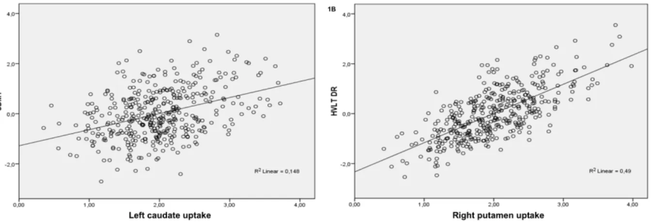
Results: At baseline, lower left caudate DAT availability was associated with worse performances in the processing speed/attention domain (β=1.535, 95%CI: 0.102 to 2.969, p=0.036), while lower right putamen uptake presented a trend towards significance for association with lower verbal recognition memory scores (β=1.067, 95%CI: -0.022 to 2.157, p=0.055). At 2-year follow up, lower left putamen DAT availability at baseline predicted greater decline in tests evaluating immediate (β=-1.42, 95%CI: -2.59 to -0.246, p=0.018) and delayed (β=-0.869, 95%CI: -1.53 to -0.210, p=0.009) verbal memory, while higher right caudate DAT availability at baseline presented a trend for greater improvement in visuo-spatial performances (β=0.326, 95%CI: -0.012 to 0.664, p=0.059).
Conclusions: This data strengthens the relationship between dopaminergic dysfunction and
Introduction
Cognitive impairment is a frequent complaint in patients with Parkinson’s disease (PD) with a negative impact on quality of life and associated caregiver burden. [1-3] Nigrostriatal dopamine depletion in PD contributes directly to motor symptoms, however there is increasing evidence that dopaminergic dysfunction in the striatum has a major role also in determining cognitive dysfunction, especially in the earliest stages of the disease. [4-8] Recent studies focused their attention on PD patients with mild cognitive impairment (PD-MCI), suggesting that striatal dopamine depletion disrupts the cortico-striatal pathway and, thus, recruitment of cortical regions involved in cognitive tasks in this subgroup of patients. [9-11] Our single-centre experience suggests a direct relationship between reduced DAT availability in both caudate and putamen and impaired cognitive performances in frontal/executive and visuospatial tasks in newly diagnosed PD. [9] However, despite the evidence of a close relationship between nigrostriatal dysfunction and cognitive impairment in PD, the role of nigrostriatal deafferentation at disease onset in predicting subsequent worsening of specific cognitive performances has been poorly investigated. [7]
In the present study, we sought to explore the relationship between nigrostriatal uptake at diagnosis and 1) cognitive scores at diagnosis and 2) change in cognitive scores from diagnosis to 2-year follow up in a large cohort of drug-naïve PD patients compared to a cohort of healthy controls (HC).
Methods
Study Participants
The data used in this study was downloaded from the PPMI database, according to guidelines (http://www.ppmi-info.org/data, Grant ID 8800, last download January 4th 2016). PPMI is an ongoing, international multicenter study designed to identify biomarkers of PD progression. Detailed descriptions of the study have been published elsewhere. [12]
PD patients were enrolled in the study if they met the following criteria: a) an asymmetric resting tremor or asymmetric bradykinesia; or evidence of (either bradykinesia or resting tremor) and rigidity; b) diagnosed within two years; c) a Hoehn and Yahr stage of I or II; d) age 30 years or older at diagnosis; e) evidence of dopamine transporter deficit on SPECT DATscan imaging; and f) drug-naïve for PD; g) diagnosis of dementia based on site investigator’s clinical impression. Demographically comparable healthy subjects were also recruited into the study if they had no current or active neurological disorder, no first-degree relative with PD and no detectable evidence of dopamine transporter deficit on the SPECT DATscan imaging and a Montreal Cognitive Assessment (MoCA) battery score>26 [13]. All participants have been performing scheduled assessments to collect biospecimens, clinical, neuropsychological and imaging data from enrollment and up to 5-year follow up.
The current analysis includes 423 newly diagnosed, drug-naïve PD patients and 196 HC and their SPECT DAT scan imaging data collected at enrollment and clinical and neuropsychological data collected at both enrollment and 2-year follow up.