For Review Only
An hypoechoic pattern of the thyroid at ultrasound does not indicate autoimmune thyroid diseases in patients with
morbid obesity
Journal: European Journal of Endocrinology Manuscript ID: EJE-10-0288.R1
mstype: Original Article Date Submitted by the
Author: 06-May-2010
Complete List of Authors: Rotondi, Mario; Unit of Internal Medicine and Endocrinology, Fondazione Salvatore Maugeri I.R.C.C.S., ISPESL Laboratory for Endocrine Disruptors, University of Pavia,
Cappelli, Carlo; University of Brescia, Medical and Surgical Sciences Leporati, Paola; University of Pavia,, Unit of Internal Medicine and Endocrinology, Fondazione Salvatore Maugeri I.R.C.C.S., ISPESL Laboratory for Endocrine Disruptors,
Chytiris, Spyridon; University of Pavia,, Unit of Internal Medicine and Endocrinology, Fondazione Salvatore Maugeri I.R.C.C.S., ISPESL Laboratory for Endocrine Disruptors,
zerbini, francesca; Unit of Internal Medicine and Endocrinology, Fondazione Salvatore Maugeri I.R.C.C.S., ISPESL Laboratory for Endocrine Disruptors, University of Pavia,
fonte, rodolfo; Unit of Internal Medicine and Endocrinology, Fondazione Salvatore Maugeri I.R.C.C.S., ISPESL Laboratory for Endocrine Disruptors, University of Pavia,
Magri, Flavia; Unit of Internal Medicine and Endocrinology, Fondazione Salvatore Maugeri I.R.C.C.S., ISPESL Laboratory for Endocrine Disruptors, University of Pavia,
Castellano, Maurizio; University of Brescia, Medical and Surgical Sciences
Chiovato, L; Unit of Internal Medicine and Endocrinology, Fondazione Salvatore Maugeri I.R.C.C.S., ISPESL Laboratory for Endocrine Disruptors, University of Pavia,
Keywords: Thyroid, Obesity, thyroid-ultrasound , morbid obesity, Autoimmunity
For Review Only
An hypoechoic pattern of the thyroid at ultrasound does not indicate
autoimmune thyroid diseases in patients with morbid obesity.
Mario Rotondi1, Carlo Cappelli2, Paola Leporati1,Spyridon Chytiris1, Francesca Zerbini1, Rodolfo Fonte1, Flavia Magri1, Maurizio Castellano2, Luca Chiovato1.
1 Unit of Internal Medicine and Endocrinology, Fondazione Salvatore Maugeri I.R.C.C.S., ISPESL
Laboratory for Endocrine Disruptors and Chair of Endocrinology, University of Pavia, Italy.
2 Department of Medical and Surgical Sciences, Internal Medicine and Endocrinology Unit,
University of Brescia, Italy.
Abbreviated title: Thyroid hypoechoic pattern in morbid obesity
Disclosure Statement: The authors have nothing to disclose
Keywords: thyroid - thyroid-ultrasound – morbid obesity –
autoimmunity
Word Count: 2944
Corresponding Author:
Luca Chiovato, MD, PhD
Unit of Internal Medicine and Endocrinology Fondazione Salvatore Maugeri I.R.C.C.S. Chair of Endocrinology, University of Pavia Via S. Maugeri 10, I-27100, Pavia, Italy Fax: +39-0382-592692
For Review Only
ABSTRACT
OBJECTIVE: Thyroid ultrasound (US) scan is a valuable tool for diagnosing thyroid diseases. In
autoimmune thyroid disease (AITD), an hypoechoic pattern of the thyroid at US is related to
circulating thyroid antibodies. Aim of this study was to evaluate the diagnostic accuracy of thyroid
US for the detection of AITD in patients with morbid obesity.
DESIGN: Thyroid US scans showing an hypoecoic pattern of the thyroid were collected from 105
patients with morbid obesity (BMI > 40 kg/m2 ) and 105 non-obese (BMI ≤30 kg/m2) patients.
RESULTS: A thyroid hypoechoic pattern at US was consistent with clinical/biochemical features of
AITD in 90/105 (85.7%) non-obese and in 22/105 (20.9%) morbid-obese patients (p<0.0001). By
performing a complete thyroid work-up, including clinical examination, thyroid morphology, serum
hormones and auto-Ab measurements, the discrepancy between the US pattern and the results of the
thyroid Ab tests was justified in 6/15 non obese patients, and only in 1/83 morbid obese patients.
Thus, an unexplained hypoechoic pattern of the thyroid at US, defined as negative tests for thyroid
Ab and absence of justifying thyroid disturbances, was found in 2/105 (1.9 %) non-obese and in
68/105 (64.8 %) patients with morbid obesity (p<0.0001).
CONCLUSIONS: Our results suggest that: 1) Morbid obesity may affect thyroid morphology 2)
an hypoechoic pattern of the thyroid at US, a well established parameter for diagnosing AITD, has a
For Review Only
INTRODUCTION
Over the last decade, ultrasound (US) scan proved to be a valuable tool for the diagnostic work-up
of thyroid diseases (1-8). Besides estimation of thyroid volume and identification of non palpable
thyroid nodules (9), US scan is able to characterize the echographic structure of thyroid tissue (10).
The normal thyroid parenchyma has a peculiar high echo density due to the typical follicle structure
(9). The interface between thyroid cells and the colloid exhibits an elevated acoustic impedance,
causing high frequency acoustic waves to be reflected back to the probe. In autoimmune thyroid
diseases (AITD), however, both lymphocytic infiltration and disruption of normal tissue
architecture cause a reduction in thyroid echogenicity (1,4,6,10-11). Thyroid hypoechogenicity is
currently viewed as an early sign of thyroid autoimmunity, which may be present even when the
thyroid disorder is not suspected from a clinical point of view (4,6-7,12-13). Rapid improvement in
US equipments and the use of standardized computerized algorithms have permitted an objective
and quantitative measurement of tissue echogenicity in thyroid diseases (11-13), as well as in other
pathological conditions (14-15). Using a subjective measure of thyroid echogenicity, previous
studies demonstrated that in patients with thyroid autoimmune diseases the presence of circulating
thyroid antibodies (Ab) as well as the development of hypothyroidism was closely correlated with
the degree of thyroid hypoechogenicity (1, 5, 11, 12, 16).
It is a common observation that a significant proportion of patients with morbid obesity display
slightly increased serum levels of TSH (17). The elevation of serum TSH, also within the normal
range, is associated with an increase in the occurrence of obesity (18) However, there is still
considerable disagreement as to the physiopathological mechanism responsible for this
phenomenon and the clinical significance of this hyperthyrotropinemia (17). A recent study from
our group questioned whether an elevated serum TSH alone provides sufficient evidence for a
diagnosis of subclinical hypothyroidism in patients with morbid obesity (19). Little has been
For Review Only
study, performed in obese children, showed for the first time that obesity is associated with
structural changes of thyroid morphology, as assessed by US, which are unrelated to thyroid
autoimmunity (23). Aim of the current study was to evaluate the diagnostic accuracy of thyroid US
for the detection of thyroid autoimmune diseases in adult patients with morbid obesity. To this
purpose, patients with an hypoechoic pattern of the thyroid at US were enrolled and stratified
according to their body weight status.
PATIENTS AND METHODS
Subjects
Obese patients and controls were recruited by searching the computerized database of thyroid US
performed at the Unit of Internal Medicine and Endocrinology of the Fondazione Salvatore Maugeri
from January 2007 to July 2009. Searching criteria were an hypoecoic pattern of the thyroid at US
and the patient’s BMI. A total of 105 consecutive patients with morbid obesity (BMI > 40) were
collected.
The control group was constituted by normo-weight and/or slightly overweight (BMI ≤30 kg/m2) patients who had performed a thyroid US scan, which revealed an hypoechoic pattern of the gland.
Starting from January 2007, consecutive non obese patients were recruited until the same number
of morbid obese patients was reached. In the control group, 76 (72.4%) patients were
normo-weight (BMI ≤25 kg/m2), and 29 (27.6%) were slightly overweight (BMI between 25 and 30 kg/m2).
A two-steps searching procedure was used: 1) All the scans performed in patients with a BMI>40
kg/m2 showing the presence of thyroid hypoechogenicity were collected. When more than one scan for a single patient was available, only the first one was considered. A total of 105 scans were
collected; 2) Starting from the date when the first obese patient was enrolled, 105 consecutive US
scans performed in patients with BMI ≤30 kg/m2 and showing the presence of thyroid hypoechogenicity were collected. The last collected scan was performed 45 days later and no
For Review Only
patient had performed more than one scan. The procedure by no means could discriminate any
clinical characteristics other than name, sex and BMI of the patient, together with the name of the
US operator. A total of 210 scans showing an hypoechoic pattern of the thyroid at US were
collected. The clinical data of the correspondent patients were obtained from computerized and
written hospital notes. These included: age, FT4, FT3, TSH, thyroglobulin Ab (Tg Ab), thyroid
peroxidase Ab (TPO Ab) TSH-receptor Ab (TR Ab) and a detailed drug history.
BMI was calculated as the weight (kg) measured to the nearest kg divided by the square of height
determined to the nearest cm (m). Isolated hyperthyrotropinemia (a condition frequently
encountered in patients with morbid obesity) (20) was defined as a raised serum level of TSH with
normal FT4 and FT3 levels, in the absence of circulating thyroid Ab.
All subjects gave their informed consent to participate in the study, which was performed in
accordance to the guidelines of the Declaration of Helsinki.
Thyroid ultrasound
Thyroid ultrasounds were performed using a real-time US device (Sonosite 180 plus) equipped with
a linear transducer operating at 7.5 MHz for morphologic study (L38 linear probe). The volume of
the thyroid, as assessed by measuring the three largest perpendicular diameters, and the US pattern
of the thyroid parenchyma are recorded for each scan. In order to minimize the operator variability
in assessing thyroid echogenicity, only those scans performed by the same operator (S.C.), who was
well trained and had a 5 years experience in thyroid imaging, were selected. According to previous
studies, echogenicity was evaluated in both thyroid lobes and in the surrounding neck muscles on
the transverse section (12, 16). The isthmus was excluded to avoid the interference of reflecting
echoes from the tracheal cartilage. Hypoechogenicity was ascertained by comparison of the echo
distribution in the thyroid parenchyma with respect to the surrounding neck muscles and only those
scans showing from mild to severegeneralized hypoechogenicity were selected (12, 16).
For Review Only
Serum concentrations of FT4 (normal range: 8.0-19.0 pg/mL), FT3 (normal range: 1.8-4.2 pg/mL),
and TSH (third generation TSH assay; normal range: 0.4-4.0 mIU/L) were measured using
immunochemoluminiscent assays by an automated analyser (Immulite 2000, DPC Cirrus, Los
Angeles, CA, USA) employing commercial kits (Diagnostic Products Corporation, Los Angeles,
CA, USA). The serum concentrations of Tg-Ab (normal range: <60 U/ml), TPO-Ab (normal range:
<60 U/ml), and TR-Ab (normal range: <1 U/ml) were measured using immunochemiluminiscent
assays employing commercial kits ( Brahms, Hennigsdorf, Germany).
Statistical analysis
Statistical analysis was performed using the SPSS software (SPSS, Inc., Evanston, IL). Between
groups comparisons were performed by Student-t test for unpaired data and by Mann–Whitney
U-test according to a normal or a nonparametric distribution of the variable U-tested. Frequencies among
groups were compared by χ2 test with Fisher’s correction, when appropriate. A p-value <0.05 was considered statistically significant.
RESULTS
The clinical and biochemical data of the patients with an hypoechoic pattern of the thyroid at US,
subdivided according to their body weight phenotype (non-obese vs. morbid obese), are reported in
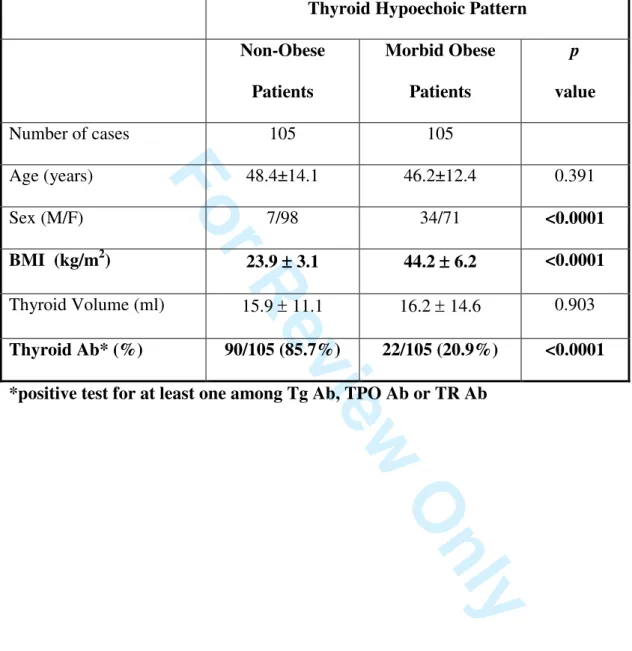
Table 1. The two groups of patients displayed significant differences for all parameters except for
age and thyroid volume. The percentage of patients in whom thyroid US hypoechogenicity was
consistent with the biochemical features of AITD (positive thyroid Ab tests) were significantly
different (p<0.0001) between the two groups. By means of US, 90 out of 105 patients (85.7%) in the
non-obese group were correctly identified as having AITD. In details, 77 out of 90 (85.5%) had a
clinical diagnosis of chronic autoimmune thyroiditis and 13 out of 90 (14.5%) were affected by
Graves’disease. In the obese group there were only 22 out of 105 (20.9%) patients showing a
correspondence between an hypoechoic pattern of the thyroid at US and positive tests for thyroid
Ab, being their clinical diagnosis chronic autoimmune thyroiditis in 21 out of 22 (95.5%) and
For Review Only
group and 83 out of 105 in the obese group who showed an hypoechoic pattern of the thyroid at US
not accompanied by positivity for any humoral immune marker of AITD. Possible causes
explaining the occurrence of an hypoechoic pattern of the thyroid at US in the absence of positive
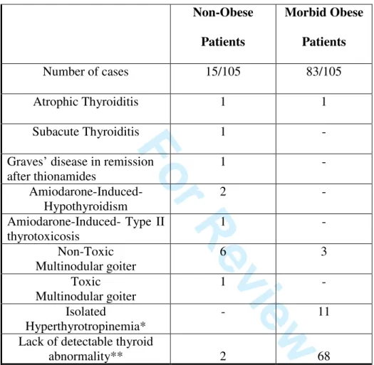
tests for thyroid Ab were searched. As shown in Table 2, in 6 out of 15 non obese patients, the
discrepancy between the US pattern and the results of the thyroid Ab tests was justified by the
following condition: amiodarone induced hypothyroidism in two cases; amiodarone induced type II
thyrotoxicosis in one patient; atrophic thyroiditis in one patient; subacute thyroiditis in one patient;
and Graves’disease in remission after medical treatment with disappearance of thyroid Ab in one
patient. By contrast, in only 1 out of 83 morbid obese patients showing a discrepancy between the
US pattern and the results for circulating thyroid Ab, the hypoechoic pattern could be explained by
the presence of atrophic thyroiditis. Other clinical conditions not potentially related to the
hypoechoic pattern of the thyroid at US were: non-toxic multinodular goiter in six patients and toxic
multinodular goiter in one case among non-obese patients. In the group of patients with morbid
obesity, three had non-toxic multinodular goiter and eleven an isolated hyperthyreotropinemia.
Thus, in 2 out of 105 (1.9 %) non obese patients and in 68 out of 105 (64.8 %) patients with morbid
obesity an hypoechoic pattern of the thyroid was found in the absence of any thyroid abnormality as
assessed by a complete thyroid work-up, including clinical examination, thyroid morphology,
hormones and auto-Ab measurements (p<0.00001).
DISCUSSION
This study shows that the finding of a thyroid hypoechoic pattern at US has a different clinical
meaning when observed in non-obese patients as compared to those with morbid obesity. In line
with previous evidences, we report that a thyroid hypoechoic pattern was strongly suggestive for the
presence of AITD, as assessed by positive thyroid Ab tests and/or clinical data, but only in
non-obese patients. A correspondence between the US hypoechoic pattern and the biochemical evidence
of AITD was found in 85.7% of the non-obese patients. On the other hand, patients with morbid
For Review Only
humoral or clinical evidence of thyroid autoimmunity. Thyroid volumes were similar between
morbid obese patients and controls. Ayturk et al in a specifically designed study reported that
euthyroid patients with metabolic syndrome have significantly higher mean thyroid volume and
nodule prevalence as compared with euthyroid controls (22). This apparent discrepancy can be
solved by considering that our patients had a wide spectrum of thyroid disease, thus preventing to
draw conclusions on thyroid volume. Indeed, the current study was specifically designed to include
only patients with an hypoechoic pattern, thus allowing to compare the underlying thyroid
conditions in US-matched patients stratified according to a different body weight status. The results
would support the concept that thyroid US, a well established tool for diagnosing AITD (1,4-6),
may be less effective when patients with morbid obesity are taken into account. A considerable
percentage (10.5%) of patients with morbid obesity had isolated hyperthyrotropinemia (a laboratory
picture which was not observed in non-obese patients). Isolated hyperthyrotropinemia is often
observed in patients with morbid obesity, but its pathogenesis remains poorly understood (17, 19).
Recent evidences showing lack of female gender prevalence, normal FT3/FT4 ratios and
normalization of serum TSH following weight loss, all support the concept that these patients
should not be considered as having subclinical hypothyroidism (19, 24-26). In agreement with this
concept, we recently demonstrated that morbid obese patients with a raised serum TSH have a
significantlylower rate of thyroid antibody positivity when comparedwith TSH-matched
normo-weight patients (19). These data suggest that autoimmunity is not a major cause of raised serum
TSH in morbid obese patients.
An hypoechoic pattern of the thyroid at US in the absence of any thyroid abnormality, as assessed
by a complete thyroid work-up, including clinical examination, thyroid morphology, hormones and
auto-Ab measurements, is a relatively rare finding in non-obese patients (1.9%), while it is observed
in the majority of patients with morbid obesity (64.8%). These data indicate profound differences
as to the clinical meaning of an hypoechoic pattern of the thyroid at US in relation to adiposity. The
For Review Only
seems clear from our and other results that thyroid hypoechogenicity is strongly related to AITD in
non-obese patients. On the other hand, the cause of thyroid hypoechogenicity in patients with
morbid obesity is difficult to be understood. The hypothesis that patients with an extreme weight
excess may have a tendency to accumulate fat in the thyroid could be an attractive one. Radetti et al,
recently reported that a thyroid hypoechoic pattern at US was found in a high proportion of obese
children with no evidence of AITD (23). Thyroid cytology performed in some of these children
turned out to be normal, showing only colloid drops and thyrocytes with no inflammatory cell
suggesting AITD (23). The issue of thyroid repercussions of fat-excess remains open as cytology
does not represent an appropriate technique for elucidating the histopathological basis of the
imaging abnormalities. Histological examination, specificallyfocused at studying fat accumulation
within thethyroid tissue, would be required to firmly demonstrate this hypothesis. Some clinically
relevant considerations stem from this study. First, the complex and yet poorly understood
repercussions of morbid obesity on the thyroid gland are not limited to thyroid function, but may
also affect thyroid morphology as evaluated by US. Only a minority of morbid obese patients with
an hypoechoic pattern of the thyroid at US display humoral signs of autoimmunity . Thus, a thyroid
hypoechoic pattern at US has a poor diagnostic accuracy for AITD when patients with morbid
For Review Only
Funding: This research did not receive any specific grant from any funding agency in the public,
commercial or not-for-profit sector.
The Authors declare that there is no conflict of interest that could be perceived as prejudicing the
For Review Only
REFERENCES
1) Marcocci C, Vitti P, Cetani F, Catalano F, Concetti R & Pinchera A. Thyroid
ultrasonography helps to identify patients with diffuse lymphocytic thyroiditis who are
prone to develop hypothyroidism. Journal of Clinical Endocrinology and Metabolism 1991
72 209-213.
2) Vitti P, Rago T, Mancusi F, Pallini S, Tonacchera M, Santini F, Chiovato L, Marcocci C &
Pinchera A. Thyroid hypoechogenic pattern at ultrasonography as a tool for predicting
recurrence of hyperthyroidism after medical treatment in patients with Graves' disease. Acta
Endocrinologica (Copenh) 1992 126 128-131.
3) Vitti P, Lampis M, Piga M, Loviselli A, Brogioni S, Rago, T, Pinchera A & Martino E.
Diagnostic usefulness of thyroid ultrasonography in atrophic thyroiditis. Journal of Clinical
Ultrasound 1994 22 375-379.
4) Pedersen OM, Aardal NP, LarssenTB, VarhaugJE, Myking O & Vik-Mo H The value of
ultrasonography in predicting autoimmune thyroid disease. Thyroid 2000 10 251-259. 5) Premawardhana LD, Parkes AB, Ammari F, John R, Darke C, Adams H & Lazarus JH.
Postpartum thyroiditis and long-term thyroid status: prognostic influence of thyroid
peroxidase antibodies and ultrasound echogenicity. Journal of Clinical Endocrinology and
Metabolism 2000 85 71-75.
6) Rago T, Chiovato L, Grasso L, Pinchera A & Vitti P. Thyroid ultrasonography as a tool for
detecting thyroid autoimmune diseases and predicting thyroid dysfunction in apparently
healthy subjects. Journal of Endocrinological Investigation 2001 24 763-769.
7) Raber W, Gessl A, Nowotny P & Vierhapper H. Thyroid ultrasound versus antithyroid
peroxidase antibody determination: a cohort study of 451 subjects. Thyroid 2002 12
For Review Only
8) Erdoğan MF, Anil C, Cesur M, Başkal N & Erdoğan G. Color flow Doppler sonography for
the etiologic diagnosis of hyperthyroidism. Thyroid 2007 17 223-228.
9) Hegedus L & Karstrup S. Ultrasonography in the evaluation of cold thyroid nodules.
European Journal of Endocrinology 1998 138 30-31.
10) Gutekunst R, Hafermann W, Mansky T & Scriba PC. Ultrasonography related to clinical
and laboratory findings in lymphocytic thyroiditis. Acta Endocrinologica (Copenh) 1989
121 129-135.
11) Loy M, Cianchetti ME, Cardia F, Melis A, Boi F & Mariotti S. Correlation of computerized
gray-scale sonographic findings with thyroid function and thyroid autoimmune activity in
patients with Hashimoto's thyroiditis. Journal of Clinical Ultrasound 2004 32 136-140.
12) Schiemann U, Gellner R, Riemann B, Schierbaum G, Menzel J, Domschke W & Hengst K.
Standardized grey scale ultrasonography in Graves' disease: correlation to autoimmune
activity. European Journal of Endocrinology 1999 141 332-336.
13) Vitti P. Grey scale thyroid ultrasonography in the evaluation of patients with Graves'
disease. European Journal of Endocrinology 2000 142 22–24.
14) Ulrich J & Voit C. Ultrasound in dermatology. Part II. Ultrasound of regional lymph node
basins and subcutaneous tumours. European Journal of Dermatology 2001 11 73-79.
15) Pedro LM, Fernandes e Fernandes J, Pedro MM, Goncalves I, Dias NV, Fernandes e
Fernandes R, Carneiro TF & Balsinha C. Ultrasonographic risk score of carotid plaques.
European Journal of Vascular and Endovascular Surgery 2002 24 492-498.
16) Mazziotti G, Sorvillo F, Iorio S, Carbone A, Romeo A, Piscopo M, Capuano S, Capuano E,
For Review Only
echogenicity in the patients with Hashimoto's thyroiditis. Clinical Endocrinology (Oxf)
2003 59 223-229.
17) Reinehr T. Obesity and thyroid function. Molecular and Cellular Endocrinology 2010 316
165-171.
18) Knudsen N, Laurberg P, Rasmussen LB, Bülow I, Perrild H, Ovesen L, Jørgensen T. Small
differences in thyroid function may be important for body mass index and the occurrence of
obesity in the population. Journal of Clinical Endocrinology and Metabolism 2005 90
4019-24.
19) Rotondi M, Leporati P, La Manna A, Pirali B, Mondello T, Fonte R, Magri F & Chiovato L.
Raised serum TSH levels in patients with morbid obesity: Is it enough to diagnose
subclinical hypothyroidism? European Journal of Endocrinology 2009 160 403-408.
20) Sari R, Balci MK, Altunbas H & Karayalcin U. The effect of body weight and weight loss
on thyroid volume and function in obese women. Clinical Endocrinology (Oxf) 2003 59
258-262.
21) Wesche MF, Wiersinga WM & Smits NJ. Lean body mass as a determinant of thyroid size.
Clinical Endocrinology (Oxf) 1998 48 701-706.
22) Ayturk S, Gursoy A, Kut A, Anil C, Nar A, Tutuncu NB.Metabolic syndrome and its
components are associated with increased thyroid volume and nodule prevalence in a
mild-to-moderate iodine-deficient area. European Journal of Endocrinology 2009 161 599-605.
23) Radetti G, Kleon W, Buzi F, Crivellaro C, Pappalardo L, di Iorgi N & Maghnie M. Thyroid
function and structure are affected in childhood obesity. Journal of Clinical Endocrinology
For Review Only
24) Moulin de Moraes CM, Mancini MC, De Melo ME, Figueiredo DA, Villares SM, Rascovski
A, Zilberstein B & Halpern A. Prevalence of subclinical hypothyroidism in a morbidly
obese population and improvement after weight loss induced by Roux-en-Y gastric bypass.
Obesity Surgery 2005 15 1287-1291.
25) Chikunguwo S, Brethauer S, Nirujogi V, Pitt T, Udomsawaengsup S, Chand B & Schauer P.
Influence of obesity and surgical weight loss on thyroid hormone levels. Surgery for Obesity
and Related Diseases 2007 3 631-635.
26) Reinehr T, de Sousa G & Andler W. Hyperthyrotropinemia in obese children is reversible
after weight loss and is not related to lipids. Journal of Clinical Endocrinology and
For Review Only
Table 1 Clinical and biochemical characteristics of patients with an hypoechoic pattern of the thyroid at US, subdivided according to their body weight status
Thyroid Hypoechoic Pattern Non-Obese Patients Morbid Obese Patients p value Number of cases 105 105 Age (years) 48.4±14.1 46.2±12.4 0.391 Sex (M/F) 7/98 34/71 <0.0001 BMI (kg/m2) 23.9 ±±±± 3.1 44.2 ±±± 6.2 ± <0.0001 Thyroid Volume (ml) 15.9 ± 11.1 16.2 ± 14.6 0.903 Thyroid Ab* (%) 90/105 (85.7%) 22/105 (20.9%) <0.0001 *positive test for at least one among Tg Ab, TPO Ab or TR Ab
For Review Only
Table 2 Thyroid condition found in non-obese and obese patients showing a thyroid hypoechoic pattern at US and negative tests for thyroid Ab
Non-Obese Patients Morbid Obese Patients Number of cases 15/105 83/105 Atrophic Thyroiditis 1 1 Subacute Thyroiditis 1 -
Graves’ disease in remission after thionamides 1 - Amiodarone-Induced- Hypothyroidism 2 - Amiodarone-Induced- Type II thyrotoxicosis 1 - Non-Toxic Multinodular goiter 6 3 Toxic Multinodular goiter 1 - Isolated Hyperthyrotropinemia* - 11
Lack of detectable thyroid
abnormality** 2 68
*defined as a raised serum TSH with normal FT4 and FT3 levels in the absence of circulating thyroid Ab
** patients in whom a complete thyroid work-up including clinical examination, circulating hormones and auto Ab measurements did not provide evidence for thyroid disease