The stiff elbow: Current concepts
Giulia Masci,1,2Gianpiero Cazzato,2,3Giuseppe Milano,4,5Gianluca Ciolli,1,2
Giuseppe Malerba,1Carlo Perisano1,
Tommaso Greco1,2Osvaldo Palmacci,1
Giulio Maccauro,1,2Francesco Liuzza1 1Department of Orthopaedics,
Fondazione Policlinico Universitario A. Gemelli IRCCS, Rome; 2Università
Cattolica del Sacro Cuore, Rome;
3Artrogruppo, Clinica San Feliciano,
Rome; 4Department of Medical and
Surgical Specialties, Radiological Sciences, and Public Health, University of Brescia;5Department of Bone and
Joint Surgery, Spedali Civili, Brescia, Italy
Abstract
Elbow stiffness is defined as any loss of movement that is greater than 30° in extension and less than 120° in flexion. Causes of elbow stiffness can be classified as traumatic or atraumatic and as congenital or acquired. Any alteration affecting the stability elements of the elbow can lead to a reduction in the arc of movement. The classification is based on the specific structures involved (Kay’s classification), anatomical location (Morrey’s classification), or on the degree of severity of rigidity (Vidal’s classification). Diagnosis is the result of a combination of medical history, physical examination (evaluating both active and passive movements), and imaging. The loss of soft tissue elasticity could be the result of bleeding, edema, granulation tissue formation, and fibrosis. Preventive measures include immobilization in extension, use of post-surgical drain, elastic compression bandage and continuous passive motion. Conservative treatment is used when elbow stiffness has been present for less than six months and consists of the use of serial casts, static or dynamic splints, CPM, physical therapy, manipulations and functional re-education. If conservative treatment fails or is not indicated, surgery is performed. Extrinsic rigidity cases are usually managed with an open or arthroscopic release, while those that are due to intrinsic causes can be managed with arthroplasties. The elbow is a joint that is particularly prone to developing stiffness due to its anatomical and biomechanical complexity, therefore the treatment of this pathology represents a challenge for the physiotherapist and the surgeon alike.
Introduction
Elbow is a joint particularly prone to stiffness due to its anatomical and biomechanical complexity.1
Elbow stiffness interferes with the primary activities of daily life, such as personal hygiene, eating, and dressing.2-4
The treatment of this pathology poses a challenge for the surgeon and the physiotherapist.
Elbow has two degrees of freedom, and the available movements in space are flexion, extension, pronation and supination. Several studies on the ROM of the elbow assessed a variation from -21° to 12° in extension, and from 122° to -164° in flexion.5 A functional joint must also
guarantee stability, which depends both on the complex conformation of the articular surfaces, and on the capsule-ligament structures and muscle components.6Morrey
et al. defined the arc of movement of the functional elbow during the daily life activity of 100°, both for flexion-extension (30° to 130°) and for pronation-supination (50° in both directions).7Elbow stiffness is
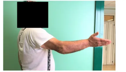
defined as the loss of movement greater than 30° in extension and loss of flexion of less than 120°.8Functional limitations can also
occur with less severe movement losses (Figure 1). Elbow stiffness is the result of morphological and structural alterations of the bone and/or soft tissues and can be the consequence of a traumatic event. Numerous atraumatic causes are observed, while at least 5% incidence from traumatic causes are reported. Its aetiology is the basis of its classification, diagnosis, prevention and treatment. Early rehabilitation, minimal immobilization and advances in surgical treatment are critical to the prevention and treatment of this condition.2,8,9
Etiopathogenesis
Causes of elbow stiffnesses are numerous and can be classified as traumatic or atraumatic; congenital or acquired.
Trauma, burns, fractures, and pathologies of the nervous system are the most frequent causes, and the resulting rigidity is directly proportional to the severity of the injury; even elbow surgery done for trauma management can be complicated by post-operative rigidity.2
Atraumatic causes include osteoarthritis (OA), inflammatory arthritis, post-septic arthritis, hemarthrosis in haemophiliacs; congenital rigidity is found in arthrogryposis and in the congenital dislocation of the radial head.2
The stability of the elbow is guaranteed
by the tight congruence of the articular surfaces, the anterior and posterior capsule, as well as by the collateral ligaments; any alteration affecting these structures can lead to a reduction in the arc of movement. For example, joint fractures, osteochondral defects and arthritic modifications alter the bone geometry of the elbow, which can lead to rigidity.2 A recent study has shown that
the development of elbow stiffness after surgery for elbow trauma displays a relatively high incidence of 8.4%.10
According to the AO classification, fractures type C2-C3 of subjects who undergo surgery 7 days after trauma have a greater risk of developing elbow stiffness; patients must be informed of the possible failure of the procedure and the possible need for reoperation.11
The evaluation of patients with substantial elbow stiffness, even following well-reduced and stabilized fractures, suggests that the thickening of soft tissues, especially of the capsule, is associated with a reduction in ROM.
Cohen et al. reported that the joint
Correspondence: Gianpiero Cazzato, Università Cattolica del Sacro Cuore, Rome; Artrogruppo, Clinica San Feliciano, Rome, Italy.
E-mail: [email protected]
Key words: Stiff elbow, elbow contracture, elbow, post-traumatic stiffness.
Contributions: All authors contributed equally to this paper with conception and design of the study, literature review and analysis, drafting, critical revision and editing of the manuscript, and in giving approval of the final version. Conflict of interest: The authors declare no potential conflict of interest.
Funding: None.
Availability of data and materials: All data and materials are in the text.
Ethics approval and consent to participate: Not applicable.
Informed consent: Not applicable. Received for publication: 11 April 2020. Accepted for publication: 17 June 2020. This work is licensed under a Creative Commons Attribution NonCommercial 4.0 License (CC BY-NC 4.0).
©Copyright: the Author(s), 2020 Licensee PAGEPress, Italy
Orthopedic Reviews 2020; 12(s1):8661 doi:10.4081/or.2020.8661
capsule of a rigid elbow is thickened, with a disorganized extracellular matrix, increased levels of inflammatory cytokines and infiltrations of fibroblasts, as a real fibrotic and inflammatory condition.12Other authors
have documented an increase in collagen cross-linking, associated with a decrease in the content of proteoglycans and water in the rigid joint, together with changes in the regulation of transforming growth factor-beta (TGF-β).13
The analysis of the joint capsule of patients who have undergone surgery has shown a thickening of the capsule, disorganization of the structure of the collagen fibers, altered levels of cytokines and enzymes, and a high number of myofibroblasts.14
Hildebrand et al. showed that the number of myofibroblasts proportionally increase in patients who had undergone capsular release; an anatomical difference has also been documented, with a greater thickening of the anterior portion of the joint capsule compared to the posterior one, which justifies the loss of extension much more frequent than the loss of flexion. The number of fibroblasts is also inversely proportional to the residual arc of movement. Myofibroblasts also appear to be absent in chronic elbow stiffness (present for more than 5 months) which suggests that their influence is prominent in the early period after trauma.4,14-16
Another cause of elbow stiffness is heterotopic ossification (HO), which is an inappropriate formation of mature lamellar bone in soft tissues, different from the most common peri-articular calcifications. HO manifests as a response to soft tissue trauma causing a physical block to the joint movement, at times creating synostoses between the radius and ulna, and abolishing the movement of pronation-supination of the forearm.9,14 About 3% of simple elbow
dislocations and 20% of fracture-dislocations are complicated by HO; in patients who have had both head injury and elbow trauma the prevalence of HO ranges from 76% to 89%.
There are many predisposing factors to post-traumatic elbow stiffness, including patient and fracture characteristics (comminuted fractures, exposed fractures, high energy trauma, infections, fragment devascularization, osteochondral defects, metabolic and cellular abnormalities), and fracture treatment (incorrect osteosynthesis, soft tissue interposition, early mobilization).
Intra and extra-articular consolidation defects and nonunions of the distal humerus, proximal ulna, and radial head are other frequent causes of functional limitation of the elbow, as well as pain and instability.
Malunion of radial head fractures occurs more often with the stiffness of the forearm, rather than with rigidity of the humeroulnar joint or OA of the proximal radio-ulnar and humeral-radial joints.14,16
Classifications
Classifications of elbow stiffness are based both on the involvement of specific anatomic structures (soft tissues, bone, and the combination of both) and on the anatomical location (intrinsic, extrinsic or combined).
Kay’s classification (1) is based on the structures involved:
• type 1, thickening of soft tissues; • type 2, thickening of soft tissues with
ossification;
• type 3, non-displaced joint fracture with thickening of soft tissues;
• type 4, displaced joint fracture with soft tissue thickening;
• type 5, presence of post-traumatic bone fragments
Morrey’s classification is based on aetiology and its anatomic location, and is subdivided into three categories: intrinsic, extrinsic, and combined rigidity;17the most
frequent rigidities have mixed aetiology. Extrinsic rigidities are determined by extra-articular causes, which include thickening of the capsule, or collateral ligaments and muscle contractures, as well as HOs, extra-articular nonunion, and thickening of soft tissues following burns. Intrinsic stiffnesses are instead caused by intra-articular adhesions, osteochondral mobile bodies, osteophytes formations and malalignments of the articular surfaces.2,7,14
Another classification criterion is based on the severity of rigidity; one of the first is that of Vidal.6The Liverpool Elbow Score
(LES) has also recently been validated to evaluate the functionality of the joint in patients with rigid elbow.18
Diagnosis
Diagnosis of elbow stiffness is the result of a combination of history, physical examination and imaging.
It is essential to know the patient’s history, which includes the onset, duration, characteristics, progression of symptoms, previous surgical interventions, or any traumatic or infectious pathologies affecting the joint. The presence or absence of comorbidities, such as inflammatory arthropathies, haemophilia, or neurological conditions should also be considered.2,9,15
Patients rarely complain of symptoms at rest, and pain is often felt in cases of extreme movement, or in patients with underlying severe OA. Resting pain is suggestive of the presence of infection, especially in patients who underwent previous surgery. In these cases, the dosage of C-reactive protein (PCR) and the erythrocyte sedimentation rate (ESR) should be given. Sometimes the pain may be due to nerve compression paraesthesia or scars with skin hypersensitivity.13,16,19Clinical examination
includes the evaluation of the shoulder, wrist and hand because they inevitably affect the functionality of the elbow. It starts with an accurate inspection, aimed at recognizing any surgical scars or burns. The clinical examination of the elbow is aimed at evaluating both active and passive flexion-extension and pronation-supination movements.
A severe joint limitation is suggestive of a bone block, while a gradual limitation is indicative of soft tissue blockage.9,15 The
presence of crackles during mobilization is indicative of degenerative changes,
synovitis, or fractures. Pain in cautious mobilization is indicative of intrinsic components of stiffness, while pain at the highest degrees of flexion-extension and prone-supination is prevalent in cases of impingement between the olecranon or the coronoid process, and in the distal part of the humerus, often being caused by osteophytes formations.14,19Particular attention must be
paid to the evaluation of ulnar nerve function, which is often involved in traumatic and degenerative processes; in cases requiring surgical treatment (release and anterior transposition), the preoperative evaluation of the ulnar nerve should always be documented.2,15 Late ulnar neuropathy
can arise due to its stretching, after the recovery of the elbow extension, in flexion contractures.20In some cases, examination
under anaesthesia is indicated to distinguish true elbow stiffness from functional limitations on an antalgic basis.13,19In most
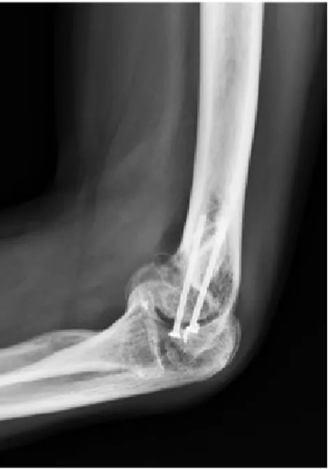
cases, an anteroposterior (AP) and lateral x-ray of the elbow are enough. Stiffnesses greater than 30° are often associated with distorted AP images, which renders oblique projections necessary.2,13,20 Also
intra-articular bone fragments can be identified with radiographs (Figures 2 and 3). Computed tomography (CT) should be required when joint damage is associated and when HO needs to be studied. Magnetic resonance imaging (MRI) is not fundamental for this type of pathology, because it does not allow an in-depth study of bone structures, still providing good information on soft tissues. Electromyography (EMG) is necessary when there is clinical suspicion of neuropathy of the ulnar nerve.2,9,14,15
Prevention
Treatment of a rigid elbow can be a difficult challenge for the orthopedic surgeon and physiotherapist, and as such the prevention of its onsets is of key importance. The loss of soft tissue elasticity could be the result of bleeding, oedema, granulation tissue formation, and fibrosis. Preventive measures could be the immobilization of the elbow in extension, the use of post-surgical drainage, and the elastic compression bandage.2,9,15 Continuous passive motion
(CPM) applied to the elbow in the immediate post-operative period and continued for about 3-4 weeks allows the drainage of fluids outside the joint and peri-articular tissues, although it should not be used when the patient underwent reconstruction and repair of the collateral ligaments.20
Inflammatory arthropathies should be controlled by effective medical therapy.
Haemophiliacs should receive appropriate coagulation factors to prevent multiple hemarthrosis. Joint degeneration due to infection could be prevented, in some cases, by irrigation and debridement.2,9,15
HOs could be prevented by using low doses of radiation within 72 hours of trauma to alter the differentiation of progenitor cells in the tissue involved, because of their high radiosensitivity. The risk of developing radiation-induced osteosarcomas is also very low in humans.16 Several studies
documented a lower incidence of re-interventions or recurrences of post-irradiation HOs. Robinson et al. reported a study of 36 patients treated with a single fraction of post-arthrolysis radiation therapy and excision of HOs. All patients showed increased ROM, and none underwent new surgical treatments.21
Although the use of nonsteroidal anti-inflammatory drugs (NSAIDs) such as indomethacin, which inhibit the formation of cyclooxygenase, has proven effective in preventing ossification in the hip, further
studies are needed to determine whether they can be effective in preventing HOs of the elbow. Recently, Costopuolus et al. demonstrated the efficacy of the use of post-operative indomethacin for the reduction of HO formation in patients undergoing biceps tendon repair.22
Indomethacin in combination with radiation therapy is considered safe and effective in the prevention of HOs. Strauss et al., in a cohort of 44 patients treated with a single fraction of radiation therapy and 10 days of indomethacin after surgery, showed the presence of small HOs in 48% of cases, but no patient had functional limitations or needed revision surgery.23The use of
COX-2 inhibitors, or coxib, (selective for cyclo-oxygenase 2) and other NSAIDs, on the other hand, is controversial, due to an undefined relationship between cardiova -scular risk and benefit.
Figure 3. X-ray views (LL) showing intra-articular bone fragments.
Figure 4. Functional re-education to pronation-supination. Figure 2. X-ray views (AP) showing
The diphosphonates are insufficient, since after their suspension there may be a resumption of the ossification process and cause gastrointestinal complications and osteomalacia as side effects. High doses of sodium etidronate can inhibit the angiogenesis necessary for mineralization of the bone matrix and reduce ossifications, but it is not recommended as it predisposes to osteomalacia and can also interfere with the production of healthy bone. Bone morphogenic protein (BMP) antagonists-based gene therapy could represent a future treatment option for the prevention of HOs, as demonstrated in studies on animal models.16In the future, gene therapy may
play an important role in the interruption of the inflammatory cycle, which is the basis of elbow stiffness, by altering the expression profile of cytokines.24The role of botulinum
toxin in the prevention of post-traumatic rigid elbow has recently been analysed. Intraoperative injection of botulinum toxin A into the elbow flexors leads to an increase in postoperative ROM and joint function in patients undergoing surgery for fractures or fracture-dislocations.15
Conservative treatment
Currently, both conservative and surgical treatment can be considered valid. Obviously, the choice will be conditioned by many factors, such as timing, mode of presentation, severity, comorbidities and patient’s compliance. Conservative treatment is used when elbow stiffness has been present for less than six months, while surgical treatment is more appropriate in those patients who have not benefited from conservative treatment, due to the persistence of pain and inadequate recovery of ROM and function.2
Conservative treatment consists of the use of serial casts, static or dynamic splints, CPM (continuous passive motion), home and assisted physical therapy, manipulations and subsequent functional re-education (Figure 4). Static immobilization of the elbow is usually used for short periods as a preventive measure after an injury or surgery, while long-term immobilization is not recommended and is therefore rarely indicated.15The use of the CPM is strongly
indicated in maintaining a satisfactory articulation. Before mobilization, the elbow should be kept raised and extended with a circular bandage and in the case of surgery, drainage should also be placed to avoid the formation of a hemarthrosis. Once the patient has passed the phase in which he cannot independently reach the complete
ROM, then the CPM can be suspended; this phase varies greatly from patient to patient and ranges from several days to a month.16
Static and dynamic splints are highly common choices for the treatment of elbow stiffness because they extend the benefit of therapy, allowing patients to promptly resume their daily lives.15 By using
adjustable static splints, a constant force is applied at intervals, which exerts stress and distraction to the tissues to temporarily reduce the risk of inflammation. Dynamic splints are the most used device in the treatment of early or late stiffness and are based on the viscoelastic properties of the peri-articular soft tissues to generate a certain degree of deformation in response to the application of a variable force.17
Moderate stiffness, which has persisted for less than a year in adults due to soft tissue contractures, have been successfully treated using dynamic splints 1. Once the pain is
reduced, the device can be applied at night with serial increases in the applied voltage. A recent study compared the use of static and dynamic splints without finding a statistically significant difference between the two groups.25Static progressive splints,
both for flexion-extension and pronation-supination, are also used for joint ROM recovery.2 As far as manipulations are
concerned, their benefit has been demonstrated, although not without risk. Some studies have shown an increase in elbow movement in 55% of patients with complications such as transient ulnar sensitive neuropathies. Other complications included peri-articular fractures, and the formation or increase of Hos.2There are few
results in the literature regarding manipulations under anaesthesia;24cases of
iatrogenic fractures have been reported in patients undergoing this procedure without having previously performed a surgical release.1Araghi et al. carried out a study on
51 patients undergoing this procedure, reporting good results.1,26The same results
were obtained by Ek et al. in a study of 12 pediatric patients.1,27In children with plexus
paralysis that causes elbow stiffness during flexion, the indicated treatment consists of electrostimulations and exercise. Furthermore, botulinum toxin A is indicated in the search for joint recovery. Basciani and Intiso have shown that the injection of botulinum toxin A followed by serial casts has increased the extension of the elbow by about 27 degrees.28
Surgical treatment
If conservative treatment fails after six months or is not indicated, surgery is
performed. The patient must be prepared and motivated to ensure extreme compliance with the post-operative rehabilitation program. Extrinsic rigidity cases are usually managed with an open or arthroscopic release, while those that are due to intrinsic causes can be managed with arthroplasties. The choice depends on the surgeon’s skills, conditions of the ulnar nerve, possible presence and location of HOs, loss of motion, and damage to the joint surfaces.14
Since the introduction of arthrolysis in 1944, numerous surgical techniques have been described. Surgical release can be performed through several accesses. It may depend not only on the variables listed above, but also on the location of previous surgical scars. Complications common to all these approaches include peripheral neuropathies, post-surgical infections, recurrence of stiffness, and Hos,5,14,19,24
triceps avulsions, fractures, and hematoma formations.29Lateral access, with an incision
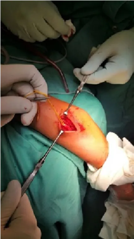
centred over the lateral humeral epicondyle (lateral column technique) allows the arthrotomy, the release of the anterior and posterior capsule, and the exposure of the lateral region of the joint. However, this access does not allow adequate exposure of the medial region and decompression of the ulnar nerve. The medial access, with an incision centred over the humeral trochlea, is used to approach the humeroulnar joint, remove heterotopic ossifications, release the medial collateral, and proceed with decompression and transposition of the ulnar nerve (Figure 5).29
This access alone is used in rare occasions, for example when performing lateral column surgery, as it does not allow exploration of the lateral region of the joint.14,19
Anterior access has limited indications since it is used for the anterior release, for anterior HOs, and isolated contractures during flexion; it can also expose the neurovascular structures of the region.14,19
Posterior access is used for extended releases. Through a posterior longitudinal incision, the medial and lateral region can be approached, and decompression of the ulnar nerve and interposition arthroplasty can be performed.14,19
Kruse et al. proposed lateral access associated with posterior mini-open access. The technique was performed on 36 patients, all undergoing previous elbow surgery.30At
an average follow-up of 38 months, all patients had displayed improvements. Specifically, the average ROM during flexion improved from 99° pre-operative to 128° at the final follow-up in the post-traumatic rigid elbow, and from 98° to 126° in the rigid degenerative elbow. Similarly, the extension improved from an average of 52° to 19° in the post-traumatic group, and from 41° to 17° in the degenerative stiffness group. The average gain in both groups was 57° in flexion-extension.30
The arthroscopic procedure allows debridement, synovectomy, removal of adhesions and osteophytes, and capsular release. The advantages of arthroscopic arthrolysis are small incisions, minimal blood loss, and minimal post-operative pain.
Historically, most authors have used arthroscopy as the first line of treatment in rigid elbows with minimal ROM limitation after the failure of the conservative treatment, because of the low rate of complications that arthroscopy brings.5
However, it must be carried out by expert hands, because it is close to neurovascular structures.
Arthroscopic arthrolysis is not indicated in severe elbow stiffness, such as in conditions of ROM lower than 80°, in the presence of previous major surgery, previous transposition of the ulnar nerve, intra-articular bone anomalies, and presence of large HOs. Complications described are neurovascular damage, infections, incomplete release, recurrence of stiffness, HOs, and synovial fistulas.14,19,31
Pederzini et al.31 reported on 212
arthroscopic elbow releases, divided into two groups. Group A patients had post-traumatic stiffness, while group B patients exhibited degenerative stiffness. A 58-month follow-up showed a reduction in pain in both the groups, with a ROM improvement of around
33° and 20° in groups A and B, respectively. The authors described the following complications: neuropathies of the posterior interosseous nerve and synovial fistulas, which resolved spontaneously, and superficial infections with resolution after antibiotic therapy. Overall, neurological complications were 2.2%, and minor complications were 10.8%.31
Management of intrinsic stiffness is challenging, especially in young patients with high functional demands. The options in these patients are, if significant joint damage is present, distraction arthroplasty, interposition arthroplasty, partial replacement, and total replacement of the elbow.
Interposition arthroplasty is used in young patients and it’s indicated when the reconstruction of the joint surface is necessary. This procedure restores the joint congruence between humerus and ulna through an osteotomy: the reconstruction of the collateral ligaments and the interposition of a fascial tissue graft secured to the distal humerus. To protect the graft, an articulated external fixator is applied. Complications of this procedure include neuropathies, donor site morbidity, muscle herniation, fixator pin infection, and long-term failures.14,19
Total elbow arthroplasty (TEA) is indicated in elderly patients with less functional demand. Complications include peri-prosthetic fractures, mobilizations, infections, triceps damage and nerve palsies.14,19
Partial arthroplasty, or hemiarthroplasty, is rarely used in patients with cartilage damage of the radio-humeral joint with an intact humeroulnar joint.
A recent literature review 5compared
798 patients and 4 different types of treatment: open arthrolysis, arthroscopic arthrolysis, arthrolysis with an external fixator (in which the open release is combined with a single-sided articulated external fixator with axis centred at the level of the condyle, and pins positioned in the ulna and humerus, then removed after 6 weeks post-surgery) and arthrolysis with distracting arthroplasty. ROM gain was significant in all four groups. The average gain of the ROM was 40° in the arthroscopic procedure, 51° in the open arthrolysis, 56° in arthrolysis with arthroplasty in distraction, and 88° in arthrolysis with the external fixator. The greatest gain was therefore in the open arthrolysis associated with the external fixator, although in 73% of cases patients reported pin infection and mobilization. Complication means in other groups was 23% in the open procedure, 5% in the arthroscopic procedure, and 58% in the open procedure with distraction arthroplasty.
The use of external fixator in open arthrolysis was described in a recent study, in which 38 patients underwent open arthrolysis and positioning of the external fixator articulated to the humerus and the distal radius (more robust than ulna and therefore less prone to fractures) for the treatment of severe post-operative joint instability. Follow-up was 31 months long, and patients maintained the fixator for an average of 43 days. At the last check, the average loss of extension significantly reduced from 46° preoperatively to 5° postoperatively, while flexion increased from 72° to 131°. Regarding complications, pin erythema formation was reported in 7 patients, and non-purulent exudation in 4 patients. No evidence of pin fracture or neurovascular injury caused by the external fixator has been reported. None of the patients needed re-intervention.32
Conclusions
The elbow is a joint that is particularly prone to developing stiffness due to its anatomical and biomechanical complexity, therefore the treatment of this pathology represents a challenge for the physiotherapist and the surgeon. The aetiology of elbow stiffness constitutes the basis of its classification, diagnosis, prevention, and treatment. Early rehabilitation, minimal immobilization, and progress in surgical treatment are crucial for the prevention and treatment of this condition. Currently, both conservative and surgical treatments are considered valid; the choice depends on many factors such as timing and mode of presentation, severity, presence of comorbidities, and patient’s compliance.
References
1. Adolfsson L. Post-traumatic stiff elbow. EFORT Open Rev 2018;3:210-6. 2. Nandi S, Maschke S, Evans PJ, Lawton
JN. The stiff elbow. Hand NY N 2009;4:368-79.
3. Bedeschi, Celli. Il gomito. Patologia traumatica. 1990th ed. Aulo Gaggi. 4. Hildebrand KA. Posttraumatic elbow
joint contractures: defining pathologic capsular mechanisms and potential future treatment paradigms. J Hand Surg 2013;38:2227-33.
5. Kodde IF, van Rijn J, van den Bekerom MPJ, Eygendaal D. Surgical treatment of post-traumatic elbow stiffness: a systematic review. J Shoulder Elbow Surg 2013;22:574-80.
6. Altissimi, Catalano. La patologia non traumatica del gomito. 1998th ed. Mattioli.
7. Morrey BF. Le patologie del gomito. Diagnosi e trattamento. 2002nd ed. Verduci editore.
8. Søjbjerg JO. The stiff elbow. Acta Orthop Scand. 1996;67:626-31. 9. Evans PJ, Nandi S, Maschke S, et al.
Prevention and treatment of elbow stiffness. J Hand Surg 2009;34:769-78. 10. Wessel LE, Gu A, Richardson SS, et al. Elbow contracture following operative fixation of fractures about the elbow. JSES Open Access 2019;3:261-5. 11. Tunalı O, Erşen A, Pehlivanoğlu T, et al.
Evaluation of risk factors for stiffness after distal humerus plating. Int Orthop 2018;42:921-6.
12. Cohen MS, Schimmel DR, Masuda K, et al. Structural and biochemical evaluation of the elbow capsule after trauma. J Shoulder Elbow Surg 2007;16:484-90. 13. Filh GM, Galvão MV.
POST-TRAUMATIC STIFFNESS OF THE ELBOW. Rev Bras Ortop 2010;45:347-54.
14. Mellema JJ, Lindenhovius ALC, Jupiter JB. The posttraumatic stiff elbow: an update. Curr Rev Musculoskelet Med 2016;9:190-8.
15. Everding NG, Maschke SD, Hoyen HA, Evans PJ. Prevention and treatment of elbow stiffness: a 5-year update. J Hand Surg 2013;38:2496-507.
16. Lindenhovius ALC, Jupiter JB. The posttraumatic stiff elbow: a review of the
literature. J Hand Surg 2007;32:1605-23. 17. Morrey BF. The posttraumatic stiff
elbow. Clin Orthop 2005;431:26-35. 18. Sun Z, Fan C. Validation of the
Liverpool Elbow Score for evaluation of elbow stiffness. BMC Musculoskelet Disord 2018;19:302.
19. Charalambous CP, Morrey BF. Posttraumatic elbow stiffness. J Bone Joint Surg Am 2012;94:1428-37. 20. Patiño JM, Saenz VP. Stiff Elbow
(Elbow Contracture). In: StatPearls [Internet]. Treasure Island (FL): StatPearls Publishing; 2020
21. Robinson CG, Polster JM, Reddy CA, et al. Postoperative single-fraction radiation for prevention of heterotopic ossification of the elbow. Int J Radiat Oncol Biol Phys 2010;77:1493-9. 22. Costopoulos CL, Abboud JA, Ramsey
ML, et al. The use of indomethacin in the prevention of postoperative radioulnar synostosis after distal biceps repair. J Shoulder Elbow Surg 2017;26:295-8.
23. Strauss JB, Wysocki RW, Shah A, et al. Radiation therapy for heterotopic ossification prophylaxis afer high-risk elbow surgery. Am J Orthop Belle Mead NJ 2011;40:400-5.
24. Zhang D, Nazarian A, Rodriguez EK. Post-traumatic elbow stiffness: Pathogenesis and current treatments. Shoulder Elb 2020;12:38-45.
25. Lindenhovius ALC, Doornberg JN, Brouwer KM, et al. A prospective randomized controlled trial of dynamic
versus static progressive elbow splinting for posttraumatic elbow stiffness. J Bone Joint Surg Am 2012;94:694-700. 26. Araghi A, Celli A, Adams R, Morrey B.
The outcome of examination (manipulation) under anesthesia on the stiff elbow after surgical contracture release. J Shoulder Elbow Surg 2010;19:202-8.
27. Ek ETH, Paul SK, Hotchkiss RN. Outcomes after operative treatment of elbow contractures in the pediatric and adolescent population. J Shoulder Elbow Surg 2016;25:2066-70.
28. Basciani M, Intiso D. Botulinum toxin type-A and plaster cast treatment in children with upper brachial plexus palsy. Pediatr Rehabil 2006;9:165-70. 29. Gauger EM, Rhee PC. Surgical
Management of the Posttraumatic Stiff Elbow: A Step-Wise Algorithm for Open Osteocapsular Release. Tech Hand Up Extrem Surg 2018;22:127-33. 30. Kruse KK, Papatheodorou LK, Weiser
RW, Sotereanos DG. Release of the stiff elbow with mini-open technique. J Shoulder Elbow Surg 2016;25:355-61. 31. Pederzini LA, Nicoletta F, Tosi M, et al.
Elbow arthroscopy in stiff elbow. Knee Surg Sports Traumatol Arthrosc Off J ESSKA 2014;22:467-73.
32. Zhou Y, Cai J-Y, Chen S, et al. Application of distal radius-positioned hinged external fixator in complete open release for severe elbow stiffness. J Shoulder Elbow Surg 2017;26:e44-51.