UNIVERSITY OF CALABRIA
DEPARTMENT OF PHARMACEUTICAL SCIENCES
PhD Thesis in
Methodologies for the Development of Molecules
of Pharmacological Interest
CHIM/09
POLYMERIC DEVICES AND NANOMATERIALS
FOR BIOMEDICAL AND PHARMACEUTICAL
APPLICATIONS
Supervisor
Dr. Francesco Puoci
PhD
Student
Giuseppe Cirillo
Coordinator
Prof. Bartolo Gabriele
TABLE OF CONTENTS
PREFACE - 1 -
PART I
MOLECULARLY IMPRINTED POLYMERS IN PHARMACEUTICAL APPLICATIONS
CHAPTER I - α-TOCOPHEROL MOLECULARLY IMPRINTED POLYMERS
1. Introduction - 5 -
2. Materials and Methods - 11 -
2.1. Materials - 11 -
2.2. Instrumentation - 11 -
2.3. Synthesis of α-tocopherol imprinted polymers - 11 -
2.4. Binding experiments - 12 -
2.5. Molecularly imprinted solid phase extraction conditions - 12 -
2.6. Molecularly imprinted solid phase extraction of vegetable sample extracts - 13 -
2.7. Method validation - 13 -
2.8. Drug Loading by the Soaking Procedure - 14 -
2.9. In Vitro Release Studies - 14 -
3. Results and discussion - 14 -
3.1. Preparation of the imprinted polymers - 14 -
3.2. Evaluation of the imprinting effect - 16 -
3.3. Optimization of the molecularly imprinted solid phase extraction - 17 -
3.4. Molecularly imprinted solid phase extraction of vegetable sample extracts - 18 -
3.5. Method validation - 20 -
II
CHAPTER II - 5-FLUOROURACIL MOLECULARLY IMPRINTED POLYMERS
1. Introduction - 23 -
2. Materials and Methods - 25 -
2.1. Materials - 25 -
2.2. Instrumentation - 26 -
2.3. Synthesis of 5-FU imprinted microparticles - 26 -
2.4. Synthesis of 5-FU spherical imprinted polymers - 27 -
2.5. Binding experiments - 27 -
2.6. Water content of spherical polymers - 27 -
2.7. Drug Loading by the Soaking Procedure - 28 -
2.8. In vitro release studies - 28 -
3. Results and Discussion - 29 -
3.1. Synthesis of bulk 5-FU imprinted polymers - 29 -
3.2. Synthesis of imprinted nanospheres - 29 -
3.3 Evaluation of the Imprinting Effect - 30 -
3.3.1. Bulk microparticles - 30 -
3.3.2. Spherical nanoparticles - 32 -
3.4. In vitro release studies - 33 -
3.4.1. Bulk microparticles - 35 - 3.4.2. Spherical nanoparticles - 37 -
III
PART II
FUNCTIONAL POLYMERS WITH
ANTIOXIDANT AND CHELATING PROPERTIES
CHAPTER III - POLYMERIC MICROSPHERES BEARING PHYTIC ACID DERIVATIVES
1. Introduction - 41 -
2. Materials and Methods - 45 -
2.1. Materials - 45 -
2.2. Instrumentation - 46 -
2.3. Synthesis of Myo-inositol orthoformate - 46 -
2.4. Synthesis of 4-vinylbenzil myo-inositol orthoformate - 46 -
2.5. Microspheres preparation - 47 -
2.6. Phosphorylation of myo-inositol residues - 47 -
2.7. Analysis of phosphate groups - 48 -
2.8. Microsomal Suspensions - 48 -
2.9. Malondialdehyde Assay - 48 -
2.10. Statistical Analysis - 49 -
2.11. Spectrophotometric determination of Fe(III) - 49 -
2.12. Spectrophotometric determination of Cu(II) - 49 -
2.13. Spectrophotometric determination of Ni(II) - 50 -
2.14. Metal ions binding experiments - 50 -
2.15. Desorption and repeated use - 50 -
3. Results and discussion - 51 -
3.1. Synthesis and characterization of polymers - 51 -
3.2. Polymers antioxidant properties - 54 -
3.3. Metal removal from aqueous solutions - 55 -
IV
PART III
CARBON NANOTUBES:
NEW MATERIALS FOR DRUG DELIVERY CHAPTER IV
CARBON NANOTUBES IN DRUG DELIVERY: FUNCTIONALIZATION AND INTERACTION WITH CELLS
1. Introduction - 61 -
2. Materials and Methods - 65 -
2.1. Instrumentation - 65 - 2.2. Synthesis of CNTs - 66 - 2.3. Oxidation of CNTs - 66 - 2.4. PEGylation of CNTs - 67 - 2.5. Synthesis of fluorescent CNTs - 67 - 2.6.Cell Studies - 67 -
3. Results and discussion - 67 -
3.1. Synthesis of MWCNTs - 67 -
3.2. Oxidation of MWCNTs - 69 -
3.3.Synthesis of fluorescent CNTs - 72 -
PREFACE
Over the last decades, micro- and nano- technologies have received great attention in medicine, pharmacy, agriculture, textiles, food, chemical, and packaging industries. Rapid advancements of these fields have been made in a wide variety of biomedical and pharmaceutical applications, including novel tissue engineered scaffolds and devices, site specific drug delivery systems, non-viral gene carriers, biosensor and screening systems, and clinical bio-analytical diagnostics and therapeutics.
To date, different kinds of materials have been employed for biomedical and pharmaceutical applications, and this PhD works reports on the preparation of some of the employed materials in this field. The thesis can be divided into three main parts. In the first two parts, our attention was focussed on two different kinds of macromolecular systems: Molecularly Imprinted Polymers (MIP) and functional polymers with antioxidant and chelating properties.
Molecular imprinting is an efficient technique for the preparation of synthetic
materials containing highly specific receptor regions with affinity for a target molecule
named template. MIP can be applied as chromatographic stationary phases, enantiomeric
separation, catalysis; as receptors, antibody, enzyme mimics, affinity and sensing materials,
as base excipients for controlled release devices of several drugs, and as stationary phases for Solid-Phase Extraction (SPE). In particular, α-Tocopherol (α-T) and 5-Fluoruracil (5-FU) MIP were synthesized by the non-covalent imprinting approach and their recognition and selectivity properties were evaluated. α-TP MIP were tested as sorbent for the selective extractions of this vitamin from vegetable sources and for its controlled/sustained release in gastrointestinal simulating fluids. 5-FU MIP, with both irregular and nanospherical shape, were applied as excipients for controlled release devices of this drug in biological simulating fluids.
Regarding the antioxidant and chelating polymers, the possibility to impart these properties to microspherical macromolecular systems were studied by the selective introduction of phytic acid derivatives in the polymeric backbone. The resulting materials
2
were checked in preventing the oxidative stress in rat liver microsomial membrane and in the heavy metals removal from environmental waste water.
In the last part of this research project, Carbon Nanotubes (CNTs) as new kind of nano-material to be applied in drug delivery field were introduced. This study was performed during the six months stage at “Leibniz Institute for Solid State and Materials Research” in Dresden (Germany) as part of “Marie Curie Research Training Network CARBIO: Multifunctional Carbon Nanotubes for Biomedical Applications”. The development of effective drug delivery systems that can transport and deliver a drug precisely and safely to its site of action is becoming a highly important research area for pharmaceutical researchers. Indeed, a great number of new delivery technologies surface each year and nearly every part of the body has been studied as a potential route for administrating both classical and novel medicines. Consequently, promising ways of delivering poorly soluble drugs, peptides and proteins have been devised. In addition, attractive drug delivery technologies, such as transdermal patches, nanodevices, bioadhesive systems, implants, micro fabricated systems, cell encapsulation devices and novel nasal drug delivery systems are currently under intensive study.
PART I
MOLECULARLY IMPRINTED POLYMERS IN
PHARMACEUTICAL APPLICATIONS
Selective Interaction
CHAPTER I α-TOCOPHEROL
MOLECULARLY IMPRINTED POLYMERS
1. Introduction
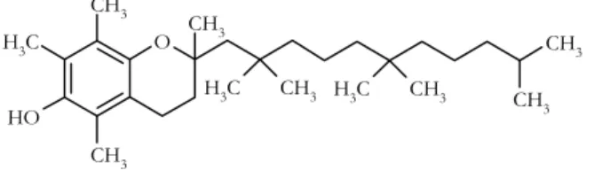
α-Tocopherol (α-TP; Figure 1.1.) is the most representative oil-soluble antioxidant of the vitamin E type, which is thought to protect cells by virtue of its ability to alleviate
oxidative stress by quenching free radicals[1].
O CH3 C H3 O H CH3 CH3 CH3 CH3 CH3 C H3 CH3 C H3
Figure 1.1. Chemical structure of α-TP
The physiological relevance of α-TP, and the severe pathological consequences of its deficiency, impose a major challenge to the living organisms for sustaining an adequate supply of this compound to different tissues, particularly those highly sensitive to α-TP
deficiency such as the brain and gonads[2]. α-TP deficiency could be overcome by dietary
supplementation, and several studies suggest that supplements of vitamin E may contribute to lowering the risk of specific chronic and degenerative diseases such as Alzheimer disease, age-related macular degeneration, some types of cancer, cataracts and ischemic
heart disease[3].
[1] J. Qian, S. Morley, K. Wilson, P. Nava, J. Atkinson, D. Manor. J. Lipid Res. 46 (2005) 2072. [2] P. Mardones, A. Rigotti. J. Nutr. Biochem. 15 (2004) 252.
[3] J.N. Hathcock, A. Azzi, J. Blumberg, T. Bray, A. Dickinson, B. Frei, I. Jialal, C.S. Johnston, F.J. Kelly, K.
6
Due to the nutritional importance of vitamin E in health, researches have been performed to develop a simple and sensitive method for its routine determination in
vegetables[4]. The α-tocopherol content in vegetables can be commonly determined by a
wide range of analytical techniques such as thin-layer chromatography (TLC), capillary gas chromatography (cGC), supercritical fluid chromatography (SFC) or high performance liquid chromatography (HPLC). The most common used technique is normal-phase HPLC with ultraviolet or fluorescence detection, but very intensive pre-treatment of the samples
is needed[5].
Our approach uses the solid phase extraction based on molecularly imprinted polymers (MISPE).
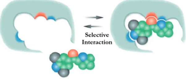
Molecular imprinting is a very useful technique for incorporating specific substrate recognition sites into polymers. The molecular recognition characteristics of these polymers are attributed to the complementary size, shape, and binding sites imparted to the
polymers by the template molecules[6,7,8,9] (Figure 1.2.).
Figure 1.2. Schematic representation of MIP
The simplicity of creating tailored recognition sites in synthetic materials by molecular imprinting, as compared with their creation by complicated multi-step organic syntheses, is very attractive from the applications point of view, and, in comparison with biomolecules, the main advantages of molecularly imprinted polymers (MIP) are their relatively high stability over a wide range of conditions (temperature, pressure, organic solvents, acidic or
basic solutes, etc.) and low cost [10,11,12].
[4] M. Lechner, B. Reiter, E. Lorbeer. J. Chromatogr. A 857 (1999) 231. [5] I.K. Cho, J. Rima, C. Ling Chang, Q.X. Li. J. Food Comp. Anal. 20 (2001) 57.
[6] E. Caro, R.M. Marcé, F. Borrull, P.A.G. Cormack, D.C. Sherrington. Trends Anal. Chem. 25 (2006) 143. [7] P.A.G. Cormack, A.Z. Elorza. J. Chromatogr. B 1 (2004) 173.
[8] K. Mosbach, Y.-H Yu, J. Andersch, L. Ye. J. Am. Chem. Soc. 123 (2001) 12420.
[9] F. Puoci, M. Curcio, G. Cirillo, F. Iemma, U.G. Spizzirri, N. Picci. Food Chem. 106 (2008) 836. [10] B. Sellergren, C.J. Allender. Adv. Drug Deliv. Rev. 57 (2005) 1733.
Selective Interaction
7
Molecular imprinting has now become an established method and has also been applied in the areas of synthetic chemistry and analytical chemistry. MIP have been used as
chromatographic stationary phases[13] for enantiomeric separations[14], and for solid-phase
extraction[15], catalysis[16] and sensor design[17], as well as for protein separation[18], as
receptor[19], antibody[20] and enzyme mimics[21], and most recently as drug delivery systems
(DDS)[22].
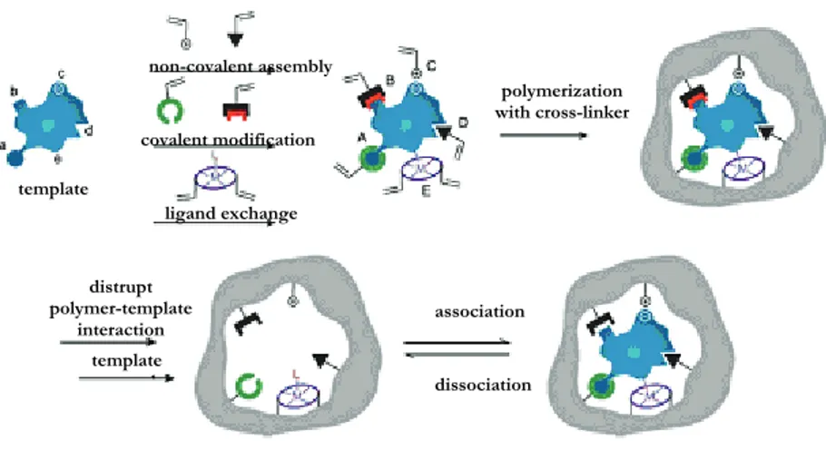
For the preparation of molecularly imprinted polymers, generally three different approaches have been developed to date: the covalent, the semi-covalent, and the non-covalent approach (Figure 1.3.).
Figure 1.3. Highly schematic representation of the molecular imprinting approaches
[11] E. Caro, N. Masqué, R.M. Marcé, F. Borrull, P.A.G. Cormack, D.C. Sherrington. J. Chromatogr. A 963 (2002)
169.
[12] K. Haupt. Analyst 126 (2001) 747.
[13] O. Brüggemann, A. Visnjevski, R. Burch, P. Patel. Anal. Chim. Acta 504 (2004) 81. [14] K. Mosbach.. Anal. Chim. Acta 435 (2001) 3.
[15] F. Puoci, C. Garreffa, F. Iemma, R. Muzzalupo, U.G. Spizzirri, N. Picci. Food Chem. 93 (2005) 349. [16] J.L. Defreese, A. Katz. Chem. Mater. 17 (2005) 6503.
[17] M. Matsuguchi,T. Uno. Sens. Actuators B 113 (2006) 94.
[18] X. Pang, S.L.G Cheng, E. Tang. Anal. Bioanal. Chem. 384 (2006) 225. [19] K. Haupt. Chem. Commun. 171 (2003) 171.
[20] M. Boopathi, M.V.S. Suryanarayana, A.K. Nigam, P. Pandey, K. Ganesan, B. Singh, K. Sekhar. Biosens.
Bioelectron. 21 ( 2006) 2339.
[21] Z. Meng, T. Yamazaki, K. Sode. Biotech. Lett. 27 (2005) 471.
[22] F. Puoci, F. Iemma, R. Muzzalupo, U.G. Spizzirri, S. Trombino, R. Cassano, N. Picci. Macromol. Biosci. 4
(2004) 22. template ligand exchange covalent modification non-covalent assembly polymerization with cross-linker distrupt polymer-template interaction template l association dissociation
8
The first, pioneered by Wulff’s group[23] involves the formation of complex between
functional monomers and template molecules via reversible covalent bond (such as boronate ester, ketal and acetal, or Schiff base) both prior to polymerization and in the rebinding experiments. The advantage of this approach is that the functional groups are only associated with the template site; however, only a limited number of compounds (alcohols (diols), aldehydes, ketones, amines and carboxylic acids) can be imprinted with this approach.
In the semi-covalent approach, firstly reported by Sellergren and Andersson[24],
covalent interactions are involved in MIP synthesis, while the subsequent rebinding phase is based on non-covalent interactions.
The non-covalent approach, pioneered and extensively developed by Mosbach and
coworkers[25] instead, involves only non-covalent interactions (such as hydrogen bonds,
ionic interactions, hydrophobic interactions, and metalion chelating interactions) for both the molecular imprinting process and the subsequent rebinding. This approach is the most widely used because of the absence of complicated synthetic processes, the subsequent possibility of using a far greater variety of functional monomers, the nature of the recognition mechanism (mimicking natural macromolecular binding), and the rapidity of
the recognition[26,27,28,29].
For these reasons, we chose the non-covalent imprinting method for the preparation of bulk imprinted polymers for the selective recognition of α-TP. The techniques involve
the pre-organization, in a suitable porogen (solvent)[30], of functional monomers around a
template molecule, which resembles the guest molecule in shape and size. Polymerization of the supramolecular assembly in the presence of an excess of cross-linker and subsequent removal of the template leads to polymers that retain the specific orientation of functional groups within the cavity created and that are capable of selective rebinding of the template.
[23] G. Wulff, A. Biffis. In Molecularly Imprinted Polymers: Man-Made Mimics of Antibodies and their
Applications in Analytical Chemistry, Techniques and Instrumentation in Analytical Chemistry. Sellergren B (ed.). Elsevier: Amsterdam. 23 (2001) 71.
[24] B. Sellergren, L. Andersson. J. Org. Chem. 55 (1990) 3381.
[25] R. Arshady, K. Mosbach. Synthesis of substrateselective polymers by host-guest polymerization. Macromol.
Chem. Phys. 182 (1981) 687.
[26] C. Alexander, H.S. Andersson, L.I. Andersson, R.J. Ansell, N. Kirsch, I.A. Nicholls, J. O’Mahony, M.J.
Whitcombe. J. Mol. Recognit. 19 (2006) 106.
[27] F. Puoci, F. Iemma, G. Cirillo, S. Trombino, R. Cassano, N. Picci. E-Polymers 13 (2007) 1.
[28] C. Chassaing, J. Stokes, R.F. Venn, F. Lanza, B. Sellergren, A. Holmberg, C. Berggren. J. Chromatogr. B 804
(2004) 71.
[29] F. Puoci, F. Iemma, G. Cirillo, R. Muzzalupo, U.G. Spizzirri, N. Picci. Macromol. Indian J. 2 (2006). [30] V.P. Joshi, S.K. Karode, M.G. Kulkarni, R.A. Mashelkar. Chem. Eng. Sci. 53 (1998) 2271.
9
In our work, new non-covalent α-TP molecularly imprinted polymers were prepared using α-TP as template, methacrylic acid (MAA) as functional monomer and ethylene glycol dimethacrylate (EGDMA) as crosslinking agent (Figure 1.4.).
O OH O O O O MAA EGDMA
Figure 1.4. Chemical structure of MAA and EGDMA
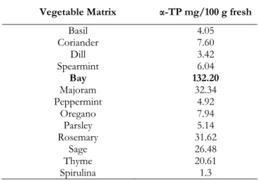
α-TP MIP were applied as sorbents in solid phase extraction (SPE) for purification and determination of α-TP in vegetable, and bay leaves were chosen as vegetable matrix because of their high α-TP content (Table 1.1.)
Table 1.1. α-TP content in different vegetable matrices
Vegetable Matrix α-TP mg/100 g fresh
Basil Coriander Dill Spearmint Bay Majoram Peppermint Oregano Parsley Rosemary Sage Thyme Spirulina 4.05 7.60 3.42 6.04 132.20 32.34 4.92 7.94 5.14 31.62 26.48 20.61 1.3
The MISPE-eluate fractions were analyzed by HPLC. The proposed MISPE protocol could overcome the drawback of traditional detection methods, which require pre-treatments of the samples, such as saponification, extraction with organic solvents, etc. The possibility to obtain the selective recognition of α-TP from natural samples in aqueous mixture without hydrophobical non-specific interactions represents the main advantage of
10
our materials[31]. Moreover, we developed a very straight-forward protocol, involving
simple steps for the pre-treatment of the samples, and the direct HPLC injection of eluate fractions without any treatment. The pre-treatment of the sample consists of a crushing step, a suspension in a suitable organic solvent and the subsequent removal of the insoluble portion by filtration. In all the steps, no toxic and biocompatible organic solvents were used. Before packing of MISPE cartriges, the capacity of the polymer to recognize selectively the template both in organic and in aqueous media was evaluated by carrying out binding experiments both in organic (i.e., acetonitrile) and in aqueous media (i.e., an ethanol/water 6/4 v/v mixture). Considerable differences in the recognition characteristics between imprinted and non-imprinted polymers have been observed, while the selectivity of the polymeric device was also evaluated using a molecule structurally similar to the template, in particular 6-hydroxy-2,5,7,8-tetramethylchroman-2-carboxylic acid (HMCA) was employed (Figure 1.5.).
O CH3 C H3 O H CH3 CH3 COOH
Figure 1.5. Chemical structure of HMCA
Then, MISPE cartridges were packed and their selectivity towards the template was studied. Finally, we investigated the ability of the MISPE cartridges to absorb selectively α-TP from bay leaves. MISPE cartridges were found to be a very efficient tool for α-α-TP
concentration and isolation from complex matrices[32].
As above reported, several studies have described the beneficial effect of oral supplementation with vitamin E on the prevention and treatment of cardiovascular
diseases and cancer[33]. For this reason, the possibility to employ α-TP imprinted polymers
[31] E. Caro, R.M. Marcé, P.A.G. Cormack, D.C. Sherrington, F. Borrull. Anal. Chim. Acta 552 (2005) 81. [32] F. Puoci, G. Cirillo, M. Curcio, F. Iemma, U.G. Spizzirri, N. Picci. Anal. Chim. Acta 593 (2007) 164. [33] M.C. Morris, D.A. Evans, C.C. Tangney, J.L. Bieinias, R.S. Wilson, N.T. Aggarwal, P.A. Scherr. Am. J. Clin.
11
as base excipients for controlled release device for α-TP oral supplementation[34] was also
evaluated.
A controlled drug delivery system would make it possible to maximize drug efficacy and safety and to provide a suitable rate of delivery of the therapeutic dose, at the most appropriate site in the body. This would prolong the duration of the drug’s pharmacological activity, to reduce side-effects, and minimize administration frequency,
thus enhancing patient compliance[35,36]. For this purpose, synthesized MIP and NIP
particles were loaded with a α-TP solution and the release profile in gastrointestinal simulating fluids was tested, showing considerable differences between imprinted and non imprinted polymers.
2. Materials and Methods
2.1. Materials
Ethylene glycol dimethacrylate (EGDMA), methacrylic acid (MAA), 2,2’-azoisobutyronitrile (AIBN), α-tocopherol (α-TP) and 6-hydroxy-2,5,7,8-tetramethylchroman-2-carboxylic acid (HMCA) were obtained from Aldrich. All solvents were reagent grade or HPLC-grade and used without further purification and they were provided by Fluka Chemie. MAA was purified before use by distillation under reduced pressure.
2.2.Instrumentation
The liquid chromatography consisted of an Jasco BIP-I pump and Jasco UVDEC-100-V detector set at 292 nm. A 250mm×4mm C-18 Hibar® Column, particle size 10 μm (Merck, Darmstadt, Germany)was employed. The mobile phase was ethanol and the flow
rate was 1.0 ml min−1. The shaker and centrifugation systems consisted of a wrist action
shaker (Burrell Scientific) and an ALC microcentrifugette 4214, respectively.
2.3. Synthesis of α-tocopherol imprinted polymers
α-TP imprinted polymers (MIP) were prepared using methacrylic acid (MAA) as functional monomers. Briefly, template α-TP, MAA, EGDMA and AIBN were dissolved
[34] F. Puoci, G. Cirillo, M. Curcio, F. Iemma, O.I. Parisi, M. Castiglione, N. Picci. Drug Deliv. 15 (2008) 253. [35] D. Cunliffe, A. Kirby, C. Alexander. Adv. Drug Del. Rev. 57 (2005) 1836.
12
in 5.25ml of chloroform in a thick-walled glass tube. The mixture was purged with nitrogen, sonicated for 10 min, and then thermo-polymerized (MIP-1, 1, MIP-2, NIP-2) under a nitrogen atmosphere for 24 h at 60 °C or photo-polymerized (MIP-3, NIP-3, MIP-4, NIP-4) for 24 h with 360 nm light at 4 °C. After the photolysis, the tubes were
incubated at 60 °C for 24 h[37]. The resultant bulk rigid polymers were crushed, grounded
into powder and sieved through a 63 nm stainless steel sieve. The sieved MIP materials were collected and the very fine powder, suspended in the supernatant solution (acetone), was discarded. The resultant MIP materials were soxhlet extracted with 200 ml of a methanol:acetic acid (8:2, v/v) mixture for at least 48 h, followed by 200 ml of methanol for others 48 h. The extracted MIP materials were dried in an oven at 60 °C overnight. The washed MIP materials were checked to be free of α-TP and any other compound by HPLC analysis.
The formulations used for the preparation of the different matrices are shown in Table 1.2. Blank polymers (to act as a control) were also prepared when polymerization was carried out in the absence of α-TP.
2.4. Binding experiments
Evaluation of the capacity of the matrices to recognize and bind the template was performed both in acetonitrile and in ethanol/water (6/4, v/v) mixture. Briefly, 50 mg of
polymer particles were mixed with 1 ml α-TP solution (0.2 mmol l-1) in a 1 ml eppendorf
and sealed. Samples were shaken in a water bath for 24 h, centrifuged for 10 min (10,000 rpm) and the α-TP concentration in the liquid phase was measured by HPLC. The amount of α-TP bound to the polymer was obtained by comparing its concentration in the MIP samples to the NIP samples. The same experiments were performed using HMCA solutions. Experiments were repeated five times and results were expressed as means (± SEM).
2.5. Molecularly imprinted solid phase extraction conditions
The 500 mg amounts of dry particles of MIP-4 and NIP-4 were packed into a 6.0 ml polypropylene SPE column. The column was attached with a stop cock and a reservoir at the bottom end and the top end, respectively. The polymer was rinsed with chloroform,
13
acetonitrile, ethanol and then with the loading solvent. α-TP was dissolved in the loading
solvent to final concentration of 0.2 mmol l−1. After conditioning, dry MISPE column was
loaded with α-TP standard solution. After loading, vacuum was applied trough the cartridges for 5 min in order to remove residual solvent. Washing solvent was then passed trough the cartridges and finally, after column drying, elution solvent was applied to perform the complete extraction of α-TP. The loading, washing and eluting fractions were analysed by HPLC to detect the α-TP amount.
Loading step: 2 ml of ethanol/water mixture (6/4, v/v), flow rate ~0.2 ml min−1;
washing step: 3 ml of ethanol/water mixture (7/3, v/v), flow rate ~0.2 ml min−1; eluting
step: 5 ml of ethanol with 5% acetic acid, flow rate ~0.1 ml min−1. In order to evaluate the
selectivity of the MIP, optimized protocol was also applied using HMCA solutions. Experiments were repeated five times and results were expressed as means (± SEM).
2.6. Molecularly imprinted solid phase extraction of vegetable sample extracts
Two grams of bay leaves were pounded and extracted with 200 ml of ethanol. To 60 ml of this mix, water was added to raise the loading solution (ethanol/water, 6/4, v/v; 100
ml). Five milliliters of this solution (8.73 x 10⁻⁸ mol) were used to load the MISPE
coloumn. Two washing steps are performed: 3 ml of an ethanol/water (7/3, v/v) mixture and 5 ml of ethanol. Finally, 4 ml of ethanol with 5% acetic acid was used as elution fraction. All the solutions were analysed by HPLC. Experiments were repeated five times and results were expressed as means (± SEM).
2.7. Method validation
The limit of detection and limit of quantization were determined using spiked bay leaves. 0.050 g of fresh bay leaves were spiked with 0.025, 0.050, 0.100, 0.200, 0.400, 0.800, 1.5, 3, 6, 15 mg of α-TP. The samples was extracted with 200 ml of ethanol and then water was added to raise the right loading solution composition (ethanol/water, 6/4, v/v), extracted using the MISPE protocol and analyzed by HPLC. Detection and quantification limits were calculated as the concentration corresponding to a signal 3 and 10 times the standard deviation of the baseline noise, respectively.
14
2.8. Drug Loading by the Soaking Procedure
Two grams of polymeric matrix were immersed in 20 ml of a α-TP solution (5.5 mM) in acetonitrile and soaked for 3 days at room temperature. During this time, the mixture was continuously stirred, and then the solvent was removed. Finally, the powder was dried under vacuum overnight at 40 °C.
2.9. In Vitro Release Studies
Release studies were done using the dissolution method described in the USP XXIV (apparatus 1-basket stirring element). To mimic the pH in the digestive tract simulated, 0.1 N HCl (pH 1.0) was used as a stimulated gastric fluid, and after 2 h, disodium hydrogen phosphate (0.4 M) was added to adjust the pH value to 6.8 to simulate a intestinal fluid. To improve the solubility of released α-TP in the simulated fluid, each testing sample contained 0.1% of sodium dodecylsulfate (SDS).
The experiments were performed as follows: 30 mg of MIP-4 and NIP-4 particles loaded with α-TP were dispersed in flasks containing 10 ml of 0.1 N HCl and maintained at
37 ± 0.5 ◦C in a water bath for 2 h under magnetic stirring (50 rpm). Disodium hydrogen
phosphate (0.4 M, 5 ml) was then added to the samples. These conditions were maintained throughout the experiment. To characterize the drug release, 2 ml of samples were drawn from the dissolution medium at designated time intervals, and the same volume of simulated fluid was supplemented. α-TP was determined by HPLC analysis, and the amount of α-TP released from five samples of each formulation was used to characterize drug release. The percentage of α-TP released was calculated considering 100% the α-TP
content in polymeric samples after drying procedure[38]. Experiments were repeated five
times and results were expressed as means (± SEM).
3. Results and discussion
3.1. Preparation of the imprinted polymers
In the synthesis of α-TP imprinted polymers, MAA was chosen as functional monomer and EGDMA was used as crosslinker. In literature, many different ratios of
template and functional monomer were used[39].
[38] G. Pitarresi, P. Pierro, G. Giammona, F. Iemma, R. Muzzalupo, N. Picci. Biomaterials 25 (2004) 4333. [39] M. Kempe, K. Mosbach. J. Chromatogr. A 694 (1995) 3.
15
Our purpose was the selective extraction of α-TP from food matrices with different water percentages, thus, we synthesized polymers at various molar ratios of template, methacrylic acid and crosslinker (Table 1.2.). Two different molar ratio of MAA/template were tested (8/1 for MIP-1,3 and NIP-1,3; 16/1 for MIP-2,4 and NIP 2,4 respectively).
The whole of the reaction conditions have to maximize the interactions between the template and the functional monomer and consequently to ensure strong and selective binding of the substrate to the polymeric matrices. α-TP is a very poor functionalized molecule, thus, when it is used as template, an increase of the strength of the interactions with the functional monomer in the pre-polymerization complex is needed. Two main parameters must be considered.
The first is the polymerization temperature. The formation of the complex is a dynamic process and, when a template with few functional groups able to create hydrogen bond is used, a low temperature is needed to reduce the kinetic energy of the system. In this case, indeed, a high temperature could drive the equilibrium away from the template-functional monomer complex toward the unassociated species, resulting in a decrease in the number of imprinted cavities and thus in the recognition properties of the final
materials[40]. Therefore, together with thermo-polymerization, a photo-polymerization
procedure, which allows a lower temperature (0-4 °C), was employed. After UV irradiation the performance of the initially formed polymer was improved by thermal stabilization at
60 °C[41].
The second parameter to be considered is the nature of the porogenic agent. The inert solvent used in the polymerization mixture may play a major role in determining the properties (surface area, internal pore volume, etc.) of the resulting polymer. Moreover, since polar solvents are more able to solvate polar molecules, this leads to the disruption of
H-bonds between the template and the functional monomer[42,43]. Thus, our general
procedure employed to improve the recognition properties of molecularly imprinted polymers was the choice of the least polar solvent in which the reagents dissolves. In our case, we chose chloroform.
[40] S.H. Cheong, S. McNiven, A. Rachkov, R. Levi, K. Yano, I. Karube. Macromolecules 30 (1997) 1317. [41] B. Sellergren, K.J. Shea. J. Chromatogr. A 635 (1993) 31.
[42] M.J. Whitcombe, M.E. Rodriguez, P. Villar, E.N. Vulfson. J. Am. Chem. Soc. 117 (1995) 105. [43] S.H. Cheong, S. McNiven, A. Rachkov, R. Levi, K. Yano, I. Karube. Macromolecules 30 (1997) 1317.
16
3.2. Evaluation of the imprinting effect
The imprinting effect in the synthesized materials was evaluated by binding experiments in which amounts of polymeric particles were incubated with an α-TP
solutions 0.2 mmol l−1 for 24 h. These preliminary experiments were performed both in
acetonitrile and in an ethanol:water mixture (6:4, v/v). A good imprinting effect was found in the materials with the higher amount of MAA synthesized with photo-initiation (MIP-4) (Table 1.2.).
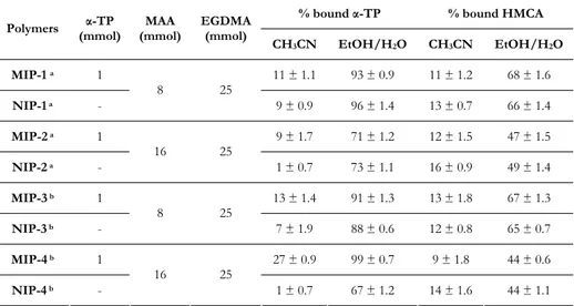
Table 1.2. Polymers composition and percentage of bound α-TP and HMCA by the imprinted and non-imprinted polymers after 24 hours in acetonitrile and
in ethanol/water (6/4 v/v) mixture.
% bound α-TP % bound HMCA
Polymers (mmol)α-TP (mmol)MAA EGDMA (mmol)
CH3CN EtOH/H2O CH3CN EtOH/H2O MIP-1 a 1 11 ± 1.1 93 ± 0.9 11 ± 1.2 68 ± 1.6 NIP-1 a - 8 25 9 ± 0.9 96 ± 1.4 13 ± 0.7 66 ± 1.4 MIP-2 a 1 9 ± 1.7 71 ± 1.2 12 ± 1.5 47 ± 1.5 NIP-2 a - 16 25 1 ± 0.7 73 ± 1.1 16 ± 0.9 49 ± 1.4 MIP-3 b 1 13 ± 1.4 91 ± 1.3 13 ± 1.8 67 ± 1.3 NIP-3 b - 8 25 7 ± 1.9 88 ± 0.6 12 ± 0.8 65 ± 0.7 MIP-4 b 1 27 ± 0.9 99 ± 0.7 9 ± 1.8 44 ± 0.6 NIP-4 b - 16 25 1 ± 0.7 67 ± 1.2 14 ± 1.6 44 ± 1.1 All polymers were synthesized in 5.25 ml of chloroform using 0.045 g of AIBN.
a thermo-polymerization; b photo-polymerization
In organic medium, the specific interactions between polymeric matrices and template are more relevant in MIP-4 than in MIP-3. In aqueous medium, MIP-4 samples bound more selectively α-TP than NIP-4 ones, while no differences were raised comparing MIP-3 and NIP-3 samples. The higher amounts of bound α-TP in aqueous medium comparing to that in acetonitrile are ascribable to the presence of more hydrophobically driven bonds, even if the higher amount of MAA probably reduces this kinds of interactions. No imprinting effect was found in α-TP MIP synthesized by thermo-polymerization: the same binding percentage for MIP and NIP particles both in organic and in aqueous media were
17
obtained (Table 1.2.), confirming that the thermo-initiation negatively interfered with the formation of the pre-polymerization complex.
To evaluate selectivity of MIP-4 towards α-TP, the same binding experiments using 6-hydroxy-2,5,7,8-tetramethylchroman-2-carboxylic acid (HMCA), instead of α-TP, were performed. The chemical differences between the two analytes drive the interactions with the polymeric matrices. The carboxylic group of HMCA makes this molecule much more hydrophilic than α- TP and the polymers practically do not interact with the analogue in organic media, while in water solution the binding percentages of the molecule are higher but considerably lower than which of the original template as reported in Table 1.2. Furthermore, in all the tested environments, the amount of HMCA bound by the imprinted and the non-imprinted polymers are practically the same, and this result clearly shows the non-specific nature of these interactions.
3.3. Optimization of the molecularly imprinted solid phase extraction
After the evaluation of MIP efficiency, MISPE cartridges were packed with the most effective materials (MIP-4) and the corresponding NIP (NIP-4), and their performances as sorbents for α-TP SPE were compared. The cartridges were packed with 500 mg of
polymer and the loading and the washing steps were optimised[44].
Acetonitrile and different ethanol/water (10/0, 8/2, 7/3 and 6/4, v/v) mixtures were employed in loading, washing and eluting step and the HPLC data were collected in Table
1.3. The best results were obtained when in the loading step 2 ml of 0.2 mmol l−1 α-TP
solution (ethanol/water, 6/4, v/v) were employed (flow rate 0.2 ml min−1). In this case,
both imprinted and non-imprinted polymers retain all the loaded α-TP. In order to minimize the non-specific component of the interaction between α-TP and polymeric matrices a washing step with ethanol/water (7/3, v/v) mixture is needed (flow rate 0.2 ml
min−1). MIP cartridges retain much more α-TP than NIP ones. In particular, only 33% of
loaded α-TP was washed out from the MIP cartridges, while in NIP ones, this percentage was 72%. The optimized elution was obtained employing ethanol containing 5% of acetic
acid (flow rate 0.1 ml min−1) and in the eluting fraction of MIP cartridges 67% of α-TP
loaded was detected, while in NIP one this percentage was only 28%. .
18
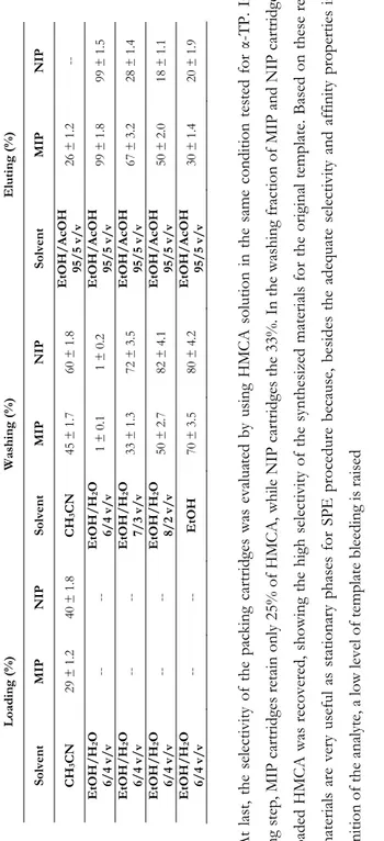
Table 1.3. Percentage of collected
α-TP
in
loading, washing and elution fractions
Loa di ng (%) Washi ng (% ) Eluti ng (% ) Solven t MIP NIP Solven t MIP NIP Solven t MIP NIP CH 3 CN 29 ± 1. 2 40 ± 1. 8 CH 3 CN 45 ± 1. 7 60 ± 1. 8 EtOH /A cOH 95 /5 v/v 26 ± 1. 2 --EtOH /H 2 O 6/ 4 v /v -- -- EtOH /H 2 O 6/ 4 v /v 1 ± 0.1 1 ± 0.2 EtOH /A cOH 95 /5 v/v 99 ± 1. 8 99 ± 1. 5 EtOH /H 2 O 6/ 4 v /v -- -- EtOH /H 2 O 7/ 3 v /v 33 ± 1. 3 72 ± 3. 5 EtOH /A cOH 95 /5 v/v 67 ± 3. 2 28 ± 1. 4 EtOH /H 2 O 6/ 4 v /v -- -- EtOH /H 2 O 8/ 2 v /v 50 ± 2. 7 82 ± 4. 1 EtOH /A cOH 95 /5 v/v 50 ± 2. 0 18 ± 1. 1 EtOH /H 2 O 6/ 4 v /v -- -- EtOH 70 ± 3. 5 80 ± 4. 2 EtOH /A cOH 95 /5 v/v 30 ± 1. 4 20 ± 1. 9 At last , the s el ec tivity of th e packing c art rid ges was evalua ted b
y using HMCA solution in the same co
ndition tested for
α-TP. In th e loading s tep , M IP ca rt rid ges r etain only 25% of HMCA , whil e NI P car tri dg es th e 33% . In th e washing fr ac tion of MIP an d NIP car tri dges, all the load ed HM CA was re cove re d, showing t he high sele ctiv ity of the s ynth esize d ma te rials
for the origina
l templa te . Bas ed on th ese res ults , our mat erial s a re v er y use ful as st ationa ry p hases for SP E proc ed ur e be ca use , b esid es th e a de qu at e s ele ctivit y an d affi nity p ro perties in the re cognition of th e anal yt e, a lo w leve l of temp late bl eeding is raised 3.4. Mo lecular ly i m printed so lid p hase ex traction of v eget ab le samp le extracts Many m edicinal and aro m ati c p lants, spi ces an d herb s have tr aditi onall y been
used in foods to improve or
modify th
eir flavour.
How
ever,
their protective effect in inhibi
ting oxi
dation reactions plays
an important rol e unk nown fo r years and it could be related to their cont ent of
19
antioxidant compounds, and in particular to the α-TP amount[45]. The aim of this work was
to develop a MISPE procedure for the clean-up of α-TP from bay. In order to measure the recovered α-TP by our method, the quantification of α-TP content in fresh bay leaves, was performed by Sànchez-Machado et al.’s method and this value was found to be 125 mg per
100 g of fresh bay leaves[46].
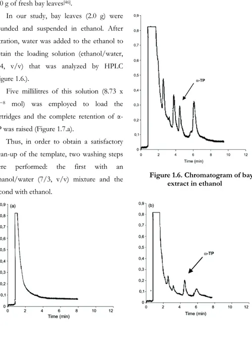
In our study, bay leaves (2.0 g) were pounded and suspended in ethanol. After filtration, water was added to the ethanol to obtain the loading solution (ethanol/water, 6/4, v/v) that was analyzed by HPLC (Figure 1.6.).
Five millilitres of this solution (8.73 x
10−8 mol) was employed to load the
cartridges and the complete retention of α-TP was raised (Figure 1.7.a).
Thus, in order to obtain a satisfactory clean-up of the template, two washing steps were performed: the first with an ethanol/water (7/3, v/v) mixture and the second with ethanol.
Figure 1.6. Chromatogram of bay extract in ethanol
[45] D.J.M. Gomez-Coronado, E. Ibanez, F.J. Ruperez, C. Barbas. J. Chromatogr. A 1054 (2004) 227. [46] D.I. Sànchez-Machado, J. Lòpez-Hernàndez, P. Paseiro-Losada. J. Chromatogr. A 976 (2002) 277.
20
Figure 1.7. Chromatograms of MISPE loading (a) and washing fractions (b and c).
In the washing fractions of MIP cartridges (Figures 1.7.b and c), not so relevant peaks were observed at the retention time of α-TP, in demonstration of the presence of imprinted cavities in polymeric matrix. The α-TP amount in these fractions represents about the 40% of the loaded one. Furthermore, an important clean-up of the extracted mixture was obtained, as showed by the presence of several peaks referable to the others compounds of the sample.
Finally, α-TP was selectively eluted using ethanol containing 5% of acetic acid (Figure 1.8.), obtaining a more than satisfactory recovery of α-TP in MIP cartridges (about the 60% of the loaded one), confirming that the compounds which co-eluted with the template in the starting solution were completely removed during the washing steps. By performing the same experiments using NIP cartridges, in washing fractions almost all the loaded α-TP was recovered (about the 98%), confirming the total non-specificity of the interactions between template and non-imprinted
Figure 1.8. Chromatogram of MISPE eluting fraction
matrices, while in the elution fraction no relevant amount of α-TP was detected
3.5. Method validation
The limit of detection and limit of quantization were determined using spiked bay leaves. 0.050 g of fresh bay leaves were spiked with 0.025, 0.050, 0.100, 0.200, 0.400, 0.800,
21
1.5, 3, 6, 15 mg of α-TP. The samples was extracted with 200 ml of ethanol and then water was added to raise the right loading solution composition (ethanol/water, 6/4, v/v), extracted using the MISPE protocol and analyzed by HPLC. Detection and quantification limits were calculated as the concentration corresponding to a signal 3 and 10 times the standard deviation of the baseline noise, respectively (American Chemical Society
Guidelines[47]) and correspond to 3.49 x 10⁻⁷ and 1.16 x 10⁻⁶ mol l−1, respectively. The
calibration curves were linear with correlation coefficients of R2 > 0.98. The intraday
precisions of the relative peak areas were below 3.3% and the interday precisions below 6.5%. Both were calculated as R.S.D.s for five measurements.
3.6. In Vitro α-TP Releasing Properties
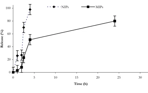
The possibility of employing the synthesized MIP-4 as devices for the controlled release of α-TP in gastrointestinal simulated fluids was investigated. In vitro release studies were performed by immersing aliquots of the microparticles loaded with α-TP at pH 1.0 (simulated gastric fluid) for 2 hours and then at pH 6.8 (simulated intestinal fluid) using the pH change method. To improve the solubility of α-TP in aqueous media, SDS was added to the solutions.
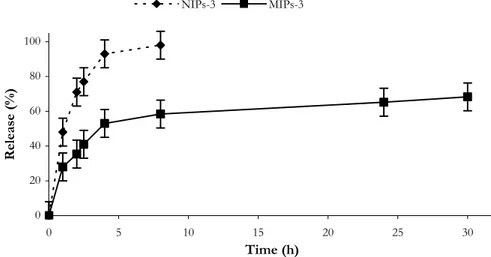
Our hypothesis was that α-TP imprinted polymers have a better ability to control drug (α-TP) release in compared with non-imprinted polymers due to the presence of specific binding sites in the polymeric network that are able to release the drug much more slowly. The experimental data confirm this hypothesis; the drug release from NIP was indeed remarkably faster than that observed from MIP (Figure 1.9.).
In particular, while in the first case the drug is completely released within 4 hours, only 50% of α-TP was released from MIP samples during the same period. After pH changing, in intestinal simulating fluids, α-TP release from MIP continues, and in 24 hours the percentage of α-TP released was about 80% (100% within 40 h). These remarkable differences depend on the different recognition properties of the two polymeric matrices.
The non-imprinted polymers, indeed, do not have specific binding cavities for the drug, while the MIP samples, because of their specific structure, strongly bound α-TP by non-covalent interactions in the cavities formed during the polymerization procedure in the presence of the analyte.
[47] American Chemical Society (ACS), Subcommitee of Environmental Analytical Chemistry. Anal. Chem. 52
22 0 20 40 60 80 100 0 5 10 15 20 25 30 Time (h) Release (%) NIPs MIPs
Figure 1.9. Release profile of α-TP from MIP-4 and NIP-4 in gastrointestinal simulating fluids.
This observation supports a model of retention mechanism, which assumes that the acid groups of the selective sites have stronger interaction with the drug than the non-selective sites. At low pH (1.0) values, the carboxylic groups are not ionized and there is a good interaction with the template. These results might help us to understand the behavior of these matrices when the pH increases. Under these conditions, that simulate the intestinal fluid, in the non-imprinted polymers the antioxidant is bound with non-covalent interactions on the surface of the matrices. At pH 6.8, the diffusion rate of the buffer on the polymer surface is fast, the carboxylic groups are ionized, and the drug is rapidly released. Instead, in the MIP case, the diffusion rate of the buffer into specific cavities of imprinted polymers is slower, and the functional groups are ionized more slowly, resulted in well controlled release. For these reason, the rate of the release was considerably different, and MIP-4 represent a very useful polymeric device for the selective and controlled release on the antioxidant agent in gastrointestinal fluids.
CHAPTER II 5-FLUOROURACIL
MOLECULARLY IMPRINTED POLYMERS
1. Introduction
5-Fluorouracil (Figure 2.1.) is a widely used antineoplastic agent for the treatment of
many types of cancers, such as colorectal, breast, head and neck malignancies[48].
N N O H O H F
Figure 2.1. Chemical structure of 5-FU
Its endurance and longevity in cancer chemoterapy is due to the fact that 5-FU is the only anticancer agent on the market that shows synergism with the most other anticancer
drugs in combination therapy[49]. On the other hand, it is effective against cancers
refractory to other treatments. Despite these advantages, the use of 5-FU is limited by its quick metabolism in the body, therefore the maintenance of high serum concentrations of this drug to improve its therapeutic activity is needed. The maintenance of these serum concentrations requires continuous administrations, but 5-FU shows severe toxic effects;
consequently reaching and/or exceeding the toxic concentration must be avoided[50].
In order to improve therapeutic index and reduce toxic effects of this drug, numerous studies in literature are aimed at design of devices for the controlled release of 5-FU, many
[48] A. Di Paolo, D. Romano, F. Vannozzi, A. Falcone, E. Mini, L. Cionini, T. Ibrahim, D. Amadori, M. Del
Tacca. Clin. Pharmacol. Ther. 72 (2002) 627.
[49] O.N. Al Safarjalani, R. Rais, J. Shi, R.F. Schinazi, F.N.M. Naguib, M.H. El Kouni. Cancer Chemother.
Pharmacol. 58 (2006) 692.
24
of them are based on polypeptidic and polysaccharidic systems[51]. On the other side, no
drug delivery systems for 5-FU were obtained starting from molecularly imprinted polymers.
The only MIP prepared using 5-FU as a template was proposed for analytical studies[52];
furthermore, it must be pointed out that the functional monomer and the crosslinker used for that studied device are not compatible with physiological conditions, thus are not suitable for pharmaceutical applications.
The purpose of this study was to investigate the possibility of employing MIP as
devices for the controlled release of 5-FU in biological fluids[53]. After MIP synthesis by
bulk polymerization procedure, which allows to obtain microparticles with irregular shape, the matrix affinity for 5-FU and its selectivity, using uracil (U; Figure 2.2.) as an analogue, were tested. N N O H O H
Figure 2.2. Chemical structure of U
The target sites of 5-FU are all the organs of the human body[54], especially the
gastrointestinal tract, therefore the release profile of this drug was evaluated both in gastrointestinal and in plasma simulating fluids. Considerable differences in the release characteristics between imprinted and non imprinted polymers (NIP) have been observed.
In addition, in order to obtain a better control on particle size and shape, and so also on the 5-FU release profile, molecularly imprinted hydrogel nanospheres have been also
synthesized applying a straightforward method such as precipitation polymerization[55], and
the possibility of employing these monodispersed imprinted nanoparticles as devices for the controlled/sustained release of 5-FU in biological fluids was tested.
[51] E. Fournier, C. Passirani, N. Colin, P. Breton, S. Sagodira, J.P. Benoit. Eur. J. Pharm. Biopharm. 57 (2004)
189.
[52] A. Kugimiya, T. Mukawa, T. Takeuchi. Analyst 126 (2001) 772.
[53] F. Puoci, F. Iemma, G. Cirillo, N. Picci, P. Matricardi, F. Alhaique. Molecules12 (2007) 805. [54] J.L. Grem, D. Nguyen, B.P. Monahan, V. Kao, F.J. Geoffrey. Biochem. Pharmacol. 58 (1999) 477. [55] G. Cirillo, F. Iemma, F. Puoci, O.I. Parisi, M. Curcio, U.G. Spizzirri, N. Picci. J. Drug Target. In press.
25
Coupling the properties of hydrogels, nanospheres and MIP it is possible to obtain very useful systems to be applied in drug delivery field. Firstly, due to their significant water content, hydrogels possess a degree of flexibility very similar to natural tissue, which
minimizes potential irritation to surrounding membranes and tissues[56]. Moreover, the high
swelling properties of these materials improved their recognition characteristics, because of the enhanced accessibility of template to the imprinted cavities. Finally, as before explained,
MIP are tailor-made materials with high selectivity for a target molecule named template[57]
and have broad application in many areas of science and the area of greatest potential, and probably an area of greatest challenge, is that of therapeutics and medical therapy. In addition, advances in nanobiotechnology have resulted in the evolution of several novel colloidal carriers such as liposomes, polymeric micelles nanoparticles, and nanoemulsions to maximize tumor cell killing effect during the tumor growth phase, and to protect the
surrounding healthy cells from unwanted exposure to the excess cytotoxic agent[58].
Polymeric nanoparticles are the most attractive colloidal carriers owing several merits such as ease of purification and sterilization, drug targeting possibility, and sustained release
action[59,60].
Also in this case, considerable differences in the release characteristics between imprinted and non imprinted spherical polymers have been raised.
2. Materials and Methods
2.1. Materials
Ethylene glycol dimethacrylate (EGDMA), methacrylic acid (MAA), 2,2-azoisobutyronitrile (AIBN), 5-fluorouracil (5-FU) and uracil (U) were obtained from Sigma–Aldrich (Sigma Chemical Co., St. Louis, MO). All solvents were reagent grade or HPLC-grade and used without further purification and were provided by Fluka Chemika-Biochemika (Buchs, Switzerland). MAA was purified before use by distillation under reduced pressure.
[56] M.E. Byrne, K. Park, N.A. Peppas. Adv. Drug Deliv. Rev. 54 (2002) 149.
[57] A. Beltran, E. Caro, R.M. Marcé, P.A.G. Cormack, D.C. Sherrington, F. Borrull. Anal. Chim. Acta 597 (2007)
6.
[58] A.K. Yadav, P. Mishra, S. Jain, P. Mishra, A.K. Mishra, G.P. Agrawal. J. Drug Target. 16 (2008) 464. [59] E. Allemann, R. Gurny, E. Doelker. Eur. J. Pharm. Biopharm. 39 (1993) 173.
26
2.2. Instrumentation
The liquid chromatography consisted of a Jasco BIP-I pump and Jasco UVDEC-100-V detector set at 266 nm. A 250 × 4 mmC-18 Hibar® column, particle size 10 μm (Merck, Darmstadt, Germany) was employed. The mobile phase was methanol/phosphate buffer 5mM, pH 6.8 (9/1, v/v) and the flow rate was 0.5 ml/min. The shaker and centrifugation systems consisted of a wrist action shaker (Burrell Scientific) and an ALC microcentrifugette 4214, respectively.
Scanning electron microscopy (SEM) photographs were obtained with a Jeol JSMT 300 A; the surface of the samples was made conductive by deposition of a gold layer on the samples in a vacuum chamber.
Approximate range in particle size was determined by measuring 300 particles per each sample with the use of an image processing and analysis system, a Leica DMRB equipped with a LEICA Wild 3D stereomicroscope.
2.3. Synthesis of 5-FU imprinted microparticles
For MIP synthesis by the non-covalent imprinting method, methacrylic acid was used as the functional monomer and EGDMA as crosslinking agent. Briefly, template 5-FU, methacrylic acid, EGDMA and AIBN were dissolved in 4 ml of dimethylformamide (DMF) in a thick-walled glass tube. The obtained solution was purged with nitrogen and sonicated for 10 min. The mixture was, then, incubated under a nitrogen atmosphere at 68 °C for 24 h. The resultant bulk rigid polymer systems were crushed, grounded into powder and sieved through a 63 μm stainless steel sieve. The fine particles were removed by repeated sedimentation from acetone (5 x 30 min). The resultant MIP materials were soxhlet extracted with 200 ml of an acetic acid:methanol (1:1) mixture for at least 48 h, followed by 200 ml of methanol for another 48 h. The extracted MIP materials were dried overnight in an oven at 60 °C. The washed MIP materials were checked to be free of 5-FU and any other compound by HPLC analysis. Reference NIP matrices (acting as a control) were prepared under the same conditions without using the template. The formulations used for the preparation of the different matrices (MIP-1, MIP-2, MIP-3) are shown in Table 2.1.
27
2.4. Synthesis of 5-FU spherical imprinted polymers
Spherical Molecularly Imprinted Polymers were prepared by precipitation polymerization using 5-fluorouracil as template, MAA as functional monomer and EGDMA as crosslinking agent. General synthetic procedure was reported: template (1 mmol) and MAA (8 mmol) were dissolved in a mixture of acetonitrile (20 ml) and methanol (20 ml), in a 100 ml round bottom flask and then EGDMA (10 mmol) and AIBN (50 mg) were added. The polymerization mixture was degassed in a sonicating water bath, purged with nitrogen for 10 min cooling with an ice-bath. The flask was then gently agitated (40 rpm) in an oil bath. The temperature was increased from room temperature to 60°C within 2 h, and then kept at 60 °C for 24 h. At the end of the reaction, the particles were filtered, washed with 100 ml of ethanol, 100 ml of acetone and then with 100 ml of diethyl ether. The template was extracted by ‘‘Soxhlet apparatus’’ using methanol-acetic acid mixture (1:1 (v/v), 100ml) for at least 48h, followed by methanol for another 48h, and monitoring the drug concentration in the extraction solvent by HPLC. Particles were
successively dried under vacuum overnight at 40 °C.Blank polymers, that act as a control,
were also prepared when polymerization was carried out in the absence of 5-fluorouracil.
2.5. Binding experiments
Binding experiments were performed both in organic (acetonitrile) and in aqueous media (water solution pH:1.0 and phosphate buffer pH:7.4). Briefly, 50 mg of polymer particles were mixed with 5 ml 5-FU solution (0.3 mM) in a 10 ml conical centrifugation tube and sealed. The tubes were oscillated by a wrist action shaker in a water bath for 24 h.
Then the mixture was centrifuged for 10 min (10000 rpm) and the 5-FU concentration in
the liquid phase was measured by HPLC. The amount of 5-FU bound to the polymer was
obtained by comparing its concentration in the imprinted samples to the non imprinted
ones. The same experiments were performed using uracil solution. Experiments were
repeated five times and results were expressed as means (± SEM).
2.6. Water content of spherical polymers
Aliquots (40–50 mg) of the nanospheres dried to constant weight were placed in a tared 5-ml sintered glass filter (Ø10 mm; porosity, G3), weighted, and left to swell by immersing the filter plus support in a beaker containing the swelling media: phosphate buffer (pH 7.4, simulated biological fluids). At predetermined times (2 - 4 - 8 -10 - 15 -20 -
28
24 h), the excess water was removed by percolation at atmospheric pressure. Then, the filter was placed in a properly sized centrifuge test tube by fixing it with the help of a bored silicone stopper, then centrifuged at 3500 rpm for 15 min and weighted. The filter tare was determined after centrifugation with only water. The weights recorded at the different times were averaged and used to give the water content percent (WR %) by the following
Eq. (1): 100 %= − × d d s W W W WR (1)
Where Ws and Wd are weights of swollen and dried spherical microparticles, respectively. Each experiment was carried out in triplicate and results were expressed as means (± SEM).
2.7. Drug Loading by the Soaking Procedure
Polymeric matrix (2.0 g) was immersed in a 5-FU solution in acetonitrile (20 mL, 5.5 mM) and soaked for 3 days at room temperature. During this time, the mixture was continuously stirred and then the solvent was removed by filtration. Finally the powder was dried under vacuum overnight at 40°C. The same experiments were performed using uracil solution.
2.8. In vitro release studies
Release studies were carried out using the dissolution method described in the USP
XXIV (apparatus 1-basket stirring element).Two parallel experiments for irregular matrices
were performed. In the first one, 30 mg of MIP and NIP micro-particles loaded of 5-FU
were dispersed in flasks containing 10 ml of 10 mM phosphate buffer solution (pH 7.4
simulating the biological fluids), while in the second one, the samples were dispersed in
flasks containing 10 ml of 0.1 N HCl (pH 1.0, simulating the gastric fluid) and maintained
at 37 ± 0.5 °C in a water bath for 2 h under magnetic stirring (50 rpm). Disodium hydrogen phosphate (0.4 M, 5 ml) was then added to the samples to adjust the pH value to 6.8 (simulated intestinal fluid). These conditions were maintained throughout the
experiment. Samples (2 ml) were drawn from the dissolution medium at appropriate time
intervals to determine the amount of drug released. A HPLC method was employed. The amount of 5-FU released from six samples of each formulation was used to characterize drug release. The same experiments were performed using particles loaded with uracil.
29
Experiments with spherical nanoparticles were performed at pH 7.4 as reported for
microparticles. Experiments were repeated five times and results were expressed as means
(± SEM).
3. Results and Discussion
3.1. Synthesis of bulk 5-FU imprinted polymers
Three kinds of MIP using different molecular ratios among template, functional monomer and crosslinker were synthesized. (Table 2.1.).
The choice of using DMF as porogen was dictated by the low solubility of 5-FU in organic solvents such as acetonitrile, chloroform and methanol. By increasing the solubility and the amount of the template in the pre-polymerization mixture it should be possible to obtain a material with more binding sites and consequently to have a better performance in the recognition profile of the matrices.
As reported in Table 2.1., MIP-1 was prepared using the typical molar ratio (1:4) of the
usual molecularly imprinted matrices[61]. Different molar ratios were used for the other two
matrices: for MIP-2 the MAA/EGDMA ratio was 1:5 while, in order to maximise the binding sites into the matrix, the MAA amount was increased for MIP-3 leading to a ratio of 4:5. After the grounding, sieving and suspending processes, the obtained materials were characterized by a dimensional size in the range of 20-63 μm.
3.2. Synthesis of imprinted nanospheres
Conventional MIP synthesized by bulk polymerization methods yield particles with limited control on particle size and shape. In literature, several attempts have been applied to produce monodisperse molecularly imprinted polymeric particles using methods such as
suspension polymerization in water[62], dispersion polymerization[63], liquid
perfluorocarbon[64], and via aqueous two-step swelling polymerization[65]. However, during
the polymerization procedure, these techniques require water or highly polar organic solvents, which frequently decrease specific interactions between functional monomers and template molecules. Precipitation technique not only allows to avoid these disadvantages,
[61] C.J. Allender, J. Richardson, B. Woodhouse, C.M. Heard, K.R. Brain. Int. J. Pharm. 195 (2000) 39. [62] J.P. Lai, X.Y. Lu, C.Y. Lu, H.F. Ju, X.W. He. Anal. Chim. Acta 442 (2001) 105.
[63] R. Say, E. Birlik, A. Ersoz, F. Yilmaz, T. Gedikbey, A. Denizli. Anal. Chim. Acta 480 (2003) 251. [64] A.G. Mayes, K. Mosbach. Anal. Chem. 68 (1996) 3769.
[65] L. Piscopo, C. Prandi, M. Coppa, K. Sparnacci, M. Laus, A. Lagana, R. Curini, G. D’Ascenzo. Macromol.
30
but also to obtain monodispersed molecularly imprinted micro- and nanospheres, without
the integrity and stability of recognition sites are compromised[66]. Moreover, spherical
shape should be advisable in order to avoid swelling anisotropic behaviour associated with other geometries.
In our protocol, MAA as functional monomer and EGDMA as crosslinking agent were used. Spherical geometry and the practically monodispersion of prepared samples were confirmed by Scanning Electron Micrographs (Figure 2.3.) and dimensional analysis. Polymerization feeds composition and mean particle sizes (dn) of the microspheres are shown in Table 2.1.
Figure 2.3. Scanning electron micrograph of SMIP
3.3 Evaluation of the Imprinting Effect 3.3.1. Bulk microparticles
Evaluation of the capacity of the matrices to recognize and bind the template was performed in acetonitrile (organic medium), in water at pH 1.0 and in a buffered water solution at pH 7.4. In table 2.1., the percentage of 5-FU bound by each matrix after 24 hours is reported.
The most powerful polymeric network was the MIP-3, and the binding ability is comparable, both in organic and in aqueous media, at pH 1.0 as well as at pH 7.4. The
differences can be related to the different interactions of the template with the solvents. In
order to evaluate the imprinting effect, the binding selectivity of MIP-3 was tested by
performing the same experiments using a molecule quite similar to 5-FU.
31
For this p
urpose uracil was used th
at diff ers fr om 5-FU only f or th e subs titu en t in position 5 of the ring. Th e binding percent ages of uracil by MIP-3 and NI P-3 are reported in Ta ble 2 .1.: as it is po ssible to note, the polymer s pra cti call y do not int er act with this mol ec ule, an d th e obtained
data are qui
te si milar in the diffe re nt environm ental con dition s. Table 2.1. Pol
ymers composition and percentage of bound
5-FU and
U by the impr
inted and non
-imprinted pol
ymeric bulk
microparticles after 24 hours in acetonitrile and in water
media at pH 1 .0 and 7.4 % bo un d 5-FU % bo un d U Polymers 5-FU (mm ol) MAA (mm ol) EGDM A (mm ol) CH 3 CN pH 1. 0 pH 7. 4 CH 3 CN pH 1 .0 pH 7. 4 MIP-1 2 5 ± 3 15 ± 3 10 ± 3 4 ± 3 12 ± 3 10 ± 3 NIP-1 - 8 32 5 ± 3 15 ± 3 10 ± 3 4 ± 3 11 ± 3 10 ± 3 MIP-2 2 10 ± 3 19 ± 3 12 ± 3 9 ± 3 15 ± 3 13 ± 3 NIP-2 - 8 40 7 ± 3 20 ± 3 8 ± 3 8 ± 3 16 ± 3 14 ± 3 MIP-3 2 30 ± 3 35 ± 3 27 ± 3 16 ± 3 11 ± 3 9 ± 3 NIP-3 - 16 20 10 ± 3 6 ± 3 9 ± 3 15 ± 3 10 ± 3 10 ± 3
All polymers were s
ynthe si ze d in 4. 0 m l of D M F us ing 0.1 g of AIBN . Anyhow, bin di ng is always significantl y lowe r than that obtai ned wi th 5-FU in the va
rious tested con
dition s. In 5-FU , in deed, f luorin e pl ays an importan t role in the formatio n of the bi ndin g sites because of its interactio n with the fun
ctional monomer (Figure 2.4.).
This
is one of the most
importan
t facto
r which l
ed to
th
e selective interaction between
the polymeri
c netwo
rk an
d 5
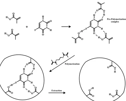
32 N N O H O H F O O H O O H N N O H O H F O O H O O O O O O H O OH N N O H O H F O O H O O H O OH O O H O O H O OH + Polymerization Extraction Pre-Polymerization complex
Figure 2.4. Schematic representation of 5-FU imprinting process.
3.3.2. Spherical nanoparticles
The imprinting effect of synthesized materials was evaluated in organic (acetonitrile) and in water (buffered water solution at pH 7.4) media. As shown in Table 2.2., in both binding media, imprinted nanosphers were able to bind much more template than the respective non imprinted ones, confirming the presence of imprinted cavities in their structure. In literature, different approaches were applied to make a quantitative
determination of the imprinting effect[67]. The imprinting efficiency α is the easiest way to
highlight the recognition properties in a MIP. In our work, α5-FU was determined as the
ratio between the amount (%) of 5-FU bound by SMIP and SNIP[68]; these values (4.6 in
CH3CN and 3.6 in water media) clearly prove the specificity of the interaction between the template and the functional groups on the polymeric nanoparticles. αU was also determined as ratio between U bound by SMIP and SNIP, respectively. The very low values (1.3 in CH3CN and 1.1 in water media) show the high chemical an spatial complementarity of SMIP binding sites toward the template, while the affinity for the analogue is very low.
[67] L. Ye, K. Mosbach. Chem. Mater. 20 (2008) 859.