Role of Ryanodine Receptors in the Assembly
of Calcium Release Units in Skeletal Muscle
Feliciano Protasi,*
§Clara Franzini-Armstrong,* and Paul D. Allen
‡§*Department of Cell and Developmental Biology, University of Pennsylvania, Philadelphia, Pennsylvania 19104-6058; ‡Department of Cardiology, Laboratory of Molecular Cardiology, Children’s Hospital, Boston, Massachusetts 02115; and §Department of Anesthesiology, Brigham and Women’s Hospital, Boston, Massachusetts 02115
Abstract.
In muscle cells, excitation–contraction (e–c) coupling is mediated by “calcium release units,” junc-tions between the sarcoplasmic reticulum (SR) and ex-terior membranes. Two proteins, which face each other, are known to functionally interact in those structures: the ryanodine receptors (RyRs), or SR calcium release channels, and the dihydropyridine receptors (DHPRs), or L-type calcium channels of exterior membranes. In skeletal muscle, DHPRs form tetrads, groups of four receptors, and tetrads are organized in arrays that face arrays of feet (or RyRs). Triadin is a protein of the SR located at the SR–exterior membrane junctions, whose role is not known. We have structurally characterized calcium release units in a skeletal muscle cell line (1B5) lacking Ry1R. Using immunohistochemistry andfreeze-fracture electron microscopy, we find that DHPR and triadin are clustered in foci in differentiating 1B5 cells. Thin section electron microscopy reveals numerous SR–exterior membrane junctions lacking foot struc-tures (dyspedic). These results suggest that components other than Ry1Rs are responsible for targeting DHPRs and triadin to junctional regions. However, DHPRs in 1B5 cells are not grouped into tetrads as in normal skel-etal muscle cells suggesting that anchoring to Ry1Rs is necessary for positioning DHPRs into ordered arrays of tetrads. This hypothesis is confirmed by finding a “restoration of tetrads” in junctional domains of sur-face membranes after transfection of 1B5 cells with cDNA encoding for Ry1R.
C
a l c i u m release units are structures that mediate one step in excitation-contraction (e–c)1 couplingin muscle cells: the rapid release of calcium from internal stores in response to depolarization of the exterior membranes (Franzini-Armstrong and Jorgensen, 1994; Flucher and Franzini-Armstrong, 1996). Calcium release units are formed by apposed junctional domains of the sar-coplasmic reticulum (SR) on one side, and of exterior membranes (plasma membrane and transverse tubules) on the other. Thin section electron microscopy reveals that the junctional regions of the SR contain foot structures disposed in ordered arrays. The feet, which span the gap
between SR and the facing junctional domains of surface membranes (Franzini-Armstrong, 1970), have been identi-fied as the cytoplasmic domains of ryanodine receptors (RyRs) (Kawamoto et al., 1986; Campbell et al., 1987; Imagawa et al., 1987; Inui et al., 1987; Block et al., 1988; Lai et al., 1988; Radermacher et al., 1994). The intramem-brane domains of RyRs form the Ca21 release channels of
the SR (for reviews see Coronado et al., 1994; Meissner, 1994; Franzini-Armstrong and Protasi, 1997; Sutko and Airey, 1997). During normal muscle contraction, the acti-vation of the SR Ca21 release channels is initiated by a
sec-ond protein, the dihydropyridine receptor (DHPR), an L-type calcium channel located in exterior membranes (Fosset et al., 1983; Pincon-Raymond et al., 1985; Rios and Brum, 1987; Tanabe et al., 1987, 1988). The direct role of DHPRs in activating muscle contraction was confirmed by lack of e–c coupling (Tanabe et al., 1988) and charge movement (Adams et al., 1990) in dysgenic skeletal mus-cle, a naturally occurring mouse mutant that fails to ex-press the a1 subunit of the DHPR (Knudson et al., 1989). In
skeletal and cardiac muscles, DHPRs are clustered in junc-tional domains of exterior membranes facing the arrays of RyRs in the junctional SR (Jorgensen et al., 1989; Flucher et al., 1990, 1994; Yuan et al., 1991; Carl et al., 1995a,b; Sun
Address all correspondence to Dr. Feliciano Protasi, Department of An-esthesia Research, Brigham and Women’s Hospital, 120 Shattuck Street, Boston, MA 02115. Tel.: (617) 278-0597. Fax: (617) 732-6927. E-mail: protasi@ zeus.bwh.harvard.edu
1. Abbreviations used in this paper: a1-DHPR, a-1 subunit of L-type volt-age-dependent Ca21 channel dihidryopyridine receptor; e-c, excitation
contraction; ES, embryonic stem; HIHS, heat-inactivated horse serum; RyR, ryanodine receptor; Ry1R, Ry2R, and Ry3R, skeletal isoform of
ry-anodine receptor, cardiac isoform of ryry-anodine receptor, and brain iso-form of ryanodine receptor; SCID, severly compromised immunodefi-cient; SR, sarcoplasmic reticulum; T, transverse.
et al., 1995; Protasi et al., 1996). In this position, DHPRs are ideally located to finely control the activation of Ca21
release from RyRs. The mechanism that allows DHPRs to control RyRs activation in skeletal muscle differs from that in cardiac and invertebrate muscles. Skeletal muscle contraction can occur in the absence of external calcium, whereas entry of extracellular calcium through the DHPRs is required for e–c coupling in cardiac and invertebrate muscles (for review see Ashley et al., 1991). The physio-logic difference in the e–c coupling mechanism between skeletal and cardiac muscles is reflected in the striking an-atomic difference in the arrangements of DHPRs within junctional domains. In skeletal muscle, DHPRs form groups of four, or “tetrads,” and tetrads are located in exact cor-respondence to the four subunits of RyRs (Franzini-Arm-strong and Nunzi, 1983; Block et al., 1988; Takekura et al., 1994; Franzini-Armstrong and Kish, 1995; Protasi et al., 1997). This arrangement suggests an e–c coupling mecha-nism of the type postulated by Schneider and Chandler (1973; for reviews see Schneider, 1981; Rios et al., 1991), in which voltage sensing by the DHPRs results in opening of RyRs by some direct molecular interaction between these two components of the junction. In cardiac muscle, on the other hand, DHPRs are clustered in proximity of RyRs, but they are not grouped into tetrads (Sun et al., 1995; Pro-tasi et al., 1996), and therefore they do not seem to be an-chored to RyRs. It is thought that in cardiac muscle Ca21
flux through the DHPRs may act as the activator of the immediately adjacent RyRs, by the “Ca21-induced Ca21
release mechanism” (Fabiato, 1983; Sham et al., 1995; San-tana et al., 1996). This mechanism does not require a DHPR–RyR molecular interaction, but would be facili-tated by immediate proximity between the two molecules. A third molecule located at the junctions between SR and surface membranes, named triadin (Brandt et al., 1990; Kim et al., 1990), has received considerable attention for its possible role in e–c coupling. It is not clear yet whether the molecule is involved in the interaction be-tween RyRs and DHPRs (Caswell et al., 1991; Fan et al., 1995a,b) or between RyRs and calsequestrin, the calcium-binding protein located in the SR lumen (Knudson et al., 1993a,b; Guo and Campbell, 1995).
A targeted mutation of the gene encoding the skeletal isoform of the ryanodine receptor (Ry1R) results in lack of
both e–c coupling and feet (hence the term “dyspedic”) in skeletal muscles (Takeshima et al., 1994; Takekura et al., 1995b; Nakai et al., 1996). We have examined the forma-tion of SR–surface membrane juncforma-tions in a dyspedic “muscle” cell line (1B5), derived from a teratoma
pro-duced by ES cells with a targeted mutation of Ry1R
(Moore et al., 1998). In these cells, as in the in vivo mus-cles from a similar targeted mutation (Takekura et al., 1995b), junctional SR vesicles dock to the surface mem-branes in the absence of feet, to form dyspedic junctions. In addition we find that in 1B5 cells triadin and DHPRs do cluster at junctional sites, despite the absence of feet, sug-gesting that RyRs are not necessary for targeting and clus-tering those molecules to junctions. However, DHPRs do not form tetrad arrays as in normal skeletal muscle cells, suggesting that some interaction with RyRs must be re-sponsible for the tetradic arrangement of DHPRs. This hy-pothesis was confirmed by restoration of tetrads after
transfection of dyspedic cells with a Ry1R cDNA. This finding provides direct structural evidence for the require-ment of RyR1 in the formation of DHPR tetrads.
Materials and Methods
The 1B5 Cell Line
The methods used to create the 1B5 cell line are described in detail else-where (Moore et al., 1998) but, briefly, it was done in four stages:
Creation of an Embryonic Stem (ES) Cell Line Possessing Two Disrupted ry1r Alleles. CCS ES cells that had successfully undergone homologous re-combination disrupting the ry1r gene at nucleotide (nt) 840 in exon 10
un-derwent a second round of selection using high G418 (3.2 mg/ml) to select for the rare homologous recombination of the disrupted allele giving ES cells where both alleles are disrupted (Mortensen et al., 1992).
Creation of Teratomas in Severely Compromised Immunodeficient (SCID) Mice. ES cells that were homozygous for the disrupted ry1r allele
were injected subcutaneously into hind quarters of SCID mice at a con-centration of 2–5 3 106 ES cells/cm3 in a volume of 1 cm3. The injections
resulted in ry1r2/ry1r2 teratocarcinomas composed of several cell types,
including 5–10% skeletal muscle. The teratomas were minced, digested with trypsin, and then the cells were plated in growth medium (DME; with 20% FBS, 100 U/ml penicillin, 100 mg/ml streptomycin, and additional 2 mM l-glutamine).
Creation of ry1r2/ry1r2 Fibroblast Cell Lines. Several primary ry1r2/
ry1r2 fibroblast clonal lines were established.
Creation of Myoblast Cell Line by myoD Infection. Several of the ry1r2/
ry1r2 fibroblast lines were infected with a retrovirus containing a
puromy-cin myoD construct. 48 h after infection, selection was started with puro-mycin (1 and 2 mg/ml) and clones were picked after 7–10 d. Puromycin-resistant clones were subcloned in growth media. One of the subclones, the 1B5 cell line, was chosen for complete characterization based on its characteristics. It exhibited contact growth inhibition at high density in growth medium and could be induced to differentiate into noncontractile multinucleated myotubes when 5% heat-inactivated horse serum (HIHS) was substituted for 20% FBS in DME medium.
Cell Culturing
The cells were grown at 378C in growth medium. After 2 d, the cells were replated on thermanox coverslips (Nunc Inc., Naperville, IL) covered with Matrigel (Collaborative Biomedical Products, Bedford, MA). The me-dium was changed every 2 d. At z70% confluence, the medium was re-placed by a low serum medium (no FBS) containing 5% HIHS to induce differentiation. The differentiation medium was changed every day and the cells were fixed 4–6 d later.
Cell Transfection
The cells were transfected during the growing phase on thermanox cover-slips when they reached z80% confluence. A transfection mixture con-taining 20 ml of lipofectamine and 3.6 mg of cDNA (in 0.1 ml of Optimem medium) per dish to be transfected was prepared 1 h before transfection and kept at room temperature. Immediately before transfection, 1 ml of Optimem medium was added to the mixture. The growth medium in the dishes was replaced with the transfection mixture and an equal amount of differentiation medium was added 6–8 h later. The transfection mixture was removed 15–20 h later and replaced with differentiation medium. The medium was changed every day and the cells were fixed 4–5 d later.
Immunohistochemistry
Cultures grown on coverslips were fixed either in methanol or acetone for 20 and 10 min, respectively at 208C, blocked in PBS containing 1% BSA and 10% goat serum for 1 h, and then incubated in primary antibodies overnight at 48C. After washing four times for 10 min in PBS/BSA, the coverslips were incubated in either FITC-conjugated (Cappel Laboratories, Malvern, PA) or CY3-conjugated (The Jackson Laboratories, Lexington, KY) goat anti–mouse secondary antibodies for 1 h at room temperature, and then washed again. The coverslips were mounted in glycerol contain-ing 0.0025% para-phenylenediamine, 0.25% 1,4 diazobicyclo 2,2,2 octane, and 5% N-propylgallate to retard photobleaching. The working dilutions and the sources of primary antibodies are listed in Table I. All antibodies
used have been previously characterized. Control slides were not incu-bated in the primary antibody. The specimens were viewed on a scanning confocal microscope (MRC-600; Biorad Laboratories, Hemel Hempstead, Hertfordshire, England).
Electron Microscopy
The cells were washed twice in PBS at 378C, fixed in glutaraldehyde, and then kept in fixative for up to 1–4 wk before further use. For thin section-ing, the cells were fixed in 3.5% glutaraldehyde in 0.1 M sodium cacody-late buffer, pH 7.2, followed by 2% OsO4 for 2 h at room temperature,
and then saturated uranyl acetate for 4 h at 608C. The samples were em-bedded in Epon 812, and the sections stained in saturated aqueous uranyl acetate followed by lead salts, both for 8 min.
For freeze fractures, the cells were fixed in glutaraldehyde as above, and infiltrated in 30% glycerol. A small piece of the coverslip was mounted with the cells facing a droplet of 30% glycerol, 20% polyvinyl al-cohol on a gold holder, and then frozen in liquid nitrogen–cooled propane (Cohen and Pumplin, 1979; Osame et al., 1981). The coverslip was flipped off to produce a fracture that followed the culture surface originally facing the coverslip. The fractured surfaces were shadowed with platinum either at 458 unidirectionally, or at 258 while rotating, and then replicated with carbon in a freeze fracture (model BFA 400; Balzers S.p.A., Milan, Italy). Sections and replicas were photographed in a EM 410 (Philips Electron Optics, Mahwah, NJ).
Measurements
The density of foci in confocal images was measured using the following procedure: (a) an area covering 100 mm2 was selected based on the
sharp-ness of the immunolabeled spots. This selection procedure accepts areas in which the optical section grazes the surface membrane and excludes ar-eas where the focal plane is below the cell surface and the spots are dif-fuse; (b) the contrast was increased to bring the spots to saturation. Under these conditions the large majority of spots are very well defined (see de-tail in Fig. 7 B); (c) the spots were counted; and (d) two or three measure-ments were performed in different areas for each cell. For more details about foci counting see Fig. 7 legend.
The width of the junctional gap between SR and surface membranes was measured along lines drawn at several random positions across the junc-tions, using a dissecting microscope fitted with an eyepiece micrometer.
The density of large membrane particles was calculated from counts of all junctional domains and several randomly selected nonjunctional do-mains in micrographs with excellent shadow quality (comparable to the one shown in Fig. 6).
The apparent diameter of intramembranous particles was measured at right angle to the direction of platinum deposition. The thickness of plati-num deposition is closely monitored using a quartz thin film monitor; thus variations in apparent particle diameter because of this factor are small. The approximate particle height was determined by measuring the length of the platinum-free “shadow.” The length of the shadow is influenced by the angle of platinum shadowing and this in turn is affected by curvature of the shadowed membrane. Variations resulting because of shadowing angle are minimized by avoiding micrographs with obviously foreshort-ened or elongated shadows. Particles clustered within junctional domains and visually identified as large were measured until a count of 100 was reached. All particles within regions of approximately equal size, but
showing no sign of large-particle clustering, were measured until the same total count was reached.
The density of large particle clusters on the surface membrane was cal-culated from counts of electron micrographs at magnifications between 20,000 and 44,500. The density of immunoreactive spots was calculated from counts of digitized images enlarged z10,000 fold.
Results
Dyspedic Calcium Release Units in 1B5 Cells
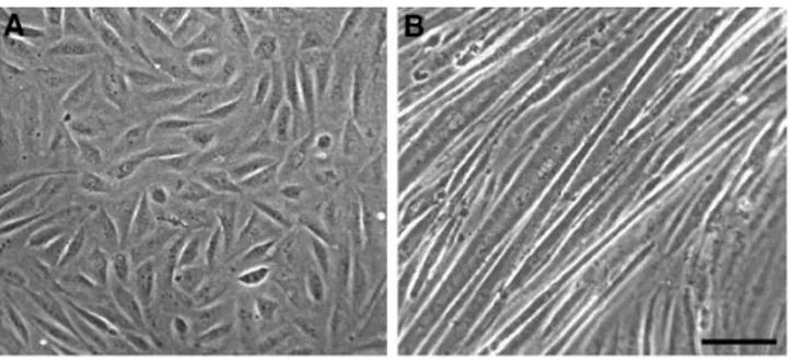
1B5 cells cultured in presence of growth factors are usually small, fusiform, and mononucleated (Fig. 1 A). After with-drawal of growth factors, most cells fuse and differentiate into aligned multinucleated myotubes, some 50 mm or more in width (Fig. 1 B).
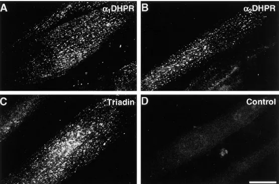
Immunohistochemistry. Differentiated 1B5 cells were immunolabeled with antibodies specific for junctional pro-teins of exterior membranes (a1 and a2 subunit of the
DHPR) and SR (triadin). 5 d after withdrawal of growth medium, about a third of the myotubes are positive for these antibodies. a1- and a2-DHPR and triadin are clus-tered in discrete foci, similar to those found in normal skeletal developing muscle and in adult skeletal and car-diac muscle (Figs. 2 and 3; compare with Yuan et al., 1991; Flucher et al., 1993b, 1994; Knudson et al., 1993a; Guo et al., 1994, 1996; Carl et al., 1995a,b). The majority of immu-nopositive spots are located on or near the surface mem-brane, on both ventral and dorsal sides of the myotubes (Fig. 2; see Fig. 3 for a through focus series). The images cannot distinguish between foci of immunofluorescence at the surface of the cell and in the cortical cytoplasm (Yuan et al., 1991). The ventral side of the cell, facing the cover-slip, is flat and thus gives the best images (Fig. 2).
Since the two antibodies used for the immunochemistry are both derived from mouse, we did not attempt double immunolabeling. However, several observations indicate that the location of DHPR- and triadin-positive foci coin-cide. First, the density of triadin-positive foci at or close to the coverslip-facing side of the cell (35.8 6 9.6/100 mm2
from 65 measurements in 25 cells, 23 confocal images, mean 6 1 SD) is very similar to the density of DHPR foci (38.8 6 7.0/100 mm2 from 102 measurements in 33 cells, 34
confocal images). Second, when the cells were colabeled with both antibodies (anti–a1-DHPR and anti-triadin) the
density of foci at or close to the coverslip-facing side of the
Table I. Antibodies Used for Immunohistochemistry
Specificity (code) Type Dilution Reference
a1-DHPR mouse 1:100 Leung et al., 1987.
(IIF7) monoclonal
a1-DHPR mouse 1:250 Morton and Froehner, 1987.
(21A6) monoclonal
a2-DHPR mouse 1:250 Morton and Froehner, 1989.
(20A) monoclonal
Triadin mouse 1:50 Caswell et al., 1991.
(GE 4.90) monoclonal
RyR mouse 1:2 Airey et al., 1990.
34C monoclonal Figure 1. (A) Undifferentiated 1B5 cells in presence of growth
medium are small, mostly fusiform, and mononucleated. (B) 6 d after withdrawal of growth medium differentiated myotubes are quite large and usually in a monolayer. Bar, 100 mm.
cells is practically identical to that obtained by single label-ing with either antibody (36.3 6 5.5/100 mm2 from 52
mea-surements in 17 cells, 16 confocal images). If the two mole-cules were not colocalized we would definitely expect a higher number of positive foci (approximately double). In addition, the intensity of foci was definitely higher in the case of cells stained with both antibodies than in cells stained in parallel with either antibody alone. A third ob-servation indicating coincidence of DHPR and triadin foci is that they are similarly disposed over the cell surface (see Figs. 7 and 8; and see below).
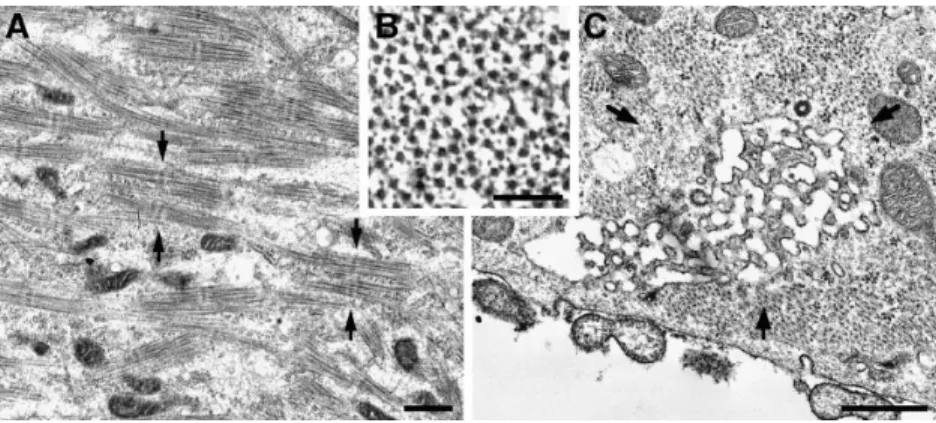
Electron Microscopy, Thin Sections. Differentiating 1B5 cells develop somewhat disordered and defective myo-fibrils containing variable amounts of myofibril compo-nents. More differentiated cells have A bands that tend to be randomly oriented and spaced, and with aligned thick filaments and a clear M line (Fig. 4 A, arrows). There is no
evidence of Z lines, and a smaller than normal comple-ment of thin filacomple-ments is present (Fig. 4 B).
Primitive transverse (T) tubules (or plasma membrane invaginations) and SR systems are present in all differenti-ating cells containing developing myofibrils, and are ab-sent in cells that do not show any evidence of myofibrillo-genesis. T tubule–like invaginations are located at the fiber periphery, mostly within a subcortical strip of cytoskeleton devoid of myofibrils. These invaginations appear most fre-quently as apparently empty, wide vesicles with a well-demarcated membrane (Fig. 5, C–E), and less frequently as labyrinthine networks resembling multiple caveolae (Fig. 4 C, arrows) (Ishikawa, 1968; Franzini-Armstrong, 1991). The continuity between these vesicles and the fiber sur-face can be directly traced in some instances (data not shown). There is no evidence of a mature T tubule net-work penetrating between the myofibrils and associating Figure 2.
Immunofluores-cence labeling of 1B5 cells 5 d after differentiation, with an-tibodies against a1-DHPR
(A), a2-DHPR (B), and
tria-din (C). No primary antibod-ies were used in the control image (D). The plane of fo-cus is close to the substrate side of the cell, which is flat. The two DHPR subunits and triadin cluster in frequent, discrete foci at or near the cell surface (see Fig. 3). About a third of the cells in differ-entiated cultures react with any of the three antibodies in the manner shown here. The densities of DHPR and tria-din foci are approximately equal (see text). Bar, 25 mm.
Figure 3. Triadin immunolocalization in a
1B5 cell at 5 d after differentiation. Through focus images at the top (A), middle (B), and bottom (C) of the cell show that the triadin foci are predominantly located on, or in prox-imity of, the plasma membrane. When the op-tical section bisects the cell, foci are at the cell edge, and few are located in the fiber interior (B, arrow). Identical results were obtained using antibodies against DHPR subunits. Bar, 10 mm.
with them. Few primitive T tubules (identifiable by their proximity to an SR element, see below) are present in the fiber interior in proximity of the Golgi regions.
Numerous small vesicles with an electron dense content are closely apposed either to the plasma membrane (Fig. 5, A–C), or to short T tubules (Fig. 5, C–E). The junctions thus formed closely resemble peripheral couplings and dy-ads (Fig. 5 F) of normal developing myotubes. We identify the vesicles as SR elements and their electron-dense con-tent presumably represents calsequestrin (Meissner, 1975). Nonjunctional or free SR is scarce. All visible T tubule profiles have at least one SR vesicle associated with them, and thus the densities of junctions along the plasmalemma
and in the subcortical and deeper regions of the cell de-pend on the extent of T tubule development. The free sur-face of the cells has numerous, short, developing T tu-bules. On the free side of the cell, 39% of the junctions are peripheral couplings, and 61% are peripherally located dy-ads (from a total of 193 junctions, from 26 cells). On the other hand, on the substrate side, T tubules are less nu-merous and peripheral couplings account for 64% of the junctions, whereas peripherally located dyads are only 36% of the total (from 152 junctions, 25 cells). Junctions within the cell interior are rare (43, or 11%, out of a total of 388 junctions in all locations), and they are usually in small clusters within some, but not all, cell profiles. The lo-Figure 4. (A) The cytoplasm of differentiated
1B5 cells 5 d after differentiation contains bundles of thick filaments arranged in com-plete A bands with evident M lines (between
arrows), but Z lines are either missing or
in-complete. (B) Incomplete sets of thin fila-ments overlap with the thick filafila-ments in this cross section of a myofibril. (C) A labyrin-thine membrane network (arrows) is an ini-tial step in the development of a T tubule sys-tem. Such networks are rarely seen. Other primitive T tubules appear as short, wide in-vaginations (see Fig. 5). Bars: (A and C) 0.5
mm; (B) 0.1 mm.
Figure 5. Dyspedic junctions in 1B5 cells at 5 d after differentiation (A–E), compared to a normal junction from in vivo mouse
dia-phragm (F). Junctions between SR and the plasma membrane (peripheral couplings; A and B), and junctions between SR and primitive T tubules (dyads; E) are present. The SR profile in C makes contact with the plasma membrane on one side, and a T tubule on the other. The SR profiles have a dense content, whereas the surface invaginations have an empty lumen. In dyspedic junctions (A–E), the junc-tional gap is too narrow to accommodate feet, so the small densities visible in the junction (A and D, arrowheads) are due to proteins other than RyRs. Note larger gap occupied by evenly spaced feet (F, arrows) in the normal junction. Bar, 0.1 mm.
cation of the junctional SR vesicles at or close to the fiber periphery agrees well with the location of the DHPRs and triadin foci seen by immunohistochemistry, which are at or near the surface of the cell (Fig. 3).
All junctions in 1B5 cells are dyspedic, i.e., “feet” are absent from the junctional gap between SR and exterior membranes, and the gap width is narrow (Fig. 5, A–E). On the average, the junctional gap is 6.5 6 2.6 nm (mean 6 1 SD, n 5 52 junctions, 37 peripheral couplings and 15 dyads, 255 measurements) and thus too small to accommodate cytoplasmic domains of RyRs or feet, which are z12-nm wide (Radermacher et al., 1994). The irregularly disposed strands that usually occupy the gap in dyspedic myotubes (Fig. 5, A and D, arrowheads) are clearly composed of a protein different from RyR. The gap in feet-containing junctions from normal myotubes is wider (Fig. 5 F, from a normal in vivo myotube). Gap widths of 10–13 nm have been previously measured in junctions from mouse myo-tubes developing in vivo (Takekura et al., 1995b) and in
vitro (Nakai et al., 1997) fixed and stained as in this study. Junctions with a 10–13-nm gap were not detected in 1B5 cells.
Electron Microscopy, Freeze Fracture. In freeze fracture replicas of undifferentiated cultures the cytoplasmic leaf-let of the plasmalemma is smooth and it shows intramem-brane particles of variable sizes, without any sign of clus-tering (Fig. 6 A).
The plasma membranes of large myotubes in differenti-ated cultures have dimples marking the mouths of caveolae and T tubules (Fig. 6 B, arrows) and plasma membrane do-mains that contain clusters of large, tall particles (Fig. 6 B,
semicircles). Small- and medium-sized particles are largely
excluded from these clusters (see details in Fig. 6, C–E). The apparent diameter and height of large particles in plasma membrane domains is 9.9 6 1.7 nm and 10.9 6 1.5 nm, respectively (mean 6 1 SD; n 5 100, 4 cells, 4 freeze fractures). The apparent diameter and height of particles, including large ones, in patches of membrane adjacent to Figure 6. Internal leaflet of the plasma
mem-brane in freeze-fracture replicas of undiffer-entiated and differundiffer-entiated cells. (A) The membrane of undifferentiated cells in the presence of growth factor is characterized by intramembrane particles that are randomly distributed and that do not form any particu-lar grouping. (B) After withdrawal of growth factors 5 d after differentiation z35% of the cells contain clusters of intramembrane parti-cles (plasma membrane domains; semicirparti-cles) that are taller and larger than the average. (C–E) Details of plasma membrane domains, 5–6 d after differentiation no order is detect-able in the distribution of large particles within these domains. Bars: (A and B) 0.2
the cluster-containing domains is 6.8 6 1.5 nm (n 5 100) and 6.5 6 1.8 nm (n 5 100), respectively. Differences be-tween diameters and heights in the two groups are ex-tremely significant (Student’s t test; P , 0.0001 in both cases). Note that the large-clustered particles are also rela-tively tall; their height to diameter ratio is 1:10, whereas the ratio in random particles is 0.95. The average density of large particles in junctional plasma membrane domains is 1,077 6 228/mm2 (n 5 63; mean 6 1 SD; n 5 number of
domains), whereas the average density of large particles in nonjunctional regions is 104 6 57/mm2 (n 5 82). The
dif-ferences between the two data are large and extremely sig-nificant (Student’s t test P , 0.0001) confirming the visual observation of clustering.
In D5–6 cultures 35% of the cells (from a total of 334 cells in 7 cultures, 11 replicas) contain plasma membrane domains with clusters of large particles (Fig. 6, B–E). The surface of the rest of the cells resemble that of cells fixed in the presence of growth medium (Fig. 6 A). We could
not determine whether the presence of particle clusters is limited to multinucleated cells.
Identification of Clusters of Particles with DHPR Foci and Clues to Their Junctional Location. Foci of DHPRs de-tected by immunolabeling and clusters of large intramem-brane particles are present at comparable densities. The two densities, related to the apparent surface area, are 38.8
6 7.0/100 mm2 (from 102 measurements in 33 cells, 34
con-focal images, detected by the a1 antibody) and 26.5 6 14.0/
100 mm2 (38 measurements in 17 cells, 38 electron
micro-graphs). We expect that the immunolabeled foci at the fiber periphery include both peripheral couplings and pe-ripherally located dyads, whereas clusters of particles de-tected in freeze-fracture correspond exclusively to periph-eral couplings. Thus the density of foci is expected to be larger than the density of clusters. The measured ratio of fluorescent foci to particle cluster densities on the sub-strate side of the cell (1.46 from the above data) is slightly lower than the ratio of total peripheral junctions (periph-Figure 7. Analogies in the distribution of triadin
and DHPR foci, and of particles clusters. (A–C) Cells immunolabeled respectively with anti-tria-din, anti–a1-DHPR, and both antibodies. The
density of clusters in all three samples is very similar (see Results) suggesting a colocalization of the two structures. Note also the tendency for an alignment of the clusters into longitudinal lines, along the long axis of the cell. The foci den-sity was measured using the procedure detailed in the Materials and Methods section. An area prepared for counting by enlarging five times and increasing the contrast is shown as a detail in
B. The 25-mm2 area contains 12 foci. Counting
was done in portions of the images where the fo-cal plane grazes the cell surface (e.g., Figs. 7, A and C, middle areas), whereas areas in which the optical section either includes the underlying cy-toplasm, or moves too far from the cell surface (e.g., Fig. 7, B, top/left region; and C, bottom) were avoided. (D) Freeze-fracture. The locations of particles clusters in the plasma membrane are marked by semicircles. The frequency of parti-cles clusters is similar to the density of foci posi-tive for triadin and a1-DHPR and they are
simi-larly aligned in longitudinal rows in some cells (arrows). The long axis of the cell is aligned ver-tically in A–C and diagonally in D, as is the ori-entation of foci and particle cluster lines. Bars: (A–C) 20 mm; (B) 0.5 mm.
eral couplings plus cortical dyads) to peripheral couplings (1.56 from counts in thin sections). The most probable cause for this minor discrepancy is that some of the fluo-rescent spots are likely to represent more than a single cluster of particles. Indeed, two or three peaks of density are often seen within some of the larger spots. Equal den-sities of DHPR foci and particle clusters suggests identity of the two structures. This is further supported by the fol-lowing observations: (a) clusters of particles and foci of DHPR immunolabeling are present on many but not all differentiating cells; (b) clusters of particles and foci of DHPRs are randomly distributed in most cells; (c) in some cells, foci that immunostain positive for DHPR anti-bodies are strikingly arranged in longitudinal rows (Figs. 7
B, and 8 B). A similar disposition is also observed for
plasma membrane domains containing clusters of particles (Fig. 7 D, arrows and semicircles, and 8 C); and (d) some DHPR foci and particle clusters are so closely spaced that they almost fuse with each other (compare Fig. 8 B with C). The above analysis suggests that the large, clustered par-ticles in the surface of 1B5 cells are DHPRs. Indeed, the large particles in 1B5 clusters are identical in size with the particles representing DHPRs in the tetrads of BC3H1
cells (Protasi et al., 1997), and are also quite similar to the slightly smaller particles representing DHPRs in cardiac muscle. The apparent diameter and height of the large, clustered particles in 1B5 cells is 9.9 and 10.9 nm, respec-tively (see above); the large particles forming tetrads in BC3H1 cells are 9.7 6 1.2-nm wide and 10.6 6 1.8-nm tall
(from 100 particles, 4 cells, 4 freeze-fractures); the parti-cles in cardiac muscle are 8.5-nm wide and 9.6-nm tall (Sun et al., 1995).
Foci of triadin detected by immunolabeling are also lo-cated in prominent longitudinal rows (Figs. 7 A, and 8 A) and have density equal to DHPR foci (see above). Thus, the three detectable components of calcium release units of 1B5 cells (DHPR and triadin foci, detected by immuno-labeling; and large particles, detected by freeze-fracture)
appear to be colocalized. Since triadin is a junctional SR protein, this colocalization occurs at the junctions between SR and exterior membranes.
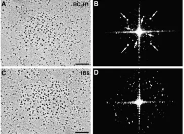
The Disposition of Skeletal DHPRs in 1B5 Cells Resem-bles That of Cardiac, Not Skeletal Muscle. Optical diffraction confirms the random disposition of DHPRs in 1B5 cells. Fig. 9 compares images from rotary shadowed replicas of
A an array of tetrads in a peripheral coupling from the
BC3H1 cell line, and C, a group of DHPRs from a 1B5 cell.
The optical transformation of the tetrad array (Fig. 9 B) shows evidence for two tetragonal arrangements indexing on the spacing between particles within the tetrad (Fig. 9
B, large arrows) and between the centers of tetrads (Fig. 9 B, small arrows; see Protasi et al., 1997). Note that tetrad
arrays in BC3H1 cells have identical parameters to those
of other skeletal muscle both in vivo and in vitro (Fran-zini-Armstrong and Kish, 1995). The optical transforma-tion of the 1B5 cluster (Fig. 9 D) shows only noise derived from the random arrangement of scattering units. This analysis confirms the visual impression that DHPRs in dyspedic junctions of 1B5 cells are randomly disposed and do not form tetradic arrays. The random disposition of DHPRs in peripheral junctional domains resembles that observed in cardiac muscle myocytes (Sun et al., 1995; Pro-tasi et al., 1996; see Fig. 12 for a diagram).
Restoration of Tetrads After Transfection of 1B5 Cells
with cDNA Encoding for the Ry1R
Immunohistochemistry. 1B5 cells transfected with the cDNA for Ry1R were immunolabeled with Ry1R-specific
anti-body. 4–5 d after withdrawal of growth medium (D4–5) Figure 8. Triadin foci (A), DHPR foci (B), and clusters of large
particles (C) are prominently aligned in a preferred orientation on the surface of some cells. The long axis of the cell is aligned along the diagonal of the images, as is the orientation of foci and particle cluster lines. Bars: (A and B) 5 mm; (C) 0.2 mm.
Figure 9. Rotary shadowing images of large particle clusters in
BC3H1 and 1B5 cells (A and C), and their respective optical
dif-fraction patterns (B and D). The tetradic arrangement of DHPRs in BC3H1 cells gives rise to a pattern (B) showing two orthogonal
arrays rotated relative to each other that represent, respectively, the spacing between tetrads in the array (small arrows) and the spacing between particles within the tetrads (large arrows; see Protasi et al., 1997 for details). The disordered arrangement of particles in the clusters of 1B5 cells results in a diffraction pattern (D) without any special features. Bars, 0.1 mm.
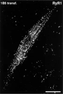
z10% myotubes are positive for Ry1R. These Ry1R
mole-cules are tightly clustered in discrete foci, similar to those found in normal developing skeletal muscle (Fig. 10; com-pare with Flucher et al., 1993b; Flucher et al., 1994). Simi-lar to DHPR and triadin foci, Ry1R immunopositive spots are mostly located on or near the surface membrane, on both ventral and dorsal sides of the myotubes. Nontrans-fected cells incubated with the same primary and second-ary antibodies and transfected cells incubated with only the secondary antibody are negative.
Freeze Fracture. Freeze fracture of transfected cells shows restoration of tetrad arrays in the surface membrane of
z10% 1B5 cells. Fig. 11, A–C shows clusters of large
parti-cles (DHPRs) in the surface membrane of a dyspedic 1B5 cell (Fig. 11 A; see also Fig. 6), of a normal developing my-otube (mouse) in vivo (Fig. 11 B), and of a transfected 1B5 cell (Fig. 11 C). In 1B5 cells, the particles have a random arrangement, whereas in normal myotubes and in trans-fected cells the particles are clearly arranged in tetrads (Fig. 11, B and C, arrows), and tetrads are part of an array. The forming arrays of tetrads in transfected 1B5 cells are always located on a slightly raised membrane mound, sug-gesting that they are located at sites where the SR is closely opposed to the surface membrane, forming a pe-ripheral coupling. Tetrads, in the domains of both normal skeletal muscle and transfected 1B5 cells, are often incom-plete, that is they miss one or more components (see also Takekura et al., 1994; Franzini-Armstrong and Kish, 1995; Protasi et al., 1997 for similar observations in a variety of muscle fibers and in the BC3H1 cell line). Examples of
Figure 10. Immunolabeling with anti-Ry1R antibodies in
trans-fected 1B5 cells (4 d after differentiation). The plane of focus is close to the substrate side of the cell, which is flat. RyRs, similar to DHPRs and triadin, cluster in frequent, discrete foci at or near the cell surface (see Figs. 2, 3, 7, and 8). Bar, 25 mm.
Figure 11. Restoration of
tetrads in transfected 1B5 cells. (A) Domain of large particles in a dyspedic 1B5 cell; the particles, represent-ing DHPRs, are randomly disposed (see also Figs. 6 and 9). (B) Array of tetrads in normal skeletal muscle (E18 mouse leg muscle); the large particles form tetrads (groups of four DHPRs;
ar-rows), and tetrads are part of
an ordered array. Several tet-rads are incomplete (formed by only two or three ele-ments). (C) Array of tetrads in a transfected 1B5 cell (4 d after differentiation); DH-PRs are clearly arranged in tetrads (arrows), as in pe-ripheral couplings of normal skeletal muscle. Details at the bottom of the figure show two sets of tetrads, complete (top row) and in-complete (bottom row). Note that: (a) complete tetrads can be slightly distorted; (b) the apparently missing compo-nents of incomplete tetrads may actually be elements (DHPRs) that broke during fracturing, leaving small stumps (see bottom left); and (c) some incomplete tetrads seem to lack one or more components (bottom right). Bars: (A–C) 0.1 mm; (inset) 0.05 mm.
complete tetrads and tetrads with one missing element from transfected 1B5 cells are shown at the bottom right of Fig. 11. Complete tetrads, formed by four large and tall particles disposed at the corners of a square, are shown in Fig. 11, bottom right, top row. A minor distortion of the precise square arrangement is sometimes introduced by the fracturing process. Incomplete tetrads with three com-ponents are shown in the bottom row. These are unequiv-ocally identified as tetrads with one missing particle, be-cause the three particles are of the same size, bebe-cause they subtend a 908 angle, and because they are part of a “do-main of large particles.” Tetrads may be incomplete for different reasons: (a) one or more particle may break dur-ing fracturdur-ing and appear as a small stump (examples at left in the bottom row), or (b) components may be missing from the array, as shown in the last two examples at right in the bottom row (Protasi et al., 1997). Both situations are found in tetrads of normal myotubes developing in vivo and in vitro (see references above).
Discussion
During normal differentiation of cardiac and skeletal mus-cle, RyRs, DHPRs, and triadin are always coclustered within calcium release units, giving the impression that these proteins and their interactions play a major role in the formation of the junctions between the SR and exte-rior membranes (Yuan et al., 1991; Flucher et al., 1993a,b, 1994; Protasi et al., 1996; for reviews see strong and Jorgensen, 1994; Flucher and Franzini-Arm-strong, 1996). However, observations of various model systems reveal that docking of the SR to exterior mem-branes may occur in the absence of either DHPRs or
RyRs (Franzini-Armstrong et al., 1991; Flucher et al., 1993b; Takekura et al., 1995b; Takeshima et al., 1995; Powell et al., 1996; Takeshima et al., 1997). Junctions are also formed in the muscle fibers of Ry1R/Ry3R double
knockout mice (Ikemoto et al., 1997), showing that the presence of a small amount of Ry3R is not needed for SR/
exterior membrane docking. In addition, in normal devel-oping cardiac muscle, docking of the SR to the surface membrane precedes the localization of DHPRs and RyRs at the junctions (Protasi et al., 1996). Clustering of SR junctional proteins at calcium release units is also indepen-dent of the presence of DHPRs, since RyRs, triadin, and calsequestrin are present at SR–surface membrane junc-tions in dysgenic myotubes lacking a1-DHPR
(Franzini-Armstrong et al., 1991; Flucher et al., 1993b).
The findings in this study of dyspedic junctions add three further clues to the formation of calcium release units. The first is that DHPRs cluster at the junctions in the absence of RyRs. It must be assumed that other pro-teins of the junctional SR, perhaps even the same compo-nents that are responsible for SR–surface docking, have the role of holding DHPRs at the junctional sites. This is confirmed by the fact that CHO cells, which are of epithe-lial origin, seem to lack the component(s) needed for both docking of ER to the surface membranes and for extensive DHPR clustering, even when expression of skeletal RyR and a cardiac–skeletal DHPR chimera is induced in these cells (Takekura et al., 1995a).
A second result is that the presence of RyRs is not needed for the clustering of triadin in calcium release units. Two quite distinct roles have been proposed for tria-din. On one hand, triadin is thought to reach out into the junctional gap between SR and surface membranes and ei-ther facilitate or directly participate in the joining of DHPRs to RyRs in skeletal muscle (Caswell et al., 1991; Fan et al., 1995a,b). On the other hand, the same molecule is de-scribed as being mostly located intraluminally, where it may help to link calsequestrin to the junctional SR mem-brane (Knudson et al., 1993a,b; Guo and Campbell, 1995). Our finding that triadin is present at the dyspedic junc-tions in the absence of RyRs does not help to solve the question of triadin function.
A third, and most important finding is that DHPRs do not associate into tetrads in the absence of Ry1R (Figs. 11
A, and 12 A) and that tetrads are restored after “rescue”
of the dyspedic phenotype by transfection of dyspedic cells with the Ry1R cDNA (Figs. 11 C, and 12 B). These results provide direct confirmation of two current hypotheses on junctional architecture. One is that DHPRs are linked to RyRs in skeletal muscle and this linkage is responsible for the formation of tetrads (Block et al., 1988; Takekura et al., 1994; Protasi et al., 1997), and the other is that the random disposition of cardiac muscle DHPRs within junctional do-mains is, on the contrary, because of a lack of a link to the cardiac RyRs (Sun et al., 1995; see Fig. 12 C).
Present and past observations lead to the conclusion that the targeting of RyRs and DHPRs, as well as triadin and calsequestrin, to the junctional areas may be indepen-dent of each other and probably depend on other junc-tional components of a still unknown identity. However, one finding during the development of junctions in avian cardiac muscle seems at odds with this conclusion. Differ-Figure 12. Diagrams showing the relation between RyRs and
DHPRs in peripheral couplings of dyspedic 1B5 cells (A), trans-fected 1B5 cells (B), and cardiac muscle (C). RyRs (gray) form ordered arrays in the junctional SR of both skeletal and cardiac muscle. The array of feet are drawn as defined in Franzini-Arm-strong and Kish (1995). Note that the array of feet in cardiac mus-cle is shown to be identical to that in skeletal musmus-cle, although this is not known in detail. (A) Dyspedic 1B5 cells do not have feet; skeletal DHPRs (black) are not capable of forming tetrads on their own, in the absence of RyRs. (B) In Ry1R-transfected
1B5 cells, as in normal skeletal muscle, DHPRs form tetrads that are located in exact correspondence of subunits of alternate feet, suggesting a link between the molecules. In B, tetrads have been drawn as they appear to be in fracture replicas; often incomplete but always associated with subunit of alternate feet. (C) In cardiac muscle, on the other hand, DHPRs do not form tetrads and their position relative to the feet subunits is variable, and thus a RyR– DHPR link is less likely.
entiation of the calcium release units in this muscle in-volves an initial docking of the SR to the surface, and then a coordinated assembly of groups of feet and DHPRs, which do not fill the entire docked area, but cluster to-gether on one side (Protasi et al., 1996).
A previous study of dyspedic skeletal muscle in vivo agrees with the results presented here in showing forma-tion of juncforma-tions that lack feet, but differs because clusters of large particles (DHPRs) were not detected in the plasma membrane of dyspedic muscle in mouse (Takekura et al., 1995b). The reason for this difference is not clear, since mRNA for the DHPR is present in the in vivo dys-pedic muscle (Takeshima et al., 1994), and the protein is
expressed (although in reduced amounts; z50% [Buck
et al., 1997]). Also, charge movement is detected in pri-mary cultures of dyspedic muscles (Nakai et al., 1996).
Muscle fibers of normal mouse express low but detect-able levels of Ry3R (Giannini et al., 1995). A few
group-ings of feet, presumably representing Ry3R, have been de-tected in dyspedic myotubes developing in vivo (Takekura et al., 1995b), confirming functional evidence for caffeine-sensitive calcium release in primary dyspedic myotubes (Takeshima et al., 1995). In 1B5 cells, on the other hand,
Ry3R cannot be detected by biochemical approaches
(Moore et al., 1998), and we have not found evidence for feet in thin sections from these cells suggesting that Ry3R
is not expressed in these cells at all.
1B5 and BC3H1, two cell lines that express skeletal
mus-cle–specific proteins of the junctional SR have three inter-esting structural defects in common: the cells either lack Z lines or have poorly developed ones; the nonjunctional sarcoplasmic reticulum is very poorly represented; periph-eral couplings and junctions between SR and primitive T tubules on the other hand are relatively frequent. Since the nonjunctional SR develops early and is associated with the Z lines of myofibrils during normal skeletal muscle dif-ferentiation (Flucher et al., 1991, 1992, 1993a), the first two defects listed above may be causally linked.
The findings of this work highlight three important structural and developmental details of Ca21 release units
of muscle cells: the requirement of Ry1R for the formation of tetrads, a structural feature of Ca21 release units that is
unique to skeletal muscle; the independence from RyRs of the association of triadin and DHPRs with the forming junctions; the independence from RyR of the initial dock-ing of SR to exterior membranes. The restoration of tet-rads in transfected 1B5 cells gives the ultimate demonstra-tion that the DHPRs arrangement in tetrads is strictly dependent on their interaction, direct or not, with Ry1R subunits.
We thank Drs. A.H. Caswell, K.P. Campbell, J.A. Airey, and J.L. Sutko for their generous gift of antibodies. We thank N. Glaser (University of Pennsylvania, Philadelphia, PA) for expert assistance and R. Moore (Uni-versity of California, Davis, CA) for helpful suggestions on transfection procedures.
This work was supported by National Institutes of Health grants R01 HL48093 to the Pennsylvania Muscle Institute (to C. Franzini-Arm-strong), and R01AR43140 (to P.D. Allen), and by a grant from The Mus-cular Dystrophy Association (to P.D. Allen).
Received for publication 5 May 1997 and in revised form 21 November 1997.
References
Adams, B.A., T. Tanabe, A. Mikami, S. Numa, and K.G. Beam. 1990. In-tramembrane charge movement restored in dysgenic skeletal muscle by in-jection of dihydropyridine receptor cDNAs. Nature. 346:569–572.
Airey, J.A., C.F. Beck, K. Murakami, S.J. Tanksley, T.J. Deerinck, M. Ellsiman, and J.L. Sutko. 1990. Identification and localization of two triad junction foot protein isoforms in mature avian fast twitch skeletal muscle. J. Biol. Chem. 265:14187–14194.
Ashely, C.C., I.P. Mulligan, and T.J. Lea. 1991. Ca21 and activation mechanisms in skeletal muscle. Rev. Biophys. 24:1–73.
Block, B.A., T. Imagawa, K.P. Campbell, and C. Franzini-Armstrong. 1988. Structural evidence for direct interaction between the molecular compo-nents of the transverse tubule/sarcoplasmic reticulum junction in skeletal muscle. J. Cell Biol. 107:2587–2600.
Brandt, N.R., A.H. Caswell, S.-N. Wen, and J.A. Talvenheimo. 1990. Molecular interactions of the junctional foot protein and dihydropyridine receptor in skeletal muscle triads. J. Membr. Biol. 113:237–251.
Buck, E.D., A.H. Nguyen, I.N. Pessah, and P.D. Allen. 1997. Dyspedic mouse skeletal muscle expresses major elements of the triadic junction but lacks ry-anodine receptor protein and function. J. Biol. Chem. 272:7360–7367. Campbell, K.P., C.M. Knudson, T. Imagawa, A.T. Leung, J.L. Sutko, S.D. Kahl,
C. Reynolds-Raab, and L. Madson. 1987. Identification and characterization of the high affinity [3H]ryanodine receptor of the junctional sarcoplasmic
reticulum Ca21 release channel. J. Biol. Chem. 262:6460–6463.
Carl, S.L, K. Felix, A.H. Caswell, N.R. Brandt, W.J. Ball, P.L. Vaghy, G. Meiss-ner, and D.G. Ferguson. 1995a. Immunolocalization of sarcolemmal dihy-dropyridine receptor and sarcoplasmic reticular triadin and ryanodine recep-tor in rabbit ventricle and atrium. J. Cell Biol. 129:672–682.
Carl, S.L, K. Felix, A.H. Caswell, N.R. Brandt, J.P. Brunschwig, G. Meissner, and D.G. Ferguson. 1995b. Immunolocalization of triadin, DHP receptors, and ryanodine receptors in adult and developing skeletal muscle of rats. Muscle Nerve. 18:1232–1243.
Caswell, A.H., N.R. Brandt, J.P. Brunschwig, and S. Purkerson. 1991. Localiza-tion and partial characterizaLocaliza-tion of the oligomeric disulfide-linked molecular weight 95,000 protein (triadin) which binds the ryanodine and dihydropyri-dine receptors in skeletal muscle triadic vesicles. Biochemistry. 30:7507– 7513.
Cohen, S.A., and D.W. Pumplin. 1979. Clusters of intramembranous particles associated with binding sites for alpha-bungarotoxin in cultured chick myo-tubes. J. Cell Biol. 82:494–516.
Coronado, R., J. Morrissette, M. Sukhareva, and D.M. Vaughan. 1994. Struc-ture and function of ryanodine receptors. Am. J. Physiol. 266:1485–1491. Fabiato, A. 1983. Calcium-induced release of calcium from cardiac
sarcoplas-mic reticulum. Am. J. Physiol. 245:1–14.
Fan, H., N.R. Brandt, M. Peng, A. Schwartz, and A.H. Caswell. 1995a. Binding sites of monoclonal antibodies and dihydropyridine receptor a1 subunit
cyto-plasmic II-III loop on skeletal muscle triadin fusion peptides. Biochemistry. 34:14893–14901.
Fan, H., N.R. Brandt, and A.H. Caswell. 1995b. Disulfide bonds, N-glycosyla-tion and transmembrane topology of skeletal muscle triadin. Biochemistry. 34:14902–14908.
Flucher, B.E., and C. Franzini-Armstrong. 1996. Formation of junctions in-volved in excitation-contraction coupling in skeletal and cardiac muscle. Proc. Natl. Acad. Sci. USA. 93:8101–8106.
Flucher, B.E., M.E. Morton, S.C. Froehner, and M.P. Daniels. 1990. Localiza-tion of the a1 and a2 subunits of the dihydropyridine receptor and ankyrin in
skeletal muscle triads. Neuron. 5:339–351.
Flucher, B.E., M. Terasaki, H. Chin, T. Beeler, and M.P. Daniels. 1991. Biogen-esis of transverse tubules in skeletal muscle in vitro. Dev. Biol. 145:77–90. Flucher, B.E., J.L. Phillips, J.A. Powell, S.B. Andrews, and M.P. Daniels. 1992.
Coordinated development of myofibrils, sarcoplasmic reticulum and trans-verse tubules in normal and dysgenic mouse skeletal muscle, in vivo and in vitro. Dev. Biol. 150:266–280.
Flucher, B.E., H. Takekura, and C. Franzini-Armstrong. 1993a. Development of the excitation-contraction coupling apparatus in skeletal muscle: associa-tion of sarcoplasmic reticulum and transverse tubules with myofibrils. Dev. Biol. 160:135–147.
Flucher, B.E., S.B. Andrews, S. Fleisher, A.R. Marks, A.H. Caswell, and J.A. Powell. 1993b. Triad formation: Organization and function of the sarcoplas-mic reticulum calcium release channel and triadin in normal and dysgenic muscle in vitro. J. Cell Biol. 123:1161–1174.
Flucher, B.E., S.B. Andrews, and M.P. Daniels. 1994. Molecular organization of transverse tubule/sarcoplasmic reticulum junctions during development of excitation-contraction coupling in skeletal muscle. Mol. Biol. Cell. 5:1105– 1118.
Fosset, M., E. Jaimovich, E. Delpont, and M. Lazdunski. 1983. [3H]nitrendipine
receptors in skeletal muscle. J. Biol. Chem. 258:6086–6092.
Franzini-Armstrong, C. 1970. Studies of the triad. J. Cell Biol. 47:488–499. Franzini-Armstrong, C. 1991. Simultaneous maturation of transverse tubules
and sarcoplasmic reticulum during muscle differentiation in the mouse. Dev. Biol. 146:353–363.
Franzini-Armstrong, C., and G. Nunzi. 1983. Junctional feet and particles in the triads of fast-twitch muscle fibers. J. Muscle Res. Cell Motil. 4:233–252. Franzini-Armstrong, C., and A.O. Jorgensen. 1994. Structure and development
of e-c coupling units in skeletal muscle. Annu. Rev. Physiol. 56:509–534. Franzini-Armstrong, C., and J.W. Kish. 1995. Alternate disposition of tetrads in
peripheral couplings of skeletal muscle. J. Muscle Res. Cell Motil. 16:319–324. Franzini-Armstrong, C., and F. Protasi. 1997. The ryanodine receptor of
stri-ated muscles, a complex capable of multiple interactions. Physiol. Rev. 77: 699–729.
Franzini-Armstrong, C., M. Pincon-Raymond, and F. Rieger. 1991. Muscle fi-bers from dysgenic mouse in vivo lack a surface component of peripheral couplings. Dev. Biol. 146:364–376.
Giannini, G., A. Conti, S. Mammarella, M. Scrobogna, and V. Sorrentino. 1995. The ryanodine receptor/calcium channel genes are widely and differentially expressed in murine brain and peripheral tissues. J. Cell Biol. 128:893–904. Guo, W., and K.P. Campbell. 1995. Association of triadin with the ryanodine
receptor and calsequestrin in the lumen of sarcoplasmic reticulum. J. Biol. Chem. 270:9027–9030.
Guo, W., A.O. Jorgensen, K.P. Campbell. 1994. Characterization and ultra-structural characterization of a novel 90kDa protein unique to skeletal mus-cle junctional sarcoplasmic reticulum. J. Biol. Chem. 269:28359–28365. Guo, W., A.O. Jorgensen, L.R. Jones, and K.P. Campbell. 1996. Biochemical
characterization and molecular cloning of cardiac triadin. J. Biol. Chem. 271: 458–465.
Ikemoto, T., S. Komazaki, H. Takeshima, M. Nishi, T. Noda, M. Iino, and M. Endo. 1997. Functional and morphological features of skeletal muscle from mutant mice lacking both type 1 and type 3 ryanodine receptors. J. Physiol. (Lond). 501:305–312.
Imagawa, T., J.S. Smith, R. Coronado, and K.P. Campbell. 1987. Purified ryan-odine receptor from skeletal muscle sarcoplasmic reticulum is the Ca21 per-meable pore of the calcium release channel. J. Biol. Chem. 262:16636–16643. Inui, M., A. Saito, and S. Fleischer. 1987. Purification of the ryanodine receptor and identity with feet structures of junctional terminal cisternae of sarcoplas-mic reticulum from fast skeletal muscle. J. Biol. Chem. 262:1740–1747. Ishikawa, H. 1968. Formation of elaborate networks of T-systems tubules in
cultured skeletal muscle with special reference to the T-system formation. J. Cell Biol. 38:51–66.
Jorgensen, A.O., A.C-Y. Shen, W. Arnold, A.T. Leung, and K.P. Campbell. 1989. Subcellular distribution of the 1,4-dihydropyridine receptor in rabbit skeletal muscle in situ: an immunofluorescence and immunocolloidal gold-labeling study. J. Cell Biol. 109:135–147.
Kawamoto, R.M., J.P. Brunschwig, K.C. Kim, and A.H. Caswell. 1986. Isola-tion, characterizaIsola-tion, and localization of the spanning protein from skeletal muscle triads. J. Cell. Biol. 103:1405–1414.
Kim, K.C., A.H. Caswell, J.A. Talvenheimo, and N.R. Brandt. 1990. Isolation of a terminal cisterna protein which may link the dihydropyridine receptor to the junctional foot protein in skeletal muscle. Biochemistry. 29:9281–9289. Knudson, C.M., N. Chaudari, A.H. Sharp, J.A. Powell, K.G. Beam, and K.P. Campbell. 1989. Specific absence of the a1 subunit of the dihydropyridine
re-ceptor in mice with muscular dysgenesis. J. Biol. Chem. 264:1345–1348. Knudson, C.M., K.K. Stang, A.O. Jorgensen, and K.P. Campbell. 1993a.
Bio-chemical characterization and ultrastructural localization of a major junc-tional sarcoplasmic reticulum glycoprotein (triadin). J. Biol. Chem. 268: 12637–12645.
Knudson, C.M., K.K. Stang, C.R. Moomaw, C.A. Slaughter, and K.P. Camp-bell. 1993b. Primary structure and topological analysis of a skeletal muscle-specific junctional sarcoplasmic reticulum glycoprotein (triadin). J. Biol. Chem. 268:12646–12654.
Lai, F.A., H.P. Erickson, E. Rousseau, Q.Y. Liu, and G. Meissner. 1988. Purifi-cation and reconstitution of the calcium release channel from skeletal mus-cle. Nature. 331:315–319.
Leung, A.T., T. Imagawa, and K.P. Campbell. 1987. Structural characterization of the 1,4-dihydropyridine receptor of the voltage-dependent Ca21 channel from rabbit skeletal muscle. Evidence for two distinct high molecular weight subunits. J. Biol. Chem. 262:7943–7946.
Meissner, G. 1975. Isolation and characterization of two types of sarcoplasmic reticulum vesicles. Biochim. Biophys. Acta. 389:51–68.
Meissner, G. 1994. Ryanodine receptor/Ca21 release channels and their regula-tion by endogenous effectors. Annu. Rev. Physiol. 56:485–508.
Moore, R.A., H. Nguyen, J. Galceran, I.N. Pessah, and P.D. Allen. 1998. A transgenic myogenic cell line lacking ryanodine receptor protein for homol-ogous expression studies: Reconstitution of Ry1R protein and function. J.
Cell Biol. 140:843–851.
Mortensen, R.M., D.A. Conner, S. Chao, A.A. Geisterfer-Lowrance, and J.G. Seidman. 1992. Production of homozygous mutant ES cells with a single tar-geting construct. Mol. Cell. Biol. 12:2391–2395.
Morton, M.E., and S.C. Froehner. 1987. Monoclonal antibody identifies a 200-kDa subunit of the dihydropyridine-sensitive calcium channel. J. Biol. Chem. 262:11904–11907.
Morton, M.E., and S.C. Froehner. 1989. The a1 and a2 polypeptides of the
dihy-dropyridine-sensitive calcium channel differ in developmental expression and tissue distribution. Neuron. 2:1499–1506.
Nakai, J., R.T. Dirksen, H.T. Nguyen, I.N. Pessah, K.G. Beam, and P.D. Allen.
1996. Enhanced dihydropyridine receptor channel activity in the presence of ryanodine receptor. Nature. 380:72–75.
Nakai, J., T. Ogura, F. Protasi, C. Franzini-Armstrong, P.D. Allen, and K.G. Beam. 1997. Functional non equality of the cardiac and skeletal ryanodine receptors. Proc. Natl. Acad. Sci. USA. 94:1019–1022.
Osame, M., A.G. Engel, C.J. Rebouche, and R.E. Scott. 1981. Freeze-fracture electron microscopic analysis of plasma membranes of cultured muscle cells in Duchenne dystrophy. Neurology. 31:972–979.
Pincon-Raymond, M., F. Rieger, M. Fosset, and M. Lazdunski. 1985. Abnormal transverse tubule system and abnormal amount of receptors for Ca21 chan-nel inhibitors of the dihydropyridine family in skeletal muscle from mice with embryonic muscular dysgenesis. Nature. 325:717–720.
Powell, J.A., L. Petherbridge, and B.E. Flucher. 1996. Formation of triads with-out the dihydropyridine receptor a1 subunits in cell lines from dysgenic
skel-etal muscle. J. Cell Biol. 134:375–387.
Protasi, F., X-H. Sun, and C. Franzini-Armstrong. 1996. Formation and matura-tion of calcium release apparatus in developing and adult avian myocardium. Dev. Biol. 173:265–278.
Protasi, F., C. Franzini-Armstrong, and B.E. Flucher. 1997. Coordinated incor-poration of skeletal muscle dihydropyridine receptors and ryanodine recep-tors in peripheral couplings of BC3H1 cells. J. Cell Biol. 137:859–870.
Radermacher, M., V. Rao, R. Grassucci, J. Frank, A.P. Timerman, S. Fleisher, and T. Wagenknecht. 1994. Cryo-electron microscopy and three-dimen-sional reconstruction of the calcium release channel/ryanodine receptor from skeletal muscle. J. Cell Biol. 127:411–423.
Pozzan, T., R. Rizzuto, P. Volpe, and J. Meldolesi. 1994. Molecular and cellular physiology of intracellular calcium stores. Physiol. Revs. 74:595–636. Rios, E., and G. Brum. 1987. Involvement of dihydropyridine receptors in
exci-tation-contraction coupling in skeletal muscle. Nature. 325:717–720. Rios, E., J. Ma, and A. Gonzales. 1991. The mechanical hypothesis of excitation
contraction coupling. J. Muscle Res. Cell Motil. 12:127–135.
Santana, L.F., H. Cheng, A.M. Gomez, M.B. Cannell, and W.J. Lederer. 1996. Relation between the sarcolemmal Ca21 current and Ca21 sparks and local control theories for cardiac excitation-contraction coupling. Circ. Res. 78: 166–171.
Sham, J.S., L. Cleemann, and M. Morad. 1995. Functional coupling of Ca21 channels and ryanodine receptors in cardiac myocytes. Proc. Natl. Acad. Sci. USA. 92:121–125.
Schneider, M.F. 1981. Membrane charge movements and depolarization-con-traction coupling. Annu. Rev. Physiol. 43:507–517.
Schneider, M.F., and W.K. Chandler. 1973. Voltage dependence charge move-ment in skeletal muscle: a possible step in excitation contraction coupling. Nature. 242:244–246.
Sun, X-H., F. Protasi, M. Takahashi, H. Takeshima, D.G. Ferguson, and C. Franzini-Armstrong. 1995. Molecular architecture of membranes involved in excitation-contraction coupling of cardiac muscle. J. Cell Biol. 129:659–671. Sutko, J.L., and J.A. Airey. 1997. Ryanodine receptor Ca21 release channel:
does diversity in form equal diversity in function? Physiol. Rev. 76:1027– 1071.
Takekura, H., L. Bennet, T. Tanabe, K.G. Beam, and C. Franzini-Armstrong. 1994. Restoration of junctional tetrads in dysgenic myotubes by dihydropyri-dine receptor cDNA. Biophys. J. 67:793–804.
Takekura, H., H. Takeshima, S. Nishimura, M. Takahashi, T. Tanabe, V. Flock-erzi, F. Hoffman, and C. Franzini-Armstrong. 1995a. Co-expression in CHO cells of two muscle proteins involved in e-c coupling. J. Muscle Res. Cell Mo-til. 16:465–480.
Takekura, H., M. Nishi, T. Noda, H. Takeshima, and C. Franzini-Armstrong. 1995b. Abnormal junctions between surface membrane and sarcoplasmic reticulum in skeletal muscle with a mutation targeted to the ryanodine re-ceptor. Proc. Natl. Acad. Sci. USA. 92:3381–3385.
Takeshima, H., M. Iino, H. Takekura, M. Nishi, J. Kuno, O. Minowa, H. Ta-kano, and T. Noda. 1994. Excitation-contraction uncoupling and muscular degeneration in mice lacking functional skeletal muscle ryanodine-receptor gene. Nature. 369:556–559.
Takeshima, H., T. Yamazawa, T. Ikemoto, H. Takekura, M. Nishi, T. Noda, and M. Iino. 1995. Ca21-induced Ca21 release in myocytes from dyspedic mice lacking the type-1 ryanodine receptor. EMBO (Eur. Mol. Biol. Organ.) J. 14:2999–3006.
Tanabe, T., H. Takeshima, A. Mikami, V. Flockerzi, H. Takahashi, K. Kan-gawa, M. Kojioma, H. Matsuo, T. Hirose, and S. Numa. 1987. Primary struc-ture of the receptor for calcium channel blockers from skeletal muscle. Na-ture. 328:313–318.
Tanabe, T., K.G. Beam, J.A. Powell, and S. Numa. 1988. Restoration of excita-tion-contraction coupling and slow calcium current in dysgenic muscle by di-hydropyridine receptor complementary DNA. Nature. 336:134–139. Yuan, S., W. Arnold, and A.O. Jorgensen. 1991. Biogenesis of transverse
tu-bules and triads: Immunolocalization of the 1,4-dihydropyridine receptor, TS28, and the ryanodine receptor in rabbit skeletal muscle developing in situ. J. Cell Biol. 112:289–301.