Sulfonimide and Amide Derivatives as Novel PPAR
α Antagonists:
Synthesis, Antiproliferative Activity, and Docking Studies
Alessandra Ammazzalorso,
*
Isabella Bruno, Rosalba Florio, Laura De Lellis, Antonio Laghezza,
Carmen Cerchia, Barbara De Filippis, Marialuigia Fantacuzzi, Letizia Giampietro, Cristina Maccallini,
Paolo Tortorella, Serena Veschi, Fulvio Loiodice, Antonio Lavecchia, Alessandro Cama,
and Rosa Amoroso
Cite This:ACS Med. Chem. Lett. 2020, 11, 624−632 Read Online
ACCESS
Metrics & More Article Recommendations*
sı Supporting InformationABSTRACT: An agonist−antagonist switching strategy was performed to discover novel PPARα antagonists. Phenyldiazenyl
derivatives of fibrates were developed, bearing sulfonimide or amide functional groups. A second series of compounds was
synthesized, replacing the phenyldiazenyl moiety with amide or urea portions. Final compounds were screened by transactivation
assay, showing good PPARα antagonism and selectivity at submicromolar concentrations. When tested in cancer cell models
expressing PPARα, selected derivatives induced marked effects on cell viability. Notably, 3c, 3d, and 10e displayed remarkable
antiproliferative effects in two paraganglioma cell lines, with CC50 lower than commercial PPARα antagonist GW6471 and a
negligible toxicity on normal fibroblast cells. Docking studies were also performed to elucidate the binding mode of these
compounds and to help interpretation of SAR data.
KEYWORDS: PPARs, sulfonimide, amide, antagonist, phenyldiazenyl, cytotoxicity
S
ince the discovery of Peroxisome Proliferator-ActivatedReceptors (PPARs), a large body of knowledge about
these nuclear receptors has been collected to date.1 PPARs
control important metabolic functions in the body, mainly implicated in lipid and glucose homeostasis, insulin sensitivity, and energetic metabolism, through the activation of three
subtypes, namely PPARα, PPARγ, and PPARδ.2 PPARα and
PPARγ agonists are currently marketed to treat metabolic
disorders, such as hyperlipidemias, hypertriglyceridemias, and
type 2 diabetes. PPARα agonists, such as fibrates, represent
therapeutic options useful to decrease lipoprotein and
triglyceride levels,3,4 whereas PPARγ agonists
thiazolidine-diones (TZDs) improve insulin sensitivity in type 2 diabetes and in metabolic disorders as obesity, dyslipidemia, and
metabolic syndrome.5 However, a moderate activation of
PPARs has been emerging as a novel therapeutic opportunity to contrast metabolic disorders; partial agonists, inverse agonists, and antagonists have been synthesized to investigate
the pharmacological actions obtained by a reduced activation
of PPARs. Several PPAR antagonists have been described,6
together with molecular mechanisms implicated in the PPAR
repression. While some antagonists were identified by a
random screening, many of these compounds have been obtained by chemical manipulation of known agonists, according to the helix12-folding inhibition hypothesis
proposed by Hashimoto.7
A reduced PPARα activity has been shown to be beneficial
in different types of cancer, where a metabolic switch from
Special Issue: In Memory of Maurizio Botta: His Vision of Medicinal Chemistry
Received: December 30, 2019 Accepted: March 3, 2020 Published: March 3, 2020
Featured Letter pubs.acs.org/acsmedchemlett
copying and redistribution of the article or any adaptations for non-commercial purposes.
glucose to fatty acid oxidation (FAO) metabolism occurs. Some tumors, including leukemia, prostate, ovarian, and renal cell carcinomas, are strongly dependent on FAO for survival
and proliferation.8 PPARα antagonists showed antitumor
effects in different cancer cell lines,9 as chronic lymphocytic
leukemia,10 renal cell carcinoma,11 glioblastoma,12 colorectal
and pancreatic cancer,13and paraganglioma.14,15
In the search for novel PPAR antagonists, in this work we
describe an agonist−antagonist switching design. The
mod-ification of the carboxylic head of PPARα agonists has been
proven to be a successful strategy to obtain antagonists: we reported in previous works the discovery of sulfonimide
derivatives of fibrates, showing antagonistic properties on
PPARα.16,17In previous studies, we synthesized novel PPAR
agonists, based on a clofibrate or gemfibrozil skeleton.18,19
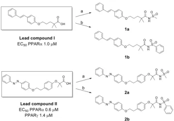
Some of these derivatives showed good PPAR activation, with submicromolar potency. We selected the stilbene derivative (Lead compound I) and the phenyldiazenyl derivative (Lead compound II) as starting compounds to obtain the corresponding methyl and phenyl sulfonimide derivatives
1a−b and 2a−b (Figure 1), in the attempt to switch the
pharmacological behavior from agonists to antagonists. Lead
compound I is a selective PPARα agonist (EC50 1.0 μM),
whereas Lead compound II is a dual PPARα/γ agonist, with a
higher PPARα efficacy and submicromolar potency (EC50
PPARα 0.6 μM, PPARγ 1.4 μM).
Lead compounds I and II were obtained as previously
described.18,19 Carboxylic acids were transformed in
sulfoni-mide derivatives 1a−b and 2a−b by treatment with
methane-or benzenesulfonamide, 1-ethyl-3-(3-(dimethylamino)propyl)-carbodiimide (EDC), and 4-dimethylaminopyridine (DMAP) (Figure 1).
These compounds were evaluated for agonist activity on the
human PPARα (hPPARα) (Table 1) and PPARγ (hPPARγ)
subtypes (data not shown). For this purpose, GAL-4 PPAR chimeric receptors were expressed in transiently transfected
HepG2 cells according to a previously reported procedure.20,21
Due to cytotoxicity exhibited by these compounds on HepG2
cells above 5 μM, their activity was evaluated at only three
concentrations (1, 2.5, and 5μM) and compared with that of
the corresponding reference agonists (Wy-14,643 for PPARα
and Rosiglitazone for PPARγ) (Supporting Information, Figure
S1) whose maximum induction was defined as 100%. Only
1a−b and 2a showed a weak selective activity toward PPARα
in the concentration range taken into consideration (Emax17−
29%), whereas no activity was observed on PPARγ (data not
shown). Given that 2b had no detectable PPARα/γ activity, it
was tested as an antagonist by conducting a competitive
binding assay in which PPARα and PPARγ activity at a fixed
concentration of the reference agonists Wy-14,643 and Rosiglitazone, respectively, was measured in cells treated with increasing concentrations of 2b. Compound 2b
completely inhibited PPARα activity with a half-maximal
inhibitory concentration of 1.2 ± 0.1 μM showing also a
simultaneous inhibition of PPARγ even though with lower
potency and activity (IC5014± 2 μM; Imax87%).
Based on these results, phenyldiazenyl compound 2b was
selected as a novel scaffold to develop novel compounds by
designing the benzenesulfonimide and amide derivatives
displayed in Figure 2. In sulfonimide derivatives 3a−g, with
the aim of probing further binding interactions inside the ligand binding domain (LBD), we introduced groups with Figure 1.From Lead compounds I and II to sulfonimide derivatives
1a−b and 2a−b. Reagents and conditions: methane- (a) or benzenesulfonamide (b), EDC, DMAP, dry dichloromethane, 0 °C−rt, 24 h, yield 44−65%.
Table 1. hPPARα Activity by GAL-4 PPAR Transactivation Assay for Synthesized Compoundsa
hPPARα hPPARα
ID Emax% Imax% IC50μM ID Emax% Imax% IC50μM
1a 17± 6 − − 4c 32± 7 − − 1b 24± 6 − − 4d i 96± 4 2.72± 0.85 2a 29± 6 − − 10a 24± 2 78± 2 7.0± 1.7 2b i 99± 1 1.2± 0.1 10b 28± 3 12± 1 − 3a i 100± 1 0.17± 0.12 10c 28± 2 62± 7 12.3± 0.9 3b i 99± 1 0.33± 0.14 10d 12± 1 71± 2 12.1± 1.1 3c i 100± 1 0.21± 0.13 10e i 100± 1 0.24± 0.04 3d i 92± 1 1.1± 0.7 13a i 93± 6 3.32± 1.31 3e i 100± 1 1.5± 0.5 13b i 87± 4 1.70± 0.25 3f i 88± 3 2.8± 0.7 13c 21± 1 67± 3 6.1± 0.8 3g i 69± 10 3.20± 0.44 13d i 94± 4 10.3± 2.7 4a i 94± 4 2.98± 1.02 13e i 100± 5 1.52± 0.22 4b 12.0± 0.3 95± 6 2.67± 1.15
ai = activity below 5% at the highest tested concentration. E
max% represents the percentage of maximum fold induction obtained with PPARα agonist Wy-14,643, taken as 100%. Imax% represents the percentage of inhibition of the maximum effect obtained with the reference agonist Wy-14,643.
different stereoelectronic properties in the para position, including hindered substituents containing an additional
aromatic ring. As amide derivatives, we selected first the
primary amide 4a and the butyl, phenyl, and benzyl secondary
amides 4b−d. Next, a second series of compounds (Figure 2)
was developed by replacing the azo moiety with amide or urea. Designed compounds were primary and secondary amides
(10a−d and 13a−d) and benzenesulfonimide derivatives (10e
and 13e).
The synthesis of benzenesulfonimides 3a−g and of amides
4a−d was performed starting from Lead compound II.
Sulfonimides 3a−g were obtained by direct coupling of
starting carboxylic acid with proper para-substituted
phenyl-sulfonamides, with EDC and DMAP, in dry CH2Cl2(Scheme
1). For compounds 3e−g, the p-substituted
phenylsulfona-mides were synthesized as previously reported.22Amides 4b−d
were synthesized by coupling Lead compound II with proper
amines, N,N′-dicyclohexylcarbodiimide (DCC),
1-hydroxy-benzotriazole hydrate (HOBt), and N-methylmorpholine (NMM) in DMF. For derivative 4a, the starting acid was reacted with ammonium chloride, under the conditions described.
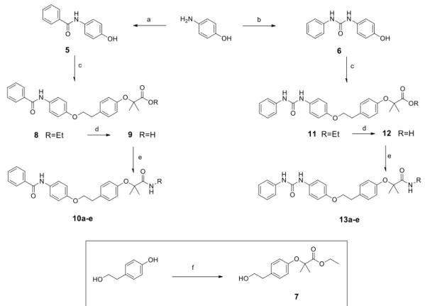
Final products 10a−e and 13a−e were obtained as depicted
in Scheme 2. Phenol 5 was synthesized by reacting
p-aminophenol with benzoyl chloride, in the presence of triethylamine (TEA) in dry DMF, whereas the reaction of p-aminophenol with phenylisocyanate, in dry acetonitrile,
afforded phenol 6. Both phenols 5 and 6 were reacted with
intermediate ester 7, synthesized by reaction of 4-(2-hydroxyethyl)phenol with ethyl 2-bromoisobutyrate.
The Mitsunobu coupling of phenols 5 and 6 with ester 7 produced 8 and 11, which were hydrolyzed in basic conditions
to acids 9 and 12. Final amides and sulfonimides 10a−e and
13a−e were obtained as previously described for compounds
3a−g and 4a−d.
All these compounds were evaluated for agonist activity on
hPPARα (Table 1) and hPPARγ (data not shown) at different
concentrations in the range 1−25 μM. Most compounds were
either poorly active or inactive on both PPAR subtypes; thus, they were tested as antagonists, as reported above. Overall,
tested compounds were completely inactive on PPARγ (data
not shown).
Sulfonimides 3a−g showed a selective good antagonist
profile on PPARα, with displacement activity toward reference
compound Wy-14,643 ranging from 69% to 100%. The IC50
calculated for these compounds displayed a low micromolar potency, with being 3a, 3b, and 3c the most potent
compounds (IC50 0.17, 0.33, and 0.21 μM, respectively).
The increased steric hindrance in the para position by introduction of an additional aromatic ring (3f and 3g)
decreased the antagonist activity (IC50 2.8 and 3.2 μM,
respectively).
As regards amides 4a−d, they were also able to selectively
antagonize PPARα exhibiting good efficacy (94−96%) and
micromolar potency (2.67−2.98 μM). Only compound 4c was
not tested as PPARα antagonist due to its residual activity
(Emax 32%) on this receptor subtype. The two series of
compounds developed by replacing the azo moiety with amide and urea exhibited similar behavior even though with small but
significant differences. All these compounds showed selective
and moderate ability to antagonize PPARα, with ureido
derivatives 13a−d being more effective and potent compared
to corresponding amides 10a−d.
Among compounds 10a−e and 13a−e, the two benzene-sulfonimide derivatives 10e and 13e turned out to be the best
PPARα antagonists, being able to completely abolish the
activation promoted by the reference agonist Wy-14,643. In this case, 10e showed higher potency than 13e (0.24 vs 1.52 μM).
Considering that 3a−e, 10e, and 13e appeared as the most
promising compounds in transactivation assay, showing a
PPARα antagonist activity ranging from 92% to 100%, together
with a potency in terms of IC50 values ranging from 0.17 to
Figure 2.Chemical structures offinal compounds 3a−g, 4a−d, 10a− e, and 13a−e.
Scheme 1. Synthesis of Compounds 3a−g and 4a−da
aReagents and conditions: (a) p-substituted benzenesulfonamide, EDC, DMAP, dry CH
2Cl2, N2, 0°C−rt, 24 h, yield 21−80%; (b) R−NH2, DCC, HOBt, NMM, DMF, rt, 24 h, yield 67−90%.
1.52 μM, we selected these compounds to perform gene
expression analysis. We analyzed whether 3a−e, 10e, and 13e
could modulate the expression of the PPARα target gene
carnitine palmitoyltransferase 1A (CPT1A), a key enzyme
involved in fatty acidβ-oxidation, considered an in vitro model
to study PPARα activation.22,23 Real-time quantitative PCR
(RTqPCR) was employed to assess the effects of the
compounds on CPT1A expression. Compounds were tested alone, or in the presence of the potent PPARα agonist GW7647, used as control. As expected, GW7647 robustly
stimulated CPT1A expression (Figure 3), whereas compounds
3a−e, 10e, and 13e induced only a weak CPT1A mRNA
expression. Notably, the combinations of GW7647 with 3a−e,
10e, or 13e were able to significantly repress CPT1A
expression, supporting the antagonistic behavior of the novel
compounds on PPARα (Figure 3).
We also explored the potential antiproliferative activity of
3a−e, 10e, and 13e in eight human cancer cell lines
representative of four distinct tumor types. We selected three pancreatic (AsPC-1, BxPC-3, Capan-2), two colorectal (HT-29, SW480), two paraganglioma (PTJ64i, PTJ86i), and one
renal (A498) cancer cell line, which express PPARα as
reported in a previous study,14 or in the Expression Atlas
database (https://www.ebi.ac.uk/gxa/home). Preliminary
MTT experiments were conducted by treatment of the eight
cancer cell lines with 3a−e, 10e, and 13e, with the PPARα
antagonist GW6471, or with the PPARα agonist Wy-14,643 for
72 h, at a single concentration (75μM) (Figure 4).
Overall, Wy-14,643 did not affect cell viability across the
tumor cell lines tested (Figure 4), whereas novel compounds,
as well as GW6471, showed antiproliferative activities, although with variable potency. Notably, all the novel
PPARα antagonists had a more marked effect on cell viability
in paraganglioma (PGL), as compared to the other cancer cell lines, with inhibition rates in PGL cells ranging from 59% to
98%, in line with the effects obtained with GW6471 in the
same cancer cell lines (inhibition rates from 85% to 92%). 3c, 3d, and 10e emerged as the compounds showing more consistent and relevant antiproliferative activities across the eight cancer cell lines, with inhibition rates from 41% to 92% in
Scheme 2. Synthesis of Compounds 10a−e and 13a−ea
aReagents and conditions: (a) benzoyl chloride, TEA, dry DMF, N
2, 0°C−rt, 24 h, yield 70%; (b) phenylisocyanate, dry ACN, N2, reflux, 5h, yield 65%; (c) 7, PPh3, DIAD (diisopropyl azodicarboxylate), dry THF, 24 h, yield 54−97%; (d) 2 N NaOH, THF, reflux, 16 h, yield 57−63%; (e) R− NH2, DCC, HOBt, NMM, DMF, rt, 24 h, yield 29−98% (for amides 10a−d and 13a−d); benzenesulfonamide, EDC, DMAP, dry CH2Cl2, N2, 0 °C−rt, 24 h, yield 34−64% (for sulfonimides 10e and 13e); (f) ethyl 2-bromoisobutyrate, K2CO3, DMF, reflux, 4 h, yield 75%.
Figure 3.Expression of PPARα target gene CPT1A. Data shown are the means± SD of three determinations (*p < 0.05; **p < 0.01; ***p < 0.001).
the pancreatic cancer cell lines, from 52% to 98% in the colon cancer cell lines, from 84% to 98% in the PGL cell lines, and
from 51% to 71% in the renal cancer cell line (Figure 4). Thus,
we selected these compounds for further characterization of
antiproliferative effects through concentration-dependent
experiments.
Pancreatic, colorectal, paraganglioma, and renal cancer cell lines were incubated with 3c, 3d, and 10e for 72 h at
concentrations from 0 μM to 24 μM (Figure 5). The
treatments significantly reduced cell viability in a
concen-tration-dependent manner, showing variable effects across the
tested cancer cell lines. In particular, 3c, 3d, and 10e drastically and significantly decreased paraganglioma cell line viability, as
shown by concentration−response curves (Figure 5, panels A,
B, C) and cytotoxic concentration (CC50) values in the low
micromolar range (Figure 5, panel D). Intriguingly, the novel
compounds showed greater antiproliferative effects and lower
CC50values than those previously obtained with the reference
compound GW6471 in the same paraganglioma cell lines.14
Remarkably, 3c, 3d, and 10e did not show toxicity against
normal HFF-1 fibroblast cells, displaying CC50 values higher
than 24μM, which was the highest concentration used in our
MTT assays, and good selectivity index (SI) values (Figure 5,
panels A, B, C, D).
Similarly, compounds 3c, 3d, and 10e showed CC50values
higher than 24μM in pancreatic, colorectal, and renal cancer
cell, except 3d that showed a CC50of 16.99μM in BxPC-3 and
10e that showed CC50 values of approximately 7 μM in
pancreatic and colorectal cancer cell lines and of 4.6 μM in
renal cancer cells (Supporting Information, Table S1).
To elucidate the binding mode of this series of compounds
and to help interpretation of structure−activity relationship
(SAR) data, we undertook docking studies using the GOLD Suite docking package (CCDC Software Limited: Cambridge,
U.K.) with the X-ray crystal structure of PPARα in complex
with the antagonist GW6471 (PDB ID: 1KKQ).24 In this
structure GW6471, bearing an amide headgroup, does not interact with Y464 and pushes the H12 to assume an inactive
and less structured conformation. The PPAR LBD is
“Y-shaped” and is composed of a polar arm I, which is extended toward H12, a hydrophobic arm II, which is located between
H3 and theβ-sheet, and a hydrophobic entrance (arm III).
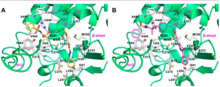
The most potent compounds 3a and 10e were chosen for docking as representative members of benzenesulfonimide derivatives bearing distal phenyldiazenyl and phenylbenzamide
moieties, respectively. As illustrated in Figure 6, both
compounds adopted a similar U-shaped configuration,
wrapping around H3. The oxygen atom of the sulfonimide
moiety of 3a (Figures 6A andS2, Supporting Information) was
engaged in an H-bond with the OH group of Y314 side chain. Moreover, the phenyl ring of the benzenesulfonimide moiety
was optimally oriented for a favorableπ−π stacking interaction
with the Y314 side chain, and the methyl group in para formed fruitful hydrophobic interactions with A441. The gem-dimethyl
substituents were projected into the lipophilic“benzophenone
pocket”,25 making further hydrophobic interactions. The
central phenoxy ring also made a π−π stacking interaction
with the F318 side chain, with the phenoxy oxygen forming a
further H-bond with Nε2 of H440. The phenyldiazenyl group
was surrounded by sulfur-containing residues such as C275,
C276, M355, and M330, forming profitable sulfur−arene
interactions.26 The ligand’s tail fitted well into arm II and
positively contributed to overall binding through hydrophobic Figure 4.Effect of compounds on the viability of pancreatic (A), colorectal (B), paraganglioma (C), and renal (D) tumor cell lines. Cell viability was assessed by MTT assay using compounds at 75μM for 72 h. Data shown are the means ± SD of duplicate experiments with quintuplicates determinations.*Statistically significant differences between control and each compound concentration (*p < 0.05; **p < 0.01; ***p < 0.001).
contacts with residues I272 of H3, L254 and L247 of H2′, and
I241, I339, V332 of theβ-sheet.
By looking at the binding mode of compound 10e (Figures
6B andS3, Supporting Information), it was observed that an
H-bond was also formed, through its carbonyl oxygen, with the OH group of the Y314 side chain, whereas the aromatic ring of the benzenesulfonamide moiety made hydrophobic
interac-tions with V444 and F273. In addition, the central phenoxy
ring was engaged in an edge-to-faceπ−π stacking interaction
with the H440 side chain. The phenylbenzamide moiety, besides the hydrophobic contacts observed for 3a, formed two additional H-bonds with the T279 OH group and the NH
backbone of A333 on theβ-sheet.
Figure 5. Compounds 3c, 3d, and 10e affect viability in paraganglioma cancer cell lines with negligible effects on normal fibroblast cells. Concentration−response curves of 3c (A), 3d (B), and 10e (C) on viability of paraganglioma cancer cell lines (PTJ86i and PTJ64i) and of normal fibroblast cells (HFF-1). Cytotoxic effects were tested by MTT assay using compounds at the indicated concentrations for 72 h. Data shown are the means± standard deviation of duplicate experiments with five replicates. Cytotoxic concentration (CC50) values are the drug concentrations required to inhibit 50% of cell viability. Selectivity index (SI) values were calculated for each compound as follows: CC50on normalfibroblast cells (HFF-1)/CC50on cancer cells (D).*Statistically significant differences between control and each compound concentration (*p < 0.05; ***p < 0.001).
Figure 6.Binding mode of compounds 3a (A, yellow sticks) and 10e (B, violet sticks) in PPARα LBD represented as green ribbon model. Only amino acids located within 4 Å of the bound ligand are displayed (white sticks) and labeled. H-bonds discussed in the text are depicted as dashed black lines.
The overlay of the docked pose of 3a and 10e with the X-ray
crystal pose of the PPARα antagonist GW6471 (Supporting
Information, Figure S4A) revealed a similar binding mode,
with analogous positioning of head groups and a similar orientation of the hydrophobic tail groups. Noteworthy, the benzenesulfonamide headgroup of 3a and 10e projected into an area that is usually occupied by the side chain of Y464 in
PPARα LBD bound to agonist ligands, such as GW409544
(Supporting Information, Figure S4B).27 Thus, the
benzene-sulfonamide derivatives do not interact with this residue that is critical for receptor activation due to steric hindrance, likely forcing H12 out of the agonist bound position and inducing a
PPARα LBD conformation that interacts efficiently with
corepressors.
Docking studies allowed deriving some clues about SAR. As
regards derivatives 3a−g, when the methyl group at position
para of 3a was replaced with methoxy (3b) or chlorine (3c),
the IC50remained in the low micromolar range, suggesting that
these compounds are able to form the same favorable interactions observed for 3a. Thus, the para position of the benzenesulfonimide moiety requires substituents with a certain degree of lipophilicity, but quite limited in size. In fact, the insertion of the more hydrophilic nitro group (3d), or the bulkier methylamide group (3e), caused a slight decrease in
potency; for derivatives 3f and 3g, a further drop in PPARα
antagonistic activity was observed, produced by the impaired accommodation of an additional aromatic ring into arm I of
PPARα. The overlay of the docked poses of 3a and 3g (Figure
S5A, Supporting Information) revealed that the benzyl amide
substituent was shifted upward in arm I and dramatically altered the interactions pattern of the benzenesulfonimide
group. Derivatives 4a−d, bearing the amide headgroup and
phenyldiazenyl tail group, turned out to be less active because
of the loss of profitable H-bonds and π−π stacking interactions
with Y314 observed in docking experiments. On comparing the
docked pose of 3a and 4c (Figure S5B, Supporting
Information), it is clear that, despite a similar positioning of
the phenyldiazenyl tail, the amide moiety was not properly oriented to engage an H bond with Y314. Also for derivatives
10a−e, the presence of the benzenesulfonamide group was
critical for the antagonistic activity, as only derivative 10e
displayed an IC50 in the low micromolar range. From the
docked pose of 10e, it can be argued that the primary amide (10a) was no longer able to form the hydrophobic interactions with residues V444 and F273, whereas both aliphatic (10b) and aromatic groups (10c and 10d) could not be placed at an optimal distance to favorably interact with such residues. As
shown inFigure S5C, the phenylbenzamide tails of both 10e
and 10b displayed the same orientation; however, the butyl amide headgroup of 10b could not properly interact with Y314, but instead was oriented toward Q277. Thus, the weak interactions formed by the headgroup were unable to induce an antagonistic conformation. This might account for the slight receptor activation and, in turn, the low antagonistic activity
shown by derivatives 10a−d. For derivatives 13a−e, the
presence of the urea moiety at the tail group improved the antagonistic activity (see 13a and 13b) due to its propensity to extend more deeply into arm II and to make an H-bond with the C275 backbone. However, introduction of phenyl and benzyl substituents (13c and 13d) on the amide headgroup
introduced steric restrictions, making it more difficult for the
ligands to interact with Y314 and with the hydrophobic residues A441, V444, and F273. Again, the introduction of the
benzenesulfonamide group increased potency (13e). As shown
inFigure S5D, this moiety well anchored the ligand into arm I
in a similar fashion to 10e. The presence of the sulfonyl group avoids the steric restrictions by rotation of the phenyl ring to a position that is better suited to interact with hydrophobic residues. Thus, the benzenesulfonamide moiety is a key structural feature in this series of derivatives to confer antagonistic activity.
In conclusion, this study led to the identification of novel
sulfonimide and amide PPARα antagonists. Most potent
compounds induced marked antiproliferative activity when
tested in in vitro cancer cells expressing PPARα (pancreatic,
colorectal, paraganglioma, and renal cancer cell lines). In addition, binding modes of representative benzenesulfonimide derivatives 3a and 10e helped to rationalize results from transactivation assay and give information about SAR of this class of compounds.
■
ASSOCIATED CONTENT*
sı Supporting InformationThe Supporting Information is available free of charge at
https://pubs.acs.org/doi/10.1021/acsmedchemlett.9b00666.
Experimental procedures, full characterization of com-pounds, 2D ligand-interaction diagrams of compounds
into the PPAR binding pocket, NMR spectra (PDF)
■
AUTHOR INFORMATIONCorresponding Author
Alessandra Ammazzalorso− Department of Pharmacy, “G.
d’Annunzio” University of Chieti-Pescara, 66100 Chieti, Italy;
orcid.org/0000-0003-4369-1772; Phone: +39 0871
3554682; Email:[email protected]
Authors
Isabella Bruno− Department of Pharmacy, “G. d’Annunzio”
University of Chieti-Pescara, 66100 Chieti, Italy
Rosalba Florio− Department of Pharmacy, “G. d’Annunzio”
University of Chieti-Pescara, 66100 Chieti, Italy
Laura De Lellis− Department of Pharmacy, “G. d’Annunzio”
University of Chieti-Pescara, 66100 Chieti, Italy
Antonio Laghezza− Department of Pharmacy-Drug Science,
University of Bari“Aldo Moro”, 70126 Bari, Italy;
orcid.org/0000-0001-6221-6155
Carmen Cerchia− Department of Pharmacy, “Drug Discovery”
Laboratory, University of Napoli“Federico II”, 80131 Napoli,
Italy
Barbara De Filippis− Department of Pharmacy, “G.
d’Annunzio” University of Chieti-Pescara, 66100 Chieti, Italy
Marialuigia Fantacuzzi− Department of Pharmacy, “G.
d’Annunzio” University of Chieti-Pescara, 66100 Chieti, Italy
Letizia Giampietro− Department of Pharmacy, “G.
d’Annunzio” University of Chieti-Pescara, 66100 Chieti, Italy;
orcid.org/0000-0002-8483-1885
Cristina Maccallini− Department of Pharmacy, “G.
d’Annunzio” University of Chieti-Pescara, 66100 Chieti, Italy;
orcid.org/0000-0003-2957-8650
Paolo Tortorella− Department of Pharmacy-Drug Science,
University of Bari“Aldo Moro”, 70126 Bari, Italy;
orcid.org/0000-0003-1358-7376
Serena Veschi− Department of Pharmacy, “G. d’Annunzio”
University of Chieti-Pescara, 66100 Chieti, Italy
Fulvio Loiodice− Department of Pharmacy-Drug Science,
University of Bari“Aldo Moro”, 70126 Bari, Italy;
orcid.org/0000-0003-3384-574X
Antonio Lavecchia− Department of Pharmacy, “Drug
Discovery” Laboratory, University of Napoli “Federico II”,
80131 Napoli, Italy; orcid.org/0000-0002-2181-8026
Alessandro Cama− Department of Pharmacy, “G. d’Annunzio”
University of Chieti-Pescara, 66100 Chieti, Italy; Center for Advanced Studies and Technology CAST, 66100 Chieti, Italy
Rosa Amoroso− Department of Pharmacy, “G. d’Annunzio”
University of Chieti-Pescara, 66100 Chieti, Italy Complete contact information is available at:
https://pubs.acs.org/10.1021/acsmedchemlett.9b00666 Funding
This work was supported by FAR funds (Italian Ministry for Instruction, University and Research) assigned to A.A. The study was also supported by the Ministry of Education, University and Research (MIUR), Progetti di Ricerca di Interesse Nazionale (PRIN) funds (Grant Number 2017EKMFTN_005), assigned to A.C.
Notes
The authors declare no competingfinancial interest.
Biography
Alessandra Ammazzalorso received her Ph.D. from the G. d’Annunzio University, Chieti-Pescara, Italy, in 2001. She is currently an Assistant Professor at Pharmacy Department, University of Chieti-Pescara. Her research interests include the design and synthesis of small-molecule drugs, mainly PPAR ligands and enzymatic inhibitors of nitric oxide synthases and aromatase. The results of these studies have been published in over 60 papers.
■
ABBREVIATIONSPPARs, Peroxisome Proliferator-Activated Receptors; TZDs, thiazolidinediones; FAO, fatty acid oxidation; EDC, 1-ethyl-3-(3-dimethylaminopropyl)carbodiimide; DMAP, 4-dimethyl-aminopyridine; LBD, ligand binding domain; DCC, N,N′-dicyclohexylcarbodiimide; HOBt, 1-hydroxybenzotriazole hy-drate; NMM, N-methylmorpholine; TEA, triethylamine; DIAD, diisopropyl azodicarboxylate; CPT1A, carnitine palmitoyltransferase 1A; RTqPCR, real-time quantitative
PCR; PGL, paraganglioma; CC50, median cytotoxic
concen-tration.
■
REFERENCES(1) Issemann, I.; Green, S. Activation of a member of the steroid hormone receptor superfamily by peroxisome proliferators. Nature 1990, 347, 645−650.
(2) Berger, J. P.; Akiyama, T. E.; Meinke, P. T. PPARs: therapeutic targets for metabolic disease. Trends Pharmacol. Sci. 2005, 26, 244− 251.
(3) Staels, B.; Dallongeville, J.; Auwerx, J.; Schoonjans, K.; Leitersdorf, E.; Fruchart, J.-C. Mechanism of action of fibrates on lipid and lipoprotein metabolism. Circulation 1998, 98, 2088−2093.
(4) Katsiki, N.; Nikolic, D.; Montalto, G.; Banach, M.; Mikhailidis, D. P.; Rizzo, M. The role of fibrate treatment in dyslipidemia: an overview. Curr. Pharm. Des. 2013, 19, 3124−3131.
(5) Sarafidis, P. A. Thiazolidinedione derivatives in diabetes and cardiovascular disease: an update. Fundam. Clin. Pharmacol. 2008, 22, 247−264.
(6) Ammazzalorso, A.; De Filippis, B.; Giampietro, L.; Amoroso, R. Blocking the Peroxisome Proliferator-Activated Receptor (PPAR): an overview. ChemMedChem 2013, 8, 1609−1616.
(7) Hashimoto, Y.; Miyachi, H. Nuclear receptor antagonists designed based on the helix-folding inhibition hypothesis. Bioorg. Med. Chem. 2005, 13, 5080−5093.
(8) Samudio, I.; Fiegl, M.; Andreeff, M. Mitochondrial uncoupling and the Warburg effect: molecular basis for the reprogramming of cancer cell metabolism. Cancer Res. 2009, 69, 2163−2166.
(9) De Lellis, L.; Cimini, A.; Veschi, S.; Benedetti, E.; Amoroso, R.; Cama, A.; Ammazzalorso, A. The anticancer potential of Peroxisome Proliferator-Activated Receptor antagonists. ChemMedChem 2018, 13, 209−219.
(10) Messmer, D.; Lorrain, K.; Stebbins, K.; Bravo, Y.; Stock, N.; Cabrera, G.; Correa, L.; Chen, A.; Jacintho, J.; Chiorazzi, N.; Yan, X. J.; Spaner, D.; Prasit, P.; Lorrain, D. A selective novel Peroxisome Proliferator-Activated Receptor (PPAR)-α antagonist induces apop-tosis and inhibits proliferation of CLL cells in vitro and in vivo. Mol. Med. 2015, 21, 410−419.
(11) Abu Aboud, O.; Donohoe, D.; Bultman, S.; Fitch, M.; Riiff, T.; Hellerstein, M.; Weiss, R. H. PPARα inhibition modulates multiple reprogrammed metabolic pathways in kidney cancer and attenuates tumor growth. Am. J. Physiol. Cell. Physiol. 2015, 308, C890−C898.
(12) Benedetti, E.; d’Angelo, M.; Ammazzalorso, A.; Gravina, G.; Laezza, C.; Antonosante, A.; Panella, G.; Cinque, B.; Cristiano, L.; Dhez, A. C.; Astarita, C.; Galzio, R.; Cifone, M. G.; Ippoliti, R.; Amoroso, R.; Di Cesare, E.; Giordano, A.; Cimini, A. PPARα antagonist AA452 triggers metabolic reprogramming and increases sensitivity to radiation therapy in human glioblastoma primary cells. J. Cell. Physiol. 2017, 232, 1458−1466.
(13) Ammazzalorso, A.; De Lellis, L.; Florio, R.; Bruno, I.; De Filippis, B.; Fantacuzzi, M.; Giampietro, L.; Maccallini, C.; Perconti, S.; Verginelli, F.; Cama, A.; Amoroso, R. Cytotoxic effect of a family of Peroxisome Proliferator-Activated Receptor antagonists in color-ectal and pancreatic cancer cell lines. Chem. Biol. Drug Des. 2017, 90, 1029−1035.
(14) Florio, R.; De Lellis, L.; di Giacomo, V.; Di Marcantonio, M. C.; Cristiano, L.; Basile, M.; Verginelli, F.; Verzilli, D.; Ammazzalorso, A.; Prasad, S. C.; Cataldi, A.; Sanna, M.; Cimini, A.; Mariani-Costantini, R.; Mincione, G.; Cama, A. Effects of PPARα inhibition in head and neck paraganglioma cells. PLoS One 2017, 12 (6), No. e0178995.
(15) Ammazzalorso, A.; De Lellis, L.; Florio, R.; Laghezza, A.; De Filippis, B.; Fantacuzzi, M.; Giampietro, L.; Maccallini, C.; Tortorella, P.; Veschi, S.; Loiodice, F.; Cama, A.; Amoroso, R. Synthesis of novel benzothiazole amides: evaluation of PPAR activity and anti-proliferative effects in paraganglioma, pancreatic and colorectal cancer cell lines. Bioorg. Med. Chem. Lett. 2019, 29, 2302−2306.
(16) Ammazzalorso, A.; Giancristofaro, A.; D’Angelo, A.; De Filippis, B.; Fantacuzzi, M.; Giampietro, L.; Maccallini, C.; Amoroso, R. Benzothiazole-based N-(phenylsulfonyl)amides as a novel family of PPARα antagonists. Bioorg. Med. Chem. Lett. 2011, 21, 4869−4872.
(17) Ammazzalorso, A.; D’Angelo, A.; Giancristofaro, A.; De Filippis, B.; Di Matteo, M.; Fantacuzzi, M.; Giampietro, L.; Linciano, P.; Maccallini, C.; Amoroso, R. Fibrate-derived N-(methylsulfonyl)amides with antagonistic properties on PPARα. Eur. J. Med. Chem. 2012, 58, 317−322.
(18) De Filippis, B.; Giancristofaro, A.; Ammazzalorso, A.; D’Angelo, A.; Fantacuzzi, M.; Giampietro, L.; Maccallini, C.; Petruzzelli, M.; Amoroso, R. Discovery of gemfibrozil analogues that activate PPARα and enhance the expression of gene CPT1A involved in fatty acids catabolism. Eur. J. Med. Chem. 2011, 46, 5218− 5224.
(19) Giampietro, L.; D’Angelo, A.; Giancristofaro, A.; Ammazzalorso, A.; De Filippis, B.; Fantacuzzi, M.; Linciano, P.; Maccallini, C.; Amoroso, R. Synthesis and structure-activity relation-ships of fibrate-based analogues inside PPARs. Bioorg. Med. Chem. Lett. 2012, 22, 7662−7666.
(20) Pinelli, A.; Godio, C.; Laghezza, A.; Mitro, N.; Fracchiolla, G.; Tortorella, V.; Lavecchia, A.; Novellino, E.; Fruchart, J. C.; Staels, B.; Crestani, M.; Loiodice, F. Synthesis, biological evaluation, and
molecular modeling investigation of new chiral fibrates with PPARα and PPARγ agonist activity. J. Med. Chem. 2005, 48, 5509−5519.
(21) Porcelli, L.; Gilardi, F.; Laghezza, A.; Piemontese, L.; Mitro, N.; Azzariti, A.; Altieri, F.; Cervoni, L.; Fracchiolla, G.; Giudici, M.; Guerrini, U.; Lavecchia, A.; Montanari, R.; Di Giovanni, C.; Paradiso, A.; Pochetti, G.; Simone, G. M.; Tortorella, P.; Crestani, M.; Loiodice, F. Synthesis, characterization and biological evaluation of ureidofi-brate-like derivatives endowed with peroxisome proliferator-activated receptor activity. J. Med. Chem. 2012, 55, 37−54.
(22) Ammazzalorso, A.; Carrieri, A.; Verginelli, F.; Bruno, I.; Carbonara, G.; D’Angelo, A.; De Filippis, B.; Fantacuzzi, M.; Florio, R.; Fracchiolla, G.; Giampietro, L.; Giancristofaro, A.; Maccallini, C.; Cama, A.; Amoroso, R. Synthesis, in vitro evaluation, and molecular modeling investigation of benzenesulfonimide Peroxisome Prolifer-ator-Activated Receptorsα antagonists. Eur. J. Med. Chem. 2016, 114, 191−200.
(23) Giampietro, L.; Laghezza, A.; Cerchia, C.; Florio, R.; Recinella, L.; Capone, F.; Ammazzalorso, A.; Bruno, I.; De Filippis, B.; Fantacuzzi, M.; Ferrante, C.; Maccallini, C.; Tortorella, P.; Verginelli, F.; Brunetti, L.; Cama, A.; Amoroso, R.; Loiodice, F.; Lavecchia, A. Novel phenyldiazenyl fibrate analogues as PPARα/γ/δ pan-agonists for the amelioration of metabolic syndrome. ACS Med. Chem. Lett. 2019, 10, 545−551.
(24) Xu, H. E.; Stanley, T. B.; Montana, V. G.; Lambert, M. H.; Shearer, B. G.; Cobb, J. E.; McKee, D. D.; Galardi, C. M.; Plunket, K.; Nolte, R. T.; Parks, D. J.; Moore, J. T.; Kliewer, S. A.; Willson, T. M.; Stimmel, J. B. Structural basis for antagonist-mediated recruitment of nuclear co-repressors by PPARα. Nature 2002, 415, 813−817.
(25) Gampe, R. T., Jr; Montana, V. G.; Lambert, M. H.; Miller, A. B.; Bledsoe, R. K.; Milburn, M. V.; Kliewer, S. A.; Willson, T. M.; Xu, H. E. Asymmetry in the PPARγ/RXRα crystal structure reveals the molecular basis of heterodimerization among nuclear receptors. Mol. Cell 2000, 5, 545−555.
(26) Forbes, C. R.; Sinha, S. K.; Ganguly, H. K.; Bai, S.; Yap, G. P. A.; Patel, S.; Zondlo, N. J. Insights into thiol-aromatic interactions: a stereoelectronic basis for S-H/π interactions. J. Am. Chem. Soc. 2017, 139, 1842−1855.
(27) Xu, H. E.; Lambert, M. H.; Montana, V. G.; Plunket, K. D.; Moore, L. B.; Collins, J. L.; Oplinger, J. A.; Kliewer, S. A.; Gampe, R. T., Jr.; McKee, D. D.; Moore, J. T.; Willson, T. M. Structural determinants of ligand binding selectivity between the peroxisome proliferator-activated receptors. Proc. Natl. Acad. Sci. U. S. A. 2001, 98, 13919−13924.