Università degli Studi di Ferrara
DOTTORATO DI RICERCA IN
FARMACOLOGIA E ONCOLOGIA MOLECOLARE
CICLO XXVII
COORDINATORE Prof. Antonio Cuneo
Altered motor phenotype and dopamine
transmission associated with mutations of the
parkinsonian gene LRRK2
Settore Scientifico Disciplinare BIO/14
Dottorando Tutore
Dott. Longo Francesco Prof. Morari Michele
1
SUMMARY
INTRODUCTION 4
LRRK2: a protein genetically implicated in Parkinson's Disease 5
Protein organization 5
Dimerization and autophosphorylation 7
Mutation 8
Parkinson’s Disease and LRRK2-associated phenotype: the clinical scenario
11
Parkinson’s Disease overview 11
Genetics of Parkinson’s Disease 13
LRRK2-associated phenotype 18
Neuropathology of LRRK2 mutation carriers 19
LRRK2: protein interaction network and pathogenic mechanisms 21
Expression 21
Interaction network and cellular function 22
LRRK2: animal models 31
LRRK2 knockout models 32
LRRK2 transgenic models 32
AIM OF THE STUDY 35
METHODS 37
Subject 38
Behavioral analysis 38
Experimental design: longitudinal study (Part 1) 38
LRRK2 kinase inhibitor administration 39
Experimental design: study on dopaminergic transmission (Part 2) 39
Drug administration 40
Bar test 40
Drag test 40
Rotarod test 41
Spontaneous motor activity 41
In vivo microdialysis 41
Neurochemical analysis using LC-MS 42
2
Synaptosomes preparation 43
[3H]-WIN 35,428 saturation binding experiments 43
Dopamine Uptake Assay 43
In vivo PK study 44
Cell cultures and treatments 44
Cells and tissue lysis 45
Western blotting 45
Immunohistochemistry 45
Stereology and neuron counting 46
Data presentation and statistical analysis 46
Drugs 47
RESULTS 48
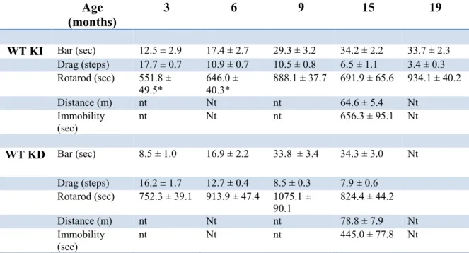
Part 1. Behavioral analysis of mice carrying mutations within the LRRK2 kinase domain
49
Characterization of motor phenotype in aging G2019S KI and WT mice 49
Characterization of motor phenotype in aging D1994S KD and WT mice 52
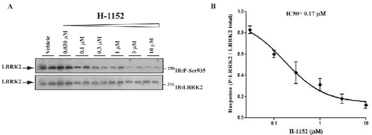
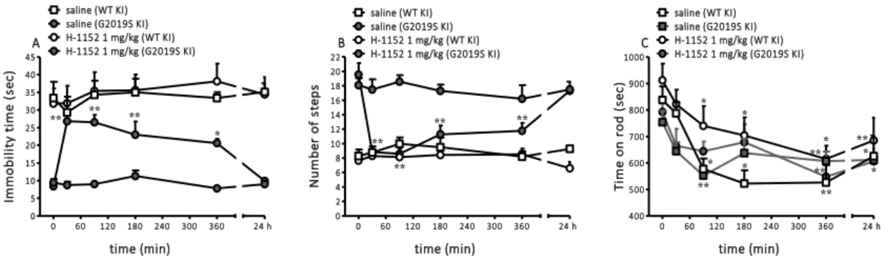
Pharmacological inhibition of the LRRK2 kinase activity 54
Effect of the LRRK2 kinase inhibitor H-1152 on motor phenotype 54
Effect of the LRRK2 kinase inhibitor H-1152 on LRRK2 phosphorylation at Ser935
57
Effect of the LRRK2 kinase inhibitor Nov-LRRK2-11 on motor phenotype in LRRK2 G2019S KI and WT mice
60
Effect of the LRRK2 kinase inhibitor Nov-LRRK2-11 on LRRK2 phosphorylation at Ser935
62
Part 2. Pharmacological manipulation of dopaminergic transmission in G2019S KI mice
65
Haloperidol 65
SCH23390 66
Pramipexole 67
Relationship between G2019S mutation and DA reuptake in phenotypic mice
68
GBR-12783 68
GBR-12783 and Nov-LRRK2-11 69
Analysis on DAT and VMAT2 protein levels 70
3 In vivo microdialysis 72 Nigrostriatal DA system 74 DISCUSSION 76 Concluding remarks 83 REFERENCES 85 ORIGINAL PAPERS 101
4
5
LRRK2: a protein implicated in Parkinson's Disease Protein organization
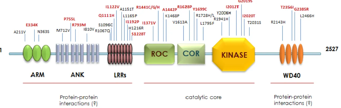
The leucine-rich repeat kinase 2 (LRRK2, PARK8) gene on chromosome 12 contains 51 exons and encodes a large multifunctional and multidomain protein containing 2527 amino acids. LRRK2 is a ubiquitous, mainly cytosolic, protein with a molecular weight of approximately 285 kDa, predominantly existing in a dimeric conformation in vivo with different points of interaction through each individual monomer (Greggio et al., 2008). According to sequence analysis, LRRK2 shows a central catalytic domain with GTPase and kinase activities. However, the N-terminal portion presents protein-protein interaction domains such as the armadillo (residues 180-660), ankyrin (residues 690-860) and the namesake leucine-rich repeats (LRRs; residues 985-1274) domains, whereas the C-terminal terminus hosts the structural repeat motif WD40 (residues 2142-2498; Fig.1).
Fig. 1. Schematic representation of LRRK2 protein linear structure.
Catalytic domains. LRRK2 encodes two different enzymatic activities: kinase and GTPase.
The central region of the protein includes the Ras of complex protein (ROC) GTPase protein domain with the adjacent C-terminal of Roc domain (COR) in conjunction with a kinase domain. Considering LRRK2 in the context of evolutionarily related protein kinases, LRRK2 originates from a different source and bears a quite different sequence from the origin (Marin, 2006; 2008). LRRK2 belongs to the TKL subfamily of human protein kinases (Manning et al., 2002), whose members show sequence similarity to both serine/threonine and tyrosine kinases. According to sequence similarity, its kinase domain shares homology with mixed-lineage kinases (MLKs) which are members of the mitogen activated protein kinase (MAPK) family (Jameel et al., 2009) However, more recent data indicate that the LRRK2 kinase domain mostly resembles the receptor-interacting protein kinase (RIPK) family (West et al., 2005), that plays a crucial role in cellular stress. The
6
presence of ROC and COR domains makes LRRK2 a member of a superfamily of proteins, named ROCO, that includes at least three other human proteins: LRRK1, DAPK1 and MASL1 (Marin, 2008). The human proteins LRRK2 and LRRK1 belong to a subgroup of ROCO proteins that always have an N-terminal LRR and a C-terminal kinase domain in addition to the ROC-COR tandem. This pair of domains is conserved throughout evolution, suggesting their functional interdependence. According to the model of the homologous domain in prokaryote C. tepidum, the COR domain (residues 1524-1837) is composed of nine α-helices and six β-sheets that are linked by a loop exposed to the solvent (Gotthardt et al., 2008). Interestingly, the prediction for human COR differs from the C. tepidum primarily in its C-terminal half, an area responsible for dimerization, thus suggesting that in human LRRK2, dimerization occurrs through its ROC domain. Indeed, the monomeric structure of the ROC domain (residues 1335-1515) comprises five α-helices and six β-sheets that are linked by loops, and the dimer form is stabilized through hydrogen bindings and hydrophobic interactions. The surface of the dimer presents only one functional ligand-binding unit, showing unique nucleotide and Mg2+ binding sites with contributions from both monomers (Deng et al., 2008). In vitro studies have shown that LRRK2 can bind GTP through the GTPase domain via a phosphate-binding motif (P-loop) region, and that it can hydrolyze GTP involving a catalytic Switch II motif that is critical for GTPase activity. A high-resolution kinase domain model, recently obtained from the homolog ROCO4, has been proposed as a platform for understanding the human LRRK2 kinase domain (residues 1183-2134) that is formed by seven α-helices and six β-sheets linked by loops, which are exposed to the solvent (Gilsbach et al., 2012). LRRK2 represents an unusual protein in which two distinct enzymatic domains within a single polypeptide chain interacts to regulate activity. In particular, the kinase activity is stimulated upon GTP binding to ROC, and it is reasonable to suppose that ROC regulates kinase activity by changing its conformations (active/inactive). Thus, a loss of GTP binding or an increased turnover of GTP to GDP should result in lowered kinase activity (Deng et al., 2008). Moreover, several studies demonstrated that LRRK2 self-regulation is a mechanism for controlling its kinase activity, which is mediated, in part, by its GTPase domain and homodimerization. Similar to MAPKKKs, which are regulated by small GTPases, LRRK2 kinase activity might be regulated by its own GTPase domain in an intrinsic manner (Lee et al., 2012a).
Repeat domains. Various investigators have identified four domains composed of
structural repeat motifs likely to be involved in regulation and localization of LRRK2. The presence of these particular domains occurring in 14% of all prokaryotic and eukariotic
7
proteins (Marcotte et al., 1999), suggests that LRRK2, in addition to its predicted protein kinase and GTPase activities, acts as a scaffold in different multiprotein signaling complex pathways. Although the presence of the armadillo repeats (ARM) domain in the N-terminus of the protein has been questioned, recent studies concluded that LRRK2 contains ARM-like repeats domain. This domain is formed by 21 α-helices grouped to form supercoil, and are linked by loops to form a curved structure (Cardona et al., 2014; Marin, 2008). The following domain, in order from N- to C-terminus, is the ankyrin repeats (ANK) domain, found in diverse bacterial and eukaryotic proteins. It is compared to cytoskeletal erythrocyte ankyrin characterized by two antiparallel α-helices followed by a loop to form a gently curved structure. According to the various models proposed by different groups, the LRRs domain is located N-terminal to the ROC domain and is composed of 14 repeats. The first 13 repeats share the LRR consensus sequence found in LRR proteins interacting with Ras, whereas the 14th repeat deviates from this consensus. These 14 repeats are grouped into two modules and each LRR subunit contributes to form a curved β-sheets linked by a large loop (Cardona et al., 2014).
Located at the C-terminal end of the protein, the predicted WD40 domain of LRRK2 comprises seven repeats formed by 30 -sheets linked by loops to form a circular bladed propeller-like structure (Cardona et al., 2014). Although WD40 has been identified in many proteins with different functions as well as proteins involved in the cytoskeletal assembly, vesicle formation and trafficking, or in proteins with enzymatic activity, the structure of this domain is well conserved and represents the most common repeat domain found in the human protein. Different studies also demonstrated that WD40 is crucial for LRRK2 physiological and pathological activities, playing an important role in the synaptic vesicular network of LRRK2 (Piccoli et al., 2011).
Dimerization and autophosphorylation
Studies using ROCO proteins from lower organisms indicate that LRRK2 predominantly exists in a dimeric conformation, and dimerization occurs in the ROC-COR bi-domain (Deng et al., 2008; Greggio et al., 2008), even though different other points of interaction through each individual monomer have been found. In particular, when the ROC domain was used as a bait in a yeast two-hybrid system, it bound to the N-terminus of LRRK2, the C-terminus of the LRR domain, the N-terminus of the ROC domain, and the C-terminus of the WD40 domain (Greggio et al., 2008). Interestingly, the deletion of the ROC domain is not sufficient to prevent dimerization, suggesting additional interaction regions within
8
LRRK2. Since a N-terminal deletion of the ANK and LRR domains does not reduce dimerization, whereas a WD40 truncated-LRRK2 could not dimerize, ROC and WD40 domains seems to be critical for LRRK2 dimerization. Dimer formation is a common phenomenon among protein kinases and can help to mediate different auto-regulation and downstream signaling pathways. In addition, allosteric regulation is mediated by homodimerization in a number of serine-threonine kinases including mitogen-activated protein (MAP) kinases, p38, c-Jun N-terminal kinase (JNK), and their upstream kinases (Leung and Lassam, 2001; Ohren et al., 2004). Despite LRRK2 monomeric form is the predominant species in cells and might have kinase activity, experimental evidence suggests that LRRK2 exists and functions as a dimer with autophosphorylation of the full-lenght protein occurring as an intramolecular (cis) event (Berger et al., 2010; Greggio et al., 2008). It was demonstrated that the autophosphorylation sites such as Thr1410, Thr1491 and Thr1503 are located mainly within the ROC domain (Gloeckner et al., 2010; Greggio et al., 2009; Kamikawaji et al., 2009; Pungaliya et al., 2010) where GTPase activity of LRRK2 is localized. In this scenario, autophosphorylation could represent an intramolecular post-translational modification that regulates the GTPase activity suggesting that the kinase activity acts as an internal modulator of the ROC domain and consequently of signal transmission. Moreover GTPase activity may be also be regulated through the recruitment of other cellular proteins (Gotthardt et al., 2008).
Mutations
The complete open reading frame of LRRK2 codes for a 2527 amino acid protein and as for many other large genes, a number of known variants can be found along the coding sequence. To date, more than 100 amino acid substitutions have been reported in human LRRK2 (Rubio et al., 2012), many of which are not currently linked to any disease. Some polymorphisms have been related to different pathologies such as leprosy (Zhang et al., 2009) or cancer (Hassin-Baer et al., 2009) and, possibly, inflammatory bowel disease (Barrett et al., 2008), whereas others are linked to neurodegenerative disease or represent an important risk factor (Paisan-Ruiz et al., 2013; Paisan-Ruiz et al., 2008). In 2004, two independent groups (Paisan-Ruiz et al., 2004; Zimprich et al., 2004) showed, for the first time, that mutations of LRRK2 were linked to dominantly inherited Parkinson’s disease (PD), corroborating a previous study which identified PARK8 as a new locus associated with PD within large Japanese family (Funayama et al., 2002). This initial discovery had a significant impact on the functional characterization of LRRK2, shedding light on disease-causing mutations in this protein, and on the genetics of PD (Fig. 2).
9
Fig. 2. Distribution of the PD-related substitutions in relation to the functional domains of LRRK2. The mutations
described as pathogenic are in red.
Of note, six missense mutations that include the p.N1437H, p.R1441C/G/H, p.Y1699C, p.S1761R, p.G2019S, and p.I2020T substitutions, clearly segregate with disease and are clustered within the enzymatic core of the protein (Cookson, 2010; Mata et al., 2006). In particular, the substitution of a glycine with a serine in position 2019 (G2019S) is located in the activation loop of the kinase domain at the conserved Mg2+-binding motif (Goldwurm et al., 2005) and represents the most common PD-related mutation. The G2019S substitution induces a stabilization of the enzyme in the active form leading to an increased kinase activity in vitro (Greggio et al., 2006; Jaleel et al., 2007; West et al., 2005) and in vivo (Sheng et al., 2012). This mutation facilitates substrates access without an enhancement of substrate affinity (West et al., 2005). The other pathogenic mutation located within the kinase domain is represented by I2020T but how it affects protein function is still debated. Some studies reported that this mutation results in an increased kinase activity (Gloeckner et al., 2006; Kamikawaji et al., 2013), even though such increase is less marked than that caused by G2019S, while other groups revealed an unchanged or a decreased function (Jaleel et al., 2007; Nichols et al., 2010). Furthermore, structure model analysis suggested that G2019S introduces a phosphorylation site (most mutations introduce a S or a T) or stabilizes an active form by hydrogen bonds, whereas the introduction of an extra hydrogen bond by I2020T could stabilize the inactive form of the kinase domain (Gilsbach et al., 2012). The second most common LRRK2 mutation, R1441C, together with two other variations of the same codon (R1441G and R1441H), has been found in the ROC/GTPase domain and is located at the interface of the ROC and COR domains (Lewis et al., 2007; Li et al., 2007). At the functional level, the R1441 mutations causes an increase of the affinity of LRRK2 for GTP which results in a reduced
10
GTPase activity, and, as a consequence, in a decrease in GTP hydrolysis (Liao et al., 2014). The ROC mutations lead to longer lasting activation of LRRK2 compared to the wild-type protein, due to the persistent GTP-bound form. However, the three mutations cause the loss of a positive charge (arginine) and affect the domain surface causing misfolding of ROC domain (Gotthardt et al., 2008). According to structure analysis, all three substitutions have an effect on protein stability, whereas only R1441C/G affects its function. In particular, the R1441H mutation induces an important effect on the tertiary structure of LRRK2 that impairs dimerization (Cardona et al., 2014). As for the R1441 mutation, also the substitution of tyrosine with cytosine at the 1699 residue located on the outer surface of the COR domain leads to a reduction in GTPase activity via alteration of the ROC and COR domains interaction (Daniels et al., 2011). Outside of the ROC/COR/Kinase enzymatic core of LRRK2, several groups have shown different amino acid variants, although most of them do not show any association with PD (Cardona et al., 2014). The identification of mutations within the WD40 and the LLR domains, together with the evidence that these domains are involved in protein-protein interactions, prompted several groups to analyze the role and the modifications occurring in these domains. At the C-terminus of the protein, the G2385R substitution lies on the surface of one of the propeller blades of the WD40 domain (Mata et al., 2006) and shows a clear genetic association with PD although its effects are controversial (Fung et al., 2006; Tan et al., 2007). Some reports showed that G2385R decreases kinase activity both in vitro and in vivo (Jaleel et al., 2007; Rudenko et al., 2012) whereas others did not find any significant difference compared to the wild-type protein (Nichols et al., 2010; West et al., 2007). However, it has recently been reported that the G2385R variant, which destabilizes LRRK2 structure, only has a mild impact on LRRK2 functional properties in the cell (Piccoli et al., 2011). On one hand, it increases the binding to Hsp90 (heat-shock protein of 90 kDa) which plays a role in maintaining LRRK2 stability (Rudenko et al., 2012). On the other hand, it decreases LRRK2 ability to bind 14-3-3 proteins, a family of conserved regulatory molecules expressed in all eukaryotic cells, able to bind a multitude of functionally diverse signaling proteins, including kinases, phosphatases, and transmembrane receptors (Fu et al., 2000). A number of variants have also been described at the N-terminus of the protein. Although three different variants are located within the ARM domain, only one of them (E334K), affecting the electrostatic surface and charging distribution in this domain, has been described as a likely PD-associated mutation (Cardona et al., 2014). Potentially pathogenic variants have been described into the ANK domain. In particular, the R793M mutation, exposed on the surface of the helix 2 of the
11
putative ankyrine repeat 4, alters the structural integrity and, consequently, the protein-protein interaction properties of LRRK2 (Mills et al., 2012). Inside the LLR repeats domain, four substitution (Q1111H, I1122V, I1192V and S1228T) are considered to be pathogenic. These amino acid substitutions, described by different groups, have been shown to alter the domain stability affecting the interaction with other proteins without altering protein function (Cardona et al., 2014).
Parkinson’s Disease and LRRK2-associated phenotype: the clinical scenario Parkinson’s Disease overview
In the last two decades there has been a substantial evolution of the classical view of the second most common progressive neurological disorder in aging populations. PD (OMIM 168600) affects 1.5 % of the population over 60 years of age, with an incidence of 17–93 in 100 000 individuals per year, rising with age (Lees, 2009).
For many years, after James Parkinson’s clinical description of the shaking palsy in 1817, the presence of a genetic component of PD has been debated, until Polymeropoulos and colleagues (Polymeropoulos et al., 1997) identified, in 1997, a pathogenic missense mutation in the α-synuclein gene (SNCA) of an Italian kindred where PD was inherited in autosomal-dominant fashion. This initial identification, followed by the finding that mutations in the same gene was associated with familial PD and work as a risk factor in sporadic PD as well (Cookson, 2010), had a significant impact on our understanding of the pathology. Indeed, the inherited and sporadic PD have common pathogenic mechanisms. Substantial evidence from postmortem human brains defined important neuropathological hallmarks of PD. Traditionally, PD has been considered a sporadic disorder pathologically characterized by the selective and progressive loss of dopaminergic neurons in the substantia nigra pars compacta (SNpc) with consequent depletion of dopamine (DA) in striatum and in other projection areas. Moreover, in the surviving neurons, α-synuclein is the main component of filamentous assemblies termed Lewy bodies (LBs) and Lewy neurites, where it accumulates in an aggregate form (Spillantini et al., 1997; Weintraub et al., 2008). Even though the mechanism behind LBs formation remains debated, further immunoreactivity studies have identified additional proteins such as ubiquitin, αB-Crystallin, Hsp70, Hsp40 as key components of LBs (Auluck et al., 2002; Pountney et al., 2005). Of note, recent evidence now suggests the possibility that α-synuclein acts as a prion-like protein for spreading PD, propagating from neuron to neuron (Olanow and
12
Brundin, 2013). Nonetheless, whether LBs are neurotoxic or represent a protective neuronal response is currently debated (Cookson, 2009; McNaught and Olanow, 2006). The typical clinical presentation of PD includes akinesia, bradykinesia, rigidity and resting tremor, which are related predominantly to the progressive neurodegeneration of DA cells. However, several studies have established that non-dopaminergic and non-motor symptoms are common, occurring across all stages of PD. Symptoms like hyposmia, constipation, depression, apathy, psychosis, cognition impairment and sleep disorders are sometimes present before diagnosis, and are often poorly recognized and inadequately treated, leading to severe disability, impaired quality of life, and shorter life expectancy (Chaudhuri and Schapira, 2009). A variety of different types of studies indicate that the onset of PD is so gradual that it is often difficult to pinpoint in an individual patient when the disease first emerges. The variable course of the disorder once the motor symptoms develop depends on the age at symptom onset, with an older age causing a more rapid disease course. Although Braak’s suggestion that the disease process may start peripherally and spread to the olfactory pathways has given support to the possibility that autonomic symptoms may be considered as clinical correlates of LBs deposition in these regions, when and where the neuropathological process begins remains debated (Gaig and Tolosa, 2009).
Clinical treatment. Despite the research in neuroprotection continues to grow, and many
cellular targets are being investigated, the possibility to pharmacologically stop or slow the progression of neurodegeneration remains elusive. To date no proven neuroprotective treatment is available for PD, and clinical approach is mainly focused on DA replacement in order to ameliorate motor symptoms and improve the quality of life and life expectancy (Goetz and Pal, 2014). Currently, L-DOPA represents the most effective symptomatic treatment of PD since its introduction over 40 years ago (Cotzias, 1969). Being a prodrug, L-DOPA is inactive until it crosses the blood-brain barrier (BBB), and is decarboxylated to DA by aromatic amino acid decarboxylase (AADC) in surviving nigrostriatal dopaminergic neuron and, in advanced disease stages, striatal serotonin terminals. Because L-DOPA can be metabolized to DA also in the periphery, leading to significant gastrointestinal adverse drug reactions, peripherally restrained AADC inhibitors, such as benserazide or carbidopa are combined to L-DOPA to prevent its peripheral breakdown (Fahn, 2008).
Unfortunately, the response to L-DOPA changes with the progression of the disease and side-effects also emerge over time. As L-DOPA dosage increases over time, dyskinesias
13
(abnormal involuntary movements) and motor fluctuations become more common in patients within a few years of therapy (Fabbrini et al., 2007). Furthermore, no treatment for preventing or eliminating dyskinesias exists, and the mechanisms underlying this motor disturbance are still poorly understood.
It is important to note that although L-DOPA remains the gold standard of symptomatic therapy of PD, other pharmacological agents are currently used. Monotherapy with dopamine agonists, which directly stimulate DA receptors, represents the logical choice to rescue the impairment in DA transmission in early PD. In addition, non-ergolinic compound (i.e. pramipexole and ropinirole), together with catechol-O-methyl transferase (COMT) inhibitors and MAO-B inhibitors (that both inhibit DA metabolism) can also be used safely in more advanced phases of the disease, in conjunction with L-DOPA, to reduce motor fluctuations and dyskinesias (Rascol et al., 2005). When motor fluctuations and dyskinesia develop, or medical therapy fails to control symptoms adequately, surgical interventions for advanced PD represent a valid option (Weiss et al., 2010). These interventions include brain invasive procedures such as deep brain stimulation (DBS) of the subthalamics nucleus (STN) or globus pallidus internus (GPi), pallidotomy and thalamotomy (Nijhawan et al., 2009; Odekerken et al., 2013; Okun and Vitek, 2004). Recently, a number of “non-pharmacological” strategies have emerged as alternative therapeutic approaches for PD, although most of them did not develop beyond the preclinical stage (Kordower, 2015). Gene transfer using viral vectors can provide long-term expression of therapeutic proteins in vivo. To date, different therapeutic approaches with adeno-associated virus (AAV) or lentiviral vectors, were undertaken to promote neuroprotection (by delivery of the neurotrophic factor neurturin), to enhance conversion of L-DOPA to DA (by delivery of AADC), and to modulate basal ganglia activity (by delivery of glutamic acid decarboxylase)(Mittermeyer et al., 2012; Muramatsu, 2010; Palfi et al., 2014). Finally, emerging evidence that differential expression of endogenous regulatory small RNAs, known as microRNAs (miRNAs), might play key regulatory roles in neurodegeneration, provides new therapeutic targets, opening a new avenues in PD therapy (Harraz et al., 2011).
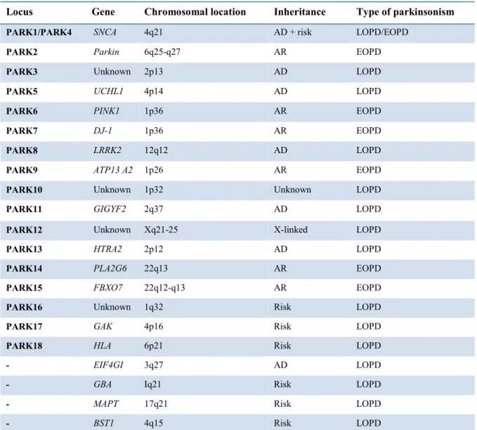
Genetics of Parkinson’s Disease
In the last two decades, using classic linkage analysis, genome-wide association studies and exome sequencing, 18 chromosomal loci (PARK1–18; Table 1) have been associated with familial PD (Lesage and Brice, 2012). To date, critical reviews argue that grouping different PD forms by pathology allow to better understand pathogenesis, nevertheless
14
most accepted classification show that gene variations causing PD are grouped in 2 categories: dominantly and recessively inherited mutations.
Autosomal dominant PD genes. Although mutations in LRRK2, represent the most
common cause of autosomal dominant PD (up to 10% of all familial forms) (Bonifati, 2014), at least three other genes (SNCA, VPS35, EIF4EG1) have been conclusively established to cause PD in the same autosomal dominant manner. The above mentioned α-synuclein is a 14 kDa protein localized to presynaptic terminals, in the nucleus, in the cytosol, in the mitochondria-associated membrane and in the endoplasmic reticulum (Guardia-Laguarta et al., 2014). The normal function of α-synuclein remains poorly understood and the precise mechanisms by which it leads to toxicity and cell death are also unclear. Substantial evidence suggests that α-syn function is related to its capacity to interact directly with particularly highly curved membranes phospholipids such as vesicles, playing a role in vesicle trafficking during exocytosis (Snead and Eliezer, 2014). Mutations in the SNCA gene (PARK1 locus), oxidative stress and post-translational modifications lead α-synuclein to adopt an oligomeric and/or fibrillary conformation that induces toxicity through several mechanism. These species could impair mitochondrial structure and complex activity (Devi et al., 2008), as well as disrupt ER-Golgi vesicular transport, which results in toxic ER stress and impairs the efficiency of some protein degradation mechanisms acting as negative regulator of DA release (Abeliovich et al., 2000).
Recently, the identification of the D620N variation on the Vacuolar Protein Sorting 35 (VPS35) gene has been shown to cause late-onset L-DOPA-responsive autosomal dominant parkinsonism which is reminiscent of sporadic PD (Trinh and Farrer, 2013). The frequency of mutation carriers is low and estimated to represent about 0.1% of the PD population. This gene encodes a subunit of the retromer system which mediates intracellular retrograde transport of endosomes to the trans-Golgi network (TGN). How mutant VPS35 causes PD is presently unknown, even though several lines of evidence indicate that mutant VPS35 may cause neurodegeneration via a gain-of-function mechanism with a dominant-negative effect on retromer assembly (Trinh and Farrer, 2013). In particular, Vps35 D620N mutants appear to disrupt the cargo sorting function of retromer, causing a deficit in retromer-dependent trafficking of CI-M6PR (cation-independent mannose-6-phosphate Receptor) and its ligand cathepsin D, likely arising from the generation and redistribution of enlarged endosome compartments that retain retromer (Follett et al., 2014).
15
Traditional linkage methods have recently identified the R1205H mutation in the EIF4G1 (eukaryotic translation initiation factor 4-gamma 1) gene as an infrequent cause of dominantly inherited PD, with an estimated frequency between 0.02–0.2% in the PD population (Chartier-Harlin et al., 2011). Of note, the eIF4G1 protein is regulated by phosphorylation of eIF4E-binding proteins (4E-BP) through the mammalian target of rapamycin (mTOR) pathway (Ramirez-Valle et al., 2008), and represents a multisubunit translation initiation complex involved in mRNA translation processes that regulates cell survival in response to stress (Chartier-Harlin et al., 2011). Interestingly, several lines of evidence point to a crucial involvement of the translation initiation process in PD. Indeed, it has been shown that LRRK2 interacts with eIF4E and directly phosphorylates 4E-BP at the postsynaptic site in vitro (Lee et al., 2012b). Furthermore the mTOR pathway and 4E-BP phosphorylation are strictly linked to PD. Activation of 4E-4E-BP by rapamycin, or its transgenic overexpression, prevent DA neuron loss in Drosophila models (Tain et al., 2009) and are associated with L-DOPA-induced dyskinesia (Santini et al., 2009). Finally, in a very recent study, OA Ross and colleagues established a link between VPS35 and EIF4G1 in α-synuclein-related neurodegeneration using a yeast model (Ross et al., 2015).
16
Table 1. Summary of genes and loci associated with PD.
Locus Gene Chromosomal location Inheritance Type of parkinsonism
PARK1/PARK4 SNCA 4q21 AD + risk LOPD/EOPD
PARK2 Parkin 6q25-q27 AR EOPD
PARK3 Unknown 2p13 AD LOPD
PARK5 UCHL1 4p14 AD LOPD
PARK6 PINK1 1p36 AR EOPD
PARK7 DJ-1 1p36 AR EOPD
PARK8 LRRK2 12q12 AD LOPD
PARK9 ATP13 A2 1p26 AR EOPD
PARK10 Unknown 1p32 Unknown LOPD
PARK11 GIGYF2 2q37 AD LOPD
PARK12 Unknown Xq21-25 X-linked LOPD
PARK13 HTRA2 2p12 AD LOPD
PARK14 PLA2G6 22q13 AR EOPD
PARK15 FBXO7 22q12-q13 AR EOPD
PARK16 Unknown 1q32 Risk LOPD
PARK17 GAK 4p16 Risk LOPD
PARK18 HLA 6p21 Risk LOPD
- EIF4GI 3q27 AD LOPD
- GBA Iq21 Risk LOPD
- MAPT 17q21 Risk LOPD
- BST1 4q15 Risk LOPD
Abbreviations: AD, autosomal dominant; AR, autosomal recessive; EOPD, early-onset PD; LOPD, late-onset PD
Autosomal recessive PD genes. Mutations, including point mutations and large
rearrangements, leading to deletions or multiplications, in each of the following three genes, Parkin (PARK2), PINK1 (PTEN induced putative kinase 1; PARK6) and DJ-1 (PARK7) have been identified worldwide.
Mutations in PARK2 alone account for almost 50% of early-onset familial recessive PD cases and are compatible with ∼15% of sporadic PD forms (Bonifati, 2014). Over a hundred mutations in PARK2 have been reported, which include homozygous or heterozygous point mutations but also deletions and duplications. The encoded protein, Parkin, belongs to the ubiquitin E3 ligases family and interacts with ubiquitin-conjugating enzymes (E2s) to catalyze the attachment of ubiquitin to protein targets (Shimura et al., 2000), thus tagging these proteins for destruction by the proteasome. In addition, Parkin is involved in mitochondrial maintenance, and mutations within this gene might be associated
17
with changes of mitochondrial autophagy and neuronal dysfunction (Park et al., 2009). Patients carrying Parkin mutations present L-DOPA-responsive PD, frequently accompanied by motor fluctuations and dyskinesias that often develop early in the course of treatment (Periquet et al., 2001). However, rarely LBs have been detected, although neural loss in nigrostriatal pathway is usually severe, suggesting pathogenic differences between the autosomal recessive and the classical forms of PD. Nonetheless, the first patient with pathogenic PINK1 mutations and LBs-positive pathology was recently reported (Bonifati, 2014).
Mutations in the PINK1 and DJ-1 gene are less common, accounting for ∼1–8%, and 1– 2% of early-onset PD cases, respectively (Bonifati, 2014). The protein encoded by PINK1 is located in the mitochondrial membranes and is involved in the mitochondrial response to cellular and oxidative stress (Valente et al., 2004a). It is clear that PINK1, by acting upstream of parkin, is involved in the elimination of damaged mitochondria, and might protect neurons against mitochondrial dysfunction and proteasome-induced apoptosis (Bonifati, 2014). The clinical phenotype is similar to that caused by mutations in Parkin, with a slow disease progression and rare additional psychiatric disturbances, particularly anxiety and depression (Valente et al., 2004b).
Interestingly DJ-1 is also likely to be involved in mitochondrial physiology; in fact it represents a sensor for oxidative stress and may mediate neuroprotection. In the presence of oxidative stress, DJ-1 has been shown to translocate to the mitochondria and exert a protective effect (Canet-Aviles et al., 2004). In particular, the L166P mutation leads to a less stable protein and to the reduction of antioxidative activity, implicating a loss-of-function mechanism (Moore et al., 2003). Finally, similar to the other recessive PD forms, the clinical picture includes an early onset, good response to L-DOPA, and slow progression. By analysis of these genes, it has come most clearly that repair of damaged mitochondria represents a common physiological function, but how these proteins are involved is not yet clear.
Of note, other more rare recessively inherited forms of PD have been identified. Mutations in the ATP13A2 (ATPase type 13A2), PLA2G6 (phospholipase A2, group VI) and FBXO7 (F-box only protein 7) genes cause an atypical PD-associated phenotype that is distinguished from the other three aforementioned forms by an earlier (juvenile) onset, partial or less sustained response to L-DOPA, and atypical clinical features such as upper motor neuron signs, pyramidal signs, dystonia, supranuclear gaze palsy, myoclonus and cognitive decline (Bonifati, 2014).
18
LRRK2-associated phenotype
The majority of clinical reports on LRRK2 PD patients consistently indicate that their clinical phenotype is not distinguishable from the idiopathic form (Healy et al., 2008; Paisan-Ruiz, 2009). PD patients carrying LRRK2 pathogenic mutations have been identified in more than 40 populations worldwide. In addition, variable degrees of population-specificity have been shown, indicating that ethnicity is an important factor that influences diagnosis (Paisan-Ruiz, 2009). Notably, penetrance of LRRK2 mutations is almost complete and age-dependent, with an increase between 17% to 85% from 50 to 70 years of age, and some G2019S mutation carriers exhibiting the disease after 80 years of age (San Luciano et al., 2010). The most frequent LRRK2 mutation, G2019S, shows incomplete penetrance and this might explain why this mutation is detectable in patients with familial PD but also in some with sporadic PD, suggesting that genetic and/or environmental factors may associate with LRRK2 to trigger dopaminergic neurodegeneration (Hulihan et al., 2008). This variant is frequent in PD patients from southern Europe, in particular in Portuguese (16%), Spanish and Italian populations. However, the highest incidence has been reported in Nord African Arabs (42%) and in Ashkenazi Jewish (28%) (Paisan-Ruiz et al., 2013). Moreover, G2019S is associated with a specific haplotype present in all subjects, suggesting that the mutation was transmitted by a single ancient founder across European population (Kachergus et al., 2005). Given the high frequency of the G2019S mutation, the clinical phenotype of LRRK2 patients is mostly associated with this mutation. Several studies consider tremor as the initial and predominant symptom of LRRK2 carriers but, in a recent study, tremor incidence was lower in patients with G2019S PD than idiopathic PD (Trinh et al., 2014). Overall, the G2019S mutation is often associated with the classical motor triad characterized by bradykinesia, rigidity and asymmetrical tremor, by a good response to L-DOPA and a slow and benign disease progression (Paisan-Ruiz, 2009). In addition, postural instability and dystonia have been reported, although dystonia may only appear due to a complication of both medical and surgical treatment (Healy et al., 2008). The impact on the susceptibility to develop L-DOPA-induced dyskinesias (LID) is also debated: some groups found it to be more frequent in G2019S carriers than idiopathic PD (Lesage et al., 2008), whereas others showed no difference, even though the time elapsed from L-DOPA treatment onset to LID appearance was significant longer in LRRK2 compared to non-LRRK2 carriers (Healy et al., 2008). Compared to sporadic PD patients, LRRK2 carriers present a lower risk of cognitive decline and psychiatric features (Alcalay et al., 2010); on the other hand, higher frequency of depression, anxiety and irritability have been reported. Interestingly, Shanker
19
and colleagues reported a trend for a greater risk of premorbid mood disorders in LRRK2 patients compared to gene-negative patients, assuming that mood disorder susceptibility genes may modify LRRK2 mutation penetrance. Of note, numerous reports identified different degrees of cognitive alterations in a number of healthy asymptomatic G2019S carriers that could represent a hopeful way to investigate early markers for pre-symptomatic PD (Thaler et al., 2012).
The distribution in age at onset and the clinical features are similar in LRRK2 R1441C patients and idiopathic or LRRK2 G2019S PD patients. Moreover G2385R carrier patients demonstrate clinical features similar to non-carrier patients, indicating that mutations in different LRRK2 domains lead to similar clinical phenotypes. Lastly, patients carrying the Y1699C mutation show unilateral leg tremors at onset, foot dystonia and then bradykinesia, rigidity, and postural instability with good responsitivity to L-DOPA (Haugarvoll et al., 2008).
Neuropathology of LRRK2 mutation carriers
The pathological features of patients with LRRK2 mutations are strikingly heterogeneous. Postmortem analysis from more than 30 LRRK2 mutation carriers displayed dopaminergic neuronal loss and gliosis in the SN, although the same mutations can cause quite different neuropathology, and LBs or Lewy neurites are not present in all cases (Wider et al., 2010). The G2019S phenotype is often characterized by the presence of α-synuclein–immune-positive LBs and, although other postmortem findings, such as tau-α-synuclein–immune-positive or ubiquitin-immunoreactive inclusions, have been found, this mutation does not always manifest as synucleinopathy or LBs disease (Ruffmann et al., 2012). Rather than distinct processes, these pathological endpoints may share the same primary cause, assuming that LRRK2 and tau/ α -synuclein crosstalk along a common neurotoxicity pathway (Rajput et al., 2006). However, the exact relationship between α–synuclein and LRRK2 is unclear as postmortem localization studies have produced conflicting results (Sharma et al., 2011). Due to the growing interest in the potential interaction of these proteins in the pathogenesis of PD, a very recent study from Guerreiro group, using diverse and more rigorous techniques, showed that endogenous LRRK2 and α-synuclein interact in cells, mouse and human brain tissue. Their data show that the G2019S mutation does not alter the ability of LRRK2 to interact with α-synuclein in HEK-293 cells, indicating that the kinase domain, and hence the phosphorylation capacity of LRRK2, does not play a major role in its interaction with α-synuclein (Guerreiro et al., 2013). Interestingly, in cell models, an increase in LRRK2 expression is associated with elevation of α-synuclein mRNA
20
(Carballo-Carbajal et al., 2010). These features are consistent with reports that the levels of LRRK2, rather than its mutations, regulate the progression of neuropathology induced by PD-related α-synuclein mutants (Lin et al., 2009). Furthermore, despite the poor specificity of the different LRRK2 antibodies (Davies et al., 2013), a number of studies in human brain tissues showed the co-localization of LRRK2 with α-synuclein-immunoreactive LBs (Guerreiro et al., 2013),. Of note, analysis of LRRK2 mRNA expression in post-mortem brain tissue from control, idiopathic PD (IPD) cases and G2019S-positive PD cases showed, in addition to a widespread neuronal localization and weak levels in SN, significant reductions in non-nigral regions (cerebellum, amygdala, frontal cortex and cingulate gyrus) of IPD brain (Sharma et al., 2011). In this scenario, further work is required to explain the contrast between the increased levels of LRRK2 in PD brain regions with pathological accumulation of α-synuclein and the pathogenic role of LRRK2 outside nigral neurons.
The most striking variability was observed in R1441 mutation carriers. DA neuron loss and gliosis in the SN, without α-synuclein-positive inclusions, have been reported in the only case examined with the R1441G substitution (Marti-Masso et al., 2009), even though some autopsies from R1441C carriers showed various neuropathological patterns. Indeed, data from four patients revealed that one case did not show synuclein and tau pathology, two cases had LBs and Lewy’s neurites (LNs), and the remaining one showed neurofibrillary tangles (NFT) without either LBs or LNs (Wszolek et al., 1997). However, no pathology reports are available for p.R1441H substitution carriers. Finally, similar finding have been reported for the I2020T and Y1699C mutations that showed tau, LBs and ubiquitin-positive cytoplasmic and nuclear inclusions (Ujiie et al., 2012).
Understanding the mechanisms that lead to dopaminergic loss in LRRK2 patients represents the unresolved challenge in the LRRK2 field. The clinical and pathological heterogeneity of LRRK2 mutation carriers, together with the widespread expression of LRRK2, supported the idea that this protein interacts with other molecules in the affected neuronal populations, and this may play an important role in the accumulation and aggregation of unfolded proteins. As extensively suggested, LRRK2 interacts with other proteins which also form protein complexes (Cookson, 2010; Greggio et al., 2011), likely acting upstream of them. Moreover, additional genetic and environmental risk factors determine the type of pathology that develops in a given individual. For example, mutation of the genes encoding α-synuclein and tau, SNCA and microtubule associated protein tau (MAPT) respectively, could be the obvious candidate to influence pathogenesis, directly or indirectly interacting with the protein LRRK2. Likewise, different environmental factors
21
such as pesticides and head trauma might lead LRRK2 either towards tau or α-synuclein pathology (Li et al., 2014). To date, the physiological and pathophysiological consequences of LRRK2 mutation remain largely unknown. Investigators have largely attempted to validate LRRK2 protein interactors by using a LRRK2- dependent phosphorylation readout, but until now all attempts have failed.
LRRK2: protein interaction network and pathogenic mechanisms
Expression
LRRK2 is found at high levels in several organs, such as liver, lung, kidney, heart, spleen, intestine and lymph nodes (Galter et al., 2006; Giasson et al., 2006; Hakimi et al., 2011), while is poorly expressed in the mammalian brain relative to most well-characterized protein kinases. Despite the lack of sensitive and specific antibodies (Davies et al., 2013), the neuroanatomical localization of LRRK2 in the brain showed a striking relation between LRRK2 gene expression and the nigrostriatal DA system affected in PD. In situ hybridization studies on LRRK2 mRNA in the mouse brain revealed that expression is highest in the cortex, moderate in striatum, olfactory tubercle, hippocampus and cerebellum, and lowest in SNpc (Biskup et al., 2006; Melrose et al., 2006).
Within the brain, LRRK2 is abundantly expressed in neurons, but it can also be detected at lower levels in astrocytes and microglia where its expression can be induced by inflammatory stimuli (Giesert et al., 2013; Paisan-Ruiz, 2009). In the cortex, striatum, and SNpc, the pattern of LRRK2 expression differs within distinct neuronal subpopulations . Throughout the striatum, in both rats and mice, LRRK2 expression appeared to be restricted to medium spiny neurons, with low to undetectable expression in larger interneurons. This is also confirmed by the overlapping of DARPP-32 and LRRK2 staining in neuronal perikarya. In the last years, the expression of LRRK2 in SNpc has been object of intensive studies. Diverse studies failed to detected protein levels, while others, after having developed more specific anti-LRRK2 polyclonal antibodies, described LRRK2 protein expression in the mouse and human SNpc, (Biskup et al., 2006; Greggio et al., 2006; Melrose et al., 2007). Interestingly, although any LRRK2 signal using similar staining conditions has been found in rat SNpc dopaminergic neurons, minimal LRRK2-immunoreactivity has been showed in the substantia nigra pars reticulata (SNpr) in both rats and mice (West et al., 2014). At a subcellular level, in striatal and cortical neurons, LRRK2 is localized throughout the cytoplasm of neuronal perikarya and dendritic
22
processes where it is associated with vesicular and intracellular membranous structures (i.e. mitochondria, endosomes, lysosomes, multivesicular bodies, lipid rafts, microtubule-associated transport vesicles, synaptosomes, Golgi complex, and endoplasmic reticulum), with the microtubule network and other membrane-bound organelles (Alegre-Abarrategui et al., 2009; Biskup et al., 2006). The synaptic localization of LRRK2 represents a contentious issue. In particular, some studies found that LRRK2 expression does not overlap with presynaptic markers, including synaptotagmin-1 (a large protein able to bind Ca2+ and involved in the presynaptic vesicle membrane fusion) in striatal and cortical neurons, but has close association with TH-positive projections, suggesting a predominantly postsynaptic localization in the striatum (West et al., 2014). Instead, others investigators suggest that the association of LRRK2 with cytoskeletal elements hints a possible role in vesicular transport, protein trafficking and presynaptic vesicle endo/exocytosis (Belluzzi et al., 2012) as well as in membrane and protein turnover, including the lysosomal degradation pathway. The LRRK2 association with outer mitochondrial membrane in rodents may suggest a causal role in PD, where mitochondrial function is impaired. Interestingly, mitochondria undergo frequent fission and fusion events especially before a cell undergoes apoptosis, and these processes are regulated by molecular machinery that includes dynamin-related GTPases and WD40 repeat-containing proteins. Therefore, LRRK2 might potentially serve as a scaffold during mitochondrial fission and fusion (Li and Beal, 2005).
Interaction network and cellular function
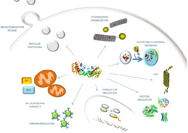
LRRK2 protein distribution within various cell compartments might reflect a functional role in multiple cellular pathways (Fig.3). The physiological cellular function of LRRK2 is not clear despite strong evolutionary conservation of this class of protein. Some evidence suggests a role in cytoskeleton organization, translation regulation, autophagy-lysosomal pathways, neurite outgrowth, vesicular trafficking, neurotransmitter release and immunoregulation, but as mentioned above, little is known about the physiological interactors and/or regulators of LRRK2, and a few substrates for its domains have been described.
23
Fig. 3. LRRK2 involvement in cellular mechanisms.
Cytoskeletal regulation. Ultrastructural analysis supports LRRK2 interaction with
cytoskeletal proteins including tubulin, tubulin-associated tau and actin. A very recent study, in line with previous evidence, demonstrated a specific, direct interaction of LRRK2 with three different β-tubulin isoforms, which is mediated by the LRRK2 ROC domain and the β-tubulin C-terminus. This interaction is dependent on guanidine nucleotide binding and modulated by ROC domain autophosphorylation. The R1441G mutation seems to disrupt this interaction, possibly by affecting tubulin acetylation. Conversely, embryonic fibroblasts (MEFs) obtained from LRRK2 knock-out mice show increased tubulin acetylation. Interestingly, Gillardon and collaborators found that recombinant human LRRK2 phosphorylates β-tubulin, and phosphorylation levels are three-fold enhanced in the presence of the G2019S mutation, with the consequential increase of microtubule polymerization (Gillardon, 2009). In addition, LRRK2 co-localizes with highly dynamic cytoskeletal structures in dopaminergic cells, and LRRK2 overexpression and mutation impact upon the morphology of growth cones (Law et al., 2014).
24
Moesin, a component of the ERM (ezrin/radixin/ moesin) family has been identified as one of the cytoskeletal LRRK2 kinase target (Parisiadou et al., 2009). These proteins play an important role in the regulation of the membrane structure and its organization by anchoring the actin cytoskeleton to the plasma membrane. Although moesin is likely to be only an efficient in vitro substrate for LRRK2, it allows the development of efficient model substrate peptides, called LRRKtide and Nictide, derived from ERK sequence, which helped improve the analysis of LRRK2 kinase activity in vitro (Jaleel et al., 2007). However, LRRK2 also plays a role in actin dynamics, interacting with actin isoforms and with the actin-associated proteins that contribute to filament assembly, organization and maintenance. A number of studies show that LRRK2 mutations lead to the accumulation of polymerized actin as well as phosphorylated ERM, and that these effects are reversed in LRRK2 knockout neurons (Parisiadou et al., 2009). Pathogenic mutations influence the interaction between LRRK2 and microtubules: some studies indicate that G2019S and R1441G/H reduce LRRK2- β-tubulin interaction (Law et al., 2014), while others an increased affinity (Kett et al., 2012). Taken together, these data suggest a role for LRRK2 in the regulation of cytoskeletal dynamics with implications for the pathogenesis of PD. Furthermore, the suggestion that LRRK2 can directly phosphorylate tubulin-associated tau is corroborated from the evidence that tau phosphorylation is reduced in LRRK2 knock-out mice (Kawakami et al., 2012). In addition, the G2019S and I2020T mutations increase tau-phosphorylation, influencing the tau’s affinity for microtubules protein and promoting its aggregation (Melrose et al., 2010).
Protein regulation and Autophagy. LRRK2 has been proposed to physically interact with
14-3-3 proteins, upon phosphorylation at residues S910 and S935 at the N-terminus of the leucine-rich repeats (Dzamko et al., 2010; Nichols et al., 2010). The 14-3-3 proteins represent a family of conserved regulatory molecules that usually assemble into either homo- or heterodimers and, in their dimeric form, bind to two phosphorylated residues which can come from the same target protein, or from two separate binding partners. These proteins have the ability to protect important regulatory sites from dephosphorylation, or to mediate dimerization of two distinct target proteins. Dzamko and colleagues showed that pathogenic LRRK2 mutations disrupt the binding between 14-3-3 and LRRK2: as a consequence, LRRK2 levels are decreased and LRKK2 is accumulated in non-cytosolic pools. How 14-3-3 interaction influences LRRK2 function or its cytoplasmic localization has not been clarified yet. It has been shown that this interaction does not control LRRK2 protein kinase activity, as mutations of Ser910 and/or Ser935 do not influence LRRK2
25
catalytic activity or dimer formation of wild type LRRK2. However, Ser910/Ser935 phosphorylation and 14-3-3 binding is markedly reduced in mouse tissues derived from homozygous R1441C knock-in mice that display impaired dopaminergic neurotransmission (Dzamko et al., 2010; Nichols et al., 2010; Tong et al., 2009). A very recent study describes an additional residue in the ROC domain involved in the regulation of the 14-3-3 binding to LRRK2. Binding of phospho-Ser1444 residue to 14-3-3 impairs LRRK2 kinase activity, and phosphorylation is abolished by pathogenic mutations in the ROC domain, even though whether this results in altered cytosolic localization of LRRK2 has not been determined yet (Muda et al., 2014). In addition, the Ser910, Ser935 and Ser1444 residues are not phosphorylated by LRRK2 itself, but rather by multiple kinases, including cAMP-dependent protein kinase (PKA). This kinase is one of key regulators in signal transduction in the brain; it is activated in response to increasing concentrations of cAMP and phosphorylates a vast number of protein kinases, including the MAPKKK c-Raf. Furthermore, PKA facilitates synaptic transmission and assumes an important function as a regulator of DA synthesis. In this context, LRRK2 kinase activity could be modulated by PKA-mediated binding of 14-3-3, suggesting that PKA acts as an upstream kinase for LRRK2 (Muda et al., 2014).
Interaction between the LRRK2 ROC-COR domain with the DVL (dishevelled) phosphoproteins family (DVL1-3) has been reported. DVLs represent key regulators of Wnt (Wingless/Int) signalling pathway, which is important for axon guidance, synapse formation and neuronal maintenance. Indeed, they can mediate the activation of small GTPases with structural similarity to the LRRK2 ROC domain. In this context, DVLs may influence LRRK2 GTPase activity, and ROC-COR domain mutations modulating LRRK2-DVL interactions indirectly influence kinase activity (Sancho et al., 2009). In addition, LRRK2 has been reported to interact with various small GTPases including Rac1 (Chan et al., 2011), rab5b (Shin et al., 2008), and rab7 (Dodson et al., 2012).
Accumulating evidence suggests that LRRK2 interacts with heat shock protein 90 (Hsp90) in vitro and in vivo, and that inhibition of Hsp90 disrupts this association, followed by LRRK2 degradation and increased cell viability. Hsp90 is a molecular chaperone essential for activating many signaling proteins and for regulating their stability in the eukaryotic cell. Moreover, Hsp90 contributes to the stabilization, activation, and/or translocation of client protein kinases, such as Src family tyrosine kinase and serine/threonine protein kinases, such as LRRK2. The binding of Hsp90 to LRRK2 seems to reflect protein instability, indeed the inhibition of Hsp90 chaperone function dramatically decreases the stability of LRRK2 in cell lines and primary neuronal cultures via a proteasome-mediated
26
degradation mechanism. Hsp90 could be a target for suppressing the accumulation of PD-related LRRK2 mutants and their pathogenic activity in neurons. Indeed, treatment of neurons with a Hsp90 inhibitor rescued the axon growth retardation defect caused by overexpression of the LRRK2 G2019S mutation (Wang et al., 2008). Furthermore, Hsp90 shows greater affinity for G2385R LRRK2 than the wild-type form, and its inhibition leads to the association of mutant LRRK2 with high molecular mass native fractions, that probably represent proteasome degradation products. Of note the G2385R mutation within the WD40 domain, which represents a risk factor of PD, causes a partial loss ofkinase activity by reducing phosphorylation at Ser910/Ser935 sites, and a concomitant diminished ability to bind 14-3-3 proteins (Rudenko et al., 2012).
Moreover, LRRK2 was found to interact with the C-terminus of Hsp70-interacting protein (CHIP), an E3 ubiquitin ligase that significantly reduces the cellular levels of LRRK2 by ubiquitination and proteasome-dependent degradation, leading to lowered LRRK2 protein levels (Ding and Goldberg, 2009). Binding to LRRK2 occurs between the charged domain and the ROC domain of LRRK2, or between the TPR domain of CHIP and the N-terminal region of LRRK2.Since the TPR domain of CHIP is well-known to bind to Hsp90, it is likely that a portion of the LRRK2-Hsp90 complex will also be associated with CHIP. Interestingly, because it has been reported that LRRK2 binds to the E3 ubiquitin ligase Parkin, enhancing its auto-ubiquitination activity, and that CHIP also forms a complex with Parkin enhancing its ubiquitin ligase activity, it is possible that the reported interaction between LRRK2 and Parkin is mediated by CHIP (Ding and Goldberg, 2009). It
has been proposed that the ability of CHIP to degrade potentially neurotoxic misfolded, damaged or mutated proteins might diminish with age. In this contest diminished CHIP-mediated degradation of LRRK2 in aged or stressed neurons may contribute to sporadic PD as well as familial PD in patients bearing LRRK2 mutations.
In either case, current data indicate that LRRK2 protein levels nicely correlate with neuronal toxicity. The interaction with Hsp70 targets LRRK2 to the lysosomal membrane for chaperone-mediated degradation thus representing a mechanism to control protein levels, (Skibinski et al., 2014). Recently, Beilina and colleagues, using protein–protein interaction arrays, identified BCL2-associated athanogene 5, Rab7L1 (RAB7, member RAS oncogene family-like 1), and Cyclin-G–associated kinase, as binding partners of LRRK2. These proteins form a complex that promotes clearance of Golgi-derived vesicles through the autophagy–lysosome system both in vitro and in vivo, although the impact of these interactions on LRRK2 levels and chaperone-mediated autophagy remains unknown (Beilina et al., 2014). Interestingly, additional studies have investigated the interaction
27
between LRRK2 and mitogen-activated kinases (MAPKs), such as extracellular signal-regulated kinase (ERK), Jun N-terminal kinase (JNK) and p38. LRRK2 induces the phosphorylation of MAPK/ERK kinases (MEK), while the G2019S mutation promotes autophagy in cells via the MEK/ERK pathway (Liou et al., 2008).
Effect on protein translation. Evidence has been provided that LRRK2 is involved in one
of the many established signaling cascades, including the mTOR and ERK pathway, and protein translation in general has been suggested to be instrumental to LRRK2 effects in fly models. In particular, both human LRRK2 and the Drosophila orthologue of LRRK2 phosphorylate eukaryotic initiation factor 4E (eIF4E)-binding protein (4E-BP), a negative regulator of eIF4E-mediated protein translation and a key mediator of various stress responses. 4E-BP is a target of mammalian target of rapamycin (mTOR), which is of interest in research on aging since recent evidence suggests that in diverse species, deletion of mTOR signalling components or treatment with the mTOR inhibitor, rapamycin, can extend lifespan. It has long been recognized that a key regulator of eIF4E function is the phosphorylation-induced release of 4E-BP from eIF4E. Although LRRK2 stimulates eIF4E-mediated protein translation both in vivo and in vitro, it attenuates resistance to oxidative stress and survival of DA neurons in Drosophila. The chronic inactivation of 4E-BP by pathogenic LRRK2 mutations might lead to deregulation of protein translation, likely resulting in age-dependent loss of DA neurons (Imai et al., 2008). mTOR signaling may link LRRK2 with aging, despite the fact that experiments have shown that 4E-BP is a relatively poor substrate for highly purified LRRK2.
Because recent genome-wide association studies (GWAS) revealed that the LRRK2 locus represents a genetic risk factor for sporadic PD (Satake et al., 2009), the potential alteration of LRRK2 expression in the etiology of sporadic PD is of outmost interest. To this purpose a number of studies have highlighted a role for LRRK2 in microRNA (miRNA) regulation. miRNAs are evolutionarily conserved small non-protein-coding transcripts that bind to partially complementary sites in the 3′-untranslated region (3′-UTR) of target messenger RNAs (mRNAs), and thereby control the translation of their target gene. A number of miRNAs have been associated with neuronal development, synaptic plasticity, memory formation and neurodegenerative diseases in the nervous system through their regulation of the translation of targeted genes (Hebert and De Strooper, 2009). Cho and colleagues, reported that the levels of LRRK2 were enhanced whereas those of miRNA-205 (miR-205) were decreased in the frontal cortex of sporadic PD patients (Cho et al., 2013). Since LRRK2 mRNA levels (i.e. LRRK2 expression) remained unchanged, a potential
post-28
transcriptional modification of the LRRK2 protein expression in the sporadic PD brain was suggested. Interestingly, miR-205 reduced LRRK2 levels in cell lines and primary neuron cultures, through targeting its binding site in the 3′-UTR of LRRK2 gene. In addition, introduction of miR-205 prevented the neurite outgrowth defects in the neurons expressing a PD-related LRRK2 R1441G mutant. In this context, the downregulation of miR-205 may play a role in the pathogenic elevation of LRRK2 protein in the sporadic PD brain (Cho et al., 2013). These findings support previous data in which LRRK2, through its kinase domain, was shown to control the production and the regulation of let-7 and miR-184, (Gehrke et al., 2010). Unfortunately, at this stage little is known about the alteration in LRRK2 function during the aging process, and no specific post-translational modifications associated with aging are known, as it is undetermined how people carrying LRRK2 mutations develop disease as adults.
Relationship to mitochondria. Mitochondrial dysfunction is widely recognized as a trigger
of PD, and LRRK2 has been found to be involved in different mitochondrial events, including mitochondrial dynamics and morphology, mitochondrial calcium buffering, ROS production and mitochondrial membrane potential maintenance. In vitro studies on primary cultures of cortical neurons found that LRRK2 colocalizes with the mitochondrial marker Cyto C and partially with the fission Dynamin like protein 1 (DLP1). Interestingly, LRRK2 G2019S overexpression in cortical neurons increases DLP-1 activity and promotes mitochondrial fission, suggesting that LRRK2 is likely to be involved in mitochondrial fission/fusion dynamics (Niu et al., 2012). In addition, cellular transfection with LRRK2 WT and LRRK2 G2019S could induce mitochondrial fragmentation and consequent cell death (Iaccarino et al., 2007). A reduction of mitochondrial membrane potential and intracellular ATP levels, accompanied by mitochondrial elongation, have been reported in skin biopsies from human LRRK2 G2019S carriers (Mortiboys et al., 2010). This is in accordance with the study of Papkovskaia and colleagues (Papkovskaia et al., 2012) that pointed to a kinase-dependent mechanism. Moreover, LRRK2 mutants are involved in the modulation of the peroxiredoxin3 activity. This enzyme, located within mitochondria, acts as antioxidant and is phosphorylated by LRRK2 (Angeles et al., 2011). Reduced cell survival and mitochondrial dysfunction, such as increased sensitivity to rotenone-mediated complex I inhibition (Ng et al., 2009), have been also shown in Drosophila flies carrying LRRK2 mutants, whereas WT LRRK2, seems to attenuate H2O2-induced oxidative stress,
suggesting a protective role for LRRK2 (Liou et al., 2008). Mutations in LRRK2 could affect mitochondrial function by increasing the vulnerability to oxidative stress. Induced
29
pluripotent stem cells (iPSCs) derived from fibroblast of G2019S LRRK2 carriers show higher levels of mitochondrial DNA (mtDNA) damage than cells from control subjects, and are characterized by a reduced mitochondrial respiration and motility (Papkovskaia et al., 2012).
LRRK2 and vesicle trafficking. Given the synaptic expression of LRRK2 and its
association with the membranous structures involved in vesicular release, including endoplasmic reticulum, Golgi apparatus, cytoskeleton, lipid raft and synaptic vesicles (Migheli et al., 2013), it is not surprising that LRRK2 is involved in vesicle trafficking and neurotransmitter release (Tong et al., 2009). Of note, the activated form of LRRK2 (LRRK2 dimer) is substantially enriched at the membrane of cells expressing endogenous or exogenous LRRK2, and the membrane-associated fraction of LRRK2 possesses greater kinase activity than cytosolic LRRK2, suggesting intense catalytic role at this site (Berger et al., 2010). Several studies show that LRRK2 acts at the synaptic site, interacting with a high number of pre-synaptic proteins including NSF (N-ethylmaleimide-sensitive fusion
factor), AP-2 (adapter protein 2), SV2A (synapticvescicle protein 2A), synapsin 1A,
syntaxin 1, clathrin (Piccoli et al., 2011), Rab5 and actin (Meixner et al., 2011). Shin and colleagues reported that the interaction between LRRK2 and Rab5 is involved in the modulation of synaptic vesicle endocytosis (Shin et al., 2008). Recently in vivo and in vitro studies revealed the interaction between LRRK2 and ArfGAP1 (ADP-ribosylation
factor GTPase-activating protein 1). ArfGAP1 is able to promote the GTPase activity of
Arf1 (ADP-ribosylation factor 1), a small protein involved in maintaining the morphology of the Golgi apparatus and the extrusion of vesicular proteins to the endoplasmic reticulum (Donaldson and Radhakrishna, 2001).
LRRK2 interacts with actin filaments and microtubules involved in vesicular transport and trafficking, regulating ERM phosphorylation via kinase activity and actin polymerization (Parisiadou et al., 2009). Interestingly, Cirnaru and coworkers (Cirnaru et al., 2014) showed a reduction of the interaction between LRRK2 and synaptic vesicles and a simultaneous enhancement of the affinity between actin and synapsin, after inhibition of kinase function.
LRRK2 interacts with SV2A, a neuronal protein involved in Ca2 + -dependent exocytosis (Chang and Sudhof, 2009). SV2A interacts with synaptotagmin, a Ca2+ sensor protein in the membrane of the pre-synaptic axon terminal that interacts with the SNARE complex (Soluble NSF Attachment Protein Receptor) under Ca2+-free conditions and translocates to the lipid membrane when is bound to Ca2+ . The interaction of SV2A with Synaptotagmin