Published online 2015 November 2. Review Article
Treatment of Acute Diarrhoea: Past and Now
Giuseppe Caramia,
1Stefania Silvi,
2Maria Cristina Verdenelli,
2and Maria Magdalena
Coman
2,3,*1Hemeritus Head Physician of Pediatrics and Neonatology, Specialized Maternal-Infantile Hospital “G. Salesi”, Ancona, Italy 2School of Biosciences and Veterinary Medicine, University of Camerino, Camerino, Italy
3School of Advanced Studies, University of Camerino, Camerino, Italy
*Corresponding author: Maria Magdalena Coman, School of Biosciences and Veterinary Medicine, University of Camerino, Camerino, Italy. Tel: +39-0737402737, Fax: +39-0737402418, E-mail: [email protected]
Received 2015 May 24; Revised 2015 June 25; Accepted 2015 July 8
Abstract
Context: Since ancient times diarrhoea has been a highly fatal disease and even today diarrhoea, the topic of this review, is a problem
affecting millions of people around the world despite the efforts of governments and professionals from the medical area. Worldwide the most common cause of children’s death is diarrhoea.
Evidence Acquisition: Diarrhoea disorders generally appear with watery stools, sometimes mixed with blood, accompanied by
abdominal pain, vomiting and fever. The symptoms depend on the content and distribution of body fluid, daily water requirements and physiological water loss in connection with age through sweating, urination and breathing, the degree of fluid and electrolyte loss in the liquid stool.
Results: Several effective interventions have been introduced as part of diarrhoea management in the last two decades such as oral
rehydration solution, zinc supplementation, vitamin A supplementation and oral administration of antibiotics and vaccines. To reduce the mortality rate, control of safe drinking water, good sanitation and vaccination against typhoid and cholera are recommended, especially in high-risk populations. Probiotics have been proposed, after more than a half of century, as additional therapy in the treatment of acute diarrhoea. Several probiotic strains showed benefit in meta-analyses of randomised controlled trials.
Conclusions: Due to the high level of evidence available, the term “oral bacteriotherapy”, used for decades in the prevention and therapy
of gastroenteritis in the growing age and adults, has expanded, but probiotics are acquiring significant scientific value based on the results from human trials. The future of probiotics depends on further explanation/elucidation of basic mechanisms, allowing scientists and physicians to maximize their health benefits.
Keywords: Probiotics, Diarrhoea, Oral Rehydration, Alternative Therapy
Copyright © 2015, Alborz University of Medical Sciences. This is an open-access article distributed under the terms of the Creative Commons Attribution-NonCom-mercial 4.0 International License (http://creativecommons.org/licenses/by-nc/4.0/) which permits copy and redistribute the material just in noncomAttribution-NonCom-mercial us-ages, provided the original work is properly cited.
1. Context
The present paper is a review on diarrhoea, including pathogens that cause several types of diarrhoea, mor-tality trend around the world and methods to detect the causative agents. The authors overviewed all the interventions to fight diarrhoea, such as oral rehydra-tion therapy (ORS), zinc and vitamin supplementa-tion, antibiotics, vaccine and probiotics in diarrhoea management.
1.1. Diarrhoea Definition
Diarrhoea is defined as stool emission with an excess weight of 200 grams per day and “pseudo diarrhoea” when faecal weight is < 200 g/24 hours (1, 2). Diar-rhoea is an alteration of normal bowel movement, variation characterized by increased water content, volume and frequency of stools. Intestinal water bal-ance results from a complex regulation involving
flammatory mediators, hormones, neuropeptides, in-testinal wall integrity, enteric nervous and circulatory system efficiency (3).
1.2. Diarrhoea Epidemiology and Trends in
Mortal-ity in WHO Regions
Worldwide, diarrhoea is the second most frequent fatal childhood disease after pneumonia, estimated to cause 1.34 million of children deaths. Different causes of death in children younger than five years vary over the world and WHO regions, although pneu-monia and diarrhoea, remain the same, significantly increase where the health systems are inefficient (4, 5) (Table 1, Figure 1).
Of all deaths from diarrhoea, 78% of children live in the African and South-East Asian regions, which are dispro-portionately loaded with infant and childhood HIV
infec-tions (7). Diarrhoeal disease occurs more frequently in HIV-infected children and their consequences are worse. Administration of antiretroviral therapy (ART) and re-storing the immune system are critical for preventing and treating diarrhoea in children affected by HIV (8). Re-search on diarrhoea in children is a priority of WHO for achieving the United Nations’ Millennium Development Goal of reducing childhood mortality.
1.3. Types of Diarrhoea
Diarrhoea, from a clinical point of view, needs to be clas-sified according to certain characteristics such as trends over time (acute or chronic) and faeces characteristics (watery, fatty, inflammatory, etc.) (3, 9).
Generally, episodes of diarrhoea can be classified into three categories including acute diarrhoea with pres-ence of three or more watery stools within 24-hours, dysentery with bloody diarrhoea and mucous presence, persistent diarrhoea when episodes of diarrhoea last more than 14 days, while chronic diarrhoea when the episode lasts for more than a month (10, 11). Diarrhoea reflects higher water content in the stool, number of stools that may reach 20 or more per day, with defeca-tion every 20 - 30 minutes.
1.4. Main Pathogens Responsible for Acute Diarrhoea
Diarrhoea is caused by infections, often spread from person-to-person or contaminated sources, water con-taminated with human or animal faeces and other causes including poor personal hygiene, food conser-vation in unhygienic conditions and malnutrition. Dy-senteric cases often have no identifiable agent (12, 13).
1.4.1. Bacteria
Bacteria may be the causative pathogens (15 to 20%) including Salmonella spp., Campylobacter spp, and Esch-erichia coli. Bacteremia, sepsis, spreading to other organs
represent systemic symptoms that complicate gastroin-testinal infection, especially by Campylobacter, E. coli, Salmonella, Shigella, Aeromonas and Yersinia (Table 2). Clostridium difficile is a pathogen that causes
uncompli-cated antibiotic-associated diarrhoea and severe, possi-bly fatal, antibiotic-associated colitis (14). Prevention is possible through infection control measures both clini-cal and epidemiologiclini-cal (15). Many pathogenic serotypes of E. coli may cause different diarrhoea conditions. Some E. coli have evolved the ability to cause a wide spectrum
of human diseases such as urinary tract infection, sepsis and/or meningitis and enteric/diarrhoeal disease. Also,
Helicobacter pylori and Vibrio cholerae can be
problemat-ic, since they are pathogens that colonize the gastric mu-cosa and cause acute inflammation, damaging the gas-tric epithelium with serious consequences as superficial gastritis, chronic atrophic gastritis and gastric cancer.
1.4.2. Fungi
One of the major causes for morbidity and mortality in immunocompromised children are represented by the invasive fungal infections (IFIs), caused by Candida
and Aspergillus species (16). Candida-induced diarrhoea
is associated with prolonged secretory diarrhoea with abdominal pain and cramping, without blood, mucus, fever, nausea and vomiting (17).
1.4.3. Protozoan Parasites
Protozoan parasites, such as Cryptosporidium parvum/hominis, Cyclospora cayetanensis, Giardia lamblia
and Isospora belli are infrequent causes of acute
diar-rhoea in healthy children (10 to 15%).
1.4.4. Parasites Helminths
Parasites Helminths affect adults and children world-wide caused by predominantly rural settings, low per-sonal hygiene, unsafe drinking water, poor sanitation, non-usage of toilets and the immune status of the child (18, 19).
1.4.5. Viruses
Adenovirus, Human Bocavirus, Human Astrovirus, Norovirus, Sapovirus and Rotavirus are known as the most common causative pathogens of gastroenteritis (50 to 70%) and compared to bacterial enteritis, usually have a better prognosis with a lower mortality, but they show a high morbidity. Toxin production, intestinal cells and tissue invasion, with consequent alteration of their function, are usually mechanisms of viral infec-tious diarrhoea.
1.5. Diagnostic Methods
Many brief episodes of diarrhoea and gastroenteritis do not require any laboratory investigations and are managed without seeking professional advice. In par-ticular circumstances, identification of the causative agent of infection is not possible only by history of disease, environmental risk factors, clinical signs and symptoms or characteristics of the faeces. Enteric infec-tions diagnosed by analysis of bacterial cultures and microscopy detecting the bacterial enteric pathogen, ova and parasites has been the main method of diag-nose for many years. Especially Campylobacter, Salmo-nella, Shigella, Vibrio, and Yersinia species are quite easily
cultivated, isolated and finally identified. Isolation of cultured organisms is essential to determine sensitiv-ity to antimicrobial agents for treatments and identify specific strains and their virulence factors (19). These traditional methods are time-consuming and since pa-tients do not have a diagnosis for days, the risk of un-treated infection and spreading of infection to other persons is high.
Polymerase chain reaction (PCR) is a shorter, but more expensive assay with clinical applications, more sensitive and accurate using just few colonies from a patient with early-infections. With newer techniques available, laboratories are able to identify up to 70% of pathogens that cause acute diarrhoea. While, multi-plex PCR allows simultaneous amplification and iden-tification of multiple organisms in a single reaction (20, 21). Moreover, molecular techniques, as sensitive ELISA and latex agglutination, are required and very important for their highly sensitivity and specificity on infection detection in very small samples and in cases of multiple infections (19).
2. Evidences Acquisition
The patient generally develops watery stools, accord-ing the pathogen type, sometimes mixed with blood, after an incubation period up to seven days, accompa-nied by vomiting and fever (Tables 3 and 4). Symptoms depend on the content and distribution of body fluid (BF) by age, daily water requirements in connection with age, physiological water loss in connection with age through sweating, urination and breathing, the degree of fluid and electrolyte loss (sodium, chloride, potassium and bicarbonate) in the liquid stool. Clini-cal estimation of the fluid deficit and dehydration, the most important symptom of acute gastroenteritis, oc-curs when all these losses are not replaced adequately altering plasmatic volume and composition of intersti-tial liquids and therefore homeostasis and metabolism of water.
Intra- and extracellular water, contained apparently completely in the lean mass, is the most important com-ponent of the organism mostly in the first months of life when it represents approximately 80% of the body weight to be reduced to approach the value of adult as 60% of the body weight, after the first year of life (2, 22). For this reason, daily water needs become more as child grows up. Dehydration is classified light if the loss of weight is below 3% in the first year of life and below 5% subsequently, moderate if it is 3%, (< 5%) -8% until 9% and serious if it is more than 9% with tachycardia, tubular necrosis to shock and coma.
However, vomiting and respiratory involvement are associated to a viral aetiology, in particular Norovirus and Rotavirus. When the infection affects children of a few months, diarrhoea tends to persist. High fe-ver (> 40°C), presence of blood in faeces, abdominal pains, and central nervous system involvements are associated more frequently to one bacterial aetiology (Tables 3 and 4).
Serious dehydration (weight loss > 10%), neurological anomalies (lethargy, convulsions, etc.) intractable or bili-ary vomit, inadequate oral rehydration and/or familiar assistance to domicile, or suspicion of a surgical patho-logical picture, are necessary for hospitalization (1, 2).
3. Results
- Preventive and therapeutic aspects are as follows:
3.1. Vaccines
For years, there are vaccines against cholera, typhoid and lately, given the high frequency and severity of Rota-virus infection, particularly during the first year of life, two orally administered Rotavirus vaccines, Rotarix™, which is a single-strain attenuated human Rotavirus vaccine, while RotaTeq is a multistrain bovine-human vaccine. They are safe and effective to prevent these en-teric diseases with a 90% to 100% efficacy rate against severe Rotavirus in Europe, Latin America and North America, (23-25). Rotavirus vaccines prevent deaths of approximately 225000 children/year in the poorest countries and in the next 20 years more than 2.5 million would be protected by the infection (Figure 2) (25-27). Omenaca and coworkers (28) showed that Rotarix™ was well-tolerated and immunogenic; however, in pre-term children in Europe, safety and reactogenicity were equal to that of full-term infant.
3.2. Probiotics
Elia Metchnikoff (1845 - 1916) was the first to introduce “oral bacteriotherapy” using “good” and efficient bac-teria present in fermented milk that prevent putrefac-tion and aging. In his book “The prolongaputrefac-tion of life. Optimistic studies”, Metchnikoff confirms that not all microbes are harmful to human health and suggests that useful microorganisms may replace the harmful one, modifying the intestinal flora. According to the FAO/WHO (29), probiotics are defined as “Live micro-organisms, which confer a health benefit on the host when administered in adequate amounts”. They have been proposed in the prevention and as adjunctive therapy in the treatment of acute diarrhoea and other pathologic conditions. The probiotics effects on the intestinal microflora and antibacterial, immunostim-ulatory and anti-inflammatory properties have been investigated in acute diarrhoea, travellers’ diarrhoea, antibiotic-associated diarrhoea (AAD), Clostridium dif-ficile gastroenteritis, chemo- or radiotherapy-induced
diarrhoea, inflammatory bowel diseases, small bowel bacterial overgrowth and irritable bowel syndrome (IBS) (30-35).
3.2.1. Probiotics as Preventive Therapy
Several studies reported the use of probiotics (such as
Lactobacillus GG) to prevent and treat acute diarrhoea
with a significant decrease in the rate of diarrhoea in-cidence (36). Invasive fungal infections are common and difficult to diagnose in infants; empirical treatment with antifungal therapy should be considered for those who fail to quickly respond to empirical antibacterial
preventive treatment. Risk factors to administer em-pirical antifungal therapy include extreme prematurity, exposure to third-generation cephalosporins and pres-ence of central venous catheters. To prevent gastrointes-tinal colonization by Candida spp., the use of probiotics, L. rhamnosus and L. reuteri, appears to be effective with
no associated adverse effects. They are effective in the protection from necrotizing enterocolitis (NEC), late-onset sepsis and reducing abnormal neurological out-comes in preterm neonates (37-39). L. reuteri probiotic
strains have potential therapeutic value in the preven-tion of experimental NEC. L. reuteri improves mucosal
barrier and anti-inflammatory properties, feeding tol-erance, probably due to its changes in intestinal flora, bowel habits and gastric motility (39). Administration of L. reuteri is always safe and well tolerated (39, 40). L. reuteri (DSM 17938) may prevent diarrhoea, especially in
children with lower nutritional status (41).
A randomized controlled clinical trial on infants treated with probiotics showed no significant effect on growth and neurodevelopmental outcomes, but con-firmed reduction in the incidence and severity of NEC (42). Another similar trial in newborns weighing below 1500 grams showed potential benefits of probiotics for premature infants in reduction of NEC risk (43). Antibi-otics, prescribed frequently in children, alter the micro-bial balance within the gastrointestinal tract and cause associated diarrhoea (AAD).
Other studies on children and teenager under 18 years old receiving antibiotics, treated with two probiotic strains, Lactobacillus rhamnosus and/or Saccharomyces boulardii showed a protective effect of probiotics (44).
Probiotics can prevent AAD by several mechanisms mostly by maintaining a healthy microflora. Another in vivo study of a probiotic formula containing Lactoba-cillus acidophilus CL1285 and LactobaLactoba-cillus casei LBC80R
(for prophylaxis of AAD and Clostridium
difficile-associ-ated diarrhoea) indicdifficile-associ-ated a lower incidence of AAD and CDAD in patients treated with probiotic combination compared with the placebo group (45).
Also, recent randomized controlled trials (RCTs) of Lac-tobacillus, Bifidobacterium, Saccharomyces, Streptococcus, Enterococcus and/or Bacillus probiotic strains indicated a
statistically significant association of probiotic adminis-tration with reduction in AAD incidence.
3.2.2. Probiotics as Therapy
Acute diarrhoea is the most investigated field in the area of probiotic use, especially in children and many systematic reviews described the probiotics role. Probi-otics demonstrated a good safety profile, significantly reduction of diarrhoea duration, reduction of stool frequency and reduction of hospitalization period (46). Using probiotics to treat diarrhoea is based on the modification of the intestinal microflora and it is well known that probiotics fight against enteric pathogens.
Moreover, beneficial effects of probiotics in acute diar-rhoea seem to be strain-specific, dose-dependent (> 1010 CFU), significantly efficient in viral gastroenteritis and more evident when probiotics treatment is initiated at the beginning of disease (46).
A recent meta-analysis designed to determine whether some probiotics (11 probiotics species and species mix-tures) are beneficial, showed a positive effect in preven-tion and/or treatment of Infectious diarrhoea, Antibiotic Associated Diarrhoea, Clostridium difficile disease, Helico-bacter pylori infection, Irritable Bowel Syndrome and
Pou-chitis, but did not show significant effects in Travellers’ Diarrhoea and Necrotizing Enterocolitis.
Probiotic strains with demonstrated beneficial effects, decreasing the frequency of diarrhoeal infections and in-creasing production of Rotavirus-specific antibodies are
L. reuteri, L. rhamnosus GG, L. casei Shirota, L. acidophilus, Bi-fidobacterium animalis ssp., B. lactis BB-12, E. coli, Enterococ-cus faecium SF68 and Saccharomyces boulardii (33). These
probiotics showed a significantly reducing incidence of acute diarrhoea when the treatment dose reaches the concentration of 2 × 10⁸ CFU/die (35).
Other studies showed that both probiotic strains L. re-uteri DSM 17938 and L. rere-uteri ATCC PTA 6475 decreased
concentrations of proinflammatory cytokines, enhanced intestinal microbiome richness, enterocyte prolifera-tion, villus repopulation and virus-specific antibodies. All these facts contribute to a better nutritional status and reduce diarrhoea duration (47).
All these studies and trials may provide understandings into the clinical application of probiotics, but it is impor-tant to elucidate the mechanisms of probiotics to maxi-mize their health benefits (48).
3.3. Oral Rehydration Therapy (ORT)
Rehydration therapy, either intravenous or oral, is a choice treatment for fluid and electrolyte losses caused by diarrhoea, decreasing the number of deaths to less than 1% from 25 - 50% (49). Oral rehydration therapy (ORT) and in particular oral rehydration solu-tion (ORS) are the standard therapies for an efficient and cost-effective management of acute gastroenteri-tis. ORS has several advantages, such as reduced need of intravenous infusions, less stool output and less vomiting. New formulations with a better taste and/ or efficacy of oral rehydration solution (ORS) are avail-able (Tavail-able 5) (50).
Among all oral solutions, the ideal one has a low os-molarity (210 - 250 mOsm/L) and a sodium content of 50 – 60 mmol/L to avoid high levels of serum sodium. The low osmolarity oral rehydration solution with reduced concentrations of sodium and glucose, (≤ 270 mOsm/L) and zinc supplementation is associated with fewer un-scheduled intravenous fluid infusions, lower stool vol-ume and less acetone vomiting than the standard ORS and is recommended in treating adults and children
(1, 51). The solution should be administered frequently and in small amounts to prevent vomiting, 50 – 100 mL/ Kg in the first four hours (11, 52).
3.4. Inhibitors of Intestinal Secretions
Several measures have been investigated as adjunct therapy to ORS including a variety of non-specific anti-diarrhoeal agents, anti-motility agents and anti-secretory agents such as racecadotril (or acetorphan). The Ameri-can Academy of Paediatrics and Centres for Disease Con-trol and Prevention published practice parameters for management of acute gastroenteritis, but studies have shown ORS underuse globally (53).
In patients with various forms of acute diarrhoea, af-ter oral administration, racecadotril is rapidly convert-ed into thiorphan, acting on the enkephalins, which inhibits pathologic (but not basal) secretion of water and electrolyte from the gut, without modifying the gastrointestinal transit time and motility. In general, racecadotril has been reported to have a good safety profile as side effects were similar with placebo and no serious events were observed in all trials in adults and children (53, 54).
Several guidelines on children with acute diarrhoea recommend a treatment with oral rehydration en-riched/supplemented with racecadotril. The cost util-ity of racecadotril with oral rehydration solution (ORS) for the treatment of acute watery diarrhoea (AWD) was studied on children younger than five years showing that racecadotril is more effective as adjuvant therapy and less costly compared to ORS (54, 55).
3.5. Antibiotic Treatment
3.5.1. Bacterial Infection
Since most of diarrhoea causes are not able to be iden-tified, empiric antibiotic treatment gives a harmful eradication of normal flora, which increases the risks of diarrhoea and other fatal infections and emergence of multidrug resistant microorganisms (3, 13). Antibiotic therapy is not indicated but is helpful for children with bloody diarrhoea (most likely shigellosis), severe dehy-dration (like suspected cholera cases) and non-intestinal associated infections (like pneumonia).
Aeromonas spp.: pathogenicity can be influenced by the
immune status of infected child, various virulence fac-tors and production of β-lactamases, which induces resis-tance to penicillin and first-generation cephalosporins (cefotaxime) (56-58).
Campylobacter infections may be treated with
ampicil-lin and azithromycin when patient has severe symptoms (59). Clostridium difficile (CDI) can be recovered from
new-borns as early as the first week of life and 71% of children below 12 months of age are colonized without traditional risk factors as antibiotic use and health-care exposure.
Recent studies suggest discontinuous treatments of the offending antibiotic, such as metronidazole, linezolid and vancomycin.
Diarrhoeagenic Escherichia coli (DEC) usually heals
it-self contrary to common E. coli gastroenteritis within a
couple of days, especially preventing dehydration. It is therefore appropriate to drink in small sips, to prevent vomiting, water and/or sports drinks in proportion to the amount of liquid stools within 24 hours, eat small meals throughout the day instead of three large meals, with some salty foods and potassium-rich foods such as ba-nanas and potatoes. Antibiotics are contraindicated and should be prescribed only on specific clinical and labora-tory indications.
Early empirical therapy of Enterotoxigenic E. coli
(ETEC) with probiotics, antibacterial drugs (rifaximin, fluoroquinolone, azithromycin) decreases the disease duration.
Helicobacter pylori causes diarrhoea, but also
gastri-tis, peptic ulcer and gastric cancer. Eradication of the pathogen includes a therapy for 7 - 14 days with lanso-prazole, omelanso-prazole, pantolanso-prazole, rabelanso-prazole, dex-lansoprazole or esomeprazole to decrease the stomach production of acid (60).
In cases of Salmonella spp. infection, several trials
sug-gest that a treatment with antibiotics, such as ampicil-lin, amoxicilampicil-lin, cefixime, azithromycin or cotrimoxazole could be appropriate (59).
Shigella spp. cause an invasive gastroenteritis and are
relatively common in children. Symptoms are mild and self-limiting. In more severe cases, diverse antibiotics (ciprofloxacin, ampicillin, cotrimoxazole, nalidixic acid, ceftriaxone and azithromycin) are recommended as the pathogens have regional differences in sensitivity.
Without treatment, severe infection of Vibrio cholerae
has a mortality rate of 30 - 50%, but by an effective thera-py mortality can decrease reaching 0.2%. The therathera-py in-cludes ampicillin, ceftriaxone, ciprofloxacin, tetracycline and trimethoprim.
Infections by Yersinia enterocolitica resolves
spontane-ously within two weeks. Y. enterocolitica is sensitive to
meropenem, ceftriaxone, ciprofloxacin, ceftazidime and amikacin, while is resistant to ampicillin (61).
The high antibiotic resistance registered worldwide el-evates a wide discussion on randomly and inappropriate use of antibiotics and surveillance of preponderance of the main pathogens and their resistance.
3.5.2. Fungal Infection
Recently three new antifungals drugs, anidulafungin, caspofungin and micafungin, have been introduced, with advantages on fluconazole and amphotericin B, which might better fit with the needs of paediatric pa-tient, neonate and preterm with invasive candidiasis and/or diarrhoea.
species, invasive candidiasis, diarrhoea, necrotizing enterocolitis and lateonset sepsis; the recommended dosage in children is 2 mg/kg/day (100 mg/day if > 40 kg body weight) (62). Caspofungin (1 mg/kg of body weight/day), as micafungin, has similar activities against natively Candida strains resistant to
flucon-azole, invasive candidiasis and diarrhoea and are well tolerated also in neonates (63).
3.5.3. Parasitic-Protozoan
The most common treatments for Cryptosporidium parvum/hominis infections are paromomycin,
azithro-mycin and nitazoxanide (NTZ) (64). The therapy for Cy-clospora cayetanensis infections is constituted by
TMP-SMX (5 - 25 mg/kg of body weight/day) or ciprofloxacin as an alternative therapy (65). Giardia lamblia, a
diarrhea-genic protozoan pathogen, is commonly treated with metronidazole (MTZ), tinidazole, albendazole, auranofin or NTZ, reducing the duration of diarrhoea (Table 6) (66, 67). Diarrhoea caused by Entamoeba histolytica can be
treated with metronidazole and other nitroimidazoles, supplemented with luminal amebicides (paramomycin). A more recent therapeutic advance is nitazoxanide de-creasing diarrhoeal illness period.
The World Health Organization classified auranofin as an antirheumatic agent, identified as 10 times more po-tent than metronidazole, in culture and a mouse model, on Entamoeba histolytica. Auranofin could represent a
promising therapy for amebiasis (64).
3.5.4. Parasitic-Helmints Nematodes
Parasitic-helmints nematodes do not exert signifi-cantly negative effects on the growth and education of children. Strongyloides stercoralis, which causes several
diseases, may be eradicated by Ivermectin therapy (68). While to treat Trichuris trichiura, mebendazole
demon-strates a good efficacy, since albendazole shows a low effect (69, 70).
Table 1. Distribution of Deaths From Diarrhoea in Children (Aged
Less Than 5 Years) by Region (Modified Rudan et al. 2008 (6))
Regions Deaths Millions Percentage Africa 4.396 16 America 0.439 12 Eastern-Mediter-ranean 1.409 17 Europe 0.263 13 Southeast Asia 3.070 18 Western Pacific 1.020 17
3.6. Alternative Therapies
3.6.1. Zinc
Zinc represents a new intervention for treating diarrhoea, which activates transcription factor NF-κB and modulates host defence and since its deficiency is associated with high inflammation and bad outcomes in fighting bacterial infections (71). Several studies showed that 10 – 20 mg zinc per day for 10 - 14 days significantly reduces the gravity and duration of diarrhoea in small children (72, 73).
Some studies found that supplementation of ORS with zinc and probiotics limits diarrhoea duration. The expla-nations for this differential effect of zinc benefits only in children from developing countries might be due to the fact that zinc deficiency (secondary to malnutrition, caus-ing decreased absorption of zinc, diarrhoeal disease itself causing excess zinc loss in stool, dietary factors causing low intake due to poor socio-economic status, etc.) is asso-ciated with impaired water and electrolyte absorption, in children from developed countries (74).
3.6.2. Vitamin A
It is well known that the relative risk of death from diar-rhoea increases twice in children with vitamin A deficiency. It has been demonstrated that a daily dose of vitamin A (400 µg) for newborns is effective in reduction of mortality (75).
0 5 10 15 20 25 Percen tage (% )
Figure 1. Causes of Child Deaths in Low-Income Countries
0 50 100 150 200 250 0 10 20 30 40 50 60 70 2007 2009 2011 2013 2015 2017 2019 2021 2025 Li ve s s aved (t hous ands ) In fa nt s vacci na te d (millio ns ) Year Infants vaccinated Lives saved
Figure 2. Potential Lives Saved by Rotavirus Vaccination (Modified Path et al. 2009 (80))
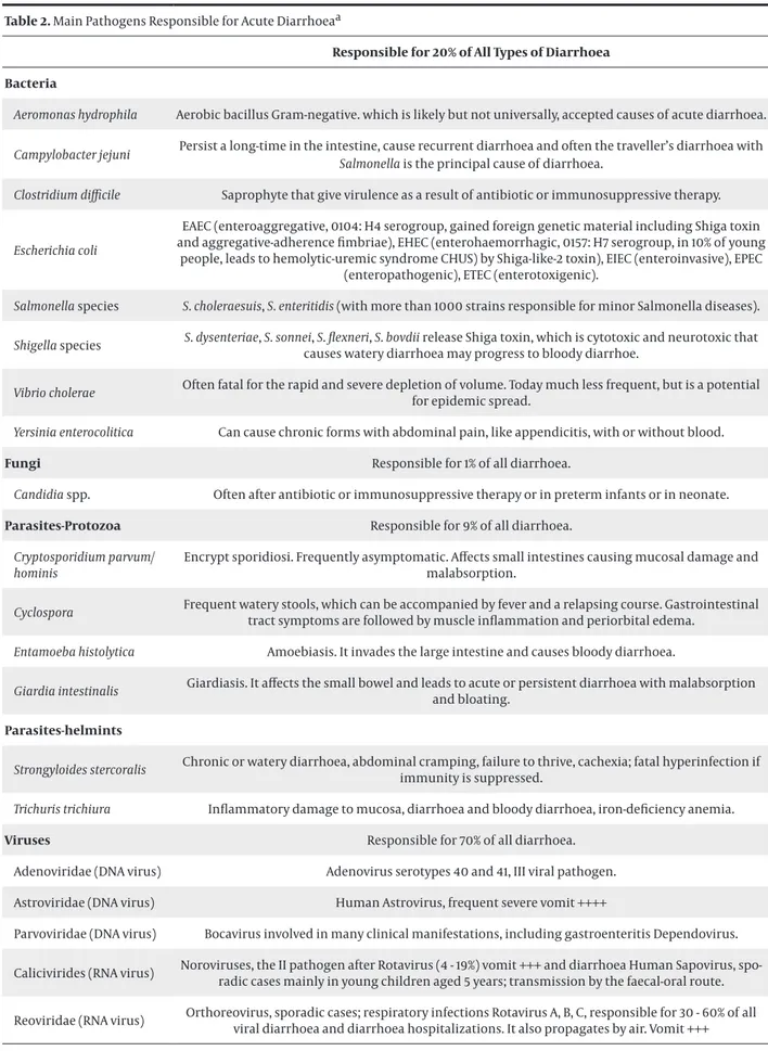
Table 2. Main Pathogens Responsible for Acute Diarrhoeaa
Responsible for 20% of All Types of Diarrhoea Bacteria
Aeromonas hydrophila Aerobic bacillus Gram-negative. which is likely but not universally, accepted causes of acute diarrhoea. Campylobacter jejuni Persist a long-time in the intestine, cause recurrent diarrhoea and often the traveller’s diarrhoea with Salmonella is the principal cause of diarrhoea. Clostridium difficile Saprophyte that give virulence as a result of antibiotic or immunosuppressive therapy. Escherichia coli
EAEC (enteroaggregative, 0104: H4 serogroup, gained foreign genetic material including Shiga toxin and aggregative-adherence fimbriae), EHEC (enterohaemorrhagic, 0157: H7 serogroup, in 10% of young
people, leads to hemolytic-uremic syndrome CHUS) by Shiga-like-2 toxin), EIEC (enteroinvasive), EPEC (enteropathogenic), ETEC (enterotoxigenic).
Salmonella species S. choleraesuis, S. enteritidis (with more than 1000 strains responsible for minor Salmonella diseases). Shigella species S. dysenteriae, S. sonnei, S. flexneri, S. bovdii release Shiga toxin, which is cytotoxic and neurotoxic that causes watery diarrhoea may progress to bloody diarrhoe. Vibrio cholerae Often fatal for the rapid and severe depletion of volume. Today much less frequent, but is a potential for epidemic spread. Yersinia enterocolitica Can cause chronic forms with abdominal pain, like appendicitis, with or without blood.
Fungi Responsible for 1% of all diarrhoea.
Candidia spp. Often after antibiotic or immunosuppressive therapy or in preterm infants or in neonate.
Parasites-Protozoa Responsible for 9% of all diarrhoea. Cryptosporidium parvum/
hominis
Encrypt sporidiosi. Frequently asymptomatic. Affects small intestines causing mucosal damage and malabsorption.
Cyclospora Frequent watery stools, which can be accompanied by fever and a relapsing course. Gastrointestinal tract symptoms are followed by muscle inflammation and periorbital edema. Entamoeba histolytica Amoebiasis. It invades the large intestine and causes bloody diarrhoea.
Giardia intestinalis Giardiasis. It affects the small bowel and leads to acute or persistent diarrhoea with malabsorption and bloating.
Parasites-helmints
Strongyloides stercoralis Chronic or watery diarrhoea, abdominal cramping, failure to thrive, cachexia; fatal hyperinfection if immunity is suppressed. Trichuris trichiura Inflammatory damage to mucosa, diarrhoea and bloody diarrhoea, iron-deficiency anemia.
Viruses Responsible for 70% of all diarrhoea.
Adenoviridae (DNA virus) Adenovirus serotypes 40 and 41, III viral pathogen.
Astroviridae (DNA virus) Human Astrovirus, frequent severe vomit ++++
Parvoviridae (DNA virus) Bocavirus involved in many clinical manifestations, including gastroenteritis Dependovirus. Calicivirides (RNA virus) Noroviruses, the II pathogen after Rotavirus (4 - 19%) vomit +++ and diarrhoea Human Sapovirus,
spo-radic cases mainly in young children aged 5 years; transmission by the faecal-oral route. Reoviridae (RNA virus) Orthoreovirus, sporadic cases; respiratory infections Rotavirus A, B, C, responsible for 30 - 60% of all viral diarrhoea and diarrhoea hospitalizations. It also propagates by air. Vomit +++
Table 3. Clinical Features of Infection With Selected Diarrhoeal Pathogens (Modified WGO 2008 (11))a
Pathogens
Clinical Features
Abdominal Pain Bloody Stool Faecal Evidence of Inflammation Fever Heme-Positive Stool Vomiting and/or Nausea
Campylobacter O V O Clostridium difficile O O O O Shiga toxin-producing E. coli N A O Salmonella O V O Shigella O V Vibrio V N V V V V Yersinia O O O O Candida O N V Cryptosporidium V O V O Cyclospora V V O Entamoeba histolytica O V V O V Giardia O N V Norovirus V V O
aCommon: O-occurs, V-variable. Not common: A-atypical, N-often not.
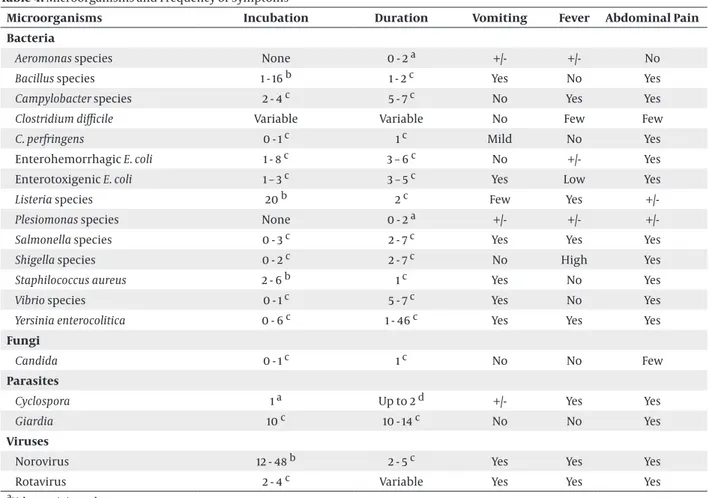
Table 4. Microorganisms and Frequency of Symptoms
Microorganisms Incubation Duration Vomiting Fever Abdominal Pain
Bacteria
Aeromonas species None 0 - 2 a +/- +/- No
Bacillus species 1 - 16 b 1 - 2 c Yes No Yes
Campylobacter species 2 - 4 c 5 - 7 c No Yes Yes
Clostridium difficile Variable Variable No Few Few
C. perfringens 0 - 1 c 1 c Mild No Yes
Enterohemorrhagic E. coli 1 - 8 c 3 – 6 c No +/- Yes
Enterotoxigenic E. coli 1 – 3 c 3 – 5 c Yes Low Yes
Listeria species 20 b 2 c Few Yes
+/-Plesiomonas species None 0 - 2 a +/- +/-
+/-Salmonella species 0 - 3 c 2 - 7 c Yes Yes Yes
Shigella species 0 - 2 c 2 - 7 c No High Yes
Staphilococcus aureus 2 - 6 b 1 c Yes No Yes
Vibrio species 0 - 1 c 5 - 7 c Yes No Yes
Yersinia enterocolitica 0 - 6 c 1 - 46 c Yes Yes Yes
Fungi
Candida 0 - 1 c 1 c No No Few
Parasites
Cyclospora 1 a Up to 2 d +/- Yes Yes
Giardia 10 c 10 - 14 c No No Yes
Viruses
Norovirus 12 - 48 b 2 - 5 c Yes Yes Yes
Rotavirus 2 - 4 c Variable Yes Yes Yes
aValues unit is week. bValues unit is hour. cValues unit is day. dValues unit is month.
Table 5. Rehydration Compounds on the Worldwide Market (Modified Caramia et al. 2009 (9))a
Glucose
mmol/L Na mEq/L K mEq/L Cl mEq/L HCOmEq/L3 o Citrate mOsm/L Kcal/L Aroma Probiotics ESPGHAN (1989/97) 74 - 111 60 20 > 25 20 200 - 250 52 - 80 No No WHO (1984/2002) 110/75 90/75 20 80 30/C.8 - 12 311/245 80 No No AMESOL 111 90 20 60 9 245 80 Lemon No DICODRAL 111 30 20 40 10 211 80 No No
DICODRAL 60 90 60 20 37 14 citrate 211 80 Banana No
DICODRAL
FORTE 111 90 20 80 30 331 80 No No
FLORIDRAL 83 60 20 37 14 citrate 214 80 Banana LGG CFU =
5 × 10⁹
GES 60 108 60 20 50 14 citrate 270 80 No No
IDRATON 245 75 75 20 65 10 citrate 245 79.1 Orange No
IDRAVITA 120 60 20 50 10 citrate 230 80 Banana No
PREREID® 77 50 20 40 10 200 79.35 Citrus No PREREID® LIQUID 1.91 50 20 57 66 230 80 Citrus No REIDRAX 75 60 20 60 10 citrate 225 60.8 No No REUTERIN BRICK 61 58.5 19.2 44.3 230 45 Apricot L. reuteri DSM 17938 CFU = 108
REUTERIN IDRO 83 61 20 46 11 220 60 No DSM 17938 L. reuteri
CFU = 108 Home solution Water 1 liter, Sugar 1 spoon (19 g), Salt 1 spoon (3 g), Bicarbonate one needle (0, 5 g).
aCFU, colony forming units; C, chloride; ESPGHAN, European Society of Paediatric Gastroenterology, HCO, Hepatology and, Nutrition; HCO3, bicarbonate; K, potassium; L, liter; LGG, Lactobacillus rhamnosus GG; L. reuteri, Lactobacillus reuteri; Na, sodium; WHO, World Health Organization.
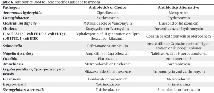
Table 6. Antibiotics Used to Treat Specific Causes of Diarrhoea
Pathogen Antibiotic/s of Choice Antibiotic/s Alternative
Aeromonas hydrophila Ciprofloxacin Meropenem
Campylobacter Azithromycin Erythromycin
Clostridium difficile Metronidazole or Vancomycin Linezolid or fidaxomicin
Cholera Doxycycline or Tetracycline Furazolidone or Erythromycin
E. coli EAEC; E. coli EHEC; E. coli EIEC; E.
coli EPEC; E. coli ETEC Cephalosporin of III generation or Cipro-floxacin or Rifaximin Colistin or Azithromycin or Meropenem
Salmonella Ceftriaxone or Ampicillin Amoxicillin or Cephalosporin of III gen-eration or Fluoroquinolones
Shigella dysentery Ampicillin or Ciprofloxacin Nalidixic Acid or Fluoroquinolones
Candida Fluconazole Amphotericin B
Amoebiasis Metronidazole or Tinidazole Paromomycin
Cryptosporidium, Cyclospora
cayeta-nensis Nitazoxanide, Cotrimoxazole Paromomycin and azithromycin
Giardiasis Tinidazole or tazoxanide Metronidazole
Isospora belli Cotrimoxazole Pirimetamina
4. Conclusions
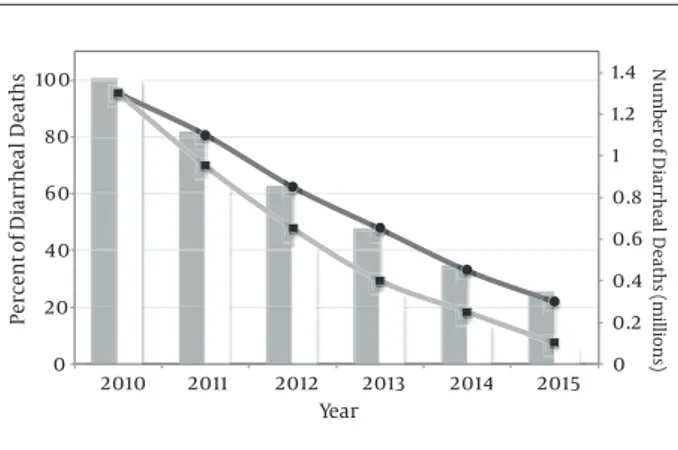
UNICEF and WHO published an exhaustive report on diarrhoea in 2009. This report includes a key package on diarrhoea prevention and treatments to reduce diarrhoea morbidity and mortality. The recommendations include improved access to safe water, promotion of sanitation, vi-tamin A and zinc supplementation, promotion of breast-feeding and treatment with ORS (76). A scale-up scenario using data from 2010 presumed a linear increase from the baseline coverage year (2010) through 2015, allowing to determinate total number and percentage of diarrhoeal deaths (Figure 3). The results of this modelling application demonstrate that diarrhoeal deaths might be decreased by at least 78% until 2015 with current available therapies.
Intensive promotion of ORS use, zinc and vitamin A sup-plementation and training health staff can be successful in reducing diarrhoeal deaths (76). On the other hand, fortifying and stimulating the community management of diarrhoea, superfluous and unnecessary antibiotic use can be significantly reduced.
It was shown that probiotics can be an effective supple-ment for diarrhoea managesupple-ment. It is very important and essential to prescribe strains documented to be effective, such as L. reuteri, Lactobacillus GG, L. acidophilus, L. bulgaricus
and Saccharomyces boulardii. Moreover, the concentration
has a significant relevance and it was demonstrated that a dose of 1010 CFU/day is effective, reducing diarrhoea dura-tion and spreading (78, 79). Furthermore, probiotic strain alone or in combination (2 or more strains) provides sig-nificant protective effects against intestinal disorders (80). Many studies showed that probiotics significantly reduce the risk of acute diarrhoea, antibiotic-associated diarrhoea and traveller’s diarrhoea. Regarding different described approach-es to control intapproach-estinal disorders, as diarrhoea, the effects ex-erted by different probiotics give promising alternatives, so clinicians should advise those probiotic strains proven to be efficacious in relevant patient groups and encourage further clinical research studies to define its proper place in the man-agement of several gastrointestinal tract disorders.
0 0.2 0.4 0.6 0.8 1 1.2 1.4 0 20 40 60 80 100 2010 2011 2012 2013 2014 2015 Nu mb er of Di arrhea l De at hs (millio ns ) Percen t of Di arrhea l De at hs Year
Figure 3. Trends in Percentage and Number of Diarrheal Deaths, Under Ambitious (●) and Universal (■) Scale-up Plans (Modified Fischer Walker et al. 2011 (77))
In conclusion, there are several tools to manage and re-duce diarrhoea incidence such as adopting practices to decrease risk factors, using feeding guidelines (including breastfeeding) and probiotics.
Footnote
Authors’ Contributions:Manuscript concept and
design Giuseppe Caramia, Stefania Silvi, and Maria Mag-dalena Coman. Data collection and interpretation: Gi-useppe Caramia, Stefania Silvi, and Maria Magdalena Co-man. Manuscript drafting: Giuseppe Caramia, Stefania Silvi, and Maria Magdalena Coman. Critical revision of the manuscript for important intellectual content: Gi-useppe Caramia, Stefania Silvi, Maria Cristina Verdenelli, and Maria Magdalena Coman.
References
1. Guarino A, Dupont C, Gorelov AV, Gottrand F, Lee JK, Lin Z, et al. The management of acute diarrhea in children in de-veloped and developing areas: from evidence base to clini-cal practice. Expert Opin Pharmacother. 2012;13(1):17–26. doi: 10.1517/14656566.2011.634800. [PubMed: 22106840]
2. Caramia G, Pompilio A, Ciuccarelli F, Moretti V. Disidratazione e reidratazione. Attualita ed interventi terapeutici. Prog Nutr. 2003;5:299–313.
3. Baldi F, Bianco MA, Nardone G, Pilotto A, Zamparo E. Focus on acute diarrhoeal disease. World J Gastroenterol. 2009;15(27):3341– 8. [PubMed: 19610134]
4. Lozano R, Wang H, Foreman KJ, Rajaratnam JK, Naghavi M, Mar-cus JR, et al. Progress towards Millennium Development Goals 4 and 5 on maternal and child mortality: an updated system-atic analysis. Lancet. 2011;378(9797):1139–65. doi: 10.1016/S0140-6736(11)61337-8. [PubMed: 21937100]
5. United Nations. The Millennium Development Goals Report 2008. 2008.
6. Rudan I, Boschi-Pinto C, Biloglav Z, Mulholland K, Campbell H. Epidemiology and etiology of childhood pneumonia. Bull World Health Organ. 2008;86(5):408–16. [PubMed: 18545744]
7. Boschi-Pinto C, Velebit L, Shibuya K. Estimating child mortality due to diarrhoea in developing countries. Bull World Health Or-gan. 2008;86(9):710–7. [PubMed: 18797647]
8. WHO . Recommendations on the Management of Diarrhoea and Pneumonia. HIV-Infected Infants and Children: Integrated Management of Childhood Illness (IMCI).. Geneva; 2010. 9. Caramia GRE, Salvatori P. Manuale di Infettivologia Neonatale:
Gas-troenteriti infettive. Società Italiana di Neonatologia. 2009;18:171–87. 10. Wanke CA. Epidemiology and causes of acute diarrhea in resource-rich countries. 2015. Available from: http://www.uptodate.com/ contents/epidemiology-and-causes-of-acute-diarrhea-in-re-source-rich-countries.
11. Organisation WG. World Gastroenterology Organisation practice guideline: Acute diarrhea. 2012. Available from: http://www.world- gastroenterology.org/guidelines/global-guidelines/acute-diar-rhea/acute-diarrhea-english.
12. Rimoldi SG, Stefani F, Pagani C, Chenal LL, Zanchetta N, Di Bar-tolo I, et al. Epidemiological and clinical characteristics of pe-diatric gastroenteritis associated with new viral agents. Arch Vi-rol. 2011;156(9):1583–9. doi: 10.1007/s00705-011-1037-5. [PubMed: 21643788]
13. Pfeiffer ML, DuPont HL, Ochoa TJ. The patient presenting with acute dysentery--a systematic review. J Infect. 2012;64(4):374–86. doi: 10.1016/j.jinf.2012.01.006. [PubMed: 22266388]
14. Alam S, Mushtaq M. Antibiotic associated diarrhea in children. Indian Pediatr. 2009;46(6):491–6. [PubMed: 19556659]
15. Bertizzolo L, Domeniconi G, Fabio G, Jacchetti G, Serafino S, For-mica S, et al. Analysis of nosocomial acquired Clostridium diffi-cile infection in an Italian research and teaching hospital. Ann
Ig. 2013;25(2):119–24. doi: 10.7416/ai.2013.1913. [PubMed: 23471449] 16. Hope WW, Castagnola E, Groll AH, Roilides E, Akova M, Arendrup
MC, et al. ESCMID* guideline for the diagnosis and management of Candida diseases 2012: prevention and management of in-vasive infections in neonates and children caused by Candida spp. Clin Microbiol Infect. 2012;18 Suppl 7:38–52. doi: 10.1111/1469-0691.12040. [PubMed: 23137136]
17. Friedman M, Ramsay DB, Borum ML. An unusual case report of small bowel Candida overgrowth as a cause of diarrhea and re-view of the literature. Dig Dis Sci. 2007;52(3):679–80. doi: 10.1007/ s10620-006-9604-4. [PubMed: 17277989]
18. Bogoch ,I, Andrews JR, Speich B, Utzinger J, Ame SM, Ali SM, et al. Mobile phone microscopy for the diagnosis of soil-transmitted helminth infections: a proof-of-concept study. Am J Trop Med Hyg. 2013;88(4):626–9. doi: 10.4269/ajtmh.12-0742. [PubMed: 23478580]
19. Pawlowski SW, Warren CA, Guerrant R. Diagnosis and treat-ment of acute or persistent diarrhea. Gastroenterology. 2009;136(6):1874–86. doi: 10.1053/j.gastro.2009.02.072. [PubMed: 19457416]
20. Sinha A, SenGupta S, Guin S, Dutta S, Ghosh S, Mukherjee P, et al. Culture-independent real-time PCR reveals extensive poly-microbial infections in hospitalized diarrhoea cases in Kolkata, India. Clin Microbiol Infect. 2013;19(2):173–80. doi: 10.1111/j.1469-0691.2011.03746.x. [PubMed: 22268636]
21. Jex AR, Stanley KK, Lo W, Littman R, Verweij JJ, Campbell BE, et al. Detection of diarrhoeal pathogens in human faeces using an automated, robotic platform. Mol Cell Probes. 2012;26(1):11–5. doi: 10.1016/j.mcp.2011.10.004. [PubMed: 22056326]
22. Wittenberg DF. Management guidelines for acute infective diar-rhoea / gastroenteritis in infants. S Afr Med J. 2012;102(2):104–7. [PubMed: 22310445]
23. de Oliveira LH, Danovaro-Holliday MC, Sanwogou NJ, Ruiz-Matus C, Tambini G, Andrus JK. Progress in the introduction of the rota-virus vaccine in Latin America and the Caribbean: four years of ac-cumulated experience. Pediatr Infect Dis J. 2011;30(1 Suppl):S61–6. doi: 10.1097/INF.0b013e3181fefdd6. [PubMed: 21183843]
24. Patel MM, Steele D, Gentsch JR, Wecker J, Glass RI, Parashar UD. Real-world impact of rotavirus vaccination. Pediatr Infect Dis J. 2011;30(1 Suppl):S1–5. doi: 10.1097/INF.0b013e3181fefa1f. [PubMed: 21183833]
25. Dennehy PH. Effects of vaccine on rotavirus disease in the pedi-atric population. Curr Opin Pediatr. 2012;24(1):76–84. doi: 10.1097/ MOP.0b013e32834ee594. [PubMed: 22189398]
26. Atherly D, Dreibelbis R, Parashar UD, Levin C, Wecker J, Rhein-gans RD. Rotavirus vaccination: cost-effectiveness and impact on child mortality in developing countries. J Infect Dis. 2009;200 Suppl 1:S28–38. doi: 10.1086/605033. [PubMed: 19817610] 27. Soares-Weiser K, MacLehose H, Bergman H, Ben-Aharon I, Nagpal
S, Goldberg E, et al. Vaccines for preventing rotavirus diarrhoea: vaccines in use. Cochrane Database Syst Rev. 2012;2:CD008521. doi: 10.1002/14651858.CD008521.pub2. [PubMed: 22336845]
28. Omenaca F, Sarlangue J, Szenborn L, Nogueira M, Suryakiran PV, Smolenov IV, et al. Safety, reactogenicity and immunogenicity of the human rotavirus vaccine in preterm European Infants: a randomized phase IIIb study. Pediatr Infect Dis J. 2012;31(5):487–93. doi: 10.1097/INF.0b013e3182490a2c. [PubMed: 22228231] 29. WHO/FAO. Evaluation of health and nutritional properties of powder
milk and live lactic acid bacteria. 2001.
30. Culligan EP, Hill C, Sleator RD. Probiotics and gastrointestinal disease: successes, problems and future prospects. Gut Pathog. 2009;1(1):19. doi: 10.1186/1757-4749-1-19. [PubMed: 19930635] 31. Allen SJ, Wareham K, Bradley C, Harris W, Dhar A, Brown H, et al. A
multicentre randomised controlled trial evaluating lactobacilli and bifidobacteria in the prevention of antibiotic-associated di-arrhoea in older people admitted to hospital: the PLACIDE study protocol. BMC Infect Dis. 2012;12:108. doi: 10.1186/1471-2334-12-108. [PubMed: 22559011]
32. Ghoshal UC, Shukla R, Ghoshal U, Gwee KA, Ng SC, Quigley EM. The gut microbiota and irritable bowel syndrome: friend or foe? Int J Inflam. 2012;2012:151085. doi: 10.1155/2012/151085. [PubMed: 22577594]
33. Shulman RJ, Smith EO. Does VSL#3 really improve symptoms in children with IBS? J Pediatr Gastroenterol Nutr. 2012;54(1):109. doi: 10.1097/MPG.0b013e31823df69b. [PubMed: 22064630]
34. Siponen SM, Ahonen RS, Kettis A, Hameen-Anttila KP. Comple-mentary or alternative? Patterns of compleComple-mentary and alter-native medicine (CAM) use among Finnish children. Eur J Clin Pharmacol. 2012;68(12):1639–45. doi: 10.1007/s00228-012-1294-6. [PubMed: 22573133]
35. Caramia G, Silvi S. Probiotics: from the ancient wisdom to the actual therapeutical and nutraceutical perspective. Probiotic bacteria and enteric infections.. New York: Springer; 2011. pp. 3–37. 36. Goldin BR, Gorbach SL. Clinical indications for probiotics:
an overview. Clin Infect Dis. 2008;46 Suppl 2:S96–100. doi: 10.1086/523333. [PubMed: 18181732]
37. Manzoni P, Mostert M, Leonessa ML, Priolo C, Farina D, Monetti C, et al. Oral supplementation with Lactobacillus casei subcies rhamnosus prevents enteric colonization by Candida spe-cies in preterm neonates: a randomized study. Clin Infect Dis. 2006;42(12):1735–42. doi: 10.1086/504324. [PubMed: 16705580] 38. Deshpande G, Rao S, Patole S, Bulsara M. Updated meta-analysis
of probiotics for preventing necrotizing enterocolitis in preterm neonates. Pediatrics. 2010;125(5):921–30. doi: 10.1542/peds.2009-1301. [PubMed: 20403939]
39. Indrio F, Riezzo G, Raimondi F, Bisceglia M, Filannino A, Cavallo L, et al. Lactobacillus reuteri accelerates gastric emptying and im-proves regurgitation in infants. Eur J Clin Invest. 2011;41(4):417–22. doi: 10.1111/j.1365-2362.2010.02425.x. [PubMed: 21114493] 40. Jones ML, Martoni CJ, Di Pietro E, Simon RR, Prakash S. Evaluation
of clinical safety and tolerance of a Lactobacillus reuteri NCIMB 30242 supplement capsule: a randomized control trial. Regul Tox-icol Pharmacol. 2012;63(2):313–20. doi: 10.1016/j.yrtph.2012.04.003. [PubMed: 22561556]
41. Agustina R, Kok FJ, van de Rest O, Fahmida U, Firmansyah A, Lukito W, et al. Randomized trial of probiotics and calcium on diarrhea and respiratory tract infections in Indonesian children. Pediat-rics. 2012;129(5):e1155–64. doi: 10.1542/peds.2011-1379. [PubMed: 22492764]
42. Sari FN, Eras Z, Dizdar EA, Erdeve O, Oguz SS, Uras N, et al. Do oral probiotics affect growth and neurodevelopmental out-comes in very low-birth-weight preterm infants? Am J Perinatol. 2012;29(8):579–86. doi: 10.1055/s-0032-1311981. [PubMed: 22566113] 43. Fernandez-Carrocera LA, Solis-Herrera A, Cabanillas-Ayon M,
Gallardo-Sarmiento RB, Garcia-Perez CS, Montano-Rodriguez R, et al. Double-blind, randomised clinical assay to evaluate the efficacy of probiotics in preterm newborns weighing less than 1500 g in the prevention of necrotising enterocolitis. Arch Dis Child Fetal Neonatal Ed. 2013;98(1):F5–9. doi: 10.1136/archdis-child-2011-300435. [PubMed: 22556209]
44. Johnston BC, Goldenberg JZ, Vandvik PO, Sun X, Guyatt GH. Probiotics for the prevention of pediatric antibiotic-associated diarrhea. Cochrane Database Syst Rev. 2011;(11):CD004827. doi: 10.1002/14651858.CD004827.pub3. [PubMed: 22071814]
45. Kamdeu Fansi AA, Guertin JR, LeLorier J. Savings from the use of a probiotic formula in the prophylaxis of antibi-otic-associated diarrhea. J Med Econ. 2012;15(1):53–60. doi: 10.3111/13696998.2011.629015. [PubMed: 22023067]
46. Bernaola Aponte G, Bada Mancilla CA, Carreazo Pariasca NY, Rojas Galarza RA, Bernaola Aponte G. Probiotics for treating persistent diarrhoea in children. Cochrane Database Syst Rev. 2010;(11):CD007401. doi: 10.1002/14651858.CD007401.pub2. [PubMed: 21069693]
47. Preidis GA, Saulnier DM, Blutt SE, Mistretta TA, Riehle KP, Major AM, et al. Host response to probiotics determined by nutritional status of rotavirus-infected neonatal mice. J Pediatr Gastroenterol Nutr. 2012;55(3):299–307. doi: 10.1097/MPG.0b013e31824d2548. [PubMed: 22343914]
48. Morrow LE, Gogineni V, Malesker MA. Probiotic, prebiotic, and syn-biotic use in critically ill patients. Curr Opin Crit Care. 2012;18(2):186– 91. doi: 10.1097/MCC.0b013e3283514b17. [PubMed: 22343306] 49. Marcos LA, DuPont HL. Advances in defining etiology and new
therapeutic approaches in acute diarrhea. J Infect. 2007;55(5):385– 93. doi: 10.1016/j.jinf.2007.07.016. [PubMed: 17825422]
50. Piescik-Lech M, Shamir R, Guarino A, Szajewska H. Review article: the management of acute gastroenteritis in children. Aliment Pharmacol Ther. 2013;37(3):289–303. doi: 10.1111/apt.12163. [PubMed: 23190209] 51. Bhatnagar S, Alam S, Gupta P. Management of acute diarrhea:
from evidence to policy. Indian Pediatr. 2010;47(3):215–7. [PubMed: 20371887]
52. Koletzko S, Osterrieder S. Acute infectious diarrhea in chil-dren. Dtsch Arztebl Int. 2009;106(33):539–47. doi: 10.3238/arz-tebl.2009.0539. [PubMed: 19738921]
53. Hao R, Michelle De Vera MD, Resurreccion E. Racecadotril in the treatment of acute diarrhea in children: a meta-analysis. PIDSP J. 2010;11(2):19–32.
54. Eberlin M, Muck T, Michel MC. A comprehensive review of the pharmacodynamics, pharmacokinetics, and clinical effects of the neutral endopeptidase inhibitor racecadotril. Front Pharma-col. 2012;3:93. doi: 10.3389/fphar.2012.00093. [PubMed: 22661949] 55. Rautenberg TA, Zerwes U, Foerster D, Aultman R. Evaluating the
cost utility of racecadotril for the treatment of acute watery di-arrhea in children: the RAWD model. Clinicoecon Outcomes Res. 2012;4:109–16. doi: 10.2147/CEOR.S31238. [PubMed: 22570557] 56. Mansour AM, Abd Elkhalek R, Shaheen HI, El Mohammady H,
Re-faey S, Hassan K, et al. Burden of Aeromonas hydrophila-associat-ed diarrhea among children younger than 2 years in rural Egyp-tian community. J Infect Dev Ctries. 2012;6(12):842–6. doi: 10.3855/ jidc.2390. [PubMed: 23276737]
57. Maetz B, Abbou R, Andreoletti JB, Bruant-Rodier C. Infections fol-lowing the application of leeches: two case reports and review of the literature. J Med Case Rep. 2012;6:364. doi: 10.1186/1752-1947-6-364. [PubMed: 23098279]
58. Wilmer A, Slater K, Yip J, Carr N, Grant J. The role of leech water sampling in choice of prophylactic antibiotics in medical leech therapy. Microsurgery. 2013;33(4):301–4. doi: 10.1002/micr.22087. [PubMed: 23417901]
59. Maragkoudakis S, Poulidaki SR, Papadomanolaki E, Alevraki G, Papadogianni M, Oikonomou N, et al. Empiric antimicrobial therapy and infectious diarrhea. Do we need local guidelines? Eur J Intern Med. 2011;22(5):e60–2. doi: 10.1016/j.ejim.2011.06.005. [PubMed: 21925045]
60. Brigic E, Hadzic D, Mladina N. Childhood and Coress model of carcinogenesis. Med Arch. 2012;66(6):375–7. [PubMed: 23409514] 61. El Qouqa IA, El Jarou MA, Samaha AS, Al Afifi AS, Al Jarousha AM.
Yersinia enterocolitica infection among children aged less than 12 years: a case-control study. Int J Infect Dis. 2011;15(1):e48–53. doi: 10.1016/j.ijid.2010.09.010. [PubMed: 21131221]
62. Scott LJ. Micafungin: a review of its use in the prophylaxis and treatment of invasive Candida infections. Drugs. 2012;72(16):2141– 65. doi: 10.2165/11209970-000000000-00000. [PubMed: 23083111] 63. Manzoni P, Benjamin DJ, Franco C, Rizzollo S, Stronati M, Watt K, et al. Echinocandins for the nursery: an update. Curr Drug Metab. 2013;14(2):203–7. [PubMed: 22935065]
64. Debnath A, Ndao M, Reed SL. Reprofiled drug targets ancient protozoans: drug discovery for parasitic diarrheal diseases. Gut Microbes. 2013;4(1):66–71. doi: 10.4161/gmic.22596. [PubMed: 23137963]
65. Ortega YR, Sanchez R. Update on Cyclospora cayetanensis, a food-borne and waterfood-borne parasite. Clin Microbiol Rev. 2010;23(1):218– 34. doi: 10.1128/CMR.00026-09. [PubMed: 20065331]
66. Granados CE, Reveiz L, Uribe LG, Criollo CP. Drugs for treating giardiasis. Cochrane Database Syst Rev. 2012;12:CD007787. doi: 10.1002/14651858.CD007787.pub2. [PubMed: 23235648]
67. Rossignol JF, Lopez-Chegne N, Julcamoro LM, Carrion ME, Bardin MC. Nitazoxanide for the empiric treatment of pediatric infec-tious diarrhea. Trans R Soc Trop Med Hyg. 2012;106(3):167–73. doi: 10.1016/j.trstmh.2011.11.007. [PubMed: 22301075]
68. Bouchaud O. [Circumstances for diagnosis and treatment of in-testinal parasitosis in France]. Presse Med. 2013;42(1):84–92. doi: 10.1016/j.lpm.2012.10.009. [PubMed: 23266344]
69. Boatin BA, Basanez MG, Prichard RK, Awadzi K, Barakat RM, Garcia HH, et al. A research agenda for helminth diseases of humans: towards control and elimination. PLoS Negl Trop Dis. 2012;6(4):e1547. doi: 10.1371/journal.pntd.0001547. [PubMed: 22545161]
70. Speich B, Ame SM, Ali SM, Alles R, Hattendorf J, Utzinger J, et al. Efficacy and safety of nitazoxanide, albendazole, and ni-tazoxanide-albendazole against Trichuris trichiura infection: a randomized controlled trial. PLoS Negl Trop Dis. 2012;6(6):e1685. doi: 10.1371/journal.pntd.0001685. [PubMed: 22679525]
71. Liu MJ, Bao S, Galvez-Peralta M, Pyle CJ, Rudawsky AC, Pavlovicz RE, et al. ZIP8 regulates host defense through zinc-mediated in-hibition of NF-kappaB. Cell Rep. 2013;3(2):386–400. doi: 10.1016/j. celrep.2013.01.009. [PubMed: 23403290]
72. Patel A, Mamtani M, Dibley MJ, Badhoniya N, Kulkarni H. Thera-peutic value of zinc supplementation in acute and persistent diarrhea: a systematic review. PLoS One. 2010;5(4):e10386. doi: 10.1371/journal.pone.0010386. [PubMed: 20442848]
73. Sabot O, Schroder K, Yamey G, Montagu D. Scaling up oral re-hydration salts and zinc for the treatment of diarrhoea. BMJ. 2012;344:e940. doi: 10.1136/bmj.e940. [PubMed: 22327358] 74. Das RR. Zinc in acute childhood diarrhea: Is it universally
ef-fective? Indian J Pharmacol. 2012;44(1):140. doi: 10.4103/0253-7613.91891. [PubMed: 22345893]
75. Sattar S, Ahmed T, Rasul CH, Saha D, Salam MA, Hossain MI. Ef-ficacy of a high-dose in addition to daily low-dose vitamin A in children suffering from severe acute malnutrition with other illnesses. PLoS One. 2012;7(3):e33112. doi: 10.1371/journal. pone.0033112. [PubMed: 22479361]
76. Wardlaw T, Salama P, Brocklehurst C, Chopra M, Mason E. Diar-rhoea: why children are still dying and what can be done. Lan-cet. 2010;375(9718):870–2. doi: 10.1016/S0140-6736(09)61798-0. [PubMed: 19833382]
77. Fischer Walker CL, Friberg IK, Binkin N, Young M, Walker N, Fontaine O, et al. Scaling up diarrhea prevention and treat-ment interventions: a Lives Saved Tool analysis. PLoS Med. 2011;8(3):e1000428. doi: 10.1371/journal.pmed.1000428. [PubMed: 21445330]
78. Jensen H, Grimmer S, Naterstad K, Axelsson L. In vitro testing of commercial and potential probiotic lactic acid bacteria. Int J Food Microbiol. 2012;153(1-2):216–22. doi: 10.1016/j.ijfoodmi-cro.2011.11.020. [PubMed: 22177712]
79. Cooke ML. Causes and management of diarrhoea in children in a clinical setting. South Afr J Clin Nutr. 2010;23(1).
80. Minocha A. Probiotics for preventive health. Nutr Clin Pract. 2009;24(2):227–41. doi: 10.1177/0884533608331177. [PubMed: 19321897]