1 23
Microbial Ecology
ISSN 0095-3628
Microb Ecol
DOI 10.1007/s00248-015-0566-y
UV Radiation and Visible Light Induce
hsp70 Gene Expression in the Antarctic
Psychrophilic Ciliate Euplotes focardii
Lorenzo Fulgentini, Valerio Passini,
Giuliano Colombetti, Cristina Miceli,
Antonietta La Terza & Roberto
1 23
Your article is protected by copyright and all
rights are held exclusively by Springer Science
+Business Media New York. This e-offprint is
for personal use only and shall not be
self-archived in electronic repositories. If you wish
to self-archive your article, please use the
accepted manuscript version for posting on
your own website. You may further deposit
the accepted manuscript version in any
repository, provided it is only made publicly
available 12 months after official publication
or later and provided acknowledgement is
given to the original source of publication
and a link is inserted to the published article
on Springer's website. The link must be
accompanied by the following text: "The final
publication is available at link.springer.com”.
ENVIRONMENTAL MICROBIOLOGY
UV Radiation and Visible Light Induce
hsp70 Gene Expression
in the Antarctic Psychrophilic Ciliate
Euplotes focardii
Lorenzo Fulgentini&Valerio Passini&Giuliano Colombetti&
Cristina Miceli&Antonietta La Terza&Roberto Marangoni
Received: 25 August 2014 / Accepted: 2 January 2015 # Springer Science+Business Media New York 2015
Abstract The psychrophilic ciliate Euplotes focardii inhabits the shallow marine coastal sediments of Antarctica, where, over millions of years of evolution, it has reached a strict molecular adaptation to such a constant-temperature environ-ment (about−2 °C). This long evolution at sub-zero tempera-tures has made E. focardii unable to respond to heat stress with the activation of its heat shock protein (hsp) 70 genes. These genes can, however, be expressed in response to other stresses, like the oxidative one, thus indicating that the molecular ad-aptation has exclusively altered the heat stress signaling path-ways, while it has preserved hsp70 gene activation in response to other environmental stressors. Since radiative stress has proved to be affine to oxidative stress in several organisms, we investigated the capability of UV radiation to induce hsp70 transcription. E. focardii cell cultures were exposed to several different irradiation regimes, ranging from visible only to a mixture of visible, UV-A and UV-B. The irradiation values of each spectral band have been set to be comparable with those recorded in a typical Antarctic spring. Using Northern blot analysis, we measured the expression level of hsp70 im-mediately after irradiation (0-h-labeled samples), 1 h, and 2 h
from the end of the irradiation. Surprisingly, our results showed that besides UV radiation, the visible light was also able to induce hsp70 expression in E. focardii. Moreover, spectrophotometric measurements have revealed no detectable endogenous pigments in E. focardii, making it difficult to propose a possible explana-tion for the visible light inducexplana-tion of its hsp70 genes. Further research is needed to conclusively clarify this point.
Keywords Euplotes focardii . hsp70 . Gene regulation . Radiative stress . UV . Pigment . Visible light gene activation
Introduction
Over the last few years, the release of anthropogenic chloro-fluorocarbons (CFCs) has caused remarkable changes in the chemistry of the atmosphere, especially regarding the ozone layer. Ozone (O3) is the only component of the atmosphere
capable of absorbing the UV-B spectral band (280–315 nm) of solar light. It is continuously destroyed by a process involving especially Cl radicals that derive from CFC photo-degradation in the stratosphere. Ozone depletion is more severe over the Antarctica, where ozone levels decline dramatically (typically >50 %) during austral spring resulting in the formation of the so-called ozone hole [1]. These changes bring about variations in spectral distribution of solar radiation reaching the terrestri-al surface, since they let the UV-B region of the solar spectrum pass through the stratospheric filter in a larger amount [2]. UV-B radiation is the most biologically active, and its absorption by cells can cause severe damage: in fact, it may lead to the production of active oxygen species and free radicals capable of changing membrane and protein structure [3]. The absorp-tion of UV-B by DNA, RNA and proteins causes mutagenesis and interferes with normal metabolic functioning. In plants, it Antonietta La Terza and Roberto Marangoni are joint last authors.
L. Fulgentini
:
G. Colombetti:
R. MarangoniCNR-Biophysics Institute, via G. Moruzzi 1, 56100 Pisa, Italy L. Fulgentini
CNR-National Institute of Optics, via G. Moruzzi 1, 56100 Pisa, Italy V. Passini
:
C. Miceli:
A. La Terza (*)School of Biosciences and Veterinary Medicine, University of Camerino, Via Gentile III da Varano, 62032 Camerino, MC, Italy e-mail: [email protected]
R. Marangoni (*)
Department of Biology, University of Pisa, via Derna 1, 56100 Pisa, Italy
e-mail: [email protected] Microb Ecol
DOI 10.1007/s00248-015-0566-y
can lead to the partial inhibition of photosynthesis. Long-term biological consequences include morphogenetic aberrations, impaired growth and restricted mobility [4].
The degree of UV sensitivity is related to the efficiency of repair mechanisms and to the existence of mitigating strate-gies, such as the presence of intracellular pigments capable of quenching and dissipating the excess energy before the for-mation of dangerous oxygen species [4,5].
Even aquatic organisms are not completely safe; they are however protected by water which can partially screen elec-tromagnetic radiation. In fact, even if light intensity decreases exponentially with the depth of the water column, biologically damaging levels of UV-B can penetrate to depths >30 m (de-pending on sea state, turbidity and vertical mixing), affecting organisms in coastal regions and the upper photic zone of open oceans [4,6,7]. In this scenario, observations by many researchers show convincing evidence of UV-B damage to phytoplankton [8].
Concerning the Antarctic aquatic environment, the polar pack is an efficient barrier for light transmission (especially when covered with snow). It reflects 40–80 % of incident radiation [4], and during austral spring, when the ozone hole reaches its climax, it is still quite extended. However, the plateau has completely ice-free local areas, calledBpolynya^: in these regions, the radiation may reach major depths. For example, some evidence indicates that Antarctic zooplankton accumulates significant DNA damage during periods of in-creased UV-B flux [1,9]. Moreover, the high concentrations of UV radiation protective mycosporine-like amino acids have been measured in sea-ice diatom samples [10].
Prolonged exposure to stress such as UV-B radiation may have negative consequences on the biochemical prop-erties of proteins: in response to UV-B-induced protein im-pairment, cells may activate an evolutionary (universally) conserved cellular defence mechanism, known as the heat shock response, which involves the rapid synthesis of a set of general stress proteins known asBheat shock proteins^ (HSPs). HSPs and in particular those belonging to the HSP70 family, behave asBmolecular chaperones^ assisting cells in chaperoning protein folding and assembly as well as in promoting damaged proteins degrading and thus con-trasting the proteotoxic effects due to exposure to heat, UV radiation as well as to a wide variety of other environ-mental stressors [11–13].
Since its discovery by Ritossa [14], heat shock protein (hsp) genes and their expression, mainly in response to heat and other different environmental stress stimuli, have been extensively analyzed in various organisms belonging to dif-ferent kingdoms (from Prokaryotes to Eukaryotes). Quite re-cently, hsp genes (and their expression) were also analyzed in organisms, living in Antarctica coastal seawater, a habitat characterized by an extremely cold (−2 °C) and constant tem-perature (for a review, see [15]).
With regard to the conservation of the heat shock response in evolution, it is interesting to note that at least three species lacking an inducible heat shock response have been found; this lack might be due to evolutionary adaptation to extremely stable temperatures (firmly in the range of−2 °C) [16]. These species are Hydra oligactis, a freshwater cnidarian in which this phenomenon has been described for the first time [17,18] and two Antarctic species: the fish Trematomus bernacchii [19] and the protozoan ciliate Euplotes focardii [20].
E. focardii is an hypotrichous bottom-dwelling ciliate, en-demic in Antarctic coastal seawater and among the various Euplotes species of Antarctic origin up to now described, is considered to be one of the most interesting to study because its adaptation to the cold does seem to be more remote than that of the other Antarctic species [21]. Evidence of this marked psychrophilic (cold-loving) behaviour comes from the observation that this species shows optimal survival and growth rates at 4–5 °C and cannot survive at a temperature above 10 °C [21]. Moreover, and with respect to the molecular adaptive strategies so far investigated, the E. focardii tubulins have evolved numerous peculiar post-translational modifica-tions, to prevent microtubules de-polymerization at sub-zero temperatures [22,23], and its hsp70 genes have lost the ca-pacity to be effectively transcriptionally activated in response to environmental thermal shock although these genes have preserved their capacity to be induced in response to other non-thermal (chemical and oxidative) shock [24].
Thus, E. focardii in view of its strict psychrophilic behav-iour represents an intriguing bio-model to explore due to its peculiar molecular adaptive strategies in extreme (cold) environments.
On a recent paper, Di Giuseppe et al. [25] investigated the sensitivity of different Euplotes species of polar (E. focardii and Euplotes nobilii) and non-polar (Euplotes quinquecarinatus and Euplotes raikovi) origin to UV radi-ations. By measuring the cell viability of the various Euplotes species under different regimes of UV-B and UV-A exposures, these authors reported that the polar spe-cies are able to face UV radiation more efficiently than non-polar ones. In particular, E. focardii showed the strongest resistance to UV-B radiation.
Given these premises, the present work was aimed at starting an investigation of the molecular basis of UV resis-tance in E. focardii. Regarding this, we examined the capacity of E. focardii to withstand UV radiations as well as other combinations of spectral components, with the activation of its hsp70 genes. Northern blot analysis of total RNA extracted from E. focardii exposed to UV-A and UV-B radiations re-vealed a consistent induction of hsp70 gene expression. Sur-prisingly, our analysis showed that besides UV radiations, visible light by itself was also able to appreciably induce E. focardii hsp70 expression. Since spectrophotometric mea-surements have revealed no detectable endogenous pigments L. Fulgentini et al.
Author's personal copy
in E. focardii; at the moment, it is difficult to propose a possible/plausible signaling pathway for the visible light in-duction of hsp70 expression, and further research is needed to clarify this phenomena.
Materials and Methods Cell Cultures
The results described in the present paper were obtained from experiments carried out on culture of E. focardii cells belong-ing to strain 95 (the same strain used by Di Giuseppe et al. [25] to analyze UV radiation effects in various Euplotes species). This strain represents type-species material chosen from a number of wild-type strains isolated from sediment and sea-water samples collected from the coastal sea-waters of Terra Nova Bay, Antarctica, where it is located in the Italian baseBMario Zucchelli^ (S 74° 42′ /E 164° 06′). In particular, this strain was collected during the X expedition to Antarctica that was held in 1995 (http://www.pnra.it).
The strain was grown in artificial sea water (33‰, pH 8.1– 8.2) in a cold room at a temperature of 4 °C as in Di Giuseppe et al. [25]. They were fed with the green algae Dunaliella tertiolecta. Stress conditions (UV and visible light exposures) were applied to cells after about 1 week of vegetative prolif-eration in the presence of food. Two days before irradiation, the cells were induced to uniformly arrest their division cycle in the G1–G0 (stationary) phase by depleting the food as de-scribed in La Terza et al. [20].
Irradiation Protocols
Immediately before applying the irradiation protocols, control (non-irradiated) and experimental (irradiated) cell samples were concentrated by mild centrifugation for 5 min at 3500 rpm (2000 rcf) at 4 °C and re-suspended in seawater at
the density of approximately 2–3000 cells per millilitre, in beakers of 40 ml (i.e. a total amount of about 12,000 cells per sample).
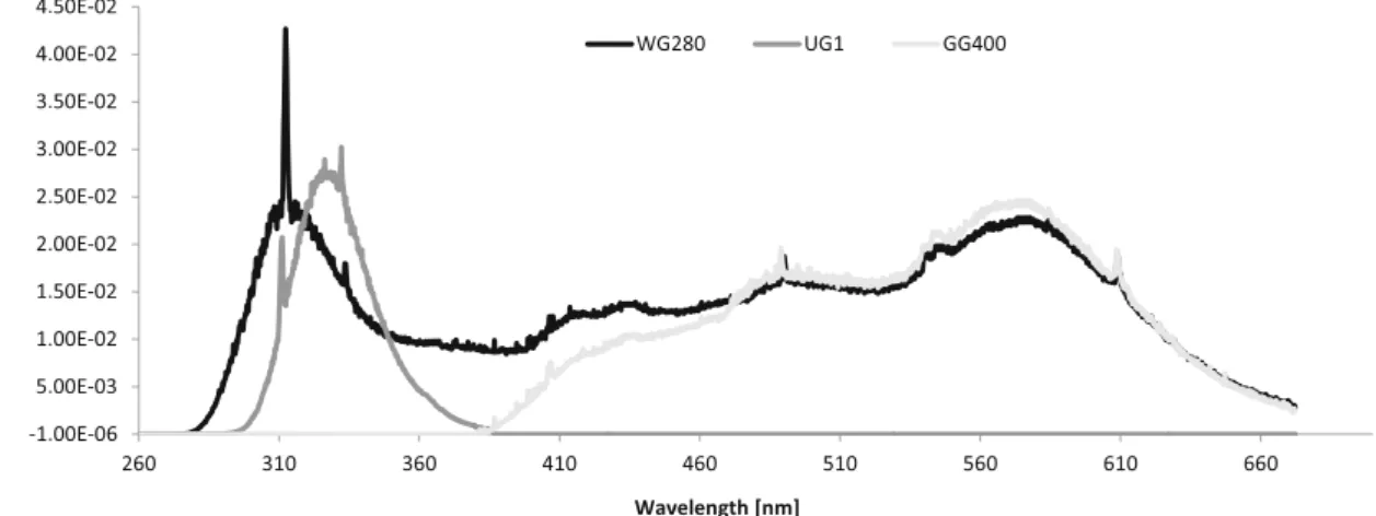
All the irradiation protocols were carried out in a temperature-controlled chamber (4 ° C). Cell cultures of E. focardii were covered with one suitable filter (chosen among the following: Schott WG280 cut-off, GG400 cut-off and UG1 broadband) in order to expose them to the following three different spectral compositions (each experimental con-dition is named after the filter used):
& GG400 samples: the samples covered by GG400 filter received exclusively visible light. Culture samples were exposed for 90 min using three UV-B (Philips TL 20 W/ 12 RS), one UV-A (Philips TLK 40 W/05) and two visible (Philips TLD 18 W/54) fluorescent tubes.
& WG280 samples: they received a combination of UV-B + UV-A + visible radiations. Culture samples were exposed for 90 min using the same combinations of fluorescent tubes used for the GG400 samples.
& UG1 samples: they received exclusively UV-B + UV-A radiations, but not the visible light component. Culture samples were exposed for 120 min using five UV-B (Philips TL 20 W/12 RS) fluorescent tubes.
It should be noted that differences in the exposure times and illumination set-up between UG1 sample and the GG400 and WG280 samples, is due to the need to treat the E. focardii cultures with the same total dose of UV band (UV-A + UV-B) and to maintain its value similar to that of an Antarctic spring morning (as recorded by “NSF Polar UV Monitoring Net-work”, see [26]).
Figure1shows the spectral distributions and the irradiance values of the irradiation reaching each sample. Spectral irra-diance was measured by means of a calibrated radiometric spectrograph (Oriel Instaspec IV). Table 1 shows UV-A, UV-B and visible radiation irradiances (in W m−2) and total
Fig. 1 UV and visible spectral distributions for each filter used in the experiment. The different curves represent the spectral composition of the polychromatic irradiation received by each sample. In the abscissa, the wavelength (in nm); in the ordinate, the irradiance value (in W m−2) UV and Visible Induces hsp70 in E. focardii
doses (in J m−2) received by each sample. Exposure times were set in order to maintain the total exposure dose in the UV-B band below the threshold of 11 kJ m−2that, in agree-ment with Di Giuseppe et al. [25], were able to ensure the survival of at least 60 % of the cells. Experiments were repeat-ed three times with four replicates for each of the assayrepeat-ed experimental condition.
RNA Extraction
Total RNA was extracted from control and irradiated E. focardii cell cultures using TRIzol reagent (Life Tech-nologies) according to the manufacturer’s protocol. E. focardii cells were harvested by mild centrifugation, and RNA was extracted just after the end of each planned irradiation period (0-h sample) and after 1 h (1-h sample) and 2 h (2-h sample).
Northern Blotting Analysis
Equal amounts of total RNA preparations (40μg/lane) were analyzed by Northern blottings after electrophoresis on 1.2 % formaldehyde denaturing agarose gel, according to standard procedures [27]. Hybridization was carried out with two different types of 32P-labeled probes [28] repre-sented by E. focardii: hsp70 gene sequences specific for the protein catalytic domain [24] and E. focardii: 17S ribosom-al DNA (rDNA) gene sequences. Blotted membranes (Hybond-N, Amersham Biosciences) were pre-hybridized for 1 h in 5× Denhardt’s solution, 0.5 % sodium dodecyl sulfate (SDS) and 10 mM EDTA. Probes were boiled for 5 min and snap cooled on ice before being added to the hybridization solution containing 6× saline-sodium citrate (SSC), 5× Denhardt’s solution, 0.1 % SDS and 100 μg/ml of denatured tRNA. All hybridizations were carried out overnight at 65 °C with a constant agitation, and mem-branes were washed twice with 0.5× SSC containing 0.1 % SDS, at the hybridization temperature for 1 h, then dried and exposed for autoradiography. Equal loading of RNA samples was evaluated by re-probing the filters firstly
hybridized with the hsp70 probes, with a homologous 17S rDNA probe. Both probes were used at the equal number of counts per minute (106). Membranes were re-probed after having been incubated with a boiling solution of 0.1 % SDS and allowed to cool to room temperatures. The intensity of hybridization bands was measured using an Imagining Densitometer GS-670 (Bio-Rad).
Spectrophotometric Pigments Assay
Cells were dissolved in acetone and centrifuged, and the su-pernatant was dried under a vacuum, re-suspended in ethanol and analyzed by means of a Jasco V550 UV/VIS spectropho-tometer. The same procedure was applied to D. tertiolecta for the extraction of its pigments.
Table 1 Irradiance values in (W m−2) (in italic) and total exposure dose in (J m−2) (plain text) for each sample in the three main spectral bands (UV-B, UV-A and visible)
UG1 WG480 GG400 UV-B 0.2 0.46 0.00 1100.00 2500.00 0.00 UV-A 1.28 1.07 0.03 6900.00 5800.00 150.00 Vis. 0.00 4.86 4.86 0.00 26,250.00 26,250.00
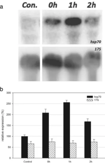
Fig. 2 a Northern blot analysis of total RNA preparations from cells covered with UG1 (UV-B + UV-A) filter and extracted 0, 1 and 2 h after the end of the irradiation. An equal amount (40μg) of each preparation was fractionated by electrophoresis, blotted and assayed, first, with the E. focardii hsp70 probe (top) and, then, with the Euplotes crassus 17S rDNA probe (bottom). b The graph shows the relative intensities (in percentage) for both hsp70 and 17S probes, normalized by taking the control expression level of hsp70 as 100 %. Error bars refer to the standard error of the mean
L. Fulgentini et al.
Author's personal copy
Results and Discussion
To assess the capacity of the psychrophilic Antarctic ciliates E. focardii to raise the expression of its hsp70 genes in response to UV radiation as well as to other spectral compositions, we exposed equal cell samples to the following different irradiation regimes: 1) UV-A and UV-B radiations (UG1 samples); 2) a mixture of visible, UV-A and UV-B radiations (WG280 sam-ples); and 3) visible light alone, without the UV-A and UV-B components (GG400 samples). For all the spectral composi-tions, the irradiation values of each spectral band were regulat-ed in order to be comparable with those recordregulat-ed in a typical Antarctic spring. From this point of view, our irradiation pro-tocols differed from those used in the study of Di Giuseppe et al. [25]. In fact, these Authors used irradiation values similar
to those recorded at noon in a mid-latitude region in summer-time. Therefore, we decided not to use the same high irradiance levels used by Di Giuseppe et al. [25] since they were too high to have an ecological relevance for E. focardii. In fact, those ciliates would never be exposed to those levels of UV-B, even in the case of a complete disruption of the Antarctic stratospher-ic ozone layer. Instead, we chose to set the irradiances to values comparable with those recorded during Antarctic spring, keep-ing the total UV-B dose supplied to the cell samples below the threshold of 11 kJ m−2, which, according to the work of Di Giuseppe et al., corresponds to a mortality of about 40 %, thus allowing us to analyze the sub-lethal effects of UV radiation at the molecular level.
The expression levels of E. focardii hsp70 transcripts fol-lowing the different irradiation regimes were measured by
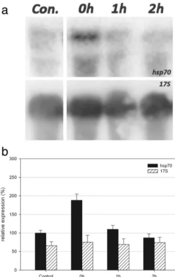
Fig. 3 a Northern blot analysis of total RNA preparations from cells covered with WG280 (UV-B + UV-A + visible) filter and extracted 0, 1 and 2 h after the end of the irradiation. An equal amount (40μg) of each preparation was fractionated by electrophoresis, blotted and assayed, first, with the E. focardii hsp70 probe (top) and, then, with the Euplotes crassus 17S rDNA probe (bottom). b The graph shows the relative intensities (in percentage) for both hsp70 and 17S probes, normalized by taking the control expression level of hsp70 as 100 %. Error bars refer to the standard error of the mean
Fig. 4 a Northern blot analysis of total RNA preparations from cells covered with GG400 (visible light) filter and extracted 0, 1 and 2 h after the end of the irradiation. An equal amount (40μg) of each preparation was fractionated by electrophoresis, blotted and assayed, first, with the E. focardii hsp70 probe (top) and, then, with the Euplotes crassus 17S rDNA probe (bottom). b The graph shows the relative intensities (in percentage) for both hsp70 and 17S probes, normalized by taking the control expression level of hsp70 as 100 %. Error bars refer to the standard error of the mean
UV and Visible Induces hsp70 in E. focardii
Northern blot analysis using as probes the E. focardii Hsp70 ATP binding domain [24]. To evaluate the effects of the dif-ferent irradiation regimes on the kinetics of hsp70 gene expression in E. focardii, samples of total RNA were extracted from irradiated cells immediately after the end of the irradia-tion period (0-h-labeled sample), 1 h (1-h sample) and 2 h (2-h sample) later.
As shown in Fig.2a, the E. focardii cells exposed to UV-B + UV-A radiations (UG1 sample) as described inBMaterials and Methods^, produced an intense 2.1-kbp band, i.e. the
molecular size expected for hsp70 mRNA, immediately after the end of the irradiation period (0-h sample). The hybridiza-tion signals peaked 1 h later (reaching about 256 % of the control sample expression level, see Fig.2b) and showed a significant decrease over the following 2 h at the end of irra-diation. To exclude any qualitative-quantitative variations be-tween RNA samples, the blots were stripped and re-hybridized with the E. focardii 17S rDNA probe, and Fig.2ashows equivalent hybridization signals.
Next, E. focardii cells were exposed to a combination of UV-B + UV-A + visible radiations (WG280 sample). Also, in this case, the hsp70 gene showed a prompt up-regulation in the 0-h samples (about 188 % with respect to the expression level of the control sample, see Fig.3b) and a gradual decrease of the transcript levels over the following 2 h (1- and 2-h samples, see Fig.3b).
Surprisingly and as shown in Fig.4, visible radiations by itself were also able to up-regulate hsp70 gene expression in E. focardii cells. Exposure to visible light produced an hsp70 transcription profile similar to that shown by the WG280 sam-ple, described above (compare Fig.3bwith Fig.4b).
Remarkably, from a behavioural point of view, the visible light-exposed cells were absolutely indistinguishable from
control (non-irradiated) cells: they in fact, maintain the same morphology and motility, while in the other two samples com-prising the UV bands, the cells showed an altered morphology with the appearance of anomalous big vacuoles inside the cytoplasm and a reduced motility (data not shown).
Our data clearly shows that Antarctic psychrophilic ciliate E. focardii is capable of reacting to radiative stress triggered by different spectral components (UV-A, UV-B or even visi-ble light alone), eliciting the transcriptional activation of its hsp70 genes. Apart from the UV-mediated hsp70 response which could be somehow expected for this Antarctic micro-organism, the most amazing results were represented by the capacity of the visible light to trigger hsp70 expression.
How can the hsp70 gene expression be activated in re-sponse to radiative stresses, as well as to visible light? Previ-ous research has showed that E. focardii is no longer capable of responding to environmental thermal shocks with an effec-tive activation of a transcriptional response of its hsp70 gene, although this gene still preserves its capacity to be activated in response to chemical as well as oxidative stressors [20]. Some hints to explain this eccentric transcriptional behaviour may reside in the peculiar organization of the 5′ regulatory regions of the E. focardii hsp70 gene [29]. The 5′ regulatory region of
hsp70 gene in E. focardii includes two sequence motifs known to be specific for stress-inducible genes: the first reg-ulation complex is identified by four motifs which are distinc-tive for theirBheat shock promoter elements^ (HSE) [30] that are targets of a specific class of transcriptional activators known asBheat shock factors^ (HSF) [16]; the second com-plex is identified by another four motifs which are distinctive forBstress response elements^ (StREs) [31,32]. Thus, at least in principle, this gene can rely on two distinct cis-acting mech-anisms to produce a stress-induced response, with HSE being
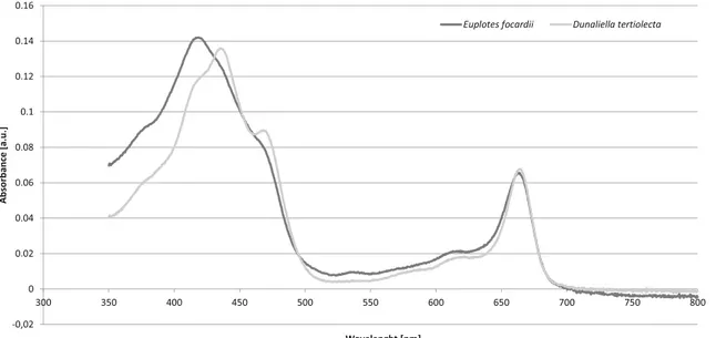
Fig. 5 Relatively normalized absorption spectra of Euplotes focardii and Dunaliella tertiolecta extracts. In the abscissa, the wavelength (in nm) of the incident light, in the ordinate the optical absorbance (in arbitrary unities)
L. Fulgentini et al.
Author's personal copy
more specific to respond to a stress of a thermal nature and StRE being responsible for the response to a broader range of stress, such as oxidative and chemical ones [33,34].
StREs could probably be the regulative elements involved in the response mediated by ultraviolet radiation, as UV-Bs are the most biologically damaging wavelengths, leading to the establishment of a strong oxidative environment inside the cell, with the production of active oxygen species and free radicals.
More puzzling is the activation of hsp70 gene transcription after irradiation with visible light only; this would need the activation of one or more endogenous chromophores, which act as photosensitizers, capable of directly or indirectly dam-aging cellular components via excitation at wavelengths at which DNA does not absorb. Among these photosensitizers, we can recall methylene blue, acridine orange, riboflavin and porphyrins [35,36]. Evenβ-carotene or one of its decompo-sition products can itself act as a photosensitizer [37]. Never-theless, the presence of such molecules in E. focardii has never been proven, and it is reasonable to think that they are present in a very small quantity in the digestive vacuoles, as sub-products of the digestion of the D. tertiolecta algae.
The activation of the hsp70 gene transcription after ir-radiation with visible light only has been found in other organisms, such as in Chlamydomonas, where the re-sponses to heat shock and light are quite similar. In partic-ular, these authors showed that the hsp70 gene expression in response to visible light was mediated through a heat shock-independent signal pathway involving a light-activated factor [38]. Chlamydomonas, cells incubated in the dark and then shifted to light intensities that caused photoinhibition, photosystem II were less damaged and recovered faster when, prior to light stress, the cultures were pre-exposed to dim light for 60 min. During this pre-incubation time, the hsp70 genes were induced [39]. Experimental evidence showed that the chlorophyll precur-sors Mg-protoporphyrin IX or its dimethyl ester play cru-cial roles as intermediates in the signaling pathway by which light activates the expression of the nuclear heat shock genes [40], once these plastidic compounds get ac-cess to the cytoplasm/nucleus [41].
E. focardii could then possess some kind of photosensi-tizers, which can absorb visible light and trigger the response, even if this ciliate appears completely rid of pigments. To deeper investigate this point, after starving the cells for several days (up to 21 days), we tried to extract this putative pigment. Results (Fig.5) show that chlorophyll deriving from the algae this ciliate feeds on is the only pigment that seems to be pres-ent inside E. focardii’s cells, even after several days of starva-tion. It is puzzling, anyway, to imagine that these pigments may have an active signaling role, considering that they are already in the digestive vacuoles, and thus are inclined to-wards the degradation pathway.
The apparent absence of any endogenous pigment and the complete absence of a synthesis pathway for the chlorophyll (as E. focardii is not a photosynthetic organism) depict a very puzzling situation: it is hard to suggest a signaling pathway for the visible light-induced transcription of hsp70. This situa-tion recalls that of the histophagous ciliate, Ophryoglena flava, that shows a strong phototactic response to visible light, even if it is apparently free of pigments and fed on non-photosynthetic tissues (in particular, beef spleen, see [42]). Maybe in both these ciliates, pigments are contained in such a small quantity, to be undetectable by our instruments. Further investigations are required to address this point. Overall, this finding aims to provide a better understanding of molecular responses upon UV hsp70 induction as well as to unveil other intriguing as-pects (visible light induction) of the adaptive strategies adopted by E. focardii to survive in the Antarctica.
References
1. Malloy KD, Holman MA, Mitchell D, Detrich HW III (1997) Solar UVB-induced DNA damage and photoenzymatic DNA repair in Antarctic zooplankton. Proc Natl Acad Sci U S A 94(4):1258–1263 2. Acevedo J, Nolan C (eds) (1993) Environmental UV radiation: causes— effects — consequences. Commission of the European Communities, Directorate-General XII for Science, Research and Development, Environment Programme, Brussels
3. Caldwell MM, Björn LO, Bornman JF, Flint SD, Kulandaivelu G, Teramura AH, Tevini M (1998) Effects of increased solar ultraviolet radiation on terrestrial ecosystems. J Photochem Photobiol B Biol 46: 40–52
4. Kennedy AD (1995) Antarctic terrestrial ecosystem response to glob-al environmentglob-al change. Annu Rev Ecol Syst 26:683–704 5. Karsten U (2008) Defense strategies of algae and cyanobacteria
against solar ultraviolet radiation. In: Amsler DC (ed) Algal chemical ecology. Springer, Berlin, pp 273–296
6. Agustí A (2008) The impact of increased ultraviolet radiation on the polar oceans. In: Duarte CM (ed) Impacts of global warming on polar ecosystems. Fundación BBVA, Bilbao, pp 25–45
7. Llabrés M, Agustí S (2010) Effects of ultraviolet radiation on growth, cell death and the standing stock of Antarctic phytoplankton. Aquat Microb Ecol 59:151–160
8. Häder DP, Helbling EW, Williamson CE, Worrest RC (2011) Effects of UV radiation on aquatic ecosystems and interactions with climate change. Photochem Photobiol Sci 10(2):242–60
9. Hansson L-A, Hylander S (2009) Effects of ultraviolet radiation on pigmentation, photoenzymatic repair, behavior, and community ecol-ogy of zooplankton. Photochem Photobiol Sci 8:1266–1275 10. Thomas DN, Dieckmann GS (2002) Antarctic sea ice—a habitat for
extremophiles. Science 295:641–644
11. Whitley D, Goldberg SP, Jordan WD (1999) Heat shock proteins: a review of the molecular chaperones. J Vasc Surg 29(4):748–751 12. Sarkar S, Singh DM, Yadav R, Arunkumar KP, Pittman GW (2011)
Heat shock proteins: molecules with assorted functions. Front Biol 6: 312–327
13. La Terza A, Barchetta S, Buonanno F, Ballarini P, Miceli C (2008) The protozoan ciliate Tetrahymena thermophila as biosensor of sublethal levels of toxicants in the soil. Fresenius Environ Bull 17:1144–1150 UV and Visible Induces hsp70 in E. focardii
14. Ritossa F (1962) A new puffing pattern induced by temperature shock and DNP in drosophila. Experientia 18:571–573
15. Clark MS, Peck LS (2009) HSP70 heat shock proteins and environ-mental stress in Antarctic marine organisms: a mini-review. Mar Genomics 2:11–18
16. Pirkkala L, Nykänen P, Sistonen L (2001) Roles of the heat shock transcription factors in regulation of the heat shock response and be-yond. FASEB J 15:1118–1131
17. Bosch TCG, Krylow SM, Bode HR, Steele RE (1988) Thermotolerance and synthesis of heat shock proteins: these responses are present in Hydra attenuata but absent in Hydra oligactis. Proc Natl Acad Sci U S A 85:7927–7931
18. Brennecke T, Gellner K, Bosch TC (1998) The lack of a stress re-sponse in Hydra oligactis is due to reduced hsp70 mRNA stability. Eur J Biochem 255:703–709
19. Hofmann GE, Buckley BA, Airaksinen S, Keen JE, Somero GN (2000) Heat-shock protein expression is absent in the Antarctic fish Trematomus bernacchii (family Nototheniidae). J Exp Biol 203: 2331–2339
20. La Terza A, Papa G, Miceli G, Luporini P (2001) Divergence be-tween two Antarctic species of the ciliate Euplotes, E. focardii and E. nobilii, in the expression of heat shock protein 70 genes. Mol Ecol 10:1061–1067
21. Valbonesi A, Luporini P (1993) Biology of Euplotes focardii, an Antarctic ciliate. Polar Biol 13:489–493
22. Pucciarelli S, Ballarini P, Miceli C (1997) Cold-adapted microtubules: characterization of tubulin posttranslational modifications in the ant-arctic ciliate Euplotes focardii. Cell Motil Cytoskeleton 38:329–340 23. Pucciarelli S, Miceli C (2002) Characterization of the cold-adapted
a-tubulin from the psychrophilic ciliate Euplotes focardii. Extremophiles 6:385–389
24. La Terza A, Miceli C, Luporini P (2004) The gene for the heat-shock protein 70 of Euplotes focardii, an Antarctic psychrophilic ciliate. Antarct Sci 16(1):23–28
25. Di Giuseppe G, Cervia D, Vallesi A (2012) Divergences in the re-sponses to ultraviolet radiation between polar and non-polar ciliated protozoa. Microb Ecol 63:634–638
26. Booth CR, Lucas TB, Morrow JH, Weiler CS, Penhale PA (1994) The United States National Science Foundation’s Polar Network for monitoring ultraviolet radiation. In: Weiler CS, Penhale PA (eds) Ultraviolet radiation in Antarctica: measurements and biological ef-fects, Antarct. Res. Ser. vol. 62. AGU, Washington, pp 17–37 27. Sambrook J, Fritsch EF, Maniatis T (eds) (1989) Molecular
clon-ing—a laboratory manual, 2nd edn. New York, Cold Spring Habour Laboratory Press
28. Feinberg AP, Vogelstein B (1984) A technique for radiolabeling DNA restriction endonuclease fragments to high specific activity. Anal Biochem 137(1):266–267
29. La Terza A, Miceli C, Luporini P (2007) Adaptive evolution of the heat-shock response in the Antarctic psychrophilic ciliate, Euplotes focardii: hints from a comparative determination of the hsp70 gene structure. Antarct Sci 16(2):239–244
30. Fernandes M, Xiao H, Lis JT (1994) Fine structure analyses of the Drosophila and Saccharomyces heat shock factor—heat shock ele-ment interactions. Nucl Acids Res 22:167–173
31. Kobayashi N, McEntee K (1993) Identification of cis and trans com-ponents of a novel heat shock stress regulatory pathway in Saccharomyces cerevisiae. Mol Cell Biol 13:248–256
32. Ruis H, Schüller C (1995) Stress signaling in yeast. BioEssays 17: 959–965
33. Feder ME, Hofmann GE (1999) Heat-shock proteins molecular chap-erones, and the stress response: evolutionary and ecological physiol-ogy. Ann Rev Physiol 61:243–282
34. Estruch F (2000) Stress-controlled transcription factors stress-induced genes and stress tolerance in budding yeast. FEMS Microb Rev 24:469–486
35. Kielbassa C, Roza L, Epe B (1997) Wavelength dependence of oxi-dative DNA damage induced by UVand visible light. Carcinogenesis 18:811–816
36. Epe B, Pflaum M, Boiteux S (1993) DNA damage induced by pho-tosensitizers in cellular and cell-free systems. Mutat Res 299:135– 145
37. Pflaum M, Kielbassa C, Garmyn M, Epe B (1998) Oxidative DNA damage induced by visible light in mammalian cells: extent, inhibi-tion by antioxidants and genotoxic effects. Mutat Res/DNA Repair 408(2):137–146
38. Kropat J, Gromoff ED, Müller FW, Beck CF (1995) Heat shock and light activation of a Chlamydomonas HSP70 gene are me-diated by independent regulatory pathways. Mol Gen Genet 248:727–734
39. Kropat J, Oster U, Rüdiger W, Beck CF (1997) Chlorophyll pre-cursors are signals of chloroplast origin involved in light induc-tion of nuclear heat-shock genes. Proc Natl Acad Sci U S A 94: 14168–14172
40. Kropat J, Beck CF (1998) Characterization of photoreceptor and signaling pathway for light induction of the Chlamydomonas heat-shock gene HSP70A. Photochem Photobiol 68:414–419
41. Kropat J, Oster U, Rüdiger W, Beck CF (2000) Chloroplast signalling in the light induction of nuclear HSP70 genes requires the accumu-lation of chlorophyll precursors and their accessibility to cytoplasm/ nucleus. Plant J 24:523–531
42. Cadetti L, Marroni F, Marangoni R, Kuhlmann H-W, Gioffre D, Colombetti G (2004) Phototaxis in the ciliated protozoan Ophryoglena flava: dose–effect curves and action spectrum determi-nation. J Photochem Photobiol B Biol 80:78–83
L. Fulgentini et al.