The Role of Magnetic Resonance Imaging in the Diagnosis of Fetal Brain Anomalies
DSJUOG DSJUOG
The Role of Magnetic Resonance Imaging in the
Diagnosis of Fetal Brain Anomalies
XXXXXXXXXXXXXXX
1,2Department of Neuroradiology, Unit, University of Bari, Bari, Italy Corresponding Author: M Resta, Department of Neuroradiology Unit University of Bari, Bari, Italy, [email protected]
10.5005/jp-journals-00000-0000
another investigation is needed, MRI provides important complementary informations.3
The MRI provides direct multiplanar imaging with a large field of view and a superior contrast resolution. The MRI visualization furthermore is not limited by oligohy-dramnios, fetal position, and maternal obesity . Currently, fetal MRI is used to confirm, in some par-ticular circumstances, a pathology, i.e., suspected by US, to improve the diagnosis, and to decide on pregnancy management or fetal surgery in utero.4,5
Fetal MRI is recommended starting from 19 weeks of gestational age. However, fetal MRI in early gestational age gives limited diagnostic information due to fetal movements and small size of fetus, while it provides limited additional information in comparison with US. TEcHNIQUE
Fetal MRI is typically performed on 1.5 T magnet. In the literature, only few centers report experimental results using strong magnetic fields (3 T),6 especially for
investi-gation on functional study for diffusion tensor imaging of fetal brain.7 These studies are limited by
excessive motion and susceptibility artifacts, but the development of new techniques will improve these problems.8
Maternal fasting is suggested to induce relative fetal hypoglycemia (the mother fasts for at least 4 hours prior to the examination), thus reducing motion fetal artifacts. The mother is supine or in the left lateral decubitus during the study.9
Usually, a phased array torso surface coil is used. A variety of fast MRI sequences are used to obtain T1- and T2-weighted images. The acronyms vary among the MRI manufacturers.
The T2 fast or Turbo spin-echo sequences are the stan-dard sequences for a fetal MRI study, acquiring a single slice. This acquisition modality reduces the fetal motion artifacts and provides excellent delineation of cerebral anatomy.10,11 However, Half Fourier Acquisition Single Shot Turbo Spin Echo (HASTE) sequences, commercially known also as Single-Shot Fast Spin Echo or Single-Shot Turbo Spin Echo, with only central lines of K-space filled, are currently the most used technique.12 This pulse sequence offers
excellent T2 images of the fetal brain with good contrast resolution in a few seconds (Figs 1 to 4, 5 to 8).
Mariachiara Resta MD*, Franca Dicuonzo MD*
*Neuroradiology, University of Bari “Aldo Moro”, Bari Italy
ABsTRAcT
Prenatal Ultrasonagraphy(US) is the mainstay modality to diagnose fetal abnormalities especially in early pregnancy. Fetal Magnetic Resonance Imaging (MRI) is a useful tool to confirm and to characterize a pathology that is suspected on US, especially in the detection of central nervous system pathologies.The use of ultrafast imaging tecniques gives additional importantant informations and optimal imaging quality, despite fetal motion, in clinical practice.
Diffusion Weighted Imaging (DWI), Diffusion Tensor Imaging (DTI), MR Spetroscopy and Functional studies have potential applications in the fetal brain imaging. Fetal MRI could recognize, in contradistinction to US, the development of fetal brain, the multilayered appearance of the cerebral parenchyma, the timing of sulci development and the myelination.
The most common indications for fetal MRI are ventriculomegaly, midline anomalies, malformations of cerebral cortical development, posterior fossa anomalies, suspected haemorraghic- ischemic lesions, tumors.
Fetal MRI is a safe and powerful complement to US for clinical management and prognostication.
Keywords: fetal MRI, MRI, fetal brain, fetal brain anomalies How to cite this article: Resta M, Dicuonzo F. The Role of Magnetic Resonance Imaging in the Diagnosis of Fetal Brain Anomalies. Donald School J Ultrasound Obstet Gynecol 2017;11(4):1-13.
Source of support: Nil Conf ict of interest: None
INTRODUcTION
Ultrasonography (US) is the first-line screening tool to detect fetal malformations.1
Over the past 20 years, magnetic resonance imaging (MRI) has been increasingly used for the visualization of fetus in utero. Fetal MRI is a useful diagnostic tool after US, in particular, for fetal brain.2
The US remains the main modality, but when it is inconclusive or when
The T1-weighted sequences with and without fat sup-pression are acquired during breath-holding. T1 is the sequence of choice for detection of hemorrhage, calcifica-tion, and fat. It could help visualize some fetal organs or pathologies with T1 hyperintensity13 (Figs
5-8). The gradient recalled echo T2 sequences are used to visualize bony structures, fat, calcifications, or the break-down products of blood or hemosiderin. Diffusion-weighted imaging (DWI) is important to detect ischemic areas of the brain and characterize
maturation of the brain.
Proton magnetic resonance spectroscopy for the detection of membranes or neuronal metabolites, such as creatine, choline, and N-acetyl aspartate in the risk of hypoxia has been used sparingly, but not routine examination.14
in
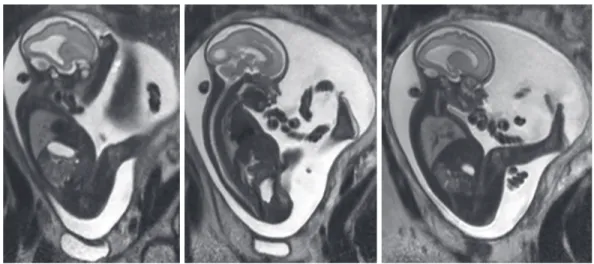
Both the brain and the spine should be imaged in all the three planes (Figs 1 to 4).
Following scout acquisition, a series of images in the axial, sagittal, and coronal planes are obtained. Fetal MRI is an interactive scanning acquisition: Each acquisition is the scout for the subsequent one.15
The contrast material administration (gadolinium) is not recommended in fetal MRI as declared by the 2013 American College of Radiology (ACR) guidance document on MRI safe practices.16 Indeed, gadolinium
could pass through the placental barrier and enter fetal circulation with potentially harmful effect on the fetus.17
About the safety, unlike X-rays and computed tomog-raphy, no maternal or fetal adverse events have been reported on MRI performed at 1.5 T.18
Theoretical adverse effects could be related to only the radiofrequency field and acoustic exposure.19
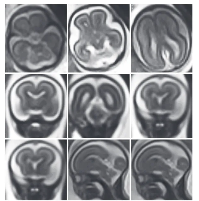
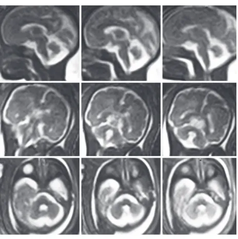
Fig. 1: 19 gestational weeks. Normal fetal brain. HASTE T2 images in selected axial, coronal, and sagittal planes. Gyration is at the beginning. In the selected sections, three layers corresponding to germinal matrix (dark line near the lateral ventricles), cortical plate (dark line), an intermediate layer (bright zone, the future myelinated white matter) are well evident
The Role of Magnetic Resonance Imaging in the Diagnosis of Fetal Brain Anomalies
DSJUOG
Fig. 2: 22 gestational weeks. Normal fetal brain. HASTE T2 images in selected axial, coronal, and sagittal planes. The images are similar to Figure 1, but the neuronal migration is going on and central sulcus is well evident. The sylvian operculum is still open. In the posterior fossa, the Reil’s vallecula is not formed
Fig. 3: 32 gestational weeks. Normal fetal brain. HASTE T2 images in selected axial and sagittal planes. The fetal brain is similar to newborn one. The gyration is almost completed, while the myelination is just at the beginning with hypointense cortex and hyperintense white matter
INDIcATIONs
Central nervous system abnormalities are the most common clinical indications for fetal MRI.20,21
Fetal MRI is the best modality to detect brain ment. The knowledge of the timing of the fetus develop-ment is mandatory for the interpretation of fetal MRI.22
In the early gestational age, there are three layers of cerebral parenchyma:
1. Germinal matrix (dark line on T2 sequences near the lateral ventricles): It could be visualized until 24 to 27 weeks of gestation (WG)
2. Cortical plate (dark line on T2 sequences, bright on T1 ones)
3. Intermediate layer (bright on T2 , dark on T1). Myelin could be detected at 22 to 40 WG.
The parieto-occipital and hippocampal sulci begin to appear at 22th WG.
The precentral and postcentral sulci appear at 27th WG.
The frontal (superior and inferior) sulci appear from 29th WG.
At 34 WG, sulcation is completed
The cellular proliferation, neuronal migration, and cortical organization are the phases of the cortex development.23-25
Fetal MRI can evaluate these developmental steps and detect cortical malformations, such as polymicrogyria, lissencephaly, or schizencephaly26 (Figs 9, 10).
The primary indications of fetal MRI according to the recommendations of the ACR and Society for Pediatric Radiology are27:
• Congenital anomalies (ventriculomegaly, corpus callosal dysgenesis, holoprosencephaly, posterior fossa anomalies, malformations of cerebral cortical development)
• Fetuses with a family risk for brain anomalies (e.g., tuberous sclerosis, corpus callosal dysgenesis, mal-formation of cerebral cortical development)
• Vascular abnormalities (in particular for complication of monochorionic twin pregnancy)
Fig. 4: Same as the case of Figure 3. HASTE T2 images in selected coronal plane
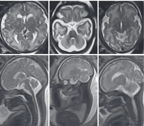
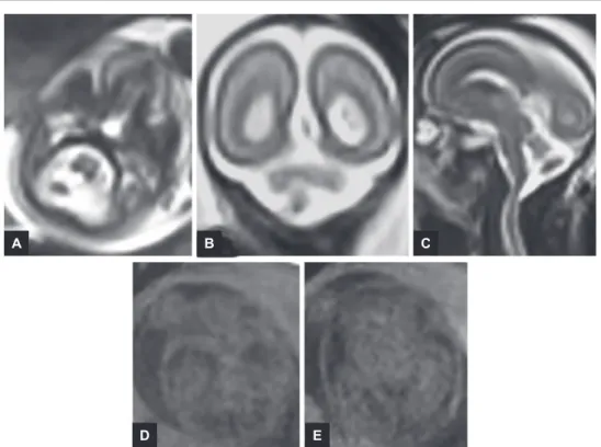
Figs 5A to E: 30 gestational weeks. (A–C) HASTE T2 images in selected axial, coronal, and sagittal planes. (D and E) T1 axial breathhold fast field echo images. Total agenesis of the corpus callosum with interhemispheric mass due to associated lipoma classically hyperintense in T1-weighted images and hypointense in T2-T1-weighted images.
A B
D
C
The Role of Magnetic Resonance Imaging in the Diagnosis of Fetal Brain Anomalies
DSJUOG
• Congenital anomalies of spine (neural tube defects, sacrococcygeal teratoma, caudal regression, sireno-melia, vertebral anomalies)
• Masses of the face and neck (venolymphatic malfor-mations, hemangiomas)
• Assessment of fetal surgery (meningomyelocele, sacrococcygeal teratomas)
Ventriculomegaly
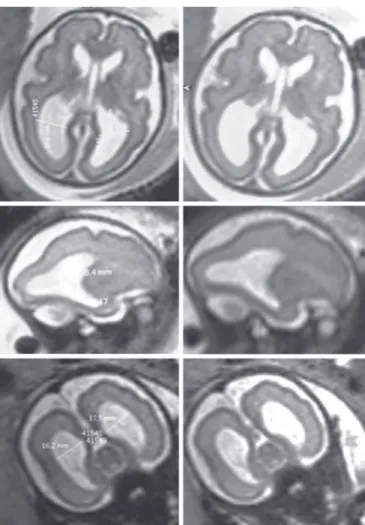
Ventriculomegalyisacommonabnormalityvisualizedon US. It is diagnosed when measurement of the atrial width is>10mm.Ventriculomegalyisdefinedasmild(atrial
width 10–12 mm), moderate (atrial width 12–15 mm), or severe (atrial width >15 mm)28 (Figs 11 to 13).
Ventriculomegalyhasvariousetiologies:Obstructive, developmental, and destructive.
Fig. 6: 22 gestational weeks. HASTE T2 images in selected axial and sagittal planes. Ultrasound diagnosis of mild bilateral ventriculomegaly. Fetal MRI confirms mild ventriculomegaly and shows in axial plane two symmetrical hyperintense lesions at level of bilateral basal ganglia. Postnatal MRI diagnosed a rare form of mitochondrial disease
In 80% of cases, ventriculomegaly is associated with other central nervous system abnormalities, which are crucial for the postnatal prognosis29-30. Some
abnormalities may be missed by US, but are well studied by MRI: They include neuronal migration, proliferation and differentia-tion disorders, delayed sulcation and gyration, heteroto-pias, hypoxic–ischemic disorders, and hemorrhages. Since most of these pathologies develop late in pregnancy, the appropriate time to perform MRI is in the late second or third trimesters.
Midline Anomalies
The most common midline fetal pathologies are corpus callosum abnormalities (Figs 14 and 15). The corpus callosum is formed by genu, body, splenium, and last the rostrum. By the 15 to 20th WG, it assumes its final appearance22 (Figs 1 to 4).
Fig. 7: 24 gestational weeks. HASTE T2 images in selected axial and sagittal planes. Isolated mild ventriculomegaly
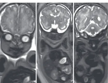
Fig. 9: 37 gestational weeks. HASTE T2 images in selected axial, coronal, and sagittal planes. Unilateral left ventriculomegaly. Smooth aspect of the left temporal cortical ribbon suggestive of lissencephaly Figs 8A to E: 22 gestational weeks. (A–C) HASTE T2 images in selected axial, coronal, and sagittal planes. (D and E) Axial breathhold fast field echo images. Extra-axial small tumor at level of cisterna magna. T1 images, even if with low contrast resolution, deny the hemorrhagic nature of the lesion. Dermoid tumor was diagnosed at postnatal imaging
A B
D
C
The Role of Magnetic Resonance Imaging in the Diagnosis of Fetal Brain Anomalies
DSJUOG
Fig. 10: Same as the case of Figure 9. Postnatal MRI (T1 and T2 selected axial and sagittal planes) and genetic evaluation confirmed the prenatal hypothesis of type I lissencephaly
Fig. 11: 24 gestational weeks. HASTE T2 images in selected axial and sagittal planes. Isolated mild ventriculomegaly
Fig. 12: Same as the case of Figure 11. HASTE T2 images in selected sagittal plane. Isolated mild ventriculomegaly
Fetal MRI confirms the US suspicion of corpus cal-losum anomalies and shows additional abnormalities (25% compared with US). There is high association with sulcation abnormalities, Chiari malformation, gray matter heterotopia, Dandy–Walker malformation (DWM), and arachnoid cysts (Fig. 16).
Also, interhemispheric cyst and lipoma can occur with corpus callosum anomalies. The diagnosis of lipoma can be done only in the third trimester on T1 sequence20 (Fig. 5).
The prognosis of corpus callosum anomalies is vari-able depending on associated malformations.
Fetal MRI is useful in diagnosing dysplastic/agenetic septum pellucidum. It could be isolated or associated with anomalies related to optic chiasma or part of septo-optic dysplasia32,33.
Posterior cranial Fossa Malformations
The MRI is useful in the diagnosis of posterior cranial fossa malformations, particularly in the third trimester when the skull ossification l imits t he U S d etection o f these structures.2
The MRI visualizes directly the cerebellar hemispheres, the vermis, the brain stem, the fourth ventricle, the cisterna magna, and the tentorium. Measurements and biometri-cal analysis can be made and compared with established nomograms.34
Posterior fossa abnormalities that can be evaluated by fetal MRI include DWM, vermian hypoplasia, Blake’s pouch cyst, mega cisterna magna, and Arnold– Chiari malformation.35
The DWM is characterized by cystic dilatation of the fourth ventricle, severely hypoplastic and upward rotated vermis, and upper displacement of the tentorium; ven-triculomegaly is frequently associated (Fig. 17); vermian hypoplasia is characterized by cystic dilatation of fourth Fig. 13: 28 gestational weeks. HASTE T2 images in selected axial,
coronal, and sagittal planes. Isolated severe ventriculomegaly This structure could be detected on fetal MRI on the midline sagittal T2 sequence as a curvilinear “C” hypoin-tense band at the superior margin of the cavum septum pellucidum.24
Abnormalities of corpus callosum include agenesis (complete or partial), hypogenesis, and hypoplasia.31
Fig. 14: 22 gestational weeks. HASTE T2 images in selected axial, coronal, and sagittal planes. Complete agenesis of corpus callosum, colpocephaly, and wide communication of the dilated third ventricle with the interhemispheric fissure
The Role of Magnetic Resonance Imaging in the Diagnosis of Fetal Brain Anomalies
DSJUOG
Fig. 15: 25 gestational weeks. HASTE T2 images in selected axial, coronal, and sagittal planes. Isolated complete agenesis of the corpus callosum without other cerebral anomalies. No markers of other fetal body or extremities malformations. During parental counseling, the potential good prognosis should be presented
Fig. 16: 29 gestational weeks. HASTE T2 images in selected axial, coronal, and sagittal planes. Large right hemispheric arachnoid cyst. Normal posterior fossa structures. Left dislocation of the midline. In parasagittal plane the genu (single black arrow) and posterior part of the body and splenium of the corpus callosum (double black arrow) are well recognizable
ventricle, mild hypoplasia, and upward rotation of the vermis and normal tentorium (Figs 18 and 19). In Blake’s pouch cyst, there is a digit-like protrusion of the fourth ventricle below an intact and mildly upward rotated vermis.
In addition, DWM can be associated with supratento-rial anomalies (agenesis of corpus callosum, polymicro-gyria) better visualized on MRI examination.
At early gestational age (<20 weeks), fetal MRI could have a low specificity in the diagnosis of isolated vermian hypoplasia, so follow-up examination is needed. Diffusion sequences DWI give quantitative information on cerebellum development: Progressive reduction in diffusivity is seen with increasing gestational age.7
The Arnold–Chiari malformation is a complex conse-quence of a defect arising from failure of normal closure of the neural tube.
There are two types of Arnold–Chiari malformation: 1. Chiari I: downward herniation of cerebellar tonsils 2. Chiari II: small posterior fossa, herniation of the vermis
and IV ventricle, hypoplastic cerebellum, supraten-torial ventricular dilatation. Myelomeningocele is always associated (Fig. 20).
Fetal MRI is important for confirmation and evaluation of the associated findings in the supratentorial brain: dysplasia of the corpus callosum, gyral
malformations, and interdigitation across the falx. Fetal MRI could detect additional key prognostic factors: degree of herniation, brain stem dysmorphology, and cerebral cortical and white matter anomalies.36 In recent years, MRI is being performed also for the planning of prenatal open or endoscopic repair of the myelomenin-gocele. There are promising results about the long-term effect of this prenatal intervention.37
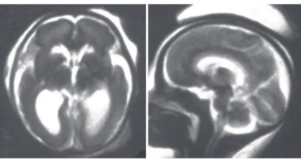
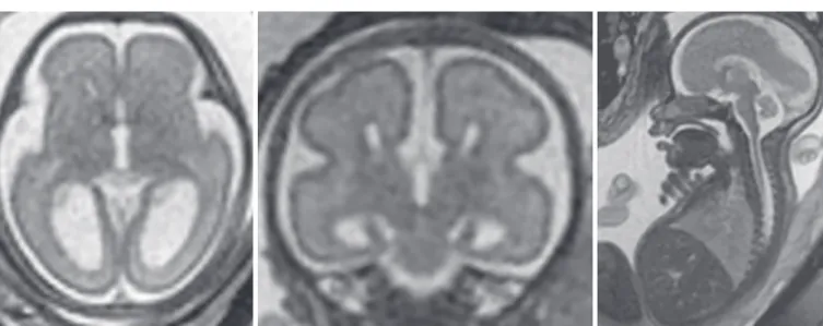
Fig. 17: 24 gestational weeks. HASTE T2 images in selected coronal and axial planes. Classical DWM with cystic dilatation of fourth ventricle and severe ventriculomegaly
Fig. 18: 22 gestational weeks. HASTE T2 images in selected axial, coronal, and sagittal planes. Vermian hypoplasia. The fourth ventricle is enlarged and communicates with cisterna magna; the cerebellum is hypoplastic and upward rotated
The Role of Magnetic Resonance Imaging in the Diagnosis of Fetal Brain Anomalies
DSJUOG
Fig. 19: 32 gestational weeks. HASTE T2 images in selected axial, coronal, and sagittal planes. Same as the case of Figure 18, MRI evolution confirms the diagnosis of vermian hypoplasia
Fig. 20: 37 gestational weeks. HASTE T2 images in selected axial, coronal, and sagittal planes. Colpocephaly, cervical ectopia of cerebellar tonsils, and lumbar spina bifida (white arrows) indicating Chiari 2 malformation
A genetic condition characterized by ponto-cerebellar atrophy/hypoplasia is Joubert syndrome, a predomi-nantly autosomal recessive disease. Prenatal US diagnosis is difficult due to the lack of specific signs and the only finding may be vermian dysplasia. The essential feature on MRI is the “molar tooth sign”: the brain stem seems like a molar tooth on axial images.
Focal Nervous Tissue Lesions (Hemorrhagic, Ischemic, and Tumors)
In suspected focal nervous tissue lesions on US, the fetal MRI is mandatory. The fetal MRI study identifies t he precise localization and can suggest the etiology.
Most of the causes of hemorrhage (particularly germinal matrix hemorrhage) can be studied by MRI (Fig. 21). A hemorrhagic lesion is typically hyperintense on T1 sequences, hypointense on T2 sequences, with susceptibility artifact on gradient echo T2 sequences15,20.
The diffusion sequences (DWI) have an important role for the detection of ischemic lesion (Fig. 22). This sequence gives information on the microscopic motion of water molecule. For example, in twin-to-twin Transfusion Syndrome, the fetal MRI is important to identify cerebral ischemic lesion in the vital fetus after the death of the cotwin.
4. Frates M, Kumar A, Benson C, Ward V, Tempany C. Fetal anomalies: comparison of MR imaging and US for diagnosis. Radiology 2004 Aug;232(2):398-404.
5. Glenn OA, Barkovich AJ. Magnetic resonance imaging of the fetal brain and spine: an increasingly important tool in prenatal diagnosis, Part 1. AJNR Am J Neuroradiol 2006 Sep;27(8):1604-1611.
6. Victoria T, Jaramillo D, Roberts TP, Zarnow D, Johnson AM, Delgado J, Rubesova E, Vossough A. Fetal MRI: jumping from 1.5 to 3 T (preliminary experience). Pediatr Radiol 2014 Apr;44(4):376-386; quiz 373-375.
7. Mignone Philpott C, Shannon P, Chitayat D, Ryan G, Raybaud CA, Blaser SI. Diffusion-weighted imaging of the cerebel-lum in the fetus with Chiari II malformation. AJNR Am J Neuroradiol 2013 Aug;34(8):1656-1660.
8. Kasprian G, Brugger PC, Weber M, Krssák M, Krampl E, Herold C, Prayer D. In utero tractography of fetal white matter development. Neuroimage 2008 Nov 1;43(2):213-224. 9. Yamashita Y, Namimoto T, Abe Y, Takahashi M, Iwamasa J,
Miyazaki K, Okamura H. MR imaging of the fetus by a HASTE sequence. AJR Am J Roentgenol 1997 Feb;168(2):513-519. 10. Chung HW, Chen CY, Zimmerman RA, Lee KW, Lee CC,
Chin SC. T2-weighted fast MR imaging with true FISP versus HASTE comparative efficacy in the evaluation of normal fetal brain maturation. AJR Am J Roentgenol 2000 Nov;175(5):1375-1380.
11. Prayer D, Brugger PC, Prayer L. Fetal MRI: techniques and protocols. Pediatr Radiol 2004 Sep;34(9):685-693.
12. Resta M, Burdi N, Resta MC. Magnetic resonance of the fetal brain. Ultrasound Rev Obstet Gynecol 2003:131-142. 13. Mirsky DM, Shekdar KV, Bilaniuk LT. Fetal MRI: head and
neck. Magn Reson Imaging Clin N Am 2012 Aug;20(3): 605-618.
Figs 22A to C: 37 gestational weeks. Axial HASTE T2 image: left frontal paraventricular cystic lesion. (A) Diffusion b1000 axial sequence, (B and C) denies ischemic etiology. Postnatal MRI diagnosed a neuro-epithelial cyst
Figs 21A to C: 22 gestational weeks. (A and B) HASTE T2 images in selected sagittal and axial planes. (C) Axial breathhold fast field echo image. Monochorionic twin pregnancy. Mild unilateral ventriculomegaly in one fetus.T1 image clearly demonstrates hemorrhage in germinal matrix (arrow)
Other etiologies of ischemic, hemorrhagic lesions include intrauterine infections, coagulopathies, and venous sinus thrombosis.
Congenital central nervous system tumors diagnosed during pregnancy are rare, and often carry a poor progno-sis. The most frequent types are the teratoma and glioma. Use of US and MRI allows the suspicion of brain tumors during pregnancy. However, the definitive diagnosis is only confirmed after birth by histology (Fig. 8).
cONcLUsION
Fetal MRI is a safe and powerful tool in prenatal evalu-ation of the fetal brain. Fetal MRI, thanks to recent technological advances, plays a very important role in determining prognosis and management. Obviously, US remains the predominant modality of evaluation of the fetal brain during pregnancy, and MRI should be performed by experienced operators only following an accurate neurosonographic examination.
REFERENcEs
1. Blondiaux E, Garel C. Fetal cerebral imaging—ultrasound vs. MRI: an update. Acta Radiol 2013 Nov;54(9):1046-1054. 2. Levine D. Ultrasound versus magnetic resonance imaging
in fetal evaluation. Top Magn Reson Imaging 2001 Feb;12(1): 25-38.
3. Saleem SN. Fetal MRI: an approach to practice: a review. J Adv Res 2014 Sep;5(5):507-523.
A
A
B
B
The Role of Magnetic Resonance Imaging in the Diagnosis of Fetal Brain Anomalies
DSJUOG
14. Girard N, Fogliarini C, Viola A, Confort-Gouny S, Le Fur Y, Viout P. MRS of normal and impaired fetal brain development. Eur J Radiol 2006 Feb;57(2):217-225.
15. Tee LM, Kan EY, Cheung JC, Leung WC. Magnetic reso-nance imaging of the fetal brain. Hong Kong Med J 2016 Jun;22(3):270-278.
16. Expert Panel on MR Safety, Kanal E, Barkovich AJ, Bell C, Borgstede JP, Bradley WG Jr, Froelich JW, Gimbel JR, Gosbee JW, Kuhni-Kaminski E, et al. ACR guidance docu-ment on MR safe practices: 2013. J Magn Reson Imaging 2013 Mar;37(3):501-530.
17. Webb JA, Thomsen HS, Morcos SK. The use of iodinated and gadolinium contrast media during pregnancy and lactation. Eur Radiol 2005 Jun;15(6):1234-1240.
18. Bulas D, Egloff A. Benefits and risks of MRI in pregnancy. Semin Perinatol 2013 Oct;37(5):301-304.
19. Stecco A, Saponaro A, Carriero A. Patient safety issues in magnetic resonance imaging: state of the art. Radiol Med 2007 Jun;112(4):491-508.
20. Manganaro L, Bernardo S, Antonelli A, Vinci V, Saldari M, Catalano C. Fetal MRI of the central nervous system: state-of-the-art. Eur J Radiol 2017 Aug;93:273-283.
21. Paladini D, Quarantelli M, Sglavo G, Pastore G, Cavallaro A, D’Armiento MR, Salvatore M, Nappi C. Accuracy of neu-rosonography and MRI in clinical management of fetuses referred with central nervous system abnormalities. Ultrasound Obstet Gynecol 2014 Aug:44(2):188-196. 22. Brisse H, Fallet C, Sebag G, Nessmann C, Blot P, Hassan M.
Supratentorial parenchyma in the developing fetal brain: in vitro MR study with histologic comparison. AJNR Am J Neuroradiol 1997 Sep;18(8):1491-1497.
23. Garel C. The role of MRI in the evaluation of the fetal brain with an emphasis on biometry, gyration and parenchyma. Pediatric Radiol 2004 Sep;34(9):694-699.
24. Amin RS, Nikolaidis P, Kawashima A, Kramer LA, Ernst RD. Normal anatomy of the fetus at MR imaging. Radiographics 1999 Oct; 19 Spec No:S201-214.
25. Griffith PD, Russell SA, Mason G, Morris J, Fanou E, Reeves MJ. The use of in utero MR imaging to delineate development brain abnormalities in multifetal pregnancies. AJNR Am J Neuroradiol 2012 Feb;33(2):359-365.
26. Rubod C, Robert Y, Tillouche N, Devisme L, Houfflin-Debarge V, Puech F. Role of fetal US and MRI in the pre-natal diagnosis of migration disorders. Prenat Diagn 2005 Dec;25(13):1181-1187.
27. American College of Radiology (ACR), Society for Pediatric Radiology (SPR). ACR-SPR practice guideline for the safe and optimal performance of fetal magnetic resonance imaging (on
line publication). Reston (VA): American College of Radiology (ACR); 2010. p. 10.
28. Levine D, Trop I, Mehta TS, Barnes PD. MR imaging appear-ance of fetal cerebral ventricular morphology. Radiology 2002 Jun;223(3):652-660.
29. Yin S, Na Q, Chen J, Li-Ling J, Liu C. Contribution of MRI to detect further anomalies in fetal ventriculomegaly. Fetal Diagn Ther 2010;27(1):20-24.
30. Hannon T, Tennant PW, Rankin J, Robson SC. Epidemiology, natural history, progression and post-natal outcome of severe fetal ventriculomegaly. Obstet Gynecol 2012 Dec;120(6): 1345-1353.
31. Conte G, Parazzini C, Falanga G, Cesaretti C, Izzo G, Rustico M, Righini A. Diagnostic value of prenatal MRI in the detection of brain malformations of fetuses before the 26th week of gestational age. AJNR Am J Neuroradiol 2016 May;37(5):946-951.
32. Glenn OA, Goldstein RB, Li KC, Young SJ, Norton ME, Busse RF, Goldberg JD, Barkovich AJ. Fetal MRI in the evaluation of fetuses referred for sonographically suspected abnormalities of the corpus callosum. J Ultrasound Med 2005 Jun;24(6):791-804.
33. Manganaro L, Bernardo S, De Vito C, Antonelli A, Marchionni E, Vinci V, Saldari M, Di Meglio L, Giancotti A, Silvestri E, et al. Role of fetal in the evaluation of isolated and non-isolated corpus callosum dysgenesis: result of a cross sectional study. Prenat Diagn 2017 Mar;37(3):244-252.
34. Ber R, Bar-Yosef O, Hoffmann C, Shahar D, Achiron R, Katorza E. Normal fetal posterior fossa in MR imaging new biometric data and possible clinical significant. AJNR Am J Neuroradiol 2015 Apr;36(4):795-802.
35. Chapman T, Mahalingam S, Ishak GE, Nixon JN, Siebert J, Dighe MK. Diagnostic Imaging of posterior fossa anomalies in the fetus and neonate: part 2, posterior fossa disorder. Clin Imaging 2015 Mar-Apr;39(2):167-175.
36. Guibaud L. Practical approach to prenatal posterior fossa abnormalities using MRI. Pediatr Radiol 2004 Sep;34(9): 700-711.
37. Egloff A, Bulas D. MRI evaluation of fetal neural tube defects. Semin Ultrasound CT MR 2015 Dec;36(6):487-500.