Synthetic long non-coding RNAs
[SINEUPs] rescue defective gene
expression in vivo
Alessia Indrieri
1, Claudia Grimaldi
1, Silvia Zucchelli
2,3, Roberta Tammaro
1,
Stefano Gustincich
2,4,5& Brunella Franco
1,6Non-coding RNAs provide additional regulatory layers to gene expression as well as the potential to being exploited as therapeutic tools. Non-coding RNA-based therapeutic approaches have been attempted in dominant diseases, however their use for treatment of genetic diseases caused by insufficient gene dosage is currently more challenging. SINEUPs are long antisense non-coding RNAs that up-regulate translation in mammalian cells in a gene-specific manner, although, so far evidence of SINEUP efficacy has only been demonstrated in in vitro systems. We now show that synthetic SINEUPs effectively and specifically increase protein levels of a gene of interest in vivo. We demonstrated that SINEUPs rescue haploinsufficient gene dosage in a medakafish model of a human disorder leading to amelioration of the disease phenotype. Our results demonstrate that SINEUPs act through mechanisms conserved among vertebrates and that SINEUP technology can be successfully applied in vivo as a new research and therapeutic tool for gene-specific up-regulation of endogenous functional proteins.
Both naturally occurring and artificial RNAs have the potential to be used as modulators of target genes. Regulation of gene expression through the activity of gene-specific artificial inhibitory nucleic acids such as siR-NAs, RNAi and morpholinos (MOs) has become a common strategy to investigate gene function and they have also extended the druggable genome to potentially all protein coding genes. An equally important approach with potentially broad-range applications could be based on natural and artificial RNAs that can increase expression of target genes and few examples have been described. For instance, degradation or inhibition in vivo of natural antisense transcripts (NATs) by single-stranded oligonucleotides or siRNAs can transiently and reversibly modu-late locus-specific gene expression1 and chemically modified mRNAs are capable to modulate gene expression in a mouse model of a lethal congenital lung disease2. Finally, a programmable transcription factor prototype has been shown to promote gene transcription in cell lines and primary cultures via an invariable transactivating domain coupled with a variable RNA domain that binds genes using sequence specificity3.
Large genomic efforts such as ENCODE4 and FANTOM5 have shown that the majority of the mammalian genome is transcribed. In addition to approximately 25000 protein-coding genes, there are at least an equal num-ber of long non-coding RNA (lncRNA) genes that generate long transcripts (over 200 base pairs) that do not encode for proteins. About one third of annotated lncRNAs overlaps with protein-coding genes and many of these are transcribed from the opposite strand forming sense/antisense (S/AS) pairs6.
We have previously shown that a natural lncRNA antisense to the Ubiquitin carboxyl-terminal ester-ase L1 (AS Uchl1), a Parkinson’s diseester-ase-associated gene, is able to increester-ase UchL1 protein synthesis at the post-transcriptional level7. We demonstrated that AS Uchl1 activity depends on two distinct RNA elements, The Binding Domain (BD) at the 5′ end, is a sequence that overlaps, in antisense orientation, to the sense protein-coding mRNA and determines target selection and AS Uchl1 specificity by RNA-RNA base pairing. The AS Uchl1 BD is 72bp long, centered across the initiating ATG with a −40/+32 configuration, spans part of the Uchl1 5′UTR and a portion of its coding sequence (CDS)7. The other functional part of the AS Uchl1 sequence is represented by the Effector Domain (ED), an inverted SINE (short interspersed nuclear elements) B2 sequence 1Telethon Institute of Genetics and Medicine (TIGEM), 80078, Via Campi Flegrei 34, Pozzuoli (NA), Italy. 2Area of Neuroscience, SISSA, 34136, Trieste, Italy. 3Dipartimento di Scienze della Salute, Universita’ del Piemonte Orientale, Novara, Italy. 4Department of Neuroscience and Brain Technologies, Italian Institute of Technology, 16163, Genova, Italy. 5TransSINE Technologies, 1-7-22 Suehiro-cho Tsurumi-ku, Yokohama, Kanagawa 230-0045 Japan. 6Medical Genetics Services, Department of Translational Medicine, Federico II University, 80131, Naples, Italy. Correspondence and requests for materials should be addressed to B.F. (email: [email protected])
received: 01 March 2016 Accepted: 18 May 2016 Published: 06 June 2016
www.nature.com/scientificreports/
embedded in the non-overlapping part of the transcript that is essential for protein synthesis up-regulation7. Its modular architecture allows to redirect translation enhancement activity to any target mRNA by swapping its BD with the appropriate antisense sequence8. The AS Uchl1 domain organization is conserved in other AS lncRNAs that overlap with protein coding genes and regulate their translation7,9. Therefore, AS Uchl1 repre-sents a new class of natural and synthetic antisense lncRNAs that can activate translation7,10. These RNAs were named SINEUPs because they require the inverted SINEB2 sequence to UP-regulate translation in a gene-specific manner10. Synthetic SINEUPs have been proven effective in vitro with a number of targets, including GFP7,10, FLAG-tagged proteins10 and secreted recombinant antibodies9, thus supporting the intrinsic scalability of SINEUP technology10.
However, to date SINEUP efficacy has only been limited to measuring target protein levels in vitro in mamma-lian cells, in experiments carried out in controlled, homogenous cell culture systems.
Here we show for the first time that SINEUPs can increase the synthesis of a functional endogenous protein in vivo and rescue haploinsufficient gene dosage in a medakafish model of a human disease.
Our study demonstrates that SINEUPs may be an effective tool for functional studies in vivo that demand increased levels of endogenous proteins. In addition, these results are a proof of principle that this technology may be applicable to design therapeutic approaches for genetic diseases in which selectively increasing the expres-sion of the target gene may be curative.
Results and Discussion
To assess the potential application of the SINEUP technology in vivo, we used the teleost medakafish (Oryzias latipes), which is particularly amenable for reverse-genetic analyses. In this model, the microinjection of early embryos with any kind of RNAs is technically easy and results in transient gene overexpression or inhibition11.
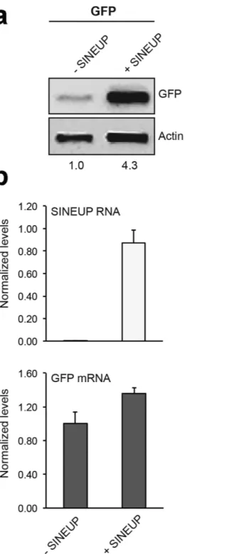
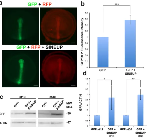
For our initial studies we utilized the SINEUP-GFP7 that results in an increase GFP protein levels in transient overexpression experiments. SINEUP-GFP was cloned into the pCS2 plasmid that allows in vitro synthesis of RNA. As expected, we observed that in human embryonic kidney (HEK) 293T/17 cells, the SINEUP-GFP pCS2 construct increased GFP protein levels by acting post-transcriptionally (Fig. 1). To test their functionality in vivo, SINEUP-GFP RNA and GFP mRNA were transcribed in vitro and then co-injected into medaka embryos. Equal amounts of RFP transcribed mRNA were also co-injected as control for SINEUP specificity. SINEUP activity was estimated as fold change in GFP protein levels, normalized for RFP protein levels, in the presence or absence of the SINEUP. Embryos injected with the SINEUP-GFP displayed increased GFP levels and unchanged RFP levels (Fig. 2a,b). Furthermore, western blot (WB) analysis confirmed increased GFP levels at 19 and 30 stage (st) embryos (Fig. 2c,d). These results demonstrate the efficacy of SINEUPs and validate the post-transcriptional mechanisms of SINEUPs in vivo. SINEB2 sequences are mouse specific and are not present in fish, in which a superfamily of vertebrate SINEs (V-SINEs) has been described12. Our results indicate that SINEUPs act through mechanisms conserved among vertebrates and suggest that the secondary structure of SINEs is functionally con-served between mice and fish.
To address the potential use of this technology in increasing expression of an endogenous protein in vivo we tested SINEUPs in a medakafish model of microphthalmia with linear skin defects (MLS) syndrome, a X-linked dominant disorder characterized by microphthalmia, brain abnormalities and skin defects in heterozygous females, and in utero lethality in males13. MLS syndrome is caused by mutations in players of the mitochon-drial respiratory chain (MRC) such as the holocytochrome c-type synthase (HCCS)14, and the subunit 7B of cytochrome c oxidase (COX), the MRC complex-IV15.
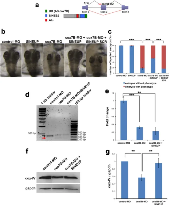
We downregulated cox7B expression using a MO-based approach. cox7B-MO was designed against the exon 2 acceptor splice-site and its injection causes exon 2 skipping resulting in frameshift15 (Fig. 3a). We designed a synthetic SINEUP against the endogenous cox7B mRNA carrying a 72bp BD starting from position −40 in the 5′UTR (before ATG) to position +32 in the CDS (Fig. 3a). We then cloned the SINEUP-cox7B sequence in the pCS2 plasmid and injected the in vitro synthesized RNA into embryos. As expected, injection of SINEUP-cox7B did not change cox7B mRNA levels (Supplementary Fig. 1a) and did not induce any aberrant phenotype in con-trol embryos (Fig. 3b,c). cox7B-morphants showed a dose-dependent phenotype characterized by microcephaly and microphthalmia15 (Fig. 3b,c). Interestingly, injection of the SINEUP-cox7B in MLS morphants fully rescued microphthalmia and microcephaly in about 50% of embryos, whereas the injection of a control SINEUP car-rying a scrambled sequence in the BD domain (SINEUP-SCR) did not result in amelioration of the phenotype (Fig. 3b,c).
To exclude that SINEUP could induce changes in cox7B transcription and/or interfere with MO function-ing in cox7B morphants, we performed RT-PCR to amplify full-length WT and mutant cox7B mRNAs from cox7B-MO injected embryos and embryos co-injected with cox7B-MO and SINEUP-cox7B. Target sequences of cox7B-MO and SINEUP-cox7B are localized in different exons of cox7B and do not overlap (Fig. 3a). As expected, SINEUP-cox7B did not alter size or abundance of WT and mutated cox-7B mRNA (Fig. 3d,e) thus confirming that SINEUPs act through a post-transcriptional mechanism.
COX7B is necessary for complex-IV formation and its downregulation in vitro induces reduction of fully assembled complex-IV and of its subunits including COX-IV15 (Supplementary Fig. 2). As expected WB analysis showed reduction of cox-IV in cox7B-MO-injected fish (Fig. 3f,g). Interestingly, SINEUP-cox7B was capable to fully rescue cox-IV levels in cox7B-morphants whereas no effect was detected using the SINEUP-SCR in cox7B morphants (Fig. 3f,g and Supplementary Fig. 3) or the SINEUP-cox7B in wt embryos (Supplementary Fig. 1b,c).
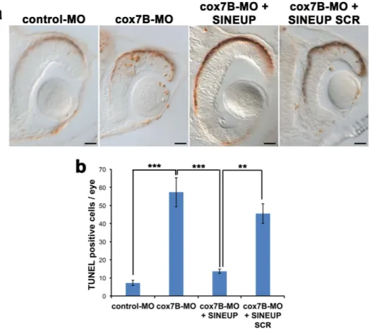
We previously demonstrated that, in hccs-morphants, MRC impairment results in increased CNS pro-grammed cell death (PCD) and that this event underlies the MLS phenotype16. Interestingly, TUNEL analy-sis revealed an increase of PCD in the eyes and brain of cox7B morphants, which was rescued by injection of SINEUP-cox7B (Fig. 4a,b).
Our data demonstrated that synthetic SINEUP-cox7B may restore MRC function thus consequently rescuing the MLS phenotype. Our results validate SINEUPs as a versatile tool for in vivo experimental biology and pave the way for its use in RNA therapeutics.
Artificial siRNAs, RNAi and MOs have become the tools of choice to inhibit gene expression and SINEUPs represent their molecular counterparts to increase protein synthesis in vivo. To our knowledge, SINEUPs rep-resent the only available tool that uses AS lncRNAs to enhance production of endogenous proteins acting on their cellular mRNAs. In this context, SINEUP technology represents a powerful tool in molecular biology to
Figure 1. pCS2 + -SINEUP-GFP activity in vitro. HEK293T/17 were transfected with pEGFP-C2 and
pCS2+/SINEUP-GFP constructs at 1:6 ratio (+SINEUP). Control cells were transfected with pEGFP-C2 and an empty control plasmid (-SINEUP). 48 hr after transfection, cells were lysed and processed for protein (a) and RNA (b) levels. Western blot was performed with anti-GFP antibody. β-actin was used as loading control. Fold-induction was calculated on Western blot images normalized to β-actin and relative to empty control samples. Expression of SINEUP-GFP (white bars) and quantity of GFP mRNA (grey bars) were monitored by Real Time PCR using specific primers. Data indicate mean ± SD. Data are representative of >3 independent replicas.
www.nature.com/scientificreports/
increase protein levels of the specific gene of interest in vivo. Given their mode of action, SINEUPs would allow a spatio-temporal regulation of protein quantities in a more physiological context, avoiding artifacts due to ectopic overexpression and targeting only cells that actively express mRNA of interest, while being ineffective if mRNA is not present or present in a mutated form. On the basis of our results, SINEUPs should be considered a new approach to treat conditions such as haploinsufficiencies due to a reduction to 50% or less of gene function in which rescue of the phenotype requires recovery of the protein levels. A large number of genetic disorders are due to reduced levels of protein products. For these conditions, an efficient SINEUP activity specific for the gene of interest would, in principle, be therapeutic. This approach presents several advantages over competing technolo-gies: a) SINEUPs can modulate translation of target mRNAs without introducing stable genomic changes; b) the induced up-regulation of specific proteins is a physiological range (~2-fold) compared to most conventional gene replacement strategies; c) SINEUPs exert their function on target mRNAs under their physiological regulation thus limiting adverse influences on cells and tissues that do not express the target transcript8,17.
In conclusion our data demonstrate that SINEUPs can selectively increase the expression of an endogenous functional protein in vivo and may be applied as a new tool in molecular biology and for nucleic acids-based therapeutics.
Methods
Plasmids.
pEGFP-C2 (Clontech), SINEUP targeting EGFP (here named SINEUP-GFP), and SINEUP con-taining a scramble sequence in the region of overlap (Binding Domain, BD) (here named SINEUP-SCR) have been previously described7.SINEUP targeting medakafish cox7B mRNA (Accession Number HE717026) was constructed using pcD-NA3-Δ5-ASUchl1 as backbone7. SINEUP-backbone lacks the BD to Uchl1 and retains AS Uchl1 effector domain (inverted SINEB2 element) embedded in AS Uchl1 non-overlapping sequence containing the partial Alu sequence and the 3′ tail. SINEUP cox7B-specific BD (72bp) was designed, in antisense orientation, around the ATG of the protein-coding sequence with a −40/+32 organization. For antisense cox7B, the method of ‘annealing
Figure 2. Synthetic SINEUP increases GFP protein levels in vivo. (a) Representative st19 embryos injected
with RFP and GFP mRNA with or without SINEUP-GFP RNA. Scale bar 100 μm. (b) Quantification of GFP/ RFP fluorescence intensity by ImageJ analysis software (n = 10, ***p < 0,001, one-tailed Student’s t-test; Error bars are SEM). (c) WB analysis of GFP in st19 and st30 embryos injected as in (a). (d) Quantification of GFP protein levels (n = 2, *p < 0,05, **p < 0,01, one-tailed Student’s t-test; Error bars are SEM).
and primer extension’ of two 3′-end overlapping oligonucleotides was used to generate the 72-bp antisense cox7B overlap region. Annealed fragment was obtained with the following oligonucleotides:
Figure 3. Synthetic SINEUP-cox7B rescues MLS phenotype in medakafish. (a) Structure of the medakafish
cox7B transcript (exon/intron gene organization is displayed) and binding sites of cox7B-MO and SINEUP-cox7B. The cox7B-MO was designed against the acceptor splice site of exon 2 and its injection causes the skipping of exon 2. The BD of SINEUP-cox7B is designed against exon 1 in position −40 in the 5′-UTR (before ATG) to position +32 in the CDS. (b) Representative st30 embryos injected with control-MO, SINEUP-cox7B, cox7B-MO with or without SINEUP-cox7B or SINEUP-SCR. Scale bars 100 μm. SINEUPs rescue microphthalmia and microcephaly in 50% of cox7B-morphants (c). (n ≥ 300 embryos/conditions, ***p < 0,001, One-way ANOVA; Error bars are SEM). (d) RT-PCR of cox7B transcripts on total RNA extracted from embryos injected with control-MO, cox7B-MO and embryos co-injected with cox7B-MO and cox7B-SINEUP. Black arrow indicates WT mRNA whereas the red arrow indicates the mutated mRNA generated by cox7B-MO-driven aberrant splicing. (e) Real Time PCR on total RNA using specific primers to amplify WT cox7B mRNA. (n = 3 independent biological samples, ***p < 0,001, **p < 0,01, two-tailed Student’s t-test; Error bars are SEM). (f) WB analysis of cox-IV in st30 embryos injected with control-MO and cox7B-MO with or without SINEUP-cox7B. (g) Quantification of cox-IV protein levels (n = 3, **p < 0,01 one-tailed Student’s t-test; Error bars are SEM).
www.nature.com/scientificreports/
AScox7BFW 5′-ATATCTCGAGATGTTAGCTGCAGCCTTTGCAAACCGGTACATGTTGTCT
GGCCTTGTG-3′
AScox7BRv 5′-GAGAGATATCCCGTTCTTCAAGGGTATCTGAAGACACAAGGCCAGACAACATGTA
CC-3′
The annealed fragment was digested with XhoI and EcoRV and ligated into antisense pcDNA3-Δ5-ASUchl1. Full-length GFP from pEGFP-C2 plasmid and full-length SINEP-GFP, SINEUP-SCR and SINEUP-cox7B from pcDNA3 plasmids were cloned into with pCS2+ vector.
pCS2+/RFP (Red Fluorescent Protein) was already described16.
Cell lines and transfection.
Human Embryonic Kidney (HEK) 293T/17 and HeLa cells were obtained from ATCC and maintained in culture with Dulbecco’s Modified Eagle Medium (GIBCO) supplemented with 10% FBS (SIGMA) and 1% antibiotics (penicillin/streptomycin), as suggested by the vendor.HEK 293T/17 cells were transfected with Fugene HD (Roche), with a 1:6 ratio between pEGFP-C2 vector and pCS2+/SINEUP-GFP plasmids and following manufacture’s instruction. Cells were collected at 48 hours after transfection and split in two samples for RNA extraction and Western Blot analysis, as previously described10.
To silence COX7B, Hela cells were transfected using siRNAs ON-TARGET against human COX7B and ON-TARGET Non-Targeting (Darmachon) to a final concentration of 50 nM using Interferin reagent as previ-ously described15. Cells were collected at 72 hr after transfection for protein extraction and Western Blot analysis.
Medakafish (Oryzias latipes) Stocks.
Wild type Oryzias latipes of the cab strain were maintained in an in-house facility (28 °C on a 14/10 hr light/dark cycle). Embryos were staged as described18.All studies on fish were conducted in strict accordance with the institutional guidelines for animal research and approved by the Italian Ministry of Health; Department of Public Health, Animal Health, Nutrition and Food Safety in accordance to the law on animal experimentation (article 7; D.L. 116/92; protocol number: 00001/11/ IGB; approval date June 6, 2011). Furthermore, all animal treatments were reviewed and approved in advance by the Ethics Committee of the Institute of Genetics and Biophysics, IGB Animal House, (Naples, Italy).
mRNAs and MOs Injections.
RNAs of SINEP-GFP, SINEUP-SCR, SINEUP-cox7B, GFP and RFP were transcribed in vitro from pCS2+ plasmids using the SP6 mMessage mMachine kit (Ambion) according to theFigure 4. cox7B downregulation induces increased cell death that is rescued by synthetic SINEUP-cox7B.
(a) TUNEL assays on retinal sections of st30 embryos injected with control-MO, cox7B-MO with or without SINEUP-cox7B or SINEUP-SCR. Scale bars 20 μm. (b) Number of TUNEL positive cells/eye (n ≥ 5 retina/ condition, **p < 0,01, ***p < 0,001, One-way ANOVA; Error bars are SEM).
manufacturer’s instructions. Control-MO (5′-CCTACCTACACCACAACAGAAATAA-3′) and cox7B-MO (5′-CCGACCTGCACGACAACACAAAGAA-3′) were purchased from Gene Tools, LLC. MOs working con-centrations, injection conditions and their specificity and efficiency were previously described15. MOs and RNAs were injected into fertilized embryos at the one/two-cell stage according to the following conditions: GFP mRNA 50 ng, RFP mRNA 50 ng, SINEUP-GFP 300 ng, control-MO and cox7B-MO 100 μM, SINEUP-cox7B and SINEUP-SCR 300 ng. At least 3 independent experiments were performed for each condition.
mRNA expression analysis.
Total RNA extraction from cells and embryos was done using RNeasy Mini kit (Quiagen) according to the manufacturer’s instructions. During the extraction protocol RNAs were digested with DNaseI (Qiagen) to remove DNA contaminating, according to the manufacturer’s instructions. At least 50 embryos were pooled in each assay. Total RNA was then reverse-transcribed into cDNA by QuantiTect Reverse Transcription Kit (Quiagen) with random hexamers oligo.For RT-PCR experiment, full length cox7B transcripts were amplified from cDNAs of control-MO, cox7B-MO and cox7B-MO + SINEUP-cox7B using the following primers designed against the exon 1 and exon 3 respectively:
cox7BEx1Fw 5′-CGCAACCATTTCCGGTATCTG-3′ cox7BEx3Rw 5′-GGTATTGGTTTGTCATCTGTAC-3′
PCR fragments were loaded on 2% agarose gel and subjected to electrophoresis.
For quantitative real time PCR analysis (qRT-PCR), cDNAs was diluited 1:2 and analyzed using Roche Light Cycler 480 system with LigthCycler 480 SYBR Green I Master (Roche). Oligonucleotide sequences of GFP and human GAPDH primers were previously described7. To specifically amplify WT medakafish cox7B mRNA, we used the following primers designed against the exon 3 and exon 2 that is skipped after the MO-driven aberrant splicing15:
cox7BEx2Fw 5′-CAGTGGAACCGTCTTCTGC-3′ cox7BEx3Rw 5′-GGTATTGGTTTGTCATCTGTAC-3′
Medakafish hprt and gapdh primers were previously described19,20 and used as endogenous controls. mRNA analysis showed comparable results with both normalization approaches. Relative expression was calculated with the ΔΔCt method21 and variation was reported as fold change (2−ΔΔCt). The results are shown as means ± SEM of three independent biological assays.
Protein extracts and Western blot analysis.
Cells or embryos (at least 100 for conditions) were lysed in RIPA buffer containing 1X protease inhibitor cocktail (SIGMA). Cleared proteins extracts were quantified by using the Bradford method (Bio-Rad). For WB, protein samples were separated on 15% SDS-PAGE and transferred to PVDF membrane (Millipore). Membranes were blocked in TBS containing 5% Non Fat Dry Milk (Cell Signaling) and incubated with the follows primary antibodies: anti-GFP 1:500 (Abcam, ab13970), anti GFP rabbit polyclonal antibody (Life technologies, a6445), used 1:1000 (for HEK 293T/17 cells experi-ments), anti-β-Actin 1:500 (Sigma), anti-GAPDH 1:1000 (Santa Cruz Biotechnology), anti-COX-IV 1:500 (Cell Signaling), anti-COX7B 1:500 (Abcam, ab137094).Proteins of interest were detected with horseradish peroxidase-conjugated goat anti-mouse or anti-rabbit IgG antibody (1:3000, GE Healthcare) and rabbit anti-chicken IgY antibody (Millipore) visualized with the Luminata Crescendo substrate (Millipore) or the Super Signal West Femto substrate (Thermo Scientific), according to the manufacturer’s protocol. Western blotting images were acquired using the Chemidoc-lt imaging system (UVP) and band intensity was calculated using ImageJ software.
TUNEL assay.
TUNEL assays were performed on st30 embryos using In Situ Cell Death Detection Kit (Roche, Mannheim, Germany) as previously reported16.Statistical Analysis.
In all experiments the significance of differences between groups was evalu-ated by ANOVA and Student’s t-test, p < 0.05 was considered significant. Quantitative data are presented as the mean ± SD (Standard Deviation) or ± SEM (Standard Error of the Mean) of at least three independent experiments.References
1. Modarresi, F. et al. Inhibition of natural antisense transcripts in vivo results in gene-specific transcriptional upregulation. Nat
Biotechnol 30, 453–459 (2012).
2. Kormann, M. S. et al. Expression of therapeutic proteins after delivery of chemically modified mRNA in mice. Nat Biotechnol 29, 154–157 (2011).
3. Fimiani, C., Goina, E. & Mallamaci, A. Upregulating endogenous genes by an RNA-programmable artificial transactivator. Nucleic
Acids Res 43, 7850–7864 (2015).
4. Djebali, S. et al. Landscape of transcription in human cells. Nature 489, 101–108 (2012). 5. Consortium, F. et al. A promoter-level mammalian expression atlas. Nature 507, 462–470 (2014).
6. Derrien, T. et al. The GENCODE v7 catalog of human long noncoding RNAs: analysis of their gene structure, evolution, and expression. Genome Res 22, 1775–1789 (2012).
7. Carrieri, C. et al. Long non-coding antisense RNA controls Uchl1 translation through an embedded SINEB2 repeat. Nature 491, 454–457 (2012).
www.nature.com/scientificreports/
8. Zucchelli, S. et al. SINEUPs: A new class of natural and synthetic antisense long non-coding RNAs that activate translation. RNA
Biol 12, 771–779 (2015).
9. Patrucco, L. et al. Engineering mammalian cell factories with SINEUP noncoding RNAs to improve translation of secreted proteins.
Gene 569, 287–293 (2015).
10. Zucchelli, S. et al. SINEUPs are modular antisense long non-coding RNAs that increase synthesis of target proteins in cells. Front
Cell Neurosci 9, 174 (2015).
11. Wittbrodt, J., Shima, A. & Schartl, M. Medaka–a model organism from the far East. Nat Rev Genet 3, 53–64 (2002).
12. Ogiwara, I., Miya, M., Ohshima, K. & Okada, N. V-SINEs: a new superfamily of vertebrate SINEs that are widespread in vertebrate genomes and retain a strongly conserved segment within each repetitive unit. Genome Res 12, 316–324 (2002).
13. Morleo, M. & Franco, B. GeneReviews(R) (eds Pagon R. A. et al.) (University of Washington, Seattle, 2011).
14. Wimplinger, I. et al. Mutations of the mitochondrial holocytochrome c-type synthase in X-linked dominant microphthalmia with linear skin defects syndrome. Am J Hum Genet 79, 878–889 (2006).
15. Indrieri, A. et al. Mutations in COX7B cause microphthalmia with linear skin lesions, an unconventional mitochondrial disease. Am
J Hum Genet 91, 942–949 (2012).
16. Indrieri, A. et al. The impairment of HCCS leads to MLS syndrome by activating a non-canonical cell death pathway in the brain and eyes. EMBO Mol Med 5, 280–293 (2013).
17. Takahashi, H. & Carninci, P. Widespread genome transcription: new possibilities for RNA therapies. Biochem Biophys Res Commun 452, 294–301 (2014).
18. Iwamatsu, T. Stages of normal development in the medaka Oryzias latipes. Mech Dev 121, 605–618 (2004).
19. Ventre, S. et al. Metabolic regulation of the ultradian oscillator Hes1 by reactive oxygen species. J Mol Biol 427, 1887–1902 (2015). 20. Carrella, S. et al. TGF-beta Controls miR-181/ERK Regulatory Network during Retinal Axon Specification and Growth. PLoS One
10, e0144129 (2015).
21. Schmittgen, T. D. & Livak, K. J. Analyzing real-time PCR data by the comparative C(T) method. Nat Protoc 3, 1101–1108 (2008).
Acknowledgements
We thank Drs. Sandro Banfi and Graciana Diez-Roux for critical reading of the manuscript and helpful discussion. We also thank the TIGEM Medakafish Facility for technical support and the TIGEM Bioinformatics Core for support in statistical analysis of data. Alessia Indrieri was the recipient of Post-doctoral Fellowships from Fondazione Umberto Veronesi. This work was supported by the Italian Telethon Foundation.
Author Contributions
A.I., S.Z., S.G. and B.F. designed the study and the experiments. A.I. and C.G. performed the experiments in fish. A.I., S.Z., C.G. and R.T. performed the experiments in vitro. A.I., S.Z., S.G. and B.F. analyzed the data and wrote the manuscript.
Additional Information
Supplementary information accompanies this paper at http://www.nature.com/srep
Competing financial interests: S.G. and S.Z. declare competing financial interests as co-founders and members
of TransSINE Technologies (www.transsine.com). S.G. and S.Z. are named inventors in patent issued in the US Patent and Trademark Office on SINEUPs and licensed to TransSINE Technologies. A.I., C.G., R.T. and B.F. declare no competing financial interests.
How to cite this article: Indrieri, A. et al. Synthetic long non-coding RNAs [SINEUPs] rescue defective gene
expression in vivo. Sci. Rep. 6, 27315; doi: 10.1038/srep27315 (2016).
This work is licensed under a Creative Commons Attribution 4.0 International License. The images or other third party material in this article are included in the article’s Creative Commons license, unless indicated otherwise in the credit line; if the material is not included under the Creative Commons license, users will need to obtain permission from the license holder to reproduce the material. To view a copy of this license, visit http://creativecommons.org/licenses/by/4.0/


