SCUOLA NORMALE SUPERIORE
PISA
and
THE INTERNATIONAL CENTRE for GENETIC ENGINEERING
and BIOTECHNOLOGY (ICGEB)
TRIESTE
Analysis of the
β
β
β
β-adducin gene: new insights into gene
structure and function
Thesis submitted for the Degree of Doctor of Philosophy
Perfezionamento in Genetica Molecolare e Biotecnologie
Candidate:
Luisa Costessi
Supervisor:
Andrés F. Muro, Ph.D
Acknowledgments
At the end of more than four years of work I want to take this opportunity to express my gratitude to everyone who has shared this important period of my life with me: this thesis is not a reflection of my efforts alone.
First of all, I would like to thank my supervisor, Andrès, for his tireless enthusiasm and wide research outlook. I am very grateful for his constant presence, guidance and support in my PhD project which allowed me to learn many useful things and to improve my scientific knowledge.
My special thanks go to all the members of Mouse Molecular Genetics group, especially to Faby and Sandra for their great friendship and sympathy, constant help and precious suggestions in any situation. Also to Fede, who worked with me everyday, side by side, sharing all the adventures of this period and becoming not only my “nearest” PhD colleague but a special and good friend.
I would also like to remember all the people of the former Molecular Pathology group who shared with me these years. Thanks to Maury and Yuna, for their precious help and support in numerous situations, and also to Cri, Erica, and Michy for their friendship. To my extraordinary friend Ely, for all the wonderful time that we have spent together in the lab and outside the institute, my PhD period would not have been the same without you.
I would like to express my gratitude to Prof. Baralle for giving me the opportunity to begin my scientific career in his group, and for his special quality of being “ever present for everyone”.
Many thanks go also to everyone in the ICGEB components, for their collaboration and for the fantastic atmosphere that they create in the institute. In particular, to Giulia, for allowing me to include a part of her work in this dissertation, and to Wendy, for her help in polishing my English.
In addition, I would like to take this chance to express my gratitude to my external collaborators, Dr. Chishti (University of Illinois College of Medicine, Chicago, USA.) and Dr. Tongiorgi (University of Trieste, Trieste, Italy), and to extend it to all the members of their groups.
A tender thank you is also deserving of my mother and father, the guiding lights in my life, and to Francesco and Martina for their constant presence, support and trust during my study.
And last, but not least, all my love and gratitude to Emy, particularly for his patience, and for everything he has said and done to help and encourage me.
ABSTRACT
Adducins are a family of membrane skeleton proteins encoded by three related genes (ADD1, ADD2 and ADD3 or α-, β- and γ-adducin genes). Both ADD1 and ADD3 are ubiquitously expressed, while the β-adducin gene shows a pattern of expression restricted to brain and haematopoietic tissues. Adducin is found as either a heterodimer or heterotetramer of α/β or α/γ subunit composition in most tissues. Human erythrocytes mainly contain α/β heterodimers, whereas the γ-subunit is also found at low levels in mouse red blood cells (RBCs). In erythrocytes, adducin is localised at the actin-spectrin junctions (junctional complexes), where it promotes the association of actin to spectrin, and bundles and caps the fast-growing ends of actin filaments. The junctional complexes comprise of other proteins, such as tropomodulin, tropomyosin, Protein 4.1, p55 and dematin, promoting and modulating spectrin–actin interactions, forming membrane associations, and regulating actin filament length. A mouse strain bearing a targeted mutation in the β-adducin gene was previously created in our lab and it was demonstrated that the mutant mice had abnormal erythrocytes, developing mild spherocytic hereditary elliptocytosis. These abnormalities were associated with alterations in the levels of several erythrocyte skeletal proteins: adducin, α-tropomyosin and actin were down-regulated, while the levels of γ-adducin and tropomodulin were increased. In addition, α- and β-subunits of the dimeric CapZ, the main muscle actin-capping protein of actin filaments barbed-ends, and which is mainly found in the cytoplasm of normal erythrocytes (ECapZ), were ~9-fold upregulated in RBC mutant skeletons, suggesting the existence of a compensatory mechanism. Moreover, purified adducin was re-incorporated into adducin deficient ghosts partially displacing ECapZ. The erythrocyte skeletal composition of dematin-adducin double knock-out mice (DAKOs) was also analysed. Despite their stronger phenotype and the presence of more significant alterations in red cell skeletons than those observed in the single mutants, the levels of ECapZ α- and β-subunits remained unaltered in the RBC skeleton of DAKOs. These results reflect the severe haemolytic anaemia manifested by the double mutant mice, and suggest the importance of adducin and dematin in the maintenance of the shape and mechanical properties of the erythrocyte membrane skeleton. Other actin-rich structures were also analysed, such as that of lens eye fibre cells, and showed analogous variations of most cytoskeletal proteins (except for CapZ
which showed no changes) suggesting different skeletal organisation between erythrocytes and lens fibre cells.
In addition to its presence in erythrocytes and eye lens cells, β-adducin is also very abundant in the brain where it is detected as a protein of similar size to the versions found in spleen, but it is translated from a peculiar brain-specific mRNA of 8-9 kb, instead of 3.5–4.5 kb of that detected in spleen. The molecular basis of this difference was discerned by determining the structure of the brain-specific β-adducin transcript in rats, mice and humans. Brain- and spleen-specific promoters and first exons, apparently not conserved in humans, were identified in rodents. It was demonstrated that β-adducin brain mRNAs are processed in all three species by a common mechanism utilising tissue-specific alternative polyadenylation sites (denominated A4 in the brain), which generates an unusually long 3'-untranslated region (3’-UTR) of about 5-6 kb. Proximal erythroid-specific polyadenylation regions in rodents (A1 and A2-3) and humans (A1, A2 and A3) were also identified. In all of these regions, the elements defining the core polyadenylation signals were identified: the hexanucleotide motif, the U-rich and GU-rich elements, and the cleavage site itself, known as the poly(A) site. A study of the β-adducin tissue-specific alternative polyadenylation mechanism was initiated by performing transient transfection assays of chimeric minigenes mimicking the structure and the arrangement of the polyadenylation sites in the β-adducin gene. It was shown that minigene-derived β-adducin transcripts were correctly polyadenylated at the A4 brain-specific site in HeLa cells. Using a deletion strategy to analyse the contribution of the core polyadenylation elements to poly(A) site selection by the chimeric minigenes, it was observed that the lack of the U-rich region of the A4 site directly decreased the efficiency of pre-mRNA processing, and that the use of an upstream cryptic poly(A)site is trigged by the absence of the A4 site hexanucleotide motif. In addition, the long brain-specific β-adducin mRNA is localised with dendrites, highlighting the importance of the 5-6 kb 3’-UTR in the subcellular localisation of the β-adducin transcript in the brain, and probably in other regulatory functions not yet addressed.
To summarise, the molecular alterations underlying the red cell shape and structure abnormalities found in mutant β-adducin mice, in double adducin-dematin KO mice, and also those found in other actin-rich structures, such as eye lens fibre cells, were characterised. Additionally, the molecular basis of the size differences in the mRNAs from brain and spleen in humans, rats and mice was determined. Preliminary experiments aimed to characterise the tissue-specific mechanisms regulating those differences were also performed.
TABLE of CONTENTS
ACKNOWLEDGMENTS
2
ABSTRACT
3
TABLE OF CONTENTS
5
LIST OF FIGURES AND TABLES
10
ABBREVIATIONS
13
1
INTRODUCTION
15
1.1 Cells membrane cytoskeleton 15
1.1.1 - Microtubules - 15
1.1.2 - Intermediate filaments - 16
1.1.3 - Actin filaments - 17
1.2 Plasma membrane and cytoskeleton of erythrocytes 19
1.2.1 - Integral RBC membrane proteins - 20
1.2.2 - Peripheral RBC membrane proteins: the erythrocyte cytoskeleton - 22 1.3 Membrane skeleton role: deformability, stability and shape 29
1.3.1 - Membrane deformability and stability - 30
1.3.2 - Cell shape - 31
1.3.3 - Cytoplasmic viscosity - 31
1.4 Disorders of the red blood cell membrane 32
1.4.1 - Hereditary spherocytosis (HS) - 32
1.4.2 - Hereditary elliptocytosis (HE) and related disorders - 33
1.4.3 - Hereditary stomatocytosis (HSt) - 34
1.5 Knock-out mouse models of erythrocyte membrane proteins 34
1.5.1 - Targeted disruption of Band 3 - 35
1.5.2 - Targeted disruption of Protein 4.1 - 35
1.5.3 - Targeted disruption of tropomodulin - 36
1.5.4 - Targeted disruption of tropomyosin - 36
1.5.5 - Skeletal and cardiac actin-deficient mice - 36
1.5.6 - Targeted disruption of Protein 4.2 - 37
1.5.7 - Targeedt disruption of dematin headpiece domain - 37
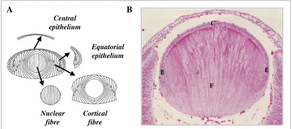
1.6 The skeleton of the eye lens fibres membranes 38
1.7 Adducin 41
1.7.1 - Adducin genes and their expression - 41
1.7.2 - β-adducin gene - 42
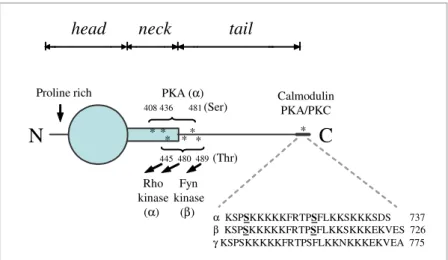
1.7.3 - Adducin protein structure - 44
1.7.4 - Adducin in vitro activities - 46
1.7.5 - Regulation of adducin functions - 46
1.7.6 - Adducin cellular localisation and in vivo activities - 47
1.7.7 - Adducin and hypertension - 48
1.8 ββββ-adducin knock-out mice 48
1.8.1 - β-adducin deficient mice and hypertension 52 1.8.2 - ΑKO mice and synaptic plasticity underlying learning and memory - 52
1.9 Dematin-adducin double knock-out mice 53
1.10 mRNA processing reactions 54
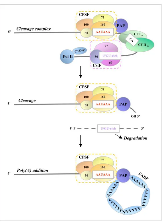
1.10.2 - Splicing - 55 1.10.3 - 3′ end formation: polyadenylation signals and machinery - 55 1.10.4 - Steps in processing: assembly, cleavage, and polyadenylation - 62
1.10.5 - Role of the poly(A) tail - 64
1.11 Crosstalk between mRNA processing events 65
1.11.1 - Capping interactions with splicing and polyadenylation - 66 1.11.2 - Connections between splicing and polyadenylation - 67 1.12 Transcription and mRNA processing of protein-encoding gene 69
1.12.1 - Transcription initiation and elongation - 69
1.12.2 - Transcription termination downstream of the poly(A) site - 70 1.13 Transcriptional and post-transcriptional regulation of gene expression 72
1.13.1 - Alternative promoters - 73
1.13.2 - Alternative splicing - 78
1.14 Untranslated regions of mRNAs 84
1.14.1 - UTR features - 85
1.14.2 - UTR function - 86
1.14.3 - Function of alternative UTRs - 91
AIMS OF THE THESIS
92
2
MATERIAL AND METHODS
93
2.1 Chemical reagents 93
2.2 Standard solutions 93
2.3 Enzymes 93
2.4 Synthetic oligonucleotides 94
2.5 Radioactive isotopes 94
2.6 Bacterial culture media and strain 94
2.7. Cell culture 94
2.8 Mice 94
2.9 Preparation of DNA 95
2.9.1 - Preparation of genomic DNA - 95
2.9.2 - Small-scale preparation of plasmid DNA from bacterial cultures - 95 2.9.3 - Large-scale preparations of plasmid DNA from bacterial cultures - 96
2.10 Preparation of total RNA 96
2.10.1 - Isolation of total RNA from organs/tissues - 96 2.10.2 - Isolation of total RNA from cells in culture - 97 2.11 Estimation of nucleic acid concentration and quality 97
2.11.1 - Spectrophotometric analysis - 97
2.11.2 - UV fluorescence of inter-calated ethidium bromide - 98
2.12 Electrophoretic separation of nucleic acid 98
2.12.1 - Agarose gel for DNA separation - 98
2.12.2 - Denaturing polyacrilamide gels for DNA separation - 98
2.12.3 - Agarose gel for RNA separation - 99
2.12.4 - Denaturing agarose gels for RNA separation- 99 2.13 Elution and purification of DNA fragments from gels 99
2.14 Enzymatic modification of DNA 100
2.14.1 - Restriction enzymes - 100
2.14.2 - Large fragment of E.Coli Polymerase I and T4 Polynucleotide Kinase- 100
2.14.3 - T4 DNA ligase - 101
2.15 Synthesis of cDNA 101
2.16 Amplification of selected DNA or cDNA fragments 101
2.17 Preparation of bacterial competent cells 103
2.18 Transformation of bacteria 103
2.20 Construction of the ββββ-adducin chimeric minigene constructs 104
2.21 In vitro site directed mutagenesis 105
2.22 Cell culture maintenance 107
2.23 Cell culture transient transfections 107
2.24 Northern Blot analysis 108
2.24.1 - Hybridisation probes preparation - 109
2.24.2 - Reutilisation of Northern blot membranes - 109 2.25 RNase protection assay and Primer extension assay 109 2.26 3’Rapid Amplification of cDNA Ends (3’RACE-PCR) 110
2.27 In situ hybridisation assay 110
2.27.1 - Tissue preparation - 111
2.27.2 - Probe preparation - 111
2.27.3 - Hybridisation - 111
2.28 Database and bioinformatics analyses 112
2.29 Preparation of RBCs protein extracts: ghosts, cytoskeletons, and cytoplasms 113
2.29.1 - Ghost and cytoplasm- 113
2.29.2 - Cytoskeleton - 113
2.30 Preparation of tissue protein extracts: total homogenates and cytoskeleton 114
2.30.1 - Total organ homogenates - 114
2.30.2 - Cytoskeleton - 114
2.31 Isolation and extraction of lens plasma membranes 115
2.32 Protein quantification 115
2.33 Electrophoretic separation of proteins 115
2.33.1 - Denaturing polyacrylamide gel electrophoresis (SDS-PAGE) - 115 2.33.2 – Bi-dimensional electrophoresis (2D-Gels) - 116
2.34 Polyacrylamide gel staining 116
2.34.1 - Coomassie blue staining - 116
2.34.2 - Silver staining - 117
2.34.3 - Stained gel analysis - 117
2.35 Proteomic analysis 117
2.35.1 - Protein digestion and extraction from SDS-PAGE - 117
2.35.2 - Mass spectrometry analysis - 118
2.36 Western blot analysis 118
2.36.1 - Antibodies - 119
2.36.2 - Reutilisation of Western blot membranes - 119 2.37 ECapZ binding assay to erythrocytes mutant and control ghosts 120
3 RESULTS
121
CHAPTER A
CHARACTERISATION OF ERYTHROID AND NON-ERYTHROID CYTOSKELETON IN βββ
β-ADDUCIN DEFICIENT MOUSE 121
3A.1 Analysis of the protein component of RBC skeletons by SDS-PAGE and mass
spectrometry 121
3A.2 2-D gels and mass spectrometry analysis of the protein component of RBC
skeleton 127
3A.3 ECapZ-αααα, -β β β β and αααα-tropomyosin: Western blot quantification in erythrocytes 130
3A.3.1 - ECapZ-α and -β - 130
3A.3.2 - α-tropomyosin - 132
3A.4 Analysis of the other major protein components of the RBC membrane
cytoskeleton 133
3A.4.1 - α and γ-adducin - 133
3A.4.2 - Actin, tropomodulin and dematin - 134
3A.5 Determination of the levels of ECapZ-α, α, α, α, -β β β β and αααα-tropomyosin in other mouse
tissues 136
3A.5.1 - CapZ-α and -β - 137
3A.5.2 - α-Tropomyosin - 138
3A.6 Cytoskeleton characterisation of ββββ-adducin and dematin double KO mice 139
3A.6.1 - Adducin subunits - 139
3A.6.2 - ECapZ-α and -β - 141
3A.6.3 - α-tropomyosin - 142
3A.6.4 - Tropomodulin - 142
3A.7 Purified adducin is incorporated into adducin deficient ghosts 143 3A.8 Eye lens fibre cell skeleton characterisation of ββββ-adducin KO mice 144
3A.8.1 - Adducins - 144
3A.8.2 - Tropomyosin - 146
3A.8.3 - CapZ-α and -β - 146
3A.8.4 - Actin - 146
3A.8.5 - p55 and Tropomodulin - 147
CHAPTER B
CHARACTERISATION OF THE ββββ-ADDUCIN GENE STRUCTUREAND ITS TISSUE-SPECIFIC
EXPRESSION 148
3B.1 Analysis of the tissue-specific expression of ββββ-adducin 63 and 97 mRNA
families during mouse development 148
3B.2 Analysis of ββββ-adducin transcripts in the brain and spleen from rodents and
humans 151
3B.3 Characterisation of the ββββ-adducin 5'-UTR in rats and mice 153 3B.4 Structure of the brain and spleen-specific promoter 159 3B.5 Analysis of the tissue-specific expression of ββββ-adducin mRNA 161 3B.6 Analysis of new putative exons and tissue-specific expression of ββββ-adducin
mRNA 164
3B.7 Mouse, rat and human brain 3’-UTR characterisation 165 3B.8 Analysis of the Add63 family of transcripts in brain and its relative expression
with respect to Add97 transcripts 170
3B.9 Identification of a highly conserved brain-specific polyadenylation site used in
mice, rats and humans 171
3B.10 Identification of several proximal spleen polyadenylation sites in mice, rats and
humans 175
3B.11 In vivo alternative polyadenylation analysis of ββββ-adducin pre-mRNA 178 3B.12 A cryptic polyadenylation site is activated by the deletion of the ββββ-adducin
hexanucleotide motif of the distal mA4 site 183
3B.13 ββββ-adducin mRNA localisation in the brain of wt mice 190
4 DISCUSSION
193
CHAPTER A
THE MEMBRANE SKELETON ORGANISATION IN CELLS OF ββ-ββ DDUCIN DEFICIENT AND
β β β
β-ADDUCIN-DEMATIN DOUBLE MUTANT MICE 193
4A.1 Cytoskeletal composition and structure in erythroid and non-erythroid cells of
β β β
β-dducin KO mice 193
4A.1.1 - The incorporation of ECapZ in RBC skeletons of mutant mice is a
compensatory mechanism triggered by the absence of β-adducin - 193 4A.1.2 - The compensatory mechanism mediated by CapZ is absent in tissues of β
-adducin deficient mice - 195
4A.1.3 - Altered levels of tropomyosin, tropomodulin, actin, adducin subunits indicate an anomalous erythrocyte skeletal architecture in mutant mice - 196
4A.2 Unaltered levels αααα-tropomyosin indicate a functional skeletal network in the
heart of mutant mice 199
4A.3 The RBC membrane skeleton organisation of dematin deficient mice and
adducin-dematin double knock-out mice 200
4A.4 Lens fibre cells of adducin deficient mice show an altered actin filament
based-skeletal organisation 202
CHAPTER B β
β β
β-ADDUCIN GENE STRUCTURE AND ITS TISSUE-SPECIFIC EXPRESSION 206
4B.1 ββββ-adducin tissue-specific expression is governed by brain- and spleen- specific
promoters in rodents but not in humans 206
4B.2 Mouse ββββ-Add63 and ββββ-Add97 mRNA families have a tissue- and
developmental-restricted expression 209
4B.3 Brain-specific ββββ-adducin pre-mRNA processing uses a distal polyadenylation
site generating a 5-6 kb 3’-UTR 211
4B.4 Different features of the proximal and distal polyadenylation signals seem to affect tissue-specific 3’-end processing of ββββ-adducin transcripts 212 4B.5 Minigene-derived β β β β-adducin transcripts are polyadenylated at mA4
brain-specific site in HeLa cells 214
4B.6 An upstream cryptic poly(A) site was used in the absence of the β β β β-adducin mA4
site hexanucleotide motif in HeLa cells 216
4B.7 The lack of USE of the β β β β-adducin mA4 site decreases the efficiency of
pre-mRNA processing 217
4B.8 ββββ-adducin mRNA tissue-specific polyadenylation could be regulated by specific
trans-acting factors 218
4B.9 Could the events responsible for the tissue-specific transcript isoforms of ββββ
-adducin be coupled? 220
4B.10 Possible roles of the long 3’-UTR of brain-specific ββββ-adducin mRNA 221 4B.10.1 - The 8-9 kb β-adducin mRNA islocalisedm with dendrites - 221 3B.10.2 - The different 3’-UTRs could influence the efficiency of translation and the
stability of spleen and brain β-adducin mRNA - 223
REFERENCES
226
LIST of FIGURES and TABLES
Figure 1.1 Cytoskeletal filaments. 16
Figure 1.2 Erythrocyte cytoskeleton. 20
Figure 1.3 Peripheral and integral RBC membrane proteins. 21
Figure 1.4 The short erythrocyte actin filament. 24
Figure 1.5 Histology of ocular lens. 38
Figure 1.6 Alternative splicing of human β-adducin pre-mRNA. 43
Figure 1.7 A model of human adducin monomer. 45
Figure 1.8 A schematic model of adducin association with actin filament and spectrin.
46
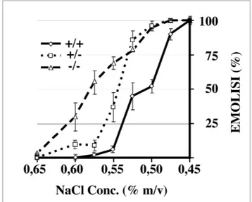
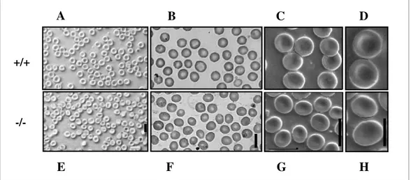
Figure 1.9 Osmotic fragility of RBCs. 49
Figure 1.10 Morphology of RBCs. 50
Figure 1.11 Analysis of ghost and skeletal proteins in β−adducin mutant mice. 51 Figure 1.12 Elements of mammalian polyadenylation signals. 56 Figure 1.13 Schematic representation of mammalian mRNA 3’-end processing. 63 Figure 1.14 Molecular inter-connections between mRNA processing reactions. 66
Figure 1.15 Core promoter consensus motifs. 74
Figure 1.16 The types and consequences of alternative promoters. 76 Figure 1.17 The 3' portions of genes leading to potential alternative poly(A) site
selection.
80 Figure 1.18 UTR-specific regulatory elements involved in post-transcriptional
regulation of gene expression.
85 Figure 3.1 SDS-PAGE analysis of RBCs skeletal proteins. 122 Figure 3.2 Mass spectrometry results of ECapZ-α (A), ECapZ-β (B) and
α-tropomyosin (C) obtained by SEQUEST analysis.
126
Figure 3.3 2D electrophoresis analysis of erythrocytes skeletal proteins. 129 Figure 3.4 ECapZ-α and -β levels in erythrocyte skeletal and cytoplasmic
preparations of mutant and control mice.
131
Figure 3.5 α-tropomyosin levels in RBC and heart cytoskeletal preparation of mutant and control mice
132
Figure 3.6 α- and β-adducin levels in erythrocyte cytoskeleton of mutant and control mice.
133
Figure 3.7 Actin, tropomodulin and dematin levels in RBC cytoskeleton of β-adducin deficient and control mice.
134
Figure 3.8 Levels of Protein 4.1 and p55 in erythrocyte skeletal preparations from mutant and control mice.
Figure 3.9 ECapZ-β levels in different mouse tissues. 137 Figure 3.10 Level of ECapZ-β in heart and brain cytoskeletal preparations of
β-adducin deficient and control mice.
138
Figure 3.11 Levels of the major RBC cytoskeletal proteins of dematin KO (DKO), adducin KO (AKO), double dematin-adducin deficient (DAKO) mice and control animals (wt).
140
Figure 3.12: In vitro purified adducin incorporation assay. 143 Figure 3.13 Levels of the major cytoskeletal proteins of lens fibres of β-adducin
mutant and control mice.
145
Figure 3.14 β-Add97 adducin mRNAs family expression in different tissues during mouse maturation.
149
Figure 3.15 Expression pattern of β-Add63 adducin mRNAs in several tissues during different mouse maturation stages.
150
Figure 3.16 Northern blot analysis of brain and spleen β-adducin transcripts. 152 Figure 3.17 Detection of brain- and spleen-specific β-adducin exons in rats. 157 Figure 3.18 Brain- and spleen-specific β-adducin exons are identified in both rats
and mice.
158
Figure 3.19 Comparison of the brain- and erythroid-specific promoters and first exons of mouse, rat and human β−adducins.
160
Figure 3.20 Analysis of tissue-specific promoter activity and the identification of a new potential β-adducin exon in mice.
162
Figure 3.21 Analysis of tissue-specific promoter activity and of the new potential β-adducin exons in humans.
163
Figure 3.22 The identification of an unusually long 3’-UTR in brain β-adducin mRNA of mouse.
166
Figure 3.23 The discovery of a 5-6 kb 3’-UTR in brain β-adducin mRNA of rats. 167 Figure 3.24 An unusually long 3’-UTR is detectable in brain β-adducin mRNA of
humans.
168
Figure 3.25 Northern blot analysis of the Add97 and Add63 β-adducin mRNA. 170 Figure 3.26 β-adducin brain-specific alternative polyadenylation sites 174 Figure 3.27 Sequence comparison of the spleen polyadenylation regions in rats,
mice and humans.
176 Figure 3.28 Schematic representation of the chimeric minigene constructs. 179 Figure 3.29 Alternative polyadenylation analysis of chimeric β-adducin mRNA in
transfected HeLa cells.
181
Figure 3.30 Deletion of the mA1 hexanucleotide motif does not affect the
polyadenylation efficiency at the brain-specific mA4 poly(A)site.
182 Figure 3.31 Effect of the deletion of the polyadenylation mA4 elements on the
3’-end processing of chimeric β-adducin transcripts in HeLa cells.
184
Figure 3.32 Deletion of the hexanucleotide motif of the canonical mA4 site in
chimeric β-adducin minigenes induces the use of a cryptic 187
polyadenylation site in HeLa cells.
Figure 3.33 Canonical and cryptic β-adducin brain-specific polyadenylation sites. 189 Figure 3.34 Conservation of the β-adducin 3’-UTR in vertebrates. 190 Figure 3.35 β-adducin transcript subcellular localisation in neuronal tissues of mice. 191 Figure 4.1 Junctional complex components of RBC skeleton of β-adducin mutant
and control mice.
195
Figure 4.2 Predicted miRNA binding sites within the 3’-UTR of β-adducin. 224
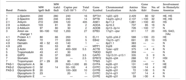
Table 1.1 Major RBC membrane proteins and their involvement in hereditary haemolytic anaemias.
32 Table 2.1 List of oligonucleotides used for mRNA analysis and for the synthesis
of dsDNA Northern blot probes.
102
Table 2.2 List of oligonucleotides used for the construction of the β-adducin chimeric minigenes.
105
Table 2.3 List of oligonucleotides used for creating deletion constructs of the β-adducin chimeric minigenes.
107
Table 3.1 Rresults of the mass spectrometry analyses of skeletal proteins separated by SDS-PAGE.
123
Table 3.2 Rresults of the mass spectrometry analyses results of skeletal proteins separated by 2D gels.
130 Table 3.3 Analysis of cytoskeletal proteins in RBCs from mutant and control
mice.
132 Table 3.4 Levels of cytoskeletal proteins in RBC from wt, DKO, AKO and
DAKO mice.
141 Table 3.5 Levels of lens membrane proteins in lens fibres from mutant mice. 146 Table 3.6 Exon and intron sizes and chromosomal location of exons in the
β-adducin genes of mice, rats and humans.
154
Table 3.7 Different β-adducin full-length transcript sizes in mouse, rat and humans.
155
Table 3.8: List of the human, mouse and rat ESTs mapping in the genomic regions upstream of the different β-adducin polyadenylation sites A1, A2-3, and
A4.
ABBREVIATIONS
The standard abbreviations used in this dissertation follow IUPAC rules. All abbreviations are defined also in the text when they are introduced for the first time. Abbreviations mentioned only once are not included in this table.
ABBREVIATION
DNA Deoxyribonucleic acid
cDNA Copy DNA
RNA Ribonucleic acid
nt Nucleotides
dNTPs Deoxynucleoside triphosphates (A, C, G and T)
N Nucleotide (A or C or G or T) R or Pu Purine (G or A) Y or Pyr Pyrimidine (T or C) bp Base pairs kb Kilobase aa Amino acid kDa Kilodalton MW Molecular weight
+/+ β-adducin homozygote wild type (controls)
+/- β-adducin heterozygote
-/- β-adducin homozygote mutant, knockout, or β-adducin deficient
KO Knockout
DAKO Dematin headpiece domain and β-adducin double knockout
RBC Red blood cell
HS Hereditary spherocytosis HE Hereditary elliptocytosis SphHE Spherocytic HE HSt Hereditary stomatocytosis BP Blood pressure OF Osmotic fragility
GPA/GPB/GPC/GPD Glycophorins A, B, C and D
DHP Dematin headpiece domain
PKC Protein kinase C
PKA Protein kinase A (cAMP dependent)
CBC Cap-binding complex
snRNP Small nuclear ribonucleoprotein particles hnRNP Heterogenous ribonuclear protein
SR Arginine-serine rich protein
USE Upstream element
DSE Downstream element
CPSF Cleavage/polyadenylation specificity factor CstF Cleavage stimulation factor
CF Cleavage factor
Pol II RNA polymerase II
CTD C-terminal domain of Pol II
PAP Poly(A) polymerase
PABP Poly(A)-binding protein TSS Transcription start site
PIC Transcription pre-initiation complex
Inr Initiator element
ORF Open reading frame
UTR Untranslated region
EST Expressed sequence tag
ARE AU-rich element
miRNA microRNA
LS mRNA localisation signal
mN1 First brain-specific exon of mouse β-adducin mE1 First erythroid-specific exon of mouse β-adducin hE1 Erythroid-specific exon of human β-adducin
Rpm Revolutions per minute
DTT Dithiothreitol
EDTA Ethylenediamine tetra-acetic acid IPTG Isopropyl-β-d-thiogalactopyranoside
TBE Tris-borate-EDTA (buffer)
SDS N-lauroylsarcosine sodium salt Tris Tris (hydroxyethyl) amino-ethane
PBS Phosphate buffer saline
IPTG Isopropyl-d-thiogalactopyranoside ddH20 Double-distilled water
ATP Adenosine triphosphate
ADP Adenosine diphosphate
c-AMP Cyclic adenosine monophosphate
R.T. Room temperature
1
INTRODUCTION
1.1 Cells membrane cytoskeleton
The cytoplasm of eukaryotic cells is spatially organised by a network of protein filaments known as the cytoskeleton. This network contains three principal types of filaments: microtubules, intermediate filaments and actin filaments (Figure 1.1 A, B, and C). They connect organelles in different regions of the cell and are used as tracks for the transport between them. In addition, filament networks provide mechanical support, which is especially important for animal cells, which do not have rigid external walls. The cytoskeleton forms an internal scaffold for the large volume of cytoplasm, holding it like a framework of girders supporting a building. Each of the three types of protein filaments is a helical polymer that has a different arrangement and function in the cell. By themselves, however, the three types of filaments could provide neither shape nor strength to the cell. Their functions depend on accessory proteins that link the filaments among them and to other cell components. Accessory proteins are also essential for the controlled assembly of the protein filaments in particular locations, and they provide the motors that either move organelles along the filaments or move the filaments themselves (Lodish, 2003).
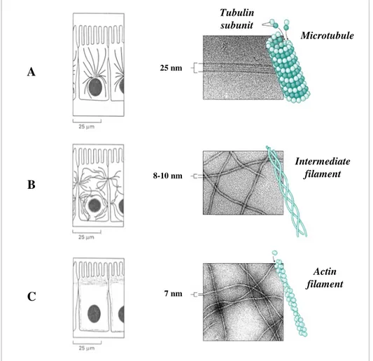
1.1.1 - Microtubules -
Microtubules are long and stiff cylindrical structures (diameter of 25 nm) (Figure 1.1 A), and usually have one end anchored in the centrosome and the other free and extend throughout the cytoplasm. They govern the location of membrane-bounded organelles and other cell components and during the M phase of the cell cycle they create the mitotic spindle. Microtubules comprise tubulin molecules, each of which is a heterodimer consisting of two closely related and tightly linked globular polypeptides called
α
- and β-tubulin. The microtubule structure is built of 13 linear protofilaments, each composed of alternating α- and β-tubulin subunits and bundled in parallel to form a cylinder. Since the 13 protofilaments are aligned in parallel with the same polarity, the microtubule itself is a polar structure which has both a plus (fast-growing) and a minus (slow-growing) end. The minus-ends of microtubules are stabilised due to being embedded in the centrosome while the fast-growing ends are free to add tubulin molecules. Therefore, each microtubule is a highly dynamic structure that alternatelygrows by the addition (polymerisation) or shrinks by the loss (depolymerisation) of tubulin subunits: this behaviour is called dynamic instability. Their polymerisation and depolymerisation are controlled by the presence of the GTP bounds to the β-tubulin and Mg2+ ions, and GTP-hydrolysis, respectively (Kreis and Vale, 1999).
A B C Microtubule Tubulin subunit Intermediate filament Actin filament 25 nm 8-10 nm 7 nm
Figure 1.1: Cytoskeletal filaments. The three major types of filaments that form the cytoskeleton:
microtubules, intermediate filaments and actin filaments are represented in Panels A, B, and C, respectively.
1.1.2 Intermediate filaments
-Intermediate filaments are tough, rope-like polymers of fibrous polypeptides that resist stretch and play a structural or tension-bearing role providing mechanical stability to cells and tissues. They are called "intermediate" because they have a diameter of 8-10 nm, between that of actin filaments and microtubules (Figure 1.1 B). In most animal cells an extensive network of intermediate filaments surrounds the nucleus and extends towards the cell periphery, where they interact with the plasma membrane. In addition, a tightly woven basketwork of intermediate filaments, the nuclear lamina, underlies the internal side of the nuclear envelope. Intermediate filaments are particularly prominent in the cytoplasm of cells that are subject to mechanical stress (for example, in epithelia, neuronal and muscle cells. A variety of tissue-specific forms are known to differ in the
type of polypeptide they comprise: the keratin filaments of epithelial cells, the
neurofilaments of nerve cells, the glial filaments of astrocytes and Schwann cells, the
desmin filaments of muscle cells, and the vimentin filaments of fibroblasts and many other cell types. Nuclear lamins, which form the nuclear lamina, are a separate family of intermediate filament proteins. The monomers of the different types of intermediate filaments differ in sequence and molecular weights, but they all comprise a homologous structure: two globular domains (N- and C- terminus) linked by a central rod domain that forms a coiled-coil structure when the protein (Kreis and Vale, 1999)
1.1.3 - Actin filaments -
Actin filaments (F-actin) are flexible helicoidal polymers with a diameter of 5-8 nm (Figure 1.1 C). They consist of a tight helix of uniformly oriented globular actin, or
G actin. Actin is the most abundant protein in many eukaryotic cells, often constituting up to 5% of total cell protein (in skeletal muscle cells it reaches about 10% of their mass). Each actin molecule is a single polypeptide of 42 kDa folded to create two structural domains divided by a central cleft where ATP and Mg2+ are bound. All higher eukaryotes have several isoforms of the protein encoded by a highly conserved family of genes that reside in different chromosomes. Four different α-actins are found in muscle cells and are associated with contractile structures. Instead, β- and γ-actins are the principal constituents of non-muscle cells, where γ-actin accounts for stress filaments and β-actin is in the front (or leading edge) of moving cells when actin filaments polymerise. Although there are subtle differences in the properties and functions of different forms of actin, their primary structures are highly conserved and all assemble into filaments that are essentially identical (Kreis and Vale, 1999). Like microtubules, actin filaments are polar structures, with two structurally different ends: a relatively inert and slow-growing minus-end (or pointed-end) and a fast-growing
plus-end (or darbed-end). The difference in elongation rates at the opposite ends of actin filaments is caused by a different G-actin critical concentration (Cc) values at the two ends. Cc is the concentration of G-actin where equilibrium between polymerisation and depolymerisation reactions is reached: Cc at the minus-end is higher than that of the plus-end. At G-actin concentration between the Cc of the two ends a particular dynamic behaviour, called treadmilling, occurs: actin molecules are added continually to the plus-end of the filament and are lost continually from the minus one, with no net change in filament length. Polymerisation of actin requires ATP as well as cations, (K+ and
Mg2+), while ATP hydrolysis weakens the bonds in the polymer and thereby promote de-polymerisation. Several actin binding proteins influence the polymerisation of actin filaments, and thus their length and stability. Capping proteins, such as tropomodulin,
CapZ and adducin bind to the ends of the filaments preventing monomer association and dissociation, while tropomyosin, stabilises F-actin interacting along its length. Other filament-binding severing and capping proteins are cofilin, severin and gelsolin: they bind to F-actin, break them into shorter filaments and remain attached at the plus-end preventing further addition or exchange of actin subunits. In addition, thymosin and
actin-depolymerising factor (ADF) inhibit the assembly of actin into filaments, while
profilin has a role in stimulating the polymerisation of actin by accelerating the exchange of ATP for ADP (Fowler, 1996; Fowler, 1997; Pollard et al., 2000). Section 1.2.2 analyses in details several of these proteins.
The dynamic modulation of the polymerisation and depolymerisation processes implies that actin filaments can form both stable and labile structures in cells. Stable actin filaments are a crucial component of the contractile apparatus of muscle cells (sarcomere) and membrane skeleton of erythrocytes, and form the core of particular cellular cell-surface protrusions such as microvilli, lamellipodia, microspikes, stereocilia, etc. Many cellular activities such as locomotion, chemotaxis, phagocytosis, and cytokinesis depend on labile structures constructed from actin filaments (Fowler, 1996; Pollard et al., 2000).
Actin filaments normally exist in different arrangements (linear bundles, two and three dimensional networks) maintained essentially by actin filament cross-linking
proteins (fimbrin,
α
-actinin, filamin, spectrin and dystrophin). Because these proteins also bind integral membrane proteins, these networks are generally located in the cortical region adjacent to the plasma membrane: actin filaments lying just beneath the plasma membrane form the cell cortex. It is mainly this actin-rich layer that gives the cell mechanical strength, and controls the shape and surface movements of cells in accordance to the cell environment (Kreis and Vale, 1999).The cell cortex network is attached to the plasma membrane in numerous ways: • Direct connections between actin filaments and membrane: integral membrane proteins directly interact with actin filaments;
• Indirect connections between actin filaments and membrane: more complex and common complex linkages that connect actin filaments to integral membrane proteins
through peripheral membrane proteins, like filamin in platelets, dystrophin in muscle cells, ERM proteins (ezrin-radixin-moesin) in epithelial cells, etc.
1.2 Plasma membrane and cytoskeleton of erythrocytes
The plasma membrane of the red blood cells (RBCs) has been extensively studied more than any other eukaryotic membrane for a number of reasons: a) RBCs are available in large numbers relatively uncontaminated by other cell types; b) since these highly specialised cells lack transcellular filaments system, nucleus and internal organelles, the plasma membrane is their only cellular structure; C) it can be easily isolated and studied avoiding the serious problem of contamination by internal membranes (in other cell types the cytoplasmic membrane typically constitutes less than 5% of the cell's membrane). The RBC membrane is basically comprised of a lipid bilayer with its associated proteins (Bennett and Gilligan, 1993).
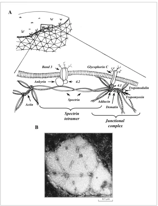
The erythrocyte cytoskeleton is the filamentous network of proteins that underlies the cytoplasmic surface of the membrane and is anchored to it. It is composed of proteins that are structurally homologous to those participating in the formation of eukaryotic cell cytoskeleton, being a similar but less elaborated filamentous network of proteins. The main components of the RBC skeleton are spectrin and actin that are organised as a hexagonal spectrin network linked to short actin filaments. The spectrin-actin junctions comprise a group of accessory proteins that promote and modulate spectrin-actin interactions, regulate the actin filament length and form membrane associations. The most important members of these junctions are tropomyosin, tropomodulin, adducin, Protein 4.1, p55, Protein 4.2 and dematin. The junctions that create the vertexes of the polygonal structure of the skeleton are known as junctional
complexes (Figure 1.2) (Bennett and Gilligan, 1993).
SDS-PAGE analysis of the proteins of red cell ghosts (a product obtained during the RBC protein purification process that contains skeletal and membrane constituents) indicates the presence of a dozen of distinct species (often identified as "Band" proteins) (Fairbanks et al., 1971). By contrast, two dimensional separation employing isoelectrofocusing and SDS-PAGE demonstrated the membrane to be composed of more than a hundred different proteins (Low et al., 2002; Rosenblum et al., 1982).
According to their location, the proteins of the RBC membranes can be divided into two general groups: integral RBC membrane proteins and peripheral RBC
Junctional complex Spectrin tetramer B A Spectrin 4.2 Ankyrin Glycophorin C Band 3 4.1 Adducin Dematin Tropomyosin Tropomodulin Actin
Figure 1.2: Erythrocyte cytoskeleton. Panel A shows a schematic model of protein organisation of
erythrocyte membranes. The scheme is adapted from Bennett and Gilligan, 1993. A RBC membrane electron microscopic image is shown in Panel B. The structural organisation of the cytoskeleton is clearly visible: spectrin tetramers appear as spokes and interact with junctional complexes (hubs) generating a hexagonal network. Ankyrin molecules are detectable as dark spots located along the spokes of spectrin. The image in Panel B is from Lodish et al., 2003.
1.2.1 - Integral RBC membrane proteins -
The integrals proteins are tightly bound to the RBC membrane through hydrophobic interactions with lipids in the bilayer (Figures 1.2 and 1.3). These proteins span the membrane and have distinct structural and functional domains, both within the bilayer and on either side of the membrane. The most abundant and well characterised proteins that belong to this group are (Tanner, 1993):
• Band 3 (anion exanger-1): is a member of a family of anion transporters. It is the major transmembrane protein of the RBCs (~106 copies of polypeptide chains per RBC arranged as dimers), constituting about 25% of total membrane proteins. It is encoded by the AE1 gene whose expression is largely restricted to erythrocytes. In addition to RBCs, Band 3 is expressed in both mouse and human kidneys as different isoforms. In RBC membranes Band 3 has a structural function in RBC membranes: it acts as an anchor site to the membrane of the skeleton, primarily through its interaction with ankyrin and secondarily binding to Proteins 4.1 or 4.2 (Bennett and Gilligan, 1993). In addition, it is as a membrane anion transporter, AE1 mediates Cl-/HCO3- exchange, thus enhancing the blood capacity for carrying CO2 from tissues to the lung, and for acid-base homeostasis (Jennings, 1989).
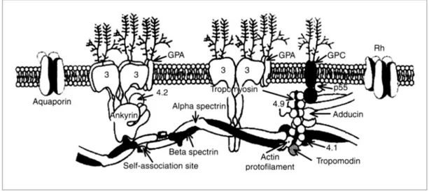
Figure 1.3: Peripheral and integral RBC membrane proteins. The figure shows a schematic
representation of the erythrocyte plasma membrane and of the cytoskeletal network located on its cytoplasmic surface. The integral proteins, Band 3, glycophorins A and C (GPA and GPC, respectively) aquaporin and palmitoylated protein Rh are represented. Spectrin tetramers, actin filaments and the protein that constitute the junctional complex creating the skeletal network, such as tropomyosin, tropomodulin, adducin, Protein 4.2 and dematin (Protein 4.9) are indicated. The direct (between skeletal and membrane proteins) and indirect (mediated by ankyrin) interactions between the skeleton and the membrane are shown. The scheme is adapted from Lux et al. 1995.
• Glycophorins: are sialic acid-rich small transmembrane glycoproteins divided into four groups: glycophorin A -GPA-, B -GPB-, C -GPC- and D -GPD- (Figures 1.2 and 1.3). They constitute ~2% of the total RBC membrane protein. Glocophorins A, B, C and D are different polypeptides (36, 20, 32, and 23 kDa, respectively; GPD is a truncated form of GPC) and are the products of three distinct genes. The majority of the carbohydrates on the RBC surface (90% sialic acids) are anchored to these proteins and impart a strong net negative charge to the cell surface. This is functionally important in reducing the interactions of erythrocytes with one another as well as with other cells,
including vascular endothelium. In RBCs, GPA is the major component present at 5-9 x 10⁶ copies per cell. GPC is also present anchoring the red cell skeleton to the membrane through the binding of Protein 4.1 and of p55 (Marfatia et al., 1995; Marfatia et al., 1997). GPC, unlike GPA and GPB, has a pattern of expression that is not limited to the erythroid lineage suggesting that it probably also has a role in cytoskeletal interactions in many other tissues (Chasis and Mohandas, 1992).
Other two abundant RBC integral-membrane proteins are Aquaporins (such as Aquaporin-1) and Palmitoylated proteins (Rh30 and Rh50) (Tanner, 1993).
1.2.2 - Peripheral RBC membrane proteins: the erythrocyte cytoskeleton -
The cytoskeletal network located on the cytoplasmic surface of the lipid bilayer of RBCs is generated by the interactions between peripheral membrane proteins (frequently called horizontal interactions). The RBC skeleton appears as a regular network in which the basic unit is composed of a hexagonal spectrin lattice. The sides of this structure consist of long, flexible spectrin tetramers, while the vertexes are formed by the attachment of the tail ends of spectrin with the short actin filaments and a group of accessory proteins that promote and modulate spectrin-actin interactions, regulate the actin filament length and form membrane associations. All the molecules that interact at the vertexes create the junctional complex (Figure 1.2 and 1.3) (Bennett, 1989; Gilligan and Bennett, 1993; Shen et al., 1986). This structural model of the erythrocytes skeleton organisation has been confirmed by high resolution electron microscopy of isolated membranes (Figure 1.2 B) (Liu et al., 1987; Shen et al., 1986). The most important members of these junctions are tropomyosin, tropomodulin, adducin, protein 4.1, p55, protein 4.2 and dematin. A brief description of the most important components of the membrane skeleton is below:
• Spectrin: is the most abundant and principal component of the cytoskeleton, in fact, it constitutes about 25% of the membrane-associated protein mass (~ 2.5 x 10⁵ copies per cell). Spectrin is a heterodimer formed by two large, structurally similar subunits encoded by two genes at different chromosomal location: α and β subunits (260 and 246 kDa, respectively). Each polypeptide is a long, thin, flexible rod (~100 nm in length) organised into a number of independently folded domains. The two antiparallel polypeptide chains, loosely intertwine and attach noncovalently to each other at multiple points. The heterodimers self-associate “head-to-head” to form ~200 nm-long tetramers. Tetrameric species of spectrin appear to predominate in the
membrane skeleton (Bennett and Gilligan, 1993). The “tail” ends of six tetramers are linked together by binding to short actin filaments and to other cytoskeletal proteins at the junctional complex (Bennett, 1989; Shen et al., 1986) (Figure 1.2 ).
• Actin filaments: as opposed to the actin filaments observed in nonmuscle cells which are generally long and vary in length, erythrocytes actin filaments (or protofilaments) are relatively short and uniform (Figure 1.4): they are composed of 12-18 monomeric units of G-actin and have an approximatelength of 33-37 or ~67 nm, depending on experimental conditions (Coleman et al., 1989; Shen et al., 1986). In each red blood cell there are approximately 3-4 x 10⁴ of these short structures (Fowler, 1996). Different mechanisms have been proposed to describe how such short actin protofilaments are generated. According to the “ruler” mechanism, several actin binding proteins regulate the assembly, the stability and especially the length of the short F-actin in RBCs: an external “molecular ruler,” made out of actin-binding proteins such as tropomodulin and tropomyosin, protect a segment of the short actin filament of about 37 nm consisting of ~12 G-actin preventing elongation and depolymerisation (Sung et al., 2000), while capping and bundling by adducin can stabilise the protofilaments (Fowler, 1996). Confoundingly, the recently discovered “helix” mechanism affirms that the intrinsic properties of actin filaments, for example turns, chemical bonds, and dimensions of the helix, may favour its fragmentation into short protofilaments under mechanical stress (Sung and Vera, 2003). Each short actin filament is linked by about six spectrin tetramers tails at the junctional complex: these interactions create the hexagonal lattice that composes the basic units of the cytoskeleton. Other skeletal proteins, such as tropomyosin, adducin tropomodulin, bind and stabilise the short protofilaments (Bennett and Gilligan, 1993).
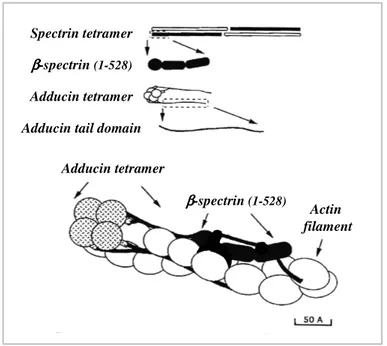
• Adducin: is a heterodimeric protein composed by the association of α, β and γ subunits. Oligomers in human erythrocytes comprise α/β subunits (Gardner and Bennett, 1986), while α/γ as well as α/β combinations of subunits are detectable in mouse RBC (Gilligan et al., 1999; Muro et al., 2000). These heterodimers are present in erythrocytes at ~3 x 10⁵ copies/cell (Gardner and Bennett, 1986).Adducin has several functions: it recruits spectrin to actin filaments (Bennett et al., 1988; Gardner and Bennett, 1987; Hughes and Bennett, 1995), bundles actin filaments (Mische et al., 1987; Taylor and Taylor, 1994), and caps the fast-growing ends of actin filaments (Kuhlman et al., 1996) (Figure 1.4). These activities indicate that adducin promotes the formation
of spectrin-actin junctions and increases their stability. More information about this protein can be found in Section 1.7.
Figure 1.4: The short erythrocyte actin filament. The figure shows a molecular model of the RBC
protofilament. Two molecules of tropomyosin are localised along each of the two grooves of the actin filament. Two tropomodulin molecules are associated with the terminal actin subunit of the F-actin pointed-ends and to the N-terminus of each of the two tropomyosin dimers. The C-terminal tail domains of adducin are associated with β subunits of spectrin along the actin filaments, whereas its N-terminal head domains cap the darbed-end of the protofilament. The scheme is adapted from Fowler et al., 1996.
• CapZ: is a capping protein for the barbed-end of actin filaments in non-erythroid cells. Its main function is to prevent monomer association and dissociation by capping F-actin barbed-ends, but it also facilitates actin polymerisation by binding to and stabilising monomers or oligomers of actin forming nucleifor filaments assembly and elongation (Casella et al., 1986; Schafer and Cooper, 1995). It is a highly conserved heterodimer formed by the association of α and β subunits (36 kDa and 32 kDa, respectively). Different isoforms of the subunits are present in a wide variety of tissues and organisms. C-terminal ends of both subunits are required for effective barbed-end capping of filaments (Casella and Torres, 1994; Schafer et al., 1994). Erythrocytes contain abundant amounts of a non-muscular isoform of CapZ (ECapZ) composed mainly by the α1β2 subunits, as described for CapZ from many other non-muscle cells (platelets, neutrophils, etc) (DiNubile et al., 1995). Despite purified ECapZ is fully functional in blocking actin elongation from barbed filament ends, as well as in nucleating actin polymerisation, this erythroid capping protein is mainly located in the cytoplasm and is not associated with protofilaments in the RBC membrane skeleton (Kuhlman and Fowler, 1997). This contrasts with what is observed in platelets, where the same CapZ is located in the cytosol and sometimes in association with actin filaments (Barkalow et al., 1996), or in striate muscle, where all the capping protein is associated with the filament at the Z disks of the sarcomere (Rybicki et al., 1988).
Therefore, erythrocytes contain two actin filament barbed-end capping proteins: ECapZ with a high affinity (Kcap ~1-5 nM) and adducin with a lower affinity (Kcap ~100 nM) for the end of actin filaments, but of the two, adducin caps and stabilises the protofilament barbed-ends. How adducin can outcompete ECapZ with its apparently higher affinity (20-100 fold) is unclear. The absence of ECapZ from the F-actin in the RBC skeleton is not due to a defect of the protein, nor to a cytosolic inhibitor factor, or even to its insufficient concentration in the cytoplasm. It was hypothesised that this peculiar phenomena is determined by the presence of additional co-factors or specific cellular conditions in the RBC that could induce (Kuhlman and Fowler, 1997):
a) an increase of adducin affinity for the protofilaments. For example, it binds more tightly to spectrin-actin complexes than to pure actin filaments (Gardner and Bennett, 1987), thus interactions with spectrin and actin in the junctional complex may increase its affinity for actin filament darbed-ends. Adducin capping activity my also be regulated by specific phosphorylation or dephosphorylation reactions(Matsuoka et al., 2000);
b) a reduction of the apparent high affinity of ECapZ for F-actin. High concentrations of ECapZ in the cytoplasm could induce its overcrowding and consequently might reduce the apparent high affinity of the capping protein for the protofilaments (Kuhlman and Fowler, 1997).
• Tropomodulin: is a globular protein (~40 kDa) present as approximately3 x 104 copies per RBC. It is a capping protein that binds to the pointed-end of the actin protofilaments (Fowler, 1996) (Figure 1.4). The C-terminal end of the protein contains an actin binding domain, important for capping activity, while the N-terminal portion contains the binding sites for the N-terminal ends of the two tropomyosin molecules that are associated with actin filament (Kostyukova et al., 2006; Sung and Lin, 1994). The presence of tropomyosin is required for tight capping of F-actin pointed-ends. In fact, in the absence of tropomyosin, tropomodulin only partially inhibits monomer association and dissociation at the minus-end of filaments (Weber et al., 1994). Therefore, the tropomodulin/tropomyosin complex blocks the elongation and depolymerisation of the actin filaments at the pointed-end contributing to the stability and length determination of the actin filament (Fowler, 1996). Tropomodulin is a highly conserved protein among species and tissues. It’s abundant in striated muscule cells where it plays an essential role in the assembly of the sarcomere and it is also a fundamental cytoskeleton component in erythrocyte membranes and eye lens cells (Fowler, 1996; Woo and
Fowler, 1994). The function of tropomodulin in other non-muscle and non-erythroid cells is still not well understood.
• Tropomyosin: is a rigid rod-like fibrous molecule composed of two small polypeptides (35 kDa) associated to form a coiled-coil structure (~33-35 nm) (Figure 1.4). RBC skeletons each contain ~7 x 10⁴ dimeric molecules of tropomyosin and they correspond to approximatelly 1% of the membrane protein (Fowler and Bennett, 1984; Shen et al., 1986). Tropomyosin exists in a large number of isoforms [high and low molecular weight (MW) isoforms] which are generated in different tissues by the alternative use of promoters and the alternative RNA processing of different genes (at least 4 distinct geneshave been characterised in mammalians: α, β, γ, and δ) (Lin et al., 1997), suggestingthat the different isoforms are not functionally equivalent (Gunning et al., 2005). Among the different tropomyosin isoforms, TM5 and TM5b (29 and 28.7 kDa, respectively) are the 2 major forms expressed in human erythrocytes. Although they are productsof 2 different genes (γ-TM and α-TM genes, respectively), theyshare several common features, including molecular weight (both are low MW isoforms), the presence of similar binding domains, and a high affinity for actin and tropomyosin (Sung et al., 2000; Sung and Lin, 1994). The rod-like structure and the actin-binding ability of dimeric tropomyosin allow it to localise, polymerised end-to-end, along each of the two grooves of the actin filament, providing structural stability and modulating the filament length (Figure 1.4). In RBCs, two dimeric tropomyosin molecules are associated with the short protofilament protecting 6-7 actin monomers in one strand (or 12-14 G-actin in the double helix) (Cooper, 2002; Fowler and Bennett, 1984). The tropomyosin ability to bind and reduce depolymerisation of actin filaments is improved when tropomodulin simultaneously interacts with both the pointed-end of the actin filament and to the terminal tropomyosin molecule: in fact, tropomodulin seems to strengthen the binding of the terminal tropomyosin molecule to the pointed-end of the protofilament (Weber et al., 1994). In erythrocytes, the binding of tropomodulin to tropomyosin also seems also to block the ability of the tropomyosin dimer to self-associate in a head-to-tail structure (Fowler, 1990; Sung and Lin, 1994). In non-erythroid cells, tropomyosin can regulate many properties of F-actin: increase filament stiffness, protect filaments from the depolymerising effects of severing and depolymerising factors (cofilin, gelsolin, ADF, etc), enhancing F-actin stability and, moreover, influence myosin mechanochemistry (Cooper, 2002; Gunning et al., 2005).
In conclusion, the combined action of tropomyosin and tropomodulin strongly influences the length and increases stability of the actin filaments.
• Protein 4.1: is a complex family of erythroid (4.1R) and non-erythroid protein 4.1 isoforms generated by the involvement of both multiple genes and alternative splicing pathways (Conboy et al., 1991; Takakuwa, 2000). Interestingly, several 4.1R pre-mRNA splicing events exhibit developmental switches in their expression patterns, implying that distinct 4.1R functions are critical at different stages of cell differentiation (Baklouti et al., 1996). Moreover, in RBCs alternative splicing regulates the expression of alternative translation initiation sites in exon 2 (AUG1) and exon 4 (AUG2) generating 135 kDa and 80 kDa isoforms of the protein, respectively (Baklouti et al., 1996; Chasis et al., 1996). Among the different 4.1R isoforms the 80 kDa one is the best characterised. It was estimated that each RBC contains ~2 x 10⁵ copies of this molecule (Bennett and Gilligan, 1993). By multiple protein-protein interactions, 4.1R promotes the formation of two ternary protein complexes in the erythrocyte membrane: spectrin-actin-4.1R and GPC-4.1R-p55 complexes (Figures 1.2 and 1.3). They are critical to the structural integrity of the skeleton and to its attachment to the membrane lipid bilayer (Bennett and Gilligan, 1993; Marfatia et al., 1994). Thus, by lateral interactions with the spectrin/actin network and vertical interactions with the cytoplasmic domain of transmembrane proteins GPC and Band 3, 4.1R is essential for maintaining erythrocyte shape and membrane mechanical properties (deformability and stability) (Chang and Low, 2001).
• p55: is the major palmitoylated phosphoprotein associated with the cytoplasmic side of the erythrocyte membrane (Bennett and Gilligan, 1993; Ruff et al., 1991). Approximately 8 x 10⁵ molecules of p55 are detectable in each mature erythrocyte. It is a member of a family of cytoskeletal and signalling proteins termed membrane-associated guanylate kinase homologues (MAGUKs). As a component of the GPC-4.1R-p55 ternary complex, p55 modulates the interactions between Protein 4.1 and GPC, regulating the stability and mechanical properties of RBC plasma membrane (Marfatia et al., 1995; Marfatia et al., 1994).
• Dematin (or Band 4.9): is an actin-bundling protein that was originally identified as a component of the junctional complex of the human erythroid membrane skeleton (Siegel and Branton, 1985) (Figure 1.2 A). It is also abundantly expressed in the human brain, heart, skeletal muscle, kidney, and lung (Kim et al., 1998). Dematin is a trimeric protein composed of two copies of a 48 kDa polypeptide and one copy of a 52 kDa