DOCTORAL THESIS
Study of cortical and hippocampal
specification using embryonic stem
cell-derived neurons
Author:
Marco Terrigno
Supervisor:
Federico Cremisi
January 2019
The only man who never
makes a mistake is the
man who never does
anything
TABLE OF CONTENTS
INTRODUCTION ... 9
Early embryonic development ... 9
Development of the nervous system ... 10
Neural induction ... 10
Morphogenesis and segmentation of the Neural Tube ... 14
Dorsoventral patterning ... 16
Rostrocaudal patterning ... 17
Local signalling centers: the Midbrain/Hindbrain boundary ... 22
Local signalling centers: the Anterior Neural Ridge ... 24
Early telencephalic patterning and development ... 27
Early cortical patterning ... 31
Neocortical development ... 35
Hippocampal development ... 39
MicroRNAs in neural development ... 42
Embryonic pluripotent stem cells ... 46
Neuralization of embryonic stem cells ... 50
Clinical applications of pluripotent stem cells ... 54
Ischemic stroke ... 56
Cell therapy in stroke ... 59
AIM OF THE RESEARCH ... 62
RESULTS ... 68
Establishing isocortical or hippocampal identity via timely manipulation of WNT and BMP ... 68
ESC-derived hippocampal cells send long range projections to Dentate Gyrus targets ... 74
Isocortical cells can send far-reaching projections when transplanted in adult motor cortex ... 75
Photothrombotic damage enhances long-range projections from transplanted isocortical cells ... 79
FGF8 regulates genes of cortical area patterning in a model of in vitro
corticogenesis ... 84
FGF8 inhibits COUP-TFI translation by acting on its 3’UTR ... 88
The FGF8-induced miRNA miR-21 inhibits COUP-TFI translation in vitro ... 92
Specificity of miR-21 interaction with the COUP-TFI 3’UTR ... 94
Complementary expression of miR-21 and COUP-TFI protein and their interaction in vivo ... 98
DISCUSSION AND CONCLUSIONS ... 104
Manipulation of WNT and BMP signaling: recapitulating cortical and hippocampal development in-vitro ... 104
Isocortical and hippocampal ESC-derived neurons: different axonal outgrowth and targeting ... 106
The pivotal role of the molecular identity acquired in vitro for establishing proper connections. ... 110
FGF8 regulates cortical area-patterning by inhibiting COUP-TF1 translation via miR-21 ... 113
MATERIALS AND METHODS ... 117
Mouse ESCs differentiation and transfection ... 117
Cortical and hippocampal primary cultures ... 119
Gene expression analysis ... 120
Microarray hybridization and data analysis ... 121
Lentiviral vector construction and use ... 122
miRNAome profiling ... 124
In silico analysis of 3′UTR putative binding sites ... 124
Western Blot ... 124
In vitro immunocytodetection (ICD) and imaging ... 125
Immunofluorescence on frozen brain tissue ... 126
RNA in situ hybridization (ISH) ... 127
In utero electroporation (IUE) ... 128
Stroke induction ... 129
In vivo grafting ... 129
Gridwalk Test ... 131
SUPPLEMENTAL MATERIALS ... 133 ACKNOWLEDGEMENTS ... 148 BIBLIOGRAPHY ... 149
TABLE OF FIGURES
Fig. 1 Timely regulation of WNT and BMP signaling affects the identity of ES
cell-derived neurons ... 69
Fig. 2 WNT signaling manipulations activate the expression of isocortical or hippocampal markers in neuralized ESCs ... 71
Fig. 3 WNT signaling induction or repression induce, respectively, hippocampal or isocortical global gene expression profile in neuralized ESCs ... 73
Fig. 4 Cell transplantation in hippocampus ... 76
Fig. 5 Cell transplantation in normal motor cortex ... 78
Fig. 6 Cell transplantation in ischemic motor cortex ... 80
Fig. 7 Cell-autonomous and environmental cues regulate axonal extension after grafting ... 83
Fig. 8 FGF8 induces anterior cortical identity in vitro ... 86
Fig. 9 FGF8 inhibits COUP-TFI translation acting on the 3’UTR ... 90
Fig. 10 FGF8 activates miRNAs targeting the COUP-TFI 3’UTR in silico ... 91
Fig. 11 FGF8-induced miRNAs repress COUP-TFI expression in vitro ... 95
Fig. 12 mir-21 and miR-132 selective binding to the COUP-TFI 3’UTR... 97
Fig. 13 miR-21 and COUP-TFI are expressed in complementary gradients in the embryonic cortex ... 100
Fig. 14 miR-21-mediated control of COUP-TFI translation in vivo ... 101
Fig. S1 Markers of hem, choroid plexus and dorsal telencephalon ... 134
Fig. S2 Staining for NeuN and synaptic markers in transplanted cells ... 136
Fig. S3 Distribution of Calbindin 1 positive fibers from transplanted CHIR8 cells ... 139
Fig. S4 Fetal transplantation in intact hippocampus ... 139
Fig. S5 Fetal transplantation in intact motor cortex ... 140
Table 1 Pathway enrichment analysis ... 141
Fig. S6 Effect of FGF signaling inhibition on A/P cortical markers and COUP-TFI protein ... 142
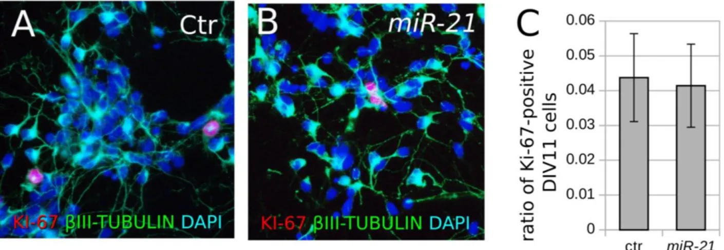
Fig. S7 Effect of miR-21 upregulation on cell proliferation. ... 142 Fig. S8 Mutagenesis of the predicted miR-21 and miR-132 binding sequences in
COUP-TFI 3’UTR ... 143
Fig. S9 In vivo IUE of miR-21 seed-mutated sponge ... 144 Fig. S10 Control LNA lipofection in vivo ... 145
Copyright disclaimer
Some sections of this manuscript, including figures, are reused with permission from previous publications (Terrigno et al., 2018a, 2018b).
9
Early embryonic development
All vertebrates, although different in shape and size, develop similarly. In mammalian embryos, starting from the fertilized egg, rapid complete cell divisions give rise to a solid symmetric sphere called morula. At the morula stage, all the cells are totipotent, thus they can give rise to any tissue of the future embryo (Wilton and Trounson, 1989). Subsequently, 3.5 days post fertilization in mice, further cell divisions generate an asymmetric hollow sphere called blastula. The cells inside the blastula are called inner cell mass and give rise to the embryonic tissues, whereas the cells on the external surface form the trophectoderm and give rise to the extra-embryonic tissues, such as the placenta (Winkel and Pedersen,
1988; Snow, 1976).
After the implantation of the blastocyst in the uterine walls, the cells of the inner cells mass, facing the internal cavity of the blastocoel, form the epiblast, while the cells below originate the hypoblast (Solnica-Krezel L and Sepich D.S., 2012; Tam P.P.L and Behringer
R.R., 1997). At this point, the relatively simple blastocyst goes through a
10 gastrulation, which defines the antero-posterior axis of the embryo and generate a more complex three-layered structure. During mammalian gastrulation, epiblast cells migrate through a small invagination called primitive streak toward the hypoblast underneath. Most of the involuting cells give rise to mesodermally derived tissues, such as muscles and bones. While the embryo begins to elongate in the antero-posterior axis, the cells that migrate first and further through the primitive streak originate the most anterior part of the embryos. At the end of the gastrulation, the superficial epiblast form the ectoderm, the migrated cells originate the mesoderm and the hypoblast give rise to the endoderm (Bellairs, 1986; Hashimoto and Nakatsuji, 1989; reviewed by Tam
et al., 1997).
Development of the nervous system
Neural induction
In the early twentieth century, classic embryological experiments showed that the nervous system of the vertebrates arise from the dorsal ectoderm. In those pioneering experiments, scientists assayed the specification of the embryonic tissues dissecting and culturing in vitro small fragments of embryo and analysing the composition of the resulting tissues. During gastrulation, as the three embryonic layers are formed, cells are committed toward specific identities. In particular, when isolated before the gastrulation, fragments of dorsal ectoderm
11 mainly originates epidermidis, whereas if isolated in gastrulating embryo, they generate neural tissues, such as brain and eyes. These observations led Hans Spemann to theorize that the default fate of the ectoderm was epidermidis, and that the tissues rearrangements occurring at the gastrulation specified the dorsal ectoderm to a neural fate (Hamburger et al., 1969). The quest for understanding the nature of such neural induction would take half a century. At the time, investigators identify as possible inducer of neural identity the involuting mesoderm. According to this model, the epiblast cells migrating through the blastopore, the equivalent of the primitive streak in the amphibian, would induce the epiblast above to acquire neural identity.
Indeed, heterotopic transplantation of the dorsal lip of the blastopore inside the cavity of another embryo induced the formation of a second body axis, complete with brain, eyes and spinal cord. Moreover, successive experiments showed that these structures arose from the host embryo and not from the tissues of the donor. Subsequently, Richard Harland (Lamb et al., 1993; Smith et al., 1993) and De Robertis (Piccolo S. et al., 1996) isolated the first two neural inducers Noggin and Chordin, respectively. These factors are secreted protein expressed by the dorsal lip of the blastopore when neural induction occurs. Both factors were able to induce neural identity in isolated fragments of dorsal ectoderm and were able to induce the formation of complete secondary axis when overexpressed in the ventral part of a developing embryo. Notably, a similar mechanism for neural induction
12 is conserved also in invertebrates. In fact, in Drosophila the short
gastrulation (sog) gene, homolog of the vertebrate Chordin, is important
for the dorso-ventral patterning of the embryo and for the induction of the neural tissue (François V and Bier E., 1995). Moreover, studies in Drosophila showed that sog gene interacts with deacapentaplegic, or dpp, a TGF-beta-like protein related to the vertebrate BMPs (Biehs B. et al.,
1996). Since these two factors actively inhibits each other, their mutation
has opposite phenotypes. Indeed, sog mutant showed an expansion of the epidermidis at the expenses of the neurogenic region of the ectoderm, while dpp mutant had an expanded neurogenic region. Moreover, ectopic overexpression of sog was able to induce neural identity in non-neurogenic region of the ectoderm. These studies in Drosophila shed light on the mechanism of neural induction in vertebrates as well, leading scientists to the conclusion that the various neural inducers shared the same mechanism of inhibition of TGF-beta signalling.
In the same years, Melton (Hemmati-Brivanlou et al., 1994) discovered a third candidate neural inducer, Follistatin, a known inhibitor of a factor member of the TGF-beta family, activin. In addition, TGF-beta signalling loss-of-function, via overexpression of a dominant-negative form of activin receptor, induced dorsal ectoderm cells to acquire neural identity. These observations suggested that neural induction was realized antagonising the epidermal default fate of the ectoderm promoted by the endogenous expression of neural inhibitors belonging to the TGF-beta family. Indeed, BMP4 is expressed in the
13 developing gastrula of the vertebrate and amphibian embryos and its expression is repressed in the neurogenic regions and near the organizer. Moreover, further biochemical studies showed that both neural inducers Noggin and Chordin bind to extracellular BMP4, inhibiting its function, while Follistatin has a greater affinity for secreted Activin and BMP7. The thesis that self-produced factors committed ectoderm toward an epidermal fate was further corroborated by ex-vivo culture experiments. In fact, as previously mentioned, a dorsal ectoderm fragment, dissected and cultured intact ex-vivo, differentiates into epidermidis, whereas dorsal ectodermal cells dissociated and cultured at low density, thus greatly diluting the secreted factors, differentiate into neurons. Moreover, BMP4 was shown to be able to antagonize the neuralizing effect of Noggin, Chordin or Follistatin on isolated dorsal ectodermal fragments and to inhibit the spontaneous differentiation of dissociated ectodermal cells into neurons (Piccolo S. et al., 1996; Thomsen
G. H., 1997). In addiction, experiments in mouse showed that Chordin
and Noggin loss-of-function during gastrulation resulted in severe defects in head development. It was therefore proposed a ‘default model’ for neural induction, where the three neural inducers acted by shielding the prospective neural ectoderm from the endogenous BMP signalling (Weinstein DC and Brivanlou A, 1997;
Hemmati-Brivanlou A and Melton D, 1997; Stern CD, 2006).
Furthermore, evidence in ascidians and chick embryo suggests that BMP inhibition alone is not sufficient for neural induction and support the important role of FGF signalling as neural inducer. In
14 fact, in both ascidians and chick FGF inhibition prevents the early phase of neural induction (Streit et al., 2000; Bertrand et al., 2003), while FGF signalling is required for the expression of Chordin and Noggin in
Xenopus (Branney et al., 2009; Delaune et al., 2005; Fletcher and Harland, 2008). Indeed, evidence suggests that FGF and BMP signalling converge
on the SMAD proteins, BMP-downstream signalling effectors, that can be inhibited by phosphorylation by FGF signalling (Pera et al., 2003). Finally, FGF and BMP signalling regulate together the expression of two transcription factors essential for the neural commitment of the ectoderm, Zic1 and Zic3 (Mizuseki et al., 1998; Marchal et al., 2009).
Morphogenesis and segmentation of the Neural Tube
Neural induction is followed by the rapid thickening of the prospective neural ectoderm, which originates a placode, the Neural Plate
(Schoenwolf, 1988). The formation of folds at the lateral extremes of the
neural plates, where the neural tissue contacts the remaining ectoderm, precedes the bending of the neural plate and the formation of the neural tube. During the first phase of the bending, the furrowing, three hinge points forms: two at the level of the lateral folds and one medial. In particular, the medial hinge is specified by the underlying notochord, which secretes the morphogen SHH, and will become the most ventral part, or floor plate, of the future neural tube (Jessell and Sanes, 2000). The second phase, the folding, involves the rotation of the neuralectoderm around the hinge points. Successively, the elevation of the lateral folds give rise to a trough-like structure, the Neural Groove, and requires both
15 changes in the cells in the neural plate and in the surrounding epidermal ectoderm (Schoenwolf, 1988; Moury and Schoenwolf, 1995). Finally, the bending of the neural plate brings the paired neural folds in contact along the dorsal midline, the future roof plate, where they adhere and fuse to each other separating the neural tissue from the surrounding ectoderm and forming the Neural Tube.
The interaction between the neural and the epidermal ectoderm specifies a third cellular population, the neural crest cells. These cells contributes to several structures such as the peripheral nervous system (including Schwann cells, neurons and glia of the sensory and autonomic ganglia and the neurons of the gastric plexus), pigment cells and the cranial skeleton (Anderson, 1999; Garcia-Castro and
Bronner-Fraser, 1999; LaBonne C and Bronner-Fraser, 1999; Le Douarin and Kalcheim, 1999). As soon as the anterior part of the neural tube closes, it
goes through a series of morphogenetic changes originating three primary vesicles that layout the basal organization of the brain:
prosencephalon (forebrain), mesencephalon (midbrain) and
rombencephalon (hindbrain). Subsequently, the forebrain is further subdivided in anterior secondary prosencephalon, including telencephalon and hypothalamus, and diencephalon. While the hindbrain give rise to the metenchepalon, which originates the cerebellum, and the myelencephalon, which form the medulla oblongata.
16
Dorsoventral patterning
The progressive regionalization of the neural tube is operated by multiple, different and overlapping patterning mechanisms generating a Cartesian map of distinct regions across the neuroepitelium, each with its own histogenetic competence. The patterning of the neural tube is realized through gradients of soluble molecules that diffuse across the neuroepithelium along its two main Cartesian axes: dorso-ventral (DV, starting as medio-lateral patterning in the two-dimensional neural plate) and antero-posterior (AP).
The ventral patterning of the neural plate is realized by the soluble morphogen Sonic Hedgehog (SHH) secreted by the underlying axial mesoderm of the notochord. At first, mesodermally derived SHH signalling activates in the most ventral region of the overlying neural epithelium a specific genetic program conferring it the identity of floor plate. Consequently, the floor plate acquires the ability to secrete SHH and acts as second organizer in the patterning of the neighbouring areas of the neural tube. (Chiang et al., 1996; Echelard et al., 1993; Ericson et al.,
1996; Hynes et al., 1995; Roelink et al., 1994, 1995; Shimamura and Rubenstein, 1997; Tanabe et al., 1995; for review see Tanabe and Jessel, 1996).
The dorso-ventral patterning of the neuroepithelium is determined by a second signalling center secreting TGF-beta (BMPs) and WNTs factors. The location of this second organizer is originally established in the neural plate at the interface between neuroectodem and epidermal ectoderm, which expresses WNTs and BMPs (BMP4 and
17 BMP7). Subsequently, the most dorsal part of the neural tube, the roof plate, acquires the identity of dorsalizing center and thus the ability to secrete dorsal morphogens. (Basler et al., 1993; Dickinson et al., 1995; Liem
et al., 1995; Shimamura and Rubenstein, 1997). These two signalling
centers create a combined interactive molecular code along the dorso-ventral axis of the neural tube, identifying, from dorso-ventral to dorsal throughout the neuroepithelium, four longitudinal domains: the floor, the basal, the alar and the roof plates. (Basler et al., 1993; Dickinson et al.,
1995; Lee and Jessell, 1999; Liemet al., 1995; Shimamura and Rubenstein, 1997).
Rostrocaudal patterning
The antero-posterior patterning of the neuroepithelium begins at the early stage of Neural Plate as the results of signals coming from the organizer, the underlying axial mesoderm and the surrounding epidermal ectoderm. Cerberus is a soluble factor, inhibitor of WNT signalling, secreted by the endomesoderm underneath the prospective forebrain, the precordal plate, and mediates the acquisition of anterior neural identity: forebrain and midbrain. Indeed, Cerberus ectopic expression in Xenopus results in the induction of complete forebrain structures (Bouwmeester et al., 1996). In addition, Lim1 is the transcription factor responsible for the production of Cerberus by the endomesoderm. In fact, Lim1 mutation results in the complete loss of anterior neural structures (Shawlot and Behringer, 1995). Moreover, Otx2 mediates the anteriorizing effect of Cerberus in the neuroepithelium (Acampora et al.,
18
1995; Ang et al., 1996; Matsuo et al., 1995; Shawlot and Behringer, 1995). In
the precordal plate, Cerberus expression colocalize with Lim1 and the neural inducer Chordin.
Indeed, several authors observed how the neural inducers secreted by the organizer Chordin, Noggin and Follistatin commit the prospective neural ectoderm toward a rostral rather than a posterior neural identity (Hemmati-Brivanlou et al., 1994; Lamb and Harland, 1995;
Liguori et al., 2003 and 2009; for review see Doniach, 1993). In addition,
classical embryological experiments of heterotopic transplantation showed that ectodermal explants gave rise to both anterior and posterior neural structures only when grafted to the posterior neural plate of a host embryo (Nieuwkoop et al., 1954). These observations have led to the formulation of the activator-transformer model for neural induction. According to this model, the neural inducing signals coming from the organizer (“activator”) originate primarily anterior neural structures, while a second signal (“transformer”) is necessary for the production of the posterior ones. Among the molecules that act as transformers of the neuroepithelium there are the Retinoic Acid (Blumberg et al., 1997;
Durston et al., 1989; Papalopulu et al., 1991; Ruiz I Altaba and Jessell, 1991a, 1991b), FGF2 (Cox and Hemmati-Brivanlou, 1995; Hemmati-Brivanlou and Melton, 1997; Kelly and Melton, 1995; Lamb and Harland, 1995) and WNT
signalling (Elkouby et al., 2010).
In addition to Cerberus, several other inhibitors of WNT signalling are expressed in the organizer region during neural induction, such as FrzB and Dickkopf (Glinka et al., 1998), creating a caudo-rostral
19 gradient of WNT/beta-catenin signalling along the AP axis of the embryo (Kiecker and Niehrs, 2001). Moreover, several studies showed that WNT inhibition synergize with the BMP inhibition in committing the prospective neural ectoderm toward an anterior neural fate. (Elkouby et
al., 2010; Mukhopadhyay et al., 2001). Indeed, Dickkopf mutant shows a
headless phenotype, lacking all the neural structures anterior to the hindbrain, close to the Noggin/Chordin double mutant (Mukhopadhyay
et al., 2001). Similarly, mutation in the WNT-inhibitor signalling
component Axin results in the complete lack of forebrain and posteriorization of the neural tube (Heisenberg et al., 2001; Masai et al.,
1997; van de Water et al., 2001)
Several transforming factors regulate the expression of Hox genes, a family of closely related genes coding for homeodomain transcription factors (Pearson et al., 2005). Hox genes show highly defined and overlapping expression domains creating, along the antero-posterior axis of the neural tube, position-specific Hox-code within the neuropithelial cells (Hooiveld et al, 1999; Marín et al., 2008). The vertebrate hindbrain is a fine example of the control exert by Hox genes on the positional identity of neurepithalial cells. The hindbrain contains sets of motor neurons that control head movements through the cranial nerves. During development, the romboencephalon is segmented in several, defined compartments (rombomers) along the AP axis, each one giving rise to a specific set of motor neurons (Lumsden and Keynes, 1989). For instance, rombomers 2 and 3 originate the motorneurons that innervates the trigeminal nerve, while motor neurons of cranial nerve VII form in
20 rombomers 4 and 5. The neat separation and compartmentalization of the rombomers is the result of a nested and partly overlapping expression of Hox genes, creating a Hox code that specifies the positional identity of each rombomer and, thus, its histogenetic competence. In particular, deletion of specific Hox genes greatly influences rombomers specification. For instance, deletion of Hoxa1, which is expressed in rombomer 4, results in the complete loss of rombomer 4 and the fusion of rombomers 4 and 5 in an single compartment. Moreover, the motor neurons of the facial nerve, originated in rombomers 4 and 5 are defective, while the abducent nerve, originated in rombomer 5, fails to develop (Carpenter et al., 1993;
Mark et al., 1993). Notably, deletion of Pbx, an important interactor of
homeodomain transcription factors, causes the loss-of-function of all Hox genes, resulting in a single romboencephalic compartment, which is misspecified as rombomer 1. (Waskiewisz et al., 2002).
One of the molecules involved in the regulation of the expression of Hox genes is the “transforming” morphogen Retionoic Acid. This factor diffuses through cell membrane and binds to an intracellular receptor (RAR), which in turns migrates into the nucleus exerting the transcriptional effect of RA signalling on the promoter of genes carrying Retinoic Acid Response Elements (RAREs). During vertebrate development, the production of RA by the posterior paraxial mesoderm, immediately adjacent to the neural tube, and its active degradation in most rostral part of the nervous system, creates a caudo-rostral gradient of RA along the AP axis of the neuroepithelium. RA
21 signalling is responsible for the expression pattern of Hox genes, such as Hoxa1 and Hoxb1. Indeed, Xenopus embryos treated with RA fail to develop the most anterior neural structures, while increasing concertation of RA activates posterior Hox genes at the expenses of the anterior ones (Durston et al, 1989).
A third family of morphogen important in the AP patterning of the neuroepithelium is the FGF family. In particular, FGF signalling acts both as a neural inducer (Streit et al., 2000; Bertrand et al., 2003;
Branney et al., 2009; Delaune et al., 2005; Fletcher and Harland, 2008) and as
a “transformer” factor. In fact, FGF-treatment steer dorsal ectoderm committed toward neural fate by BMP inhibition to a posterior neural identity (Slack and Tannahill, 1992). Moreover, similarly to RA, higher concertation of FGF activates more posterior neural and Hox genes both in amphibian and in chick (Kengaku and Okamoto, 1995; Liu et al, 2001).
The regionalization of the neural tube creates several compartments of neuroepithelial cells along the AP axis with different histogenetic competence and response to specific inductive signals.
(Ericson et al., 1995; Hynes et al., 1995; Shimamura and Rubenstein, 1997; Simon et al.,1995). For instance, although SHH expression is ubiquitous
in the ventral neural tube, it activates specific genetic networks at different axial levels. In fact, the expression of Nkx2.1 and Nkx2.6 is confined to the anterior and the posterior neural plate, respectively. As a result, SHH signalling specify a ventral column of oligodendroglial cells only in the more posterior regions of the neural tube, such as the spinal cord (Garcia-Lopez and Martinez, 2010; Perez-Villegas et al., 1999).
22 Finally, FGF8 is yet another example of the different competence of the regionalized neuroepithelium toward an inductive signal. In fact, FGF signalling activates the expression of the anterior neural marker FoxG1 in isolated fragments of prospective forebrain, while it induces more posterior markers, such as En2, in the midbrain. (Crossley et al., 1996;
Shimamura and Rubenstein, 1997).
Local signalling centers: the Midbrain/Hindbrain boundary
In the rostral neuroepithelium other homeodomain transcription factors functionally replace the Hox genes, whose expression domains end at the anterior border of the metencephalon, as main regulators of positional identity. In fact, already in the early neural plate Gbx2 and Otx2, two homeodomain proteins, show defined and adjacent expression domains. Gbx2 is expressed from the posterior neural plate to the midbrain/hindbrain boundary, while Otx2 domain is complementary and localized in the anterior regions of the brain (Joyneret al., 2000; Hidalgo-sanchez et al., 2005). As previously mentioned Otx2
is among the firsts anterior neural markers expressed in the neural plate and mediates the anteriorizing effect of the transforming factor Cerberus. Indeed, Otx2 deletion results in the loss of the forebrain, while gbx2-mutant shows an opposite phenotype and lacks the hindbrain
(Matsuo et al., 1995; Acampora et al., 1995; Millert et al., 1999; Wassarman et al., 1997).
Another important family of genes in the early pattering of the neural plate is the Iroquois family (Irx). Most vertebrates have six
23 Iroquois genes expressed in specific domains along the AP axis of the embryo (de la Calle-Mustienes et al., 2005; Rodríguez-Seguel et al., 2009). The mutual interaction among these genes is responsible for their pattern of expression. For instance, Irx1 activates both Otx2 and Gbx2, which in turn repress each other creating the neat boundary at the border between mesencephalon and metencephalon (mes-met; Glavic et al,
2002).
During the early neural patterning specific regions of the neuroepithelium acquire special developmental functions; likewise roof and floor plates, these cells act as second organizers, secreting morphogens and influencing the patterning of the surrounding neuroepithelium. In particular, early heterotopic transplantation experiments showed that the midbrain/hindbrain boundary (MHB) greatly influences the development of the surrounding tissues
(Alvaro-Mallart; 1993). In fact, fragments of quail metencephalon grafted in to
the chick prosencephalon generated both ectopic cerebellum and midbrain in the host tissue. Therefore, the hindbrain fragment respecified part of the of the chick forebrain to a more posterior identity. WNT1, FGF8 and En1 are key factors for the formation of the mes-met organizer and its activity. In particular, deletion of WNT1, which is expressed in the MHB and in the dorsal mesencephalon, results in the complete loss of midbrain and cerebellum (McMahon and Bradley, 1990). Moreover, WNT1 signalling at the mes-met boundary activates the expression of transcription factor En1, which is also critical for the development of this region. Consistently, En1 mutant has a defective
24 phenotype close to the WNT1-deficent mouse (Wurst et al., 1994). Finally, FGF8 is necessary for both the induction of the MHB and its instructive role on the surrounding neuroepithelium. In fact, similarly to the WNT1 and En1 mutants, FGF8-defiecient mice show defects in the specification of both hindbrain and midbrain (Meyers et al., 1998; Meyers
and Martin, 1999). Moreover, beads imbibed of FGF8 and grafted into the
chick prosenchepalon induce an ectopic and MHB, cerebellum and midbrain (Crossley et al., 1996). Therefore, FGF8 signalling is not only necessary for its formation and maintenance of the MHB, but it is also sufficient to induce the repatterning of the anterior regions into midbrain and hindbrain. Furthermore, all the factors important for the activity of the mes-met organizer interact with each other in a genetic network of cross-regulation and mutual repression.
According to the current model, irx1 activates the expression of Otx2 and Gbx2, whose mutual repression defines the position of the MHB boundary. In addition, the interaction between Gbx2 and Oxt2 maintains the expression of FGF8, which in turn activates En1 in the cells coexpressing Irx1 and Oxt2 (Rhinn and Brand, 2001).
Local signalling centers: the Anterior Neural Ridge
Foxg1 is the key transcription factor for the specification of the telencephalon (Hebert and McConnell, 2000; Shimamura and Rubenstein,
1997; Shimamura et al., 1995; Tao and Lai, 1992). Its expression requires
inductive signals from a local patterning center, the anterior neural ridge (ANR), which forms early at the rostral end of the neural plate; possibly
25 through inductive BMP signalling from the surrounding non-neural ectoderm (Barth et al., 1999; Houart et al., 2002). Indeed, the ablation of the ANR results in the loss of the telencephalon (Shimamura and
Rubenstein, 1997) and telencephalic markers, such as Foxg1, Emx1, Emx2
and Dlx2 (Houart et al., 1998). Conversely, ANR heterotopic transplantation in the posterior prosencephalon induces ectopic expression of telencephalic markers (Houart et al., 1998, 2002). Therefore, the ANR acts as a second organiser and its function is both necessary and sufficient for the induction of telencephalic identity in the surrounding neural tissue.
Interestingly, FGF signalling plays a central role both at MHB and in the ANR. In fact, fragments of prospective anterior neural plate cocultured with FGF8-imbibed beads activates the expression of FoxG1
(Shimamura and Rubenstein, 1997). Moreover, several FGFs are expressed
at the ANR, such as FGF3, FGF8, FGF15, FGF17 and FGF18 (Crossley and
Martin, 1995; Maruoka et al., 1998; McWhirter et al., 1997; Shinya et al., 2001). Consequently, the effect of FGF8 deletion on telencephalic
development is relatively small and limited to the ventral forebrain
(Storm et al., 2006; Theil et al., 2008; Gutin et al., 2006; Storm et al., 2006).
However, when FGF signalling is tuned down, via ablation of the three main FGF receptors (FGFR1, FGFR2, FGFR3), FoxG1 expression disappears and most telecephalic cells undergo apoptosis, thus mimicking the ablation of the ANR (Houart et al., 1998; Paek et al., 2009). Therefore, Foxg1 and FGF signalling mutually regulate each other and support cell survival and proliferation in the developing forebrain
26
(Martynoga et al., 2005; Paek et al., 2009; Shimamura and Rubenstein, 1997; Storm et al., 2003).
The double function of FGF signaling in specifying different regions anticipates a central concept about the mechanisms regulating neural patterning: the same molecular machinery of intracellular signaling, when activated in different times and/or in different regions exerts distinct effects. The outcome results form the interaction of different signalings acting in the cell and its previous developmental history. Indeed, the competence of the cell to respond to signaling and the type of modification depends on the current pattern of gene expression.
Finally, as previously mentioned, WNT signalling repression in necessary for the specification of the forebrain (Heisenberg et al., 2001;
Masai et al., 1997; van de Wateret al., 2001). Indeed, Spemann’s organizer
expresses several WNT signalling inhibitors, such as Cerberus, Dikkopf and FrzB, which act in synergy with the neural inducers in the commitment of the prospective neuroectoderm toward and anterior neural identity (Glinka et al., 1998; Kiecker and Niehrs, 2001; Elkouby et al.,
2010; Mukhopadhyay et al., 2001). In addition, the expression of Tlc, a
WNT signalling inhibitor member of Frizzled-related protein (sFrp), in the anr of the zebrafish is necessary and sufficient for FGF8 expression and telencephalic induction (Houart et al., 2002).
27
Early telencephalic patterning and development
Several patterning mechanisms establish, prior the closure of the neural tube, two main dorsal (pallial) and ventral (subpallial) domains in the telencaphalic neural plate. The dorsal pallium is subdivied along its dorso-ventral axis in three regions: the medial pallium, which originates the hippocampus and the hem, the dorsal pallium, containing the neocortical primordium, the lateral pallium, where the piriform cortex forms, and the ventral pallium, which contributes to the claustroamygdaloid complex. Moreover, three main regions compose the ventral domain of the telencephalon: the medial ganglionic eminescence (MGE) and the lateral and caudal ganglionic eminescences (LGE, CGE). Each region give rise to different type of interneurons that either form the basal ganglia of the forebrain, including amygdala and accumbens nucleus, or migrate and populate the entire cortex.
After the closure of the neural tube at E9, the anteromedial cerebral pole (ACP) replace the ANR as main organizer of the rostral forebrain. The ACP secretes several FGF factors, such as FGF8, FGF3, FGF15, FGF17 and FGF18, (Cholfin and Rubenstein, 2008) which, diffusing along the anteromedial to posterolateral axis of the telencephalon, pattern the surrounding pallial and subpallial tissue (Cholfin and
Rubenstein, 2007, 2008; Fukuchi-Shimogori and Grove, 2001; Garel et al., 2003; Toyoda et al., 2010). In particular, the expression of FGF15 and
FGF3, excluded from the dorsal midline of the pallium, extends in the ventral telecephalon and is involved in the patterning of the subpallium
28 development, the ACP contributes to the formation of the medial prefrontal cortex, the septum and the commissural plate, which is an anteromedial structure channelling the major commissure of the hemispheres (Moldrich et al., 2010; Toyoda et al., 2010)
Consistently with the development of the posterior neural tube, the ventral pattering of the telencephalon is mediated by SHH signalling. Initially expressed by the prechordal mesoderm, underling the prosencephalic neural plate, SHH becomes later expressed also by the ventral midline of the neural plate and establishes the ventral domain of the telencephalon (Echelard et al., 1993; Rubenstein and Beachy,
1998). Indeed, mutation in the SHH signalling profoundly affects
forebrain development. In particular, SHH-null mice do not form ventral telencephalic structures nor express ventral markers such as Dlx2, Gsx2, and Nkx2.1 (Chiang et al., 1996; Fuccillo et al., 2004; Ohkubo et
al., 2002; Rallu et al., 2002; Rash and Grove, 2007). Whereas, overexpression
of SHH in the dorsal telecephalon can induce ectopic expression of ventral markers both in mouse and fish (Macdonald and Barth, 1995;
Ericson et al., 1995; Hauptmann and Gerster, 1996; Kohtz et al., 1998; Shimamura and Rubenstein, 1997). Moreover, since SHH is involved in cell
proliferation and survival, SHH-null mice show underdeveloped brains and high cell mortality (Ericson et al., 1995; Litingtung and Chiang, 2000;
Ohkubo et al., 2002; Rowitch et al., 1999). In addition, SHH expression is
essential for the correct formation of bilateral structures such as eyes and hemispheres; therefore, in both mouse and human, mutation in SHH
29 cause a condition where the forming hemisphere fail to separate, the holoprosencephaly.
The specification of the pallial domain is determined by the transcription factor Gli3. Although it is initially expressed in the entire forebrain, Gli3 becomes progressively restricted to the dorsal telecephalon (Aoto et al., 2002; Corbin et al., 2003). Moreover, the Gli3-mutant mouse lacks both neocortex and hippocampus (Grove et al., 1998;
Kuschel et al., 2003; Theil et al., 1999; Tole et al., 2000). Notably, the
phenotype of the SHH-null mouse can be rescued by Gli3 loss-of-function. In fact, SHH/Gli3-double mutant mice show normal specification of the ventral telencephalic structures (Aoto et al., 2002;
Rallu et al., 2002; Rash and Grove, 2007). Thus, SHH and Gli3 seem to
establish the dorsoventral patterning of the telencephalon by mutually repressing each other.
In addition to SHH, the specification of the subpallium requires FGF signalling and the expression of FoxG1. In particular, FoxG1 appears to act downstream SHH; In fact, in both mouse and zebrafish, abrogation of FoxG1 expression results in the loss of the ventral telencephalic identity (Danesin et al., 2009; Dou et al., 1999;
Martynoga et al., 2005; Xuan et al., 1995) and cannot be rescue by Gli3
loss-of-function Hanashima et al., 2007). Consistently, SHH overexpression does not rescue the phenotype of Foxg1-null mice, although Foxg1 is able, at least partly, to recover ventral development when SHH signalling is repressed (Danesin et al., 2009). Furthermore, telencephalic development is completely abolished in Foxg1/Gli3 double mutant mice,
30 thus both Foxg1 and SHH are necessary for the development of the ventral and dorsal telencephalon, respectively (Hanashima et al., 2007).
Indeed, FGF signalling plays an important role in both the antero-posterior and in the dorso-ventral patterning of the forebrain. In fact, as previously mentioned, FGF8 and Foxg1 mutually activates each other by creating a positive feedback loop that promotes the development of the ventral forebrain (Martynoga et al., 2005; Paek et al.,
2009; Shimamura and Rubenstein, 1997; Storm et al., 2003). Moreover, FGF8
and FGF3 double knockdown completely impairs ventral telencephalic development in zebrafish (Shanmugalingam et al., 2000; Shinya et al., 2001;
Walshe and Mason, 2003). Consistently, mice deprived of the two main
FGF receptors FGFR1 and FGFR2 and FGF8-null or FGF8 hypomorphic mice share a similar phenotype (Gutin et al., 2006; Storm et al., 2006). Notably, the phenotype of FGFR1/FGFR2 double mutant mice is not rescue by Gli3 loss of function (Gutin et al., 2006). Instead, FGF8 overexpression partly restores ventral development in SHH-null mice (Okada et al., 2008).
It appears that both Foxg1 and FGF signalling act downstream the SHH-Gli3 network. In fact, SHH induces both Foxg1 expression and FGF signalling in the developing telecephalon by repressing the dorsal marker Gli3. Moreover, Foxg1 downregulation observed in SHH-null mice is rescued by Gli3 loss-of-function (Rash and
Grove, 2007). Similarly, SHH signalling maintains the expression of
FGF3, FGF8, FGF15, FGF17, and FGF18 in the anterior medial telecephalon by repressing Gli3 (Aoto et al., 2002; Kuschel et al., 2003; Rash
31
and Grove, 2007; Theil et al., 1999). Overall, SHH signalling promotes
ventral development by repressing Gli3, thus shielding foxg1 and FGF signalling, in the subpallium.
Early cortical patterning
The patterning along the anteroposterior axis of the cerebral cortex is mediated by the FGF factors produced by the ACP, the rostral signalling center. In particular, FGF8 forms a rostro-caudal gradient across neocortical epithelium. Moreover, if the expression of FGF8 by the ACP is tuned down, the anterior cortical areas are disrupted and the posterior ones result shifted forward (Fukuchi-Shimogori and Grove, 2001). Coherently, ectopic electroporation of FGF8 in the posterior cortex creates a mirror duplication of the anterior cortical fields
(Fukuchi-Shimogori and Grove, 2001). Therefore, FGF8 acts as a morphogen and the
ACP functions as the main organizer in the cortical area patterning
(Toyoda et al., 2010).
Early after neural induction, the entire neural plate expresses the transcription factor Pax6, which then becomes restricted to the pallium, while the subpallium expresses Nkx2.1 (Inoue et al., 2000; Corbin
et al., 2003). Initially, the domains of expression of the two transcription
factors meet at the pallial-subpallial border (PBS), while later the homeodomain transcription factor Gsx2 starts to be expressed at the interface (Corbin et al., 2003). The opposite and partly overlapping gradients of Pax6 and Gsx2 define the position of the PBS (Corbin et al.,
32 Pax6 loss of function results in the ventral and lateral pallium expressing subpallial markers, such as Mash1, Gsx2 and Dlx2 (Kim et al., 2001;
Stoykova et al., 1996, 1997, 2000; Toresson et al., 2000; Yun et al., 2001).
Consistently, in Gsx2-null mice a portion of the subpallium acquires dorsal identity (Corbin et al., 2000; Stoykova et al., 2000; Toresson
et al., 2000; Yun et al., 2001). Moreover, the double knockout mice for
Pax6 and Gsx2 only show minor disruption at the PBS, suggesting the mutual antagonistic function of these two transcription factors (Toresson
et al., 2000). Notably, Pax6 loss-of-function can rescue the dorsalized
phenotype of the SHH-null mice, although lo a lesser extent compared to the loss of Gli3 (Fuccillo et al., 2006). In addition, Gli3 is required for Pax6 expression (Aoto et al., 2002; Kuschel et al., 2003; Theil et al., 1999). Finally, Gli3 and Pax6 interact with each other and with the transcription factor Emx2 in promoting pallial development (Fuccillo et al., 2006). In fact, the expression of Emx2 in the cortical primordium is lost in the Gli3-null mice (Theil et al., 1999), while Gli3 promotes dorsal expression of Emx2 via BMP and WNT signalling (Theil et al., 2002).
As previously mentioned, the roof plate acts as a second organizer in the patterning of the neural tube secreting BMP and WNT factors essential for the acquisition of dorsal identity (Chizhikov and
Millen, 2005; Grove et al., 1998; Basler et al., 1993; Dickinson et al., 1995; Lee and Jessell, 1999; Liem et al., 1995, 1997; Shimamura and Rubenstein, 1997).
In fact, the telencephalic roof plate originates two dorso-medial structures important in the patterning of the cortical epithelium: the choroid plexus and the cortical hem. The hem expresses the WNT factors
33 2a, 3a and 5a (Grove et al., 1998), while the expression of several BMP factors comprises the adjacent hippocampal primordium and the choroid plexus, which, in particular, expresses BMP2, 4, 6 and 7 and the BMP signaling downstream targets Msx1 and Msx2, as well (Furuta et al.
1997; Grove et al. 1998). Indeed, choroid plexus and cortical hem fail to
form in mice where BMP signaling is blocked (Hebert et al. 2003;
Fernandes et al. 2007). Furthermore, BMP-soaked beads injected in
forebrain of the developing chick disrupt ventral telecephalic development (Golden et al., 1999; Spoelgen et al., 2005).
The loss of two WNT signaling downstream molecules, Lef1 and Lrp6, disrupts the development of the Dentate Gyrus, while the entire hippocampus is missing if the hem is removed or in WNT3a-null mice (Galceran et al., 2000; Zhou et al., 2004; Yoshida et al., 2006, Lee et al.,
2000b). Moreover, constitutive activation of canonic WNT signaling in
the developing murine forebrain causes the expansion of the dorsal domain, whereas WNT signaling repression leads to reduced pallial development (Backman et al., 2005). Finally, in chick, WNT signaling appears to be necessary and sufficient for the induction of dorsal telencephalic identity (Gunhaga et al., 2003). Thus, early in neural development WNT signaling represses telencephalic specification and promotes posterior neural identities (Heisenberg et al., 2001; Masai et al.,
1997; van de Water et al., 2001), whereas later induces dorsal telencephalic
identities (Gunhaga et al., 2003; Backman et al., 2005). The interaction of Pax6 and Emx2 refines the position of the cortical hem and hippocampus within the caudomedial domain (Kimura et al., 2005). In particular, Emx1
34 and Emx2 have a partly redundant function in specifying the hem and, consistently, the double knockout mice for Emx1 and Emx2 lacks both hem and hippocampus (Shinozaki et al., 2004). Notably, Emx2 appears to be important both in establishing the domain of the hem and as an effector of hem-derived WNT signalling in the surrounding cortical epithelium (Muzio et al., 2005).
Furthermore, the mutual repressive interaction between the FGF factors expressed by the ACP and the BMP factors of the hem further refines the anterior border of the hem/hippocampal domain
(Ohkubo et al., 2002; Shimogori et al., 2004; Storm et al., 2006). Indeed, FGF
signaling appears to be dispensable for hem specification since the triple knockout mice for the three main FGF receptors (FGFR1, FGFR2 and
FGFR3) show a telencephalon with dorsal fates correctly specified,
although hypomorphic (Paek et al., 2009). In addition, early in utero electroporation of FGF8 abolishes WNT expression in the dorsal midline and disrupt hem specification Shimogori et al., 2004. Finally, in the dorsal midline, the expression of Neurogenin (Ngn) and Hes genes regulates the border between hem and choroid plexus (Imayoshiet al.,
2008). In fact, Ngn is switched off in the choroid plexus, which starts
expressing the Hes genes, while the hem maintains high levels of Ngn expression.
The antihem is a cortical signalling center positioned at the level of the pallial-subpallial border, flanking the cortical epithelium opposite to the hem. The location of the antihem is identified by the expression of different members of the EGF family (Tgfa, Nrg1,and
35 Nrg3), FGF7 and the WNT signalling antagonist Sfrp2 (Assimacopoulos et
al., 2003; Kim et al., 2001; Kornblum et al., 1997; Kawano and Kypta, 2003; Ladher et al., 2000; Rattner et al., 1997). Notably, the rostrolateral (high) to
caudomedial (low) graded expression of Pax6 and the opposite gradients of Emx2 and Lhx2 in the cortical epithelium parallel, respectively, the position of the antihem, which extends rostrolaterally, and of the hem, located in caudomedial position (Assimacopoulos et al.,
2003; Grove et al., 1998; Mangale et al., 2008; Bishop et al., 2000; Nakagawa and O’Leary, 2001). Moreover, Pax6 and Foxg1 are required for the
specification of the antihem (Assimacopoulos et al., 2003; Kim et al., 2001;
Dou et al., 1999; Muzio and Mallamaci, 2005). In fact, Foxg1 represses hem
specification and Foxg1-null mice show expansion of dorsal fates, while the latero-ventral ones are missing (Dou et al., 1999; Muzio, L. and
Mallamaci A.J., 2005). On the contrary, Lhx2 expression represses
antihem and hem fate; in fact, both structures expand at the expenses of the cortical epithelium in Lhx2-null mice (Bulchand et al., 2001; Mangale
et al., 2008; Monuki et al., 2001). Interestingly, when Lhx2 is removed only
in a few patches of cells, those closer to the dorsal midline acquire the identity of hem, while the ones in lateral position, expressing both Foxg1 and Pax6, become antihem (Mangale et al., 2008).
Neocortical development
The neocortex takes up the most part of the cerebral cortex, which includes also the paleocortex, formed by olfactory and piriform cortex, and the archicortex, including entorhinal cortex, subiculum and
36 hippocampus. Unlike the other regions, the neocortex shows a laminar pattern composed of six layers of neurons, each layer with its own distinct connectivity, morphology and function. In addition, the neocortex comprises different areas and functionally specialized cortical fields, which are closely interconnected with each other although each one is characterized by distinct cytoarchitecture, connections and pattern of gene expression (O’Leary and Nakagawa, 2002; O’Leary et al.,
2007; Rash and Grove, 2006; Sur and Rubenstein, 2005). Three of the main
cortical areas process primary sensory information: the visual (V1), the somatosensory (S1) and the auditory (A1) areas. The fourth primary cortical area is the motor (M1) area, which controls voluntary movements. The olfactory information is pre-processed by the olfactory bulb and then sent to the olfactory cortex. Between the primary cortices there are several higher order cortical areas, specialized in further processing information related to specific features of a particular modality (e. g. movement perception, motor coordination).
Virtually all the glutamatergic neurons of the neocortex originate from Emx1-positive progenitors in the cortical ventricular zone (VZ). These progenitors give rise to the six cortical layers in an inside-out fashion; thus, the deep layer 6, 5 and 4 are generated first and, subsequently, the upper cortical layers 3 and 2 arise from the VZ and the intermediate progenitors of the subventricular zone (SVZ) (Gorski et al.,
2002; Kriegstein and Noctor, 2004; Mione et al., 1994; Molyneaux et al., 2007).
The Cajal–Retzius (CZ) neurons, that populate layer 1, are generated in specific niches, such as hem, antihem, septum and subpallium, rather
37 than the VZ and then migrate tangentially spreading across the entire neocortex. Most of the GABAergic neurons of the neocortex, which makes up to 20% of the total neurons, originate in the ventral telencephalon (LGE and CGE) and migrate during development populating the entire pallium (Ang et al., 2003; Marin and Rubenstein,
2003; Nery et al., 2002), although in primates a significant proportion of
cortical interneurons are generated locally (Letinic et al., 2002). Several transcription factors show overlapping and graded pattern of expression in the VZ and SVZ along the A/P and M/L axes, uniquely encoding the position and specifying the area identities of the cortical progenitors. Among these transcription factors of particular relevance in the cortical arealization are: Emx2, Sp8, CoupTF1 and Pax6 (reviewed in:
O’Leary, D.D. and Sahara O, 2008).
Several morphogens, secreted in a timely and localized fashion by different cortical patterning centers, are responsible for the fine regulation of these patterns of transcription. For instance, the FGF factors (FGF8, 17 and 18) secreted by the commissural plate (CoP), a structure derived from the anterior neural ridge, promote the expression of anterior cortical markers, such as Sp8, while repressing the expression of the posterior cortical markers Emx2 and COUP-TF1 (Crossley and
Martin, 1995; Garel et al., 2003; Storm et al., 2006). Consistently with its
expression in the posterior cortical areas, Emx2 loss of function results in the expansion of the rostro-lateral cortex at the expenses of the caudo-medial (visual) areas, which, in turns, are greatly reduced in size
38 Moreover, conditional knockout of Sp8, normally expressed by progenitors in a high anterior- medial to low posterior-lateral gradient, results in an anterior shift of cortical markers, suggesting that Sp8 specifies identities of frontal/motor cortical areas (Hamasaki et al., 2004). Interestingly, Sp8 is required for the maintenance of FGF8 expression in the CoP (Sahara et al., 2007; Zembrzycki et al., 2007). Therefore, since FGF signalling expression greatly influences cortical area patterning, the shift in markers observed in the Sp8 conditional KO could either be due to a direct role of Sp8 in specifying the anterior cortical progenitors, or to a decreased secretion of FGF8 by the CoP. Consistently, loss of Emx2 results in the expansion of the FGF8/17 domain in the forebrain
(Fukuchi-Shimogori and Grove, 2003). Furthermore, Emx2 represses the ability of
Sp8 to bind to the upstream elements in FGF8 promoter, thus suppressing the transcriptional activation activity of Sp8 and limiting the expression of FGF8 to the CoP (Sahara et al., 2007; Zembrzycki et al.,
2007). COUP-TFI is an orphan nuclear receptor expressed in a high
posterior-lateral to low anterior-medial expression gradient by both progenitors and cortical neurons. Coup-TF1 conditional knockout by crosses to an Emx1-Cre line in early, E10, cortical progenitors results in a massive expansion of the frontal/motor areas at the expenses of most of the parietal and occipital cortex (Liu et al., 2000). Thus, these results suggest that COUPTF1 is required to balance the patterning of the neocortex repressing the frontal/motor cortical identities in the parietal and occipital cortex (Armentano et al., 2007). Finally, the autocrine secretion of FGF10 by cortical progenitors promotes their symmetric division, thus increasing their number and delaying differentiation
39
(Sahara and O’Leary, 2009). Consequently, manipulation of the
FGF-signaling greatly influence both cortical arealization and brain size.
(Fukuchi-Shimogori and Grove, 2001; Garel et al., 2003; Storm et al., 2006; Sahara and O’Leary, 2009).
Hippocampal development
The hippocampus arises from the invagination of the dorsal midline of the telencephalon, adjacent to the cortical hem, at E8.5. The boundaries between the choroid plexus, cortical hem and hippocampus are defined early in development by the non-overlapping expression of molecular markers signatures. In fact, Msx1 and BMP4/7 are strongly expressed by the choroid plexus (Grove et al., 1998, Bulchand et al., 2001), while the cortical hem expresses high level of WNT3a, WNT2 and WNT5, whose signalling is essential for the correct development of the hippocampus
(Grove et al.,1998). In particular, the analysis of the Gli3-mutant mouse is
paradigmatic of the interplay of different signaling pathways in the development of the nervous system. In fact, the expression of Gli3 by the cranial mesenchyme represses SHH signaling in the dorsal telencephalon thus regulating also BMP and WNT expression. Indeed, in the Gli3-null mouse WNT3a expression by the cortical hem is compromised and the hippocampal development is defective. (Grove et
al., 1998; Grove and Tole, 1999; Lee et al., 2000). Consistently, experiments
in which the cortical hem is ablated or hem-dependent WNT expression is abrogated showed the crucial role of the cortical hem in the hippocampal development. In addition, if the transcription factor Lhx2
40 is ablated in the neocortical cells surrounding the cortical hem, these acquire a cortical hem identity. Indeed, Lhx2 expression specifies the neuroepithelium toward a neocortical identity and represses alternative fates (Bulchand et al., 2001; Mangale et al., 2008).
The hippocampus comprises three different fields: the Dentate Gyrus, the CA1 and CA3 layers. Each one of these regions has its own cellular composition and morphology, connectivity and expresses specific molecular markers. In particular, KA1 expression, a glutamate receptor subunit, is detectable as early as E14.5 in the CA3, while SCIP, a POU-domain transcription factor, is expressed in the CA1 starting from E15.5. However, the specification of the different hippocampal fields precedes the expression of any known marker. In fact, hippocampal explants harvested from E12.5 embryos correctly and autonomously upregulate the expression of the field-specific markers.
(Lee et al., 2000; Tole and Grove, 2001). The cortical hem is also the main
source of Cajal Retzius cells (CZ). These are among the first neurons to appear at E10 and later migrate dorsolaterally populating the preplate and influencing the development of the hippocampal connections (Del
Rìo et al., 1997) and the organization of the surrounding cortical tissues (König et al., 1977, 1981; Del Rìo et al., 1995). Moreover, the CZ cells,
arising also from the PSPB and the septum, secrete the glycoprotein Reelin, which is crucial for the correct neurogenesis and lamination of the cortex (Zhao et al., 2004).
Likewise, hippocampal pyramidal neurons are generated starting from E10 in partially overlapping waves and gradients (Bayer,
41
1980); however, unlike cortical neurogenesis, in rodents new neurons
are generated in the hippocampus long after birth. The pyramidal neurons of the CA1-3 originate in the VZ of the hippocampus and then migrate toward the pial surface on a glial scaffold (Nadarajah et al., 2001), while the DG cells arise from a smaller and specialized region close to the fimbria, the dentate neuroepithelium (Danglot et al., 2006).
Neurogenesis in the DG starts at E10 (Deguchi et al., 2011), reaches a peak at E16 and continues in the first postnatal week (Bayer,
1980). In addition, the generation of the first, deep layer neurons of CA1
and CA2 starts at E11 and peaks at E15 (Stanfield and Cowan, 1979; Bayer,
1980), while CA3 neurons arise later (E12) and reach their maximum one
day earlier (E14) (Angevine, 1965). Upon leaving the VZ, migrating hippocampal neurons display an multipolar morphology (Tabata and
Nakajima, 2003) and sojourn for 2-4 days in the subvetricular zone before
continuing their migration (Altman and Bayer, 1990b). Moreover, CZ cells play an important role in orchestrating neuronal migration. In fact, the DG cells originated in the dentate neuroepithelium, migrate tangentially in a sub-pial stream, attracted to the DG region by the Sdf1 factor secreted by CZ cells and become visible in the DG at E18. Just before birth, at E20, DG cells migrate radially and form the upper blade of the DG (Barry et al., 2008). Finally, five days after birth (P5) both DG blades are formed and proliferating neuroblasts reside in the inner sub-granular zone of the DG.
In addition, interneurons residing in the hippocampus are generated between E12 and E13 in the medial and the caudal ganglionic
42 eminences (MGE and CGE, respectively) of the ventral telencephalon
(Pleasure et al., 2000; Wichterle et al., 2001; Nery et al., 2002; Tricoire et al., 2011) and migrate toward the hippocampal primordium following
similar routes of migration of the cortical interneurons. The first interneurons reach the CA1-CA3 fields at E16 and later, at E17, the DG primordium (Manent et al., 2006). Consistently with their common ventral origins, Dlx1-null mice show almost complete loss of both hippocampal and cortical interneurons.
MicroRNAs in neural development
MiRNAs, small non-coding RNAs, are transcribed as primary miRNAs (pri-miRNAs) and then processed into precursor miRNAs (pre-miRNAs) and mature miRNAs with an average 22 nucleotides in length. Most miRNAs target the 3′ UTR of mRNAs suppressing their translation
(reviewed by Ha and Kim, 2014; Krol et al., 2010); although both miRNAs
binding to other regions of target mRNAs (Broughton JP et al., 2016) and small RNAs having positive effect on gene expression (Vasudevan S;
2012) have been reported. In addition, miRNAs play a critical role in
animal development (reviewed by Fu G. et al., 2013; Ines Alvarez-Garcia and
Miska E.A., 2005; Stefani G. and Slack F.J., 2008) and several human
disease are linked to miRNAs aberrant expression (reviewed by Tüfekci et
43 In particular, miRNAs have been described regulating several processes occurring during vertebrate neural development (Krol
et al., 2010; Rajman M. and Schratt G., 2017), such as patterning and
specification of neural progenitors, neurogenesis and plasticity of mature neurons (Coolen and Bally-Cuif, 2009; Fineberg et al., 2009; Bian and
Sun, 2011; Luikart et al., 2012; Schouten et al., 2012). For instance, miR-7a
has an expression gradient opposed to Pax6 in the ventricular walls of the developing telencephalon, restricts Pax6 expression in the dorsal forebrain and controls the development of dopaminergic neurons in the olfactory bulb (De Chevigny et al., 2012). Nurr1, a transcription factor important in the specification of dopaminergic neurons, is similarly regulated by miR-132 (Yang et al., 2012). Consistently, some miRNAs help establishing specific cell types during neural development. In fact, inhibition of miR-181a and miR-125b impairs the differentiation of human stem cells differentiation in dopaminergic neurons (Stappert et
al., 2013), while miR-9 regulates FoxP1 expression in motor neuron
development and altered miR-9 expression changes the columnar identities in developing chick spinal cords (Otaegi et al., 2011). In addition, several studies have analysed global miRNA expression during in-vivo neurogenesis identifying time specific (Barca-Mayo and De
Pietri Tonelli, 2014; Lv et al., 2014; Miska et al., 2004; Nielsen et al., 2009; Yao et al., 2012), regionally restricted (Anderegg et al., 2013) and cell
type-specific (Paridaen and Huttner, 2014; Ghosh et al., 2014) miRNAs and suggesting a post-transcriptional miRNA-mediated regulation of the switch from neurogenesis to gliogenesis in the radial glia.
44 Global loss of miRNA regulation via conditional knock-out (CKO) of Dicer shed light on the role of miRNAs in cortical and retinal development (De Pietri Tonelli et al., 2008; Kawase-Koga et al., 2009;
Nowakowski et al., 2011). Dicer CKO studies revealed profound effects on
cortical histogenesis and layering; although early inactivation of Dicer in the developing brain generally leads to induction of apoptosis and extensive cell death, since several components of the DNA-damage response signal-transduction network are regulated by miRNAs (Bailey
et al., 2010). Early (E8) Dicer deletion under FoxG1 promoter leads to
decreased expression of radial glia markers Nestin, Sox9 and ErbB2, abnormal migration of newborn neurons and expansion of Tbr2-expressing basal cortical progenitors (Nowakowski et al., 2011). Consistently, loss of function of the Tbr2-targeting miRNA miR-92b results in an increase of Tbr2-positive neural progenitors, while miR-92b gain of function shows opposite effects (Nowakowski et al., 2013). Similarly, Dicer CKO around E10 under Emx1 or Nestin promoters showed overproduction of early-born subcortical projection neurons and reduced number of late-born upper-layer callosal cortical neurons compared to control (De Pietri Tonelli et al., 2008; Kawase-Koga et al., 2009). Moreover, Dicer removal in post-mitotic neurons did not impair cortical layering and only resulted in reduced dendritic development (Davis et
al., 2008). Thus, miRNAs are involved in fine-tune cell fate during
corticogenesis and miRNAs loss-of-function affects different cell types and layers at different times.
45 Finally, during retinal development in Xenopus several key transcription factors specifying late retinal cell identity are regulated at the translational level. In fact, multipotent retinal progenitor cells express since early retinogenesis the transcript of the transcription factors Xotx5b, homolog of the mammalian homeobox gene Crx and specifying the late photoreceptor fate, Xotx2 and Xvsx1, homolog of Otx2 and Vsx2, respectively, and specifying bipolar cells fate (Viczian et
al., 2003; D’Autilia et al., 2006; Decembrini et al., 2006). However, the
translation of these transcription factors is inhibited until later stages, when photoreceptor and bipolar cells are generated, by a cell-cycle dependent post-transcriptional mechanism mediated by 129, miR-155, miR-214, and miR-222 (Decembrini et al., 2006). These four miRNAs are highly expressed in early retinal progenitors and progressively downregulated as retinogenesis procedes and the cell-cycle of the retinal progenitors lengthens (Alexiades and Cepko, 1996, Decembrini et al., 2009;
Pitto and Cremisi, 2010).
In conclusion, several miRNAs regulate the translation of key transcription factors involved in the ordered generation of the different types of neurons during corticogenesis and retinogenesis. In fact, competence of retinal progenitor in Xenopus is strictly regulated by a network of miRNAs, while Dicer loss-of-function in CKO mice strongly suggest miRNAs control of the competence of cortical progenitor cells as well. These results lead to the intriguing possibility that the transcripts of key genes specifying for different neuronal identities may be already present in early neural progenitors, conferring them