BIOMATERIALS AND
BIOACTIVE MOLECULES TO
DRIVE DIFFERENTIATION IN
STRIATED MUSCLE TISSUE
ENGINEERING
EDITED BY : Valentina Di Felice, Giancarlo Forte and Dario Coletti
PUBLISHED IN : Frontiers in Physiology
Media SA. All rights reserved. All content included on this site, such as text, graphics, logos, button icons, images, video/audio clips, downloads, data compilations and software, is the property of or is licensed to Frontiers Media SA (“Frontiers”) or its licensees and/or subcontractors. The copyright in the text of individual articles is the property of their respective authors, subject to a license granted to Frontiers. The compilation of articles constituting
this e-book, wherever published, as well as the compilation of all other content on this site, is the exclusive property of Frontiers. For the conditions for downloading and copying of e-books from Frontiers’ website, please see the Terms for Website Use. If purchasing Frontiers e-books from other websites or sources, the conditions of the website concerned apply. Images and graphics not forming part
of user-contributed materials may not be downloaded or copied without permission. Individual articles may be downloaded
and reproduced in accordance with the principles of the CC-BY licence subject to any copyright or other notices. They may not be re-sold as an e-book. As author or other contributor you grant a CC-BY licence to others to reproduce your articles, including any graphics and third-party materials supplied by you, in accordance with the Conditions for Website Use and subject to any copyright notices which you include in connection with your articles and materials. All copyright, and all rights therein,
are protected by national and international copyright laws. The above represents a summary only. For the full conditions see the Conditions for Authors and the Conditions for Website Use.
ISSN 1664-8714 ISBN 978-2-88919-841-2 DOI 10.3389/978-2-88919-841-2
Frontiers is more than just an open-access publisher of scholarly articles: it is a pioneering approach to the world of academia, radically improving the way scholarly research is managed. The grand vision of Frontiers is a world where all people have an equal opportunity to seek, share and generate knowledge. Frontiers provides immediate and permanent online open access to all its publications, but this alone is not enough to realize our grand goals.
Frontiers Journal Series
The Frontiers Journal Series is a multi-tier and interdisciplinary set of open-access, online journals, promising a paradigm shift from the current review, selection and dissemination processes in academic publishing. All Frontiers journals are driven by researchers for researchers; therefore, they constitute a service to the scholarly community. At the same time, the Frontiers Journal Series operates on a revolutionary invention, the tiered publishing system, initially addressing specific communities of scholars, and gradually climbing up to broader public understanding, thus serving the interests of the lay society, too.
Dedication to Quality
Each Frontiers article is a landmark of the highest quality, thanks to genuinely collaborative interactions between authors and review editors, who include some of the world’s best academicians. Research must be certified by peers before entering a stream of knowledge that may eventually reach the public - and shape society; therefore, Frontiers only applies the most rigorous and unbiased reviews.
Frontiers revolutionizes research publishing by freely delivering the most outstanding research, evaluated with no bias from both the academic and social point of view. By applying the most advanced information technologies, Frontiers is catapulting scholarly publishing into a new generation.
What are Frontiers Research Topics?
Frontiers Research Topics are very popular trademarks of the Frontiers Journals Series: they are collections of at least ten articles, all centered on a particular subject. With their unique mix of varied contributions from Original Research to Review Articles, Frontiers Research Topics unify the most influential researchers, the latest key findings and historical advances in a hot research area! Find out more on how to host your own Frontiers Research Topic or contribute to one as an author by contacting the Frontiers Editorial Office: [email protected]
MOLECULES TO DRIVE
DIFFERENTIATION IN STRIATED
MUSCLE TISSUE ENGINEERING
Topic Editors:Valentina Di Felice, University of Palermo, Italy
Giancarlo Forte, St. Anne’s University Hospital Brno, Czech Republic Dario Coletti, Pierre and Marie Curie University, France
Tissue engineering is an innovative, multidisciplinary approach which combines (bio)materials, cells and growth factors with the aim to obtain neo-organogenesis to repair or replenish damaged tissues and organs. The generation of engineered tissues and organs (e. g. skin and bladder) has entered into the clinical practice in response to the chronic lack of organ donors. In particular, for the skeletal and cardiac muscles the translational potential of tissue engineering approaches has clearly been shown, even though the construction of this tissue lags behind others given the hierarchical, highly organized architecture of striated muscles.
Cardiovascular disease is the leading cause of death in the developed world, where the yearly incidence of Acute MI (AMI) is approx 2 million cases in Europe. Recovery from AMI and reperfusion is still less than ideal. Stem cell therapy may represent a valid treatment. However, delivery of stem cells alone to infarcted myocardium provides no structural support while the myocardium heals, and the injected stem cells do not properly integrate into the myocardium because they are not subjected to the mechanical forces that are known to drive myocardial cellular physiology.
On the other hand, there are many clinical cases where the loss of skeletal muscle due to a trau-matic injury, an aggressive tumour or prolonged denervation may be cured by the regeneration of this tissue.
In vivo, stem or progenitor cells are sheltered in a specialized microenvironment (niche), which regulates their survival, proliferation and differentiation. The goal of this research topic is to highlight the available knowledge on biomaterials and bioactive molecules or a combination of them, which can be used successfully to differentiate stem or progenitor cells into beating car-diomyocytes or organized skeletal muscle in vivo. Innovations compared to the on-going trials may be: 1) the successful delivery of stem cells using sutural scaffolds instead of intracoronary or intramuscular injections; 2) protocols to use a limited number of autologous or allogeneic stem
methods to tailor the scaffolds to the elastic properties of the muscle; 5) studies which suggest how to realize scaffolds that optimize tissue functional integration, through the combination of the most up-to-date manufacturing technologies and use of bio-polymers with customized degradation properties.
Citation: Di Felice, V., Forte, G., Coletti, D., eds. (2016). Biomaterials and Bioactive Molecules to Drive
Differentiation in Striated Muscle Tissue Engineering. Lausanne: Frontiers Media. doi: 10.3389/978-2-88919-841-2
Table of Contents
05 Editorial : Biomaterials and bioactive molecules to drive differentiation in
striated muscle tissue engineering
Valentina Di Felice, Giancarlo Forte and Dario Coletti
07 Simple silicone chamber system for in vitro three-dimensional skeletal muscle
tissue formation
Celia Snyman, Kyle P. Goetsch, Kathryn H. Myburgh and Carola U. Niesler
13 Angiogenesis as a novel therapeutic strategy for Duchenne muscular
dystrophy through decreased ischemia and increased satellite cells Yuko Shimizu-Motohashi and Atsushi Asakura
20 3D hydrogel environment rejuvenates aged pericytes for skeletal muscle tissue
engineering
Claudia Fuoco, Elena Sangalli, Rosa Vono, Stefano Testa, Benedetto Sacchetti, Michael V. G. Latronico, Sergio Bernardini, Paolo Madeddu, Gianni Cesareni, Dror Seliktar, Roberto Rizzi, Claudia Bearzi, Stefano M. Cannata, Gaia Spinetti and Cesare Gargioli
28 A multistep procedure to prepare pre-vascularized cardiac tissue constructs
using adult stem sells, dynamic cell cultures, and porous scaffolds
Stefania Pagliari, Annalisa Tirella, Arti Ahluwalia, Sjoerd Duim, Marie-Josè Goumans, Takao Aoyagi and Giancarlo Forte
40 Native extracellular matrix: a new scaffolding platform for repair of damaged
muscle
Laura Teodori, Alessandra Costa, Rosa Marzio, Barbara Perniconi, Dario Coletti, Sergio Adamo, Bhuvanesh Gupta and Attila Tarnok
49 Targeting pleiotropic signaling pathways to control adult cardiac stem cell fate
and function
Stefania Pagliari, Jakub Jelinek, Gabriele Grassi and Giancarlo Forte
59 Skeletal muscle tissue engineering: best bet or black beast?
Barbara Perniconi and Dario Coletti
61 Skeletal muscle tissue engineering: strategies for volumetric constructs
Giorgio Cittadella Vigodarzere and Sara Mantero
74 Cardiac tissue engineering: a reflection after a decade of hurry
Valentina Di Felice, Rosario Barone, Giorgia Nardone and Giancarlo Forte
78 Muscle acellular scaffold as a biomaterial: effects on C2C12 cell differentiation
and interaction with the murine host environment
Barbara Perniconi, Dario Coletti, Paola Aulino, Alessandra Costa, Paola Aprile, Luigi Santacroce, Ernesto Chiaravalloti, Laura Coquelin, Nathalie Chevallier, Laura Teodori, Sergio Adamo, Massimo Marrelli and Marco Tatullo
Biomaterials and bioactive molecules to drive
differentiation in striated muscle tissue engineering
Valentina Di Felice1,2*, Giancarlo Forte3and Dario Coletti4,5,61
Department of Experimental Medicine and Clinical Neurosciences, University of Palermo, Palermo, Italy
2Dipartimento di Medicine e Terapie d’avanguardia, Strategie Biomolecolari e Neuroscienze, Istituto Euro-Mediterraneo di Scienza e Tecnologia, Palermo, Italy 3
Integrated Center for Cell Therapy and Regenerative Medicine (ICCT), International Clinical Research Center, St. Anne’s University Hospital, Brno, Czech Republic 4
B2A Biological Adaptation and Ageing, Université Pierre-et-Marie-Curie, Paris, France 5
Department of Anatomical, Histological, Forensic Sciences and Hortopedics, Sapienza University of Rome, Rome, Italy 6Interuniversity Institute of Myology, Rome, Italy
*Correspondence: [email protected]; [email protected]
Edited by:
Paul M. L. Janssen, Ohio State University, USA
Reviewed by:
Michelle M. Monasky, Humanitas Research Hospital, Italy
Keywords: cardiac tissue engineering, regenerative medicine, scaffolds, vasculature niche, stem cell transplantation, skeletal muscle
The generation of engineered tissues and organs has entered into the clinical practice in response to the chronic lack of organ donors. In particular, for the skeletal and cardiac muscles the translational potential of tissue engineering approaches has clearly been shown, even though the construction of these tissues lags behind others given the hierarchical, highly organized archi-tecture of striated muscles. Failure of the cardiac tissue leads to cardiovascular diseases, which are the leading cause of death in the developed world (Di Felice et al., 2014). On the other hand, there are many clinical cases where the loss of skeletal muscle due to a traumatic injury, an aggressive tumor, or prolonged den-ervation may be cured by the regeneration of the muscle tissue (Perniconi and Coletti, 2014).
In this volume, we have included articles from renowned researchers in the fields of skeletal and cardiac muscle engineer-ing who have contributed with methods, original research, and review articles covering various aspects of native and synthetic biomaterials or three-dimensional (3D) structures able to induce stem cell differentiation and which may be used in pre-clinical and clinical studies.
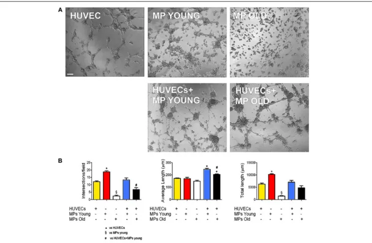
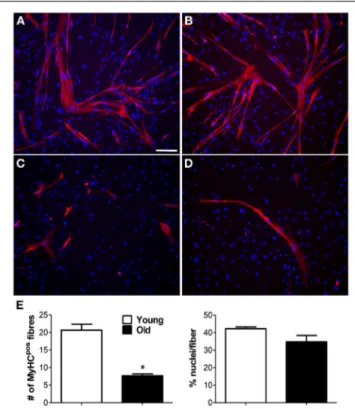
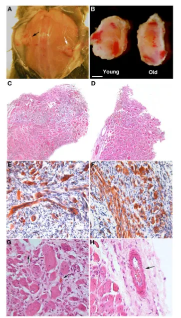
Of the two bio-artificial systems described, one is a silicon chamber system for the generation of skeletal muscle constructs described bySnyman et al. (2013). This inexpensive and read-ily adaptable system (distance between pins, cell number, and matrix-cell volume can be readily changed) may be used with different hydrogels and applied to any existing culture cham-bers. The other is a PEG-fibrinogen (PF)-based hydrogel scaffold used to rejuvenate aged adult skeletal muscle-derived pericytes (MP) from pig skeletal muscle. In this 3D environment peri-cytes were able to recover their differentiation potential toward myogenic differentiation and vessel formation. Fuoco and col-leagues demonstrated that the 3D PF environment was beneficial for swine-derived MP by mimicking the stiffness and mechani-cal properties of young muscle extracellular matrix (ECM), thus, “rejuvinating” aged MP may be an alternative to the use of myo-genic progenitors; indeed, swine derived MP represent a valid
alternative to build human size comparable artificial muscle units from the swine muscle (Fuoco et al., 2014).
The method of choice to seed stem cells in 3D cultures is important, in order to form a densely packaged proto-tissue, independently from the scaffold used. This step is fundamental for the cells of the proto-tissue to complete their maturation pro-cess and interconnect with the native tissue upon in vivo implan-tation.Pagliari et al. (2014a)showed a method for sequential cell seeding, where first gelatin scaffolds were colonized with human mesenchymal stem cells in a static condition to favor endothe-lialization; then the scaffolds were loaded with pre-committed cardiac progenitors and cultured in perfusion bioreactor in car-diogenic conditions. The authors obtained a well-packed cardiac proto-tissue, rich in vessels, but without functional contractile or vascular structures. They concluded that even the more complex 3D dynamic culture system needs to be improved with stretching or electric stimulation to obtain mature, functional cardiac tissue. A valid alternative to biosynthetic scaffolds are de-cellularized organs, for example the muscle acellular scaffold (MAS) from skeletal muscle.Perniconi et al. (2014)demonstrated that MAS may be used to induce the differentiation of cultured myoblasts and this is an excellent support for 3D myogenic cultures also
in vivo in orthotopical engraftments. MAS is histocompatible,
porous, degradable and non-toxic. This structure is stable in anatomical sites other than the skeletal musculature, but does not provide enough signals to trigger myogenesis by the colonizing cells. The possibility to use MAS for the reconstruction of tis-sue different from the skeletal muscle should be investigated. For example, considering similarities between the skeletal and the car-diac tissue, MAS might be used as an autologous native scaffold for cardiac tissue transplantation.
The same authors also contributed a review article on the use-fulness of the MAS as a scaffolding platform able to re-create the natural structure of the muscle. MAS is naturally embedded with active native molecules which remain active after the decellular-ization process and help the implanted proto-tissue to integrate
in the host organ. Tissue-derived ECM helps in structuring niches rich in adhesive and signaling molecules supporting stem cells self-renewal and differentiation (Teodori et al., 2014).
When the muscle lesion is so extensive that the function of the muscle is impaired, the definition of volumetric muscle losses (VMLs) applies. In this case the muscle should be replaced by devices able to re-establish the function of the musculature and which preserve force transmission and continuity of the architec-ture within the host tissues. Hence,Cittadella Vigodarzere and Mantero (2014)described the architecture of the native skele-tal tissue, identifying all the single elements which should be taken into consideration in a skeletal muscle tissue engineer-ing construct. They focused the attention on the vasculature structures of constructs and, since decellularized ECM promotes vascularization of the implanted construct, they concluded that decellularized scaffolds are ready for a clinical application on human.
A clinical example of the latter is described in an inter-esting review discussing the importance of angiogenesis and the pro-angiogenic and pro-myogenic effects of the vascular endothelial growth factor (VEGF)/VEGF receptor pathway as a therapeutic strategy to cure muscle weakness and cardiomy-opathy in Duchenne muscular dystrophy (DMD) patients (Shimizu-Motohashi and Asakura, 2014).
Apart from the structures of ECM and vasculature, also growth factors, cytokines, pleiotropic signaling pathways, and cell-specific regulators play an important role in the differentia-tion and self-renewal of cardiac as well as skeletal muscle stem cells.Pagliari et al. (2014b)described the most important signal-ing pathways which influence the in vivo differentiation of cardiac progenitor cells. Many of these pathways and factors have differ-ent and distant effects on the differdiffer-entiation process (JAK/STAT, Hippo pathway, Wnt and Notch signaling, etc.).
This book provides a comprehensive up-to-date review on car-diac and skeletal muscle tissue engineering and highlights the several elements which have to be taken into consideration when engineering a functional proto-tissue.
REFERENCES
Cittadella Vigodarzere, G., and Mantero, S. (2014). Skeletal muscle tissue engineering: strategies for volumetric constructs. Front. Physiol. 5:362. doi: 10.3389/fphys.2014.00362
Di Felice, V., Barone, R., Nardone, G., and Forte, G. (2014). Cardiac tissue engineering: a reflection after a decade of hurry. Front. Physiol. 5:365. doi: 10.3389/fphys.2014.00365
Fuoco, C., Sangalli, E., Vono, R., Testa, S., Sacchetti, B., Latronico, M. V., et al. (2014). 3D hydrogel environment rejuvenates aged pericytes for skele-tal muscle tissue engineering. Front. Physiol. 5:203. doi: 10.3389/fphys.2014. 00203
Pagliari, S., Jelinek, J., Grassi, G., and Forte, G. (2014a). Targeting pleiotropic sig-naling pathways to control adult cardiac stem cell fate and function. Front.
Physiol. 5:219. doi: 10.3389/fphys.2014.00219
Pagliari, S., Tirella, A., Ahluwalia, A., Duim, S., Goumans, M. J., Aoyagi, T., et al. (2014b). A multistep procedure to prepare pre-vascularized cardiac tissue constructs using adult stem sells, dynamic cell cultures, and porous scaffolds. Front. Physiol. 5:210. doi: 10.3389/fphys.2014. 00210
Perniconi, B., and Coletti, D. (2014). Skeletal muscle tissue engineering: best bet or black beast? Front. Physiol. 5:255. doi: 10.3389/fphys.2014.00255
Perniconi, B., Coletti, D., Aulino, P., Costa, A., Aprile, P., Santacroce, L., et al. (2014). Muscle acellular scaffold as a biomaterial: effects on C2C12 cell differen-tiation and interaction with the murine host environment. Front. Physiol. 5:354. doi: 10.3389/fphys.2014.00354
Shimizu-Motohashi, Y., and Asakura, A. (2014). Angiogenesis as a novel thera-peutic strategy for Duchenne muscular dystrophy through decreased ischemia and increased satellite cells. Front. Physiol. 5:50. doi: 10.3389/fphys.2014. 00050
Snyman, C., Goetsch, K. P., Myburgh, K. H., and Niesler, C. U. (2013). Simple silicone chamber system for in vitro three-dimensional skeletal muscle tissue formation. Front. Physiol. 4:349. doi: 10.3389/fphys.2013.00349
Teodori, L., Costa, A., Marzio, R., Perniconi, B., Coletti, D., Adamo, S., et al. (2014). Native extracellular matrix: a new scaffolding platform for repair of damaged muscle. Front. Physiol. 5:218. doi: 10.3389/fphys.2014. 00218
Conflict of Interest Statement: The authors declare that the research was
con-ducted in the absence of any commercial or financial relationships that could be construed as a potential conflict of interest.
Received: 20 December 2014; accepted: 05 February 2015; published online: 23 February 2015.
Citation: Di Felice V, Forte G and Coletti D (2015) Biomaterials and bioactive molecules to drive differentiation in striated muscle tissue engineering. Front. Physiol.
6:52. doi: 10.3389/fphys.2015.00052
This article was submitted to Striated Muscle Physiology, a section of the journal Frontiers in Physiology.
Copyright © 2015 Di Felice, Forte and Coletti. This is an open-access article dis-tributed under the terms of the Creative Commons Attribution License (CC BY). The use, distribution or reproduction in other forums is permitted, provided the original author(s) or licensor are credited and that the original publication in this jour-nal is cited, in accordance with accepted academic practice. No use, distribution or reproduction is permitted which does not comply with these terms.
Simple silicone chamber system for in vitro
three-dimensional skeletal muscle tissue formation
Celia Snyman1†, Kyle P. Goetsch1†, Kathryn H. Myburgh2and Carola U. Niesler1*1Discipline of Biochemistry, School of Life Sciences, University of KwaZulu-Natal, Pietermaritzburg, South Africa 2Department of Physiological Sciences, University of Stellenbosch, Stellenbosch, South Africa
Edited by:
Valentina Di Felice, University of Palermo, Italy
Reviewed by:
Laszlo Csernoch, University of Debrecen, Hungary
Toshia Fujisato, Osaka Institute of
*Correspondence:
Carola U. Niesler, Discipline of Biochemistry, School of Life Sciences, University of KwaZulu-Natal, Private Bag X01, Scottsville, 3209 Pietermaritzburg, South Africa
e-mail: [email protected] †These authors have contributed equally to this work.
Bioengineering skeletal muscle often requires customized equipment and intricate casting techniques. One of the major hurdles when initially trying to establish in vitro tissue engineered muscle constructs is the lack of consistency across published methodology. Although this diversity allows for specialization according to specific research goals, lack of standardization hampers comparative efforts. Differences in cell type, number and density, variability in matrix and scaffold usage as well as inconsistency in the distance between and type of adhesion posts complicates initial establishment of the technique with confidence. We describe an inexpensive, but readily adaptable silicone chamber system for the generation of skeletal muscle constructs that can readily be standardized and used to elucidate myoblast behavior in a three-dimensional space. Muscle generation, regeneration and adaptation can also be investigated in this model, which is more advanced than differentiated myotubes.
Keywords: three-dimensional assays, tissue engineering, hydrogel constructs, in vitro skeletal muscle tissue
INTRODUCTION
Three-dimensional (3D) skeletal muscle constructs can be bio-engineered in vitro. 3D models have advantages over 2D cell cultures in mimicking in vivo conditions as they allow for the study of dimensionality, cellular architecture, cell polarity and function. Constructs can be adapted for the generation of in vitro drug screening assays as well as in vivo tissue repair following transplantation of constructs (Vandenburgh et al., 2008; Corona et al., 2012). If genetically modified to express recombinant pro-tein, these constructs can be used for therapeutic protein delivery (Vandenburgh et al., 1996).
An assortment of methods for the generation of bio-artificial skeletal muscle have been previously described (Table 1), with variations on aspects including chamber construction, matrix composition and ultimate tissue size generated. The chambers employed for 3D culture of skeletal muscle may be divided into two main categories: the uncomplicated silicone tubing model and the more intricate models constructed in chamber slides and multi-well plates (Table 1A) and chambers and micro-patterned wells that are precast via photolithographic moulds (Table 1B). Confluent myoblast monolayers cultured in a matrix-coated petri dish under differentiating conditions may also form scaffold-free 3D muscle tissue due to contractility of the differentiating fibers (Table 1C). These methods naturally reflect the thrust of the particular research group. While each model has specific advan-tages, key methodological aspects differ considerably between the various models which may potentially hamper efficient compar-ison. A critical overview of the various models is required before describing our simple chamber system.
In general culture vessels for 3D skeletal muscle constructs consist of tubes, standard pre-fabricated laboratory-based culture
plates or dishes that contain two tissue adhesion points that mimic tendons and consist of either cast silicone posts, metal pins and mesh, sutures or Velcro pads (Vandenburgh et al., 1996, 2008; Dennis and Kosnik, 2000; Powell et al., 2002). Some models require post-modification with custom-made inserts that replace pins or Velcro adhesion points. In the models consisting of a sil-icone tube or a precast chamber slide, the adhesion points are on average 18–30 mm apart, which may dictate factors including the volume and concentration of cells initially seeded (Table 1A) (Powell et al., 2002; Vandenburgh et al., 2008). The cell seeding number ranged from 1× 10⁶to 6× 10⁶cells per tube, and the hydrogel-cell suspension volume varied between 400µl required for the tube models and 3.2 ml for the adapted single well-chamber slide (Table 1A) (Vandenburgh et al., 1996; Hinds et al., 2011; Smith et al., 2012). The matrix mixture and culture periods are similarly diverse. This is, however, also an indication of the ease with which tissue-engineering models that use silicone tubes with adhesion points may be constructed and adapted for a range of purposes (Vandenburgh et al., 2008; Hinds et al., 2011; Smith et al., 2012).
While Vandenburgh initially generated in vitro 3D mus-cle tissue constructs from C2C12 myoblasts in silicone tubing containing Velcro pads∼25 mm apart (Table 1A), this group sub-sequently employed a more complex custom-built silicone mould cast around a Teflon template with two flexible silicone posts inserted into a standard 96-well plate 4 mm apart (Vandenburgh et al., 1996, 2008). In other more recently-developed models these anchorage points and the distance between them in custom-built models varied from 4 to 50 mm, according to the size of the wells and aspects required for the experiment. The flexibility of the cantilever posts in the more advanced models Technology, Japan
Table 1 | A comparison of published methods for the generation of bio-artificial skeletal muscle. Model description Purpose Anchor points Cell type and seeding
conditions
Matrix (final concentrations)
References
(A) CAST CHAMBERS CONTAINING TWO ADHESION POINTS
Silicone rubber tubes cut lengthwise in 35 mm dishes
Reversible gene therapy
Velcro pads or stainless steel mesh
C2C12 mouse myoblasts (1–4× 10⁶cells/well) Collagen (1.6 mg/ml) and Matrigel (Ratio of 6:1 v/v) Vandenburgh et al., 1996
Sylgard moulds produced in a vacuum-moulding process around Teflon spacers; 96-well plates
In vitro drug screening Flexible silicone posts Primary mouse myoblasts (0.2 × 10⁶cells/well) Collagen (1 mg/ml) and Matrigel (Ratio of 6:1 v/v) Fibrinogen–thrombin (0.5 mg/ml) and thrombin (1 U/ml) Vandenburgh et al., 2008
Moulds cast from 2% agarose in PBS around Teflon spacers; 24-well plates
Heart muscle kinetics
Flexible Sylgard posts
Neonatal rat heart cells (0.62 × 10⁶cells/well) Fibrinogen (5 mg/ml) and Matrigel (100µl/ml) polymerized with thrombin (32:1 v/v) Hansen et al., 2010
Rectangular casting moulds (see
Hansen et al., 2010)
Interaction between cells and surrounding matrix
Silicone pins Primary human
myoblasts
(0.66× 10⁶cells/well)
Fibrin-based matrix Chiron et al., 2012
Mechanical Cell Stimulator, version 4.0 (MCS4)
Silicone rubber tissue moulds; 6-well plates Mechanical stimulation to improve tissue-engineered human skeletal muscle
Stainless steel pins Primary human skeletal muscle cells
(1× 10⁶cells/well)
Collagen 1 (0.8 mg/ml) and Matrigel (Ratio of 6:1 v/v)
Powell et al., 2002
Silicone tube cut lengthwise (ends sealed with PDMS); 6-well plates Hydrogel matrix combinations; influence on contractile function of engineered muscle tissue
Velcro adhesion pads Primary rat skeletal myoblasts (6× 10⁶cells/well) Collagen 1 (1.4 mg/ml) and Matrigel Fibrinogen (2, 4 or 6 mg/ml) and Matrigel (10%, 20% or 40% v/v) Hinds et al., 2011
Commercially available single-well chamber slides Optimized culture parameters improved reproducibility and the cellular architecture
Polyethylene mesh Primary rat muscle derived cells (Cell count not stated; 3.2 ml/well)
Collagen 1 Smith et al.,
2012
(B) PHOTOLITHOGRAPHIC MOULDS AND MICRO-PATTERNED WELLS WITH POSTS
Sylgard tissue moulds cast from patterned master templates of coated photo-resistant silicone wafers Muscle cell alignment Array of silicone posts C2C12 mouse myoblasts (1× 10⁶cells/well) Primary rat skeletal myoblasts (2× 10⁶cells/well) Collagen I (1 mg/ml) and fibrinogen (2 mg/ml) (ratio of 1:0, 3:1, 1:1, 1:3, 0:1) Thrombin (0.4 U/mg fibrinogen) Matrigel added to all combinations
Bian and Bursac, 2009
Precast micro-patterned wells Formation of muscle for use in bioactuators
PDMS cantilevers C2C12 mouse myoblasts (400 cells/micro-patterned well) Collagen 1 (2 mg/ml) and Matrigel Sakar et al., 2012 (Continued)
Table 1 | Continued
Model description Purpose Anchor points Cell type and seeding conditions
Matrix (final concentrations)
References
(C) SCAFFOLD-FREE CONFLUENT MONOLAYER CONTRACTION INTO CYLINDER BETWEEN PINNED SUTURES
Sylgard-based 35 mm culture dish coated with laminin
Excitability and contractile properties of muscle engineered from co-cultured primary cells
Silk sutures coated with 50µg/ml laminin
Co-culture of primary myogenic precursors, fibroblasts and all related cell types
Laminin base layer (1µg/cm2)
Dennis and Kosnik, 2000
Sylgard-coated 35 mm culture dish coated with laminin
Skeletal muscle construct from C2C12 myoblasts; AIM-V media
Silk sutures C2C12 myoblasts AIM-V
serum-free medium; 0.02 × 10⁶cells / dish
Laminin base layer (2µg/ml) Laminin top layer (10µg/ml)
Fujita et al., 2009b
The chambers employed for three-dimensional culture of skeletal muscle may be divided into three main categories: Cast chambers such as the uncomplicated silicon tubing model (A), Photolithographic moulds and micro-patterned wells (B), and Scaffold-free confluent monolayers (C).
These are fitted into the wells of standard culture plates.
The combination of hydrogel components as well as cell type, number and volume seeded per well vary considerably between published methods, and are summarized. The concentration of Matrigel is 10% (v/v) unless otherwise indicated.
is an improvement from the originally employed metal pins or large, fixed Velcro pads or metal mesh (Vandenburgh et al., 2008). The advantage of these custom-built systems with standardized dimensions and with flexible cantilever posts is that the cultured constructs could be used to investigate muscle kinetics (force generation, internal strain or contractile forces) on mechanical stimulation, or in response to compounds such as insulin-like growth factor 1 and cholesterol-lowering statins (Eastwood et al., 1998; Nirmalanandhan et al., 2007; Vandenburgh et al., 2008; Smith et al., 2012).
The actual vessels used for myooid culture may also be custom-built via micro-pattern technology or photolithography (Table 1B) (Dennis and Kosnik, 2000; Vandenburgh et al., 2008; Fujita et al., 2009a; Hansen et al., 2010; Smith et al., 2012). Using this technology a negative template is generated and a mould is subsequently cast from biological grade silicone (Bian and Bursac, 2009; Sakar et al., 2012). The use of an array of posts in a wafer pattern rather than simply two adhesion points permits the for-mation of a sheet-like culture that allows for the investigation of muscle cell alignment. This is in contrast to the 3D construct generated between two cantilevers.
Even though reproducibility is improved, these casting meth-ods and inserts demand specialized equipment, which commonly translates into an increase in cost. In addition the system may not easily be adaptable to different well-types or tissue sizes.
Currently, various hydrogel components are routinely used, both individually and in combination, to successfully engi-neer muscle tissue containing striated and aligned myotubes. These include collagen 1, Matrigel from Engelbrecht-Holm-Swarm (EHS) sarcoma cells, laminin I, and fibrin-based gels (Table 1) (Vandenburgh et al., 2008; Bian and Bursac, 2009; Hansen et al., 2010). The hydrogels differ in their molecular composition, macromolecular orientation and the degree of
cross-linking (Dennis and Kosnik, 2000; Hinds et al., 2011). This has practical implications; for instance, Matrigel polymer-ization is temperature-sensitive, while laminin I and collagen 1 are able to spontaneously form 3D gels at room temperature (Yurchenco et al., 1992). Furthermore, myogenesis itself is dif-ferentially affected by these matrix factors; collagen 1 has been shown to suppress differentiation, whereas Matrigel promotes myotube formation in both 2D and 3D models (Langen et al., 2003; Grefte et al., 2012). Laminin I promotes myoblast adher-ence, proliferation and myotube fusion (Schuler and Sorokin, 1995; Vachon et al., 1996). Finally, the matrix combinations as reported for the different models vary considerably (Table 1). The most conventional combination consists of collagen 1 in combination with Matrigel in a ratio of 6:1 (v/v) or a 10–20% Matrigel component (Table 1). The concentration of fibrinogen on its own or used in combination with other matrix components also varies considerably (Table 1) (Vandenburgh et al., 2008; Hinds et al., 2011). Such diversity may hamper direct comparison of results.
Successful formation of scaffold-free cylindrical 3D myooids in 35 mm culture dishes coated first with Sylgard and then laminin has also been described (Dennis and Kosnik, 2000; Fujita et al., 2009b) (Table 1C). After an initial low seeding density (2–10× 103 cells) in a coated 35 mm dish, cells are cultured in a 2D monolayer to confluence. This is followed by replacement of the growth media (GM) with differentiation media (DM) to stimu-late differentiation into muscle fibers. Subsequent formation of cylindrical myooids is due to contraction of the fibers and release from the underlying Sylgard coating (Huang et al., 2005; Fujita et al., 2009a). Silk sutures pinned into the Sylgard base act as handling points and mimic flexible tendons for the cylindrical myooid. The laminin matrix employed merely forms a separating layer between the Sylgard coating and the cultured cells, while
the final histology and molecular characteristics reflect that of skeletal muscle. It is believed that the interaction between co-cultured primary fibroblasts and myoblasts in this model allows for the investigation of functional and molecular development, as reflected by the contractile properties and expression of tran-scription factors MyoD and muscle-related myosin heavy chain of such fabricated skeletal muscle (Dennis and Kosnik, 2000; Huang et al., 2005; Fujita et al., 2009b). The culture period to establish this model is, however, considerably longer than the previously-mentioned models.
Although numerous methods for bioengineering skeletal mus-cle exist, currently no standardized culture vessel and proto-col has been proposed. Also, many are prohibitive due to the required customized equipment and intricate casting techniques. Below we describe an inexpensive, accessible hydrogel-based sys-tem that may be readily standardized, yet is easily customized to reflect desired matrix combinations and tissue size. This model is ideal for laboratories expanding into three-dimensional assays.
MATERIALS AND METHODS
SELECTION AND CONSTRUCTION OF CHAMBER
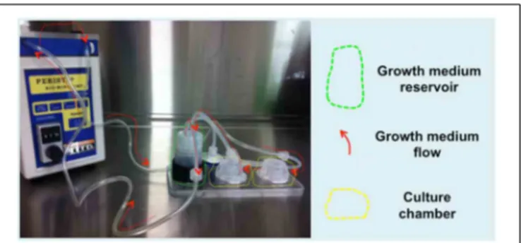
An adaptable chamber system was generated by using 18 mm sec-tions of biological grade silicone tubing (outer diameter: 5 mm) which was cut in half (lengthwise) (Figure 1A). Surgical grade stainless steel pins (3 mm long, outer diameter: 0.2 mm) were inserted into the tubes to act as adhesion points; distances between the pins ranged from 4 to 8 mm, depending on the size of the muscle tissue required. The tubes were fitted into each well of a 4- or 24-well-culture plate and secured within the wells using Sylgard 182 (Dow Corning Corporation, cat. 3097358-1004) (Figure 1A). Sylgard was allowed to cure for 24 h and the plates sterilized overnight under an ultraviolet light.
SELECTION AND PREPARATION OF CELLS
Murine C2C12 myoblasts (ATCC, cat. CRL-1772) were main-tained in GM containing Dulbecco’s Modified Eagle Serum (DMEM, Highveld, cat.CN3193-9), L-glutamine (2% v/v, Cambrex, 605E), PenStrep (2% v/v, Cambrex, cat.17-602E), and Fetal Calf Serum (FCS; 10% v/v, Invitrogen, cat.10108165). Primary cultured human skeletal myoblasts (HSKM, Lonza, cat. CC-2561) were cultured in Ham’s-F10 (Gibco, cat.15140), FCS (20% v/v), Penstrep (2% v/v), L-glutamine (2% v/v), fibroblast growth factor (FGF; 2.5 ng/ml, Promega, cat.G507A).
PREPARATION OF HYDROGEL/MATRIX
Rat tail collagen 1 (3.6 mg/ml; Sigma, cat. C9791) was neutral-ized with 10% NaOH (∼30 µl per ml of collagen 1) until a color change was observed (yellow to pink due to pH indica-tor in DMEM). All solutions were kept on ice to restrict matrix polymerization prior to seeding.
CELL-HYDROGEL SUSPENSION
Initially a cell suspension (27µl; in GM) containing 3.2 × 10⁶ HSKM cells or 6.4 × 10⁶C2C12 cells and 33µl 10X DMEM was added to the neutralized collagen 1 solution (43µl) (total volume:
FIGURE 1 | Details on simple silicone tube chamber construction and hydrogel-cell preparation. (A) Biological grade silicone tubing is cut to fit
the diameter of the well; the tube is then cut lengthwise in half (1). Surgical grade stainless steel pins are inserted through the silicone wall at predefined distances from each other (2). The silicone tube is secured in place within the well with Sylgard 182 to form a chamber (3). The hydrogel-cell suspension is pipetted into the silicone chamber around the pins (4) and the well is flooded with growth media once the gel construct has set (5).(B) Calculations for preparation of the hydrogel-cell mixture
(Ratio of 6:1).(C) A multi-well plate containing a hydrogel-C2C12 mix seeded in silicone tube chambers (arrows indicate pins positioned 4 mm apart).
103µl). To achieve a cell-collagen 1:Matrigel ratio of 6:1 (v/v) as is often used in the generation of skeletal muscle (Vandenburgh et al., 1996, 2008; Powell et al., 2002), 17µl Matrigel (10.1 mg/ml stock concentration, BD Biosciences, cat.356231) was added to the cell-collagen 1 suspension to achieve a final volume of 120µl (Figure 1B).
HSKM cells are larger than C2C12 myoblasts; this accounts for the lower number of human myoblasts in the hydrogel mix when compared to mouse myoblasts. The cell/hydrogel suspension con-taining C2C12 or HSKM cells was pipetted into each silicone tube chamber around the pins. Constructs were subsequently incubated at 37◦C overnight and the wells then flooded with 350µl GM (Figure 1A). Twenty-four hours later, culture GM was replaced with DM, which contained DMEM, L-glutamine (Lonza; 2% v/v), PenStrep (Lonza; 2% v/v) and horse serum (HS; 1% v/v, Invitrogen, cat.16050-130).
PREPARATION FOR IMAGE COLLECTION
Brightfield images were captured at various stages of muscle development using a Motic 3.0 MP camera and an Olympus stereo microscope (VMZ, Japan). For histochemical investigation of desmin expression and the actin cytoskeleton, muscle con-structs were fixed for 2 h in the wells in paraformaldehyde (4% prepared in PBS). Subsequent removal of the pins allowed con-structs to be transferred from the chambers to culture wells where they were incubated with either a polyclonal rabbit anti-desmin antibody (1/600, Abcam cat.AB15200) for 2 h at room tem-perature followed by a Dylight488 donkey anti-rabbit antibody (1/1000, Jackson, cat.711-485-152) for 1 h at room temperature,
or TRITC-conjugated Phalloidin (0.5 ng/ml, Sigma; cat. P1951) for 2 h at room temperature. Nuclei were stained with Hoechst (2.5µg/ml, Sigma, cat C8890) for 10 min at room temperature and constructs mounted on glass slides with Moviol and viewed with the Zeiss 710 confocal microscope. Constructs cultured for 15 days in DM were also fixed with glutaraldehyde (2%, 2 h at room temperature), dehydrated with a graded series of alcohols and embedded in Spurr’s resin. Thin sections were cut and DIC images were obtained with the 710 Zeiss confocal microscope.
This adapted technique proved to be advantageous for skele-tal muscle formation with constructs from both C2C12 and HSKM cells successfully spanning the pins after a 3–7 day culture period (Figures 1C, 2A–C). After 12–15 days in culture, differ-entiated C2C12 myotubes showed clear formation of actin fibers (Figure 2D). In addition, aligned myotubes expressed desmin (Figure 2E) and longitudinal sections showed evidence of orga-nization into multinucleated myotubes (Figure 2F), which is an initial requirement for functionality.
DISCUSSION
Muscle tissue engineering is no longer in its infancy, nor is it the prerogative of only a few laboratories. Nonetheless, only a few research groups have made major advances in this field (Vandenburgh et al., 2008; Hansen et al., 2010; Vandenburgh, 2010; Chiron et al., 2012; Corona et al., 2012; Sakar et al., 2012; Smith et al., 2012). One of the major hurdles when initially try-ing to establish in vitro tissue engineered muscle constructs is the lack of consistency across published methodology. Such lack of uniformity was highlighted by a summary of the range of moulds already employed in muscle tissue engineering (Table 1). A care-ful view of Table 1 indicates that particular moulds have been designed with specific purposes in mind (Powell et al., 2002; Bian and Bursac, 2009; Sakar et al., 2012). However, differences in cell type, seeding density as well as variability in hydrogel/scaffolds used, and also variations in the distance between and type of adhesion posts (i.e., stainless steel, sutures, Velcro, etc.) hampers initial establishment of the technique with confidence. In the cur-rent study we describe an inexpensive, readily adaptable silicone chamber system for the generation of skeletal muscle constructs.
We highlight the basic steps and requirements needed to form differentiated muscle tissue from either C2C12 or HSKM myoblasts in a collagen 1/Matrigel hydrogel. This model is adapt-able to fit into any existing culture dish or chamber used in most laboratories. Despite this simplicity, variations in the dis-tance between pins, as well as cell number and matrix-cell volume can be readily achieved. In addition, the use of appropriate pins as anchor points allows for future mechanical stimulation to investigate contractile forces and allows for the study of internal stresses during muscle differentiation in 3D cultures. It is also use-ful for investigations into genetic manipulation, drug therapy or co-culture of complimentary cell phenotypes.
This model has several practical advantages. Biological grade silicone is inexpensive, readily available and allows for ease of pin insertion and subsequent tissue manipulation. In addition, after initial use, the various components of the chamber system may also be reused following de-cellularization with ammonium hydroxide and cleaning with alcohol and sonication. This model,
FIGURE 2 | Successful generation of mouse and human skeletal muscle constructs using the simple silicone chamber system. (A)
When seeded in a matrix of collagen 1 and Matrigel (14%), mouse C2C12 cells formed tissue (day 7 in differentiation media) between pins placed 4 mm apart.(B) When seeded in a matrix of collagen 1 and Matrigel (14%),
mouse C2C12 cells formed tissue (day 3 in differentiation media) between pins placed 8 mm apart.(C) When seeded in a matrix of collagen 1 and
Matrigel (14%), human skeletal muscle (HSKM) cells formed tissue (day 3 in differentiation media) between pins placed 4 mm apart.(D) After 12 days
in differentiation media, actin fibers stained with TRITC-phalloidin and were clearly visible in the differentiated mouse C2C12 myotubes. Nuclei were stained with Hoechst (scale bar= 50 µm). (E) After 15 days in differentiation media, elongated myotubes were aligned and contained desmin, an intermediate filament required for myotube contractile function (scale bar= 50 µm). (F) Thin sections of resin-embedded C2C12 myoblasts culture for 15 days in differentiation media showed the formation of multi-nucleated (arrows) myotubes (scale bar= 20 µm).
with standardized parameters, may be used as an optimized sys-tem for initial evaluation of factors involved in skeletal muscle generation from both primary cultured myoblasts and established cell lines.
Imaging of myoblasts functioning in a three-dimensional space is also more closely aligned to in vivo behavior. With this model we have described the expression of desmin, an inter-mediate filament that plays a key role in the integration of striated muscle morphology and function (Capetanaki et al., 2007). We also showed the transition of myoblasts into elon-gated, multi-nucleated myotubes, one of the relevant steps during myoblast differentiation. It must be noted, however, that the use of C2C12 myoblasts to generate functional striated tissue may require electrical stimulation during the generation of the
bioengineered tissue; this is not the case for primary muscle progenitor cells (Langelaan et al., 2011). Engineered tissue may further be processed for histological purposes or immunocy-tochemical investigation of transcription factors and expressed proteins. We propose that the method we describe may allow skeletal muscle research groups utilizing 2D cell culture models to move into 3D tissue models with relative ease. This will be impor-tant to enable more rapid enhancement of our understanding of muscle synthesis, repair and adaptation in vitro, in a model more advanced than differentiated myotubes.
ACKNOWLEDGMENTS
The work was supported by the South African National Research Foundation, South African Medical Research Council and University of KwaZulu-Natal. The authors also thank the UKZN Microanalysis and Microscopy Unit (Pietermaritzburg) as well as Graeme Marwick (Denel Dynamics) for all their assistance. REFERENCES
Bian, W., and Bursac, N. (2009). Engineered skeletal muscle tissue net-works with controllable architecture. Biomaterials 30, 1401–1412. doi: 10.1016/j.biomaterials.2008.11.015
Capetanaki, Y., Bloch, R. J., Kouloumenta, A., Mavroidis, M., and Psarras, S. (2007). Muscle intermediate filaments and their links to membranes and membranous organelles. Exp. Cell Res. 313, 2063–2076. doi: 10.1016/j.yexcr.2007.03.033 Chiron, S., Tomczak, C., Duperray, A., Lainé, J., Bonne, G., Eder, A., et al. (2012).
Complex Interactions between Human Myoblasts and the Surrounding 3D Fibrin-Based Matrix. PLoS ONE 7:e36173. doi: 10.1371/journal.pone.0036173 Corona, B. T., Machingal, M. A., Criswell, T., Vandhavkar, M., Dannahower, A. C.,
Bergmen, C., et al. (2012). Further development of a tissue engineered muscle repair construct in vitro for enhanced functional recovery following implanta-tion in vivo in a murine model of volumetric muscle loss injury. Tissue Eng. Part
A 18, 1213–1228. doi: 10.1089/ten.tea.2011.0614
Dennis, R. G., and Kosnik, P. E. (2000). Excitability and isomet-ric contractile properties of mammalian skeletal muscle constructs engineered in virto. In Vitro Cell. Dev. Biol. 36, 327–335. doi: 10.1290/1071-2690(2000)036<0327:EAICPO>2.0.CO;2
Eastwood, M., Mudera, V. C., McGrouther, D. A., and Brown, R. A. (1998). Effect of precise mechanical loading on fibroblast populated collagen lattices: mor-phological changes. Cell Motil. Cytoskeleton 40, 13–21. doi: 10.1002/(SICI)1097-0169(1998)40:1<13::AID-CM2>3.0.CO;2-G
Fujita, H., Shimizu, K., and Nagamori, E. (2009a). Application of a cell sheet-polymer film complex with temperature sensitivity for increased mechanical strength and cell alignment capability. Biotechnol. Bioeng. 103, 370–377. doi: 10.1002/bit.22251
Fujita, H., Shimizu, K., and Nagamori, E. (2009b). Novel method for fabrication of skeletal muscle construct from the C2C12 myoblast cell line using serum-free medium AIM-V. Biotechnol. Bioeng. 103, 1034–1041. doi: 10.1002/bit.22318 Grefte, S., Vullinghs, S., Kuijpers-Jagtman, A. M., Torensma, R., and Von Den Hoff,
J. W. (2012). Matrigel, but not collagen I, maintains the differentiation capacity of muscle derived cells in vitro. Biomed. Mater. 2, 055004. doi: 10.1088/1748-6041/7/5/055004
Hansen, A., Eder, A., Bönstrup, M., Flato, M., Mewe, M., Schaaf, S., et al. (2010). Development of a drug screening platform based on engineered heart tissue.
Circ. Res. 107, 35–44. doi: 10.1161/CIRCRESAHA.109.211458
Hinds, S., Bian, W., Dennis, R. G., and Bursac, N. (2011). The role of extracellular matrix composition in structure and function of bioengineered skeletal muscle.
Biomaterials 32, 3575–3583. doi: 10.1016/j.biomaterials.2011.01.062
Huang, Y.-C., Dennis, R. G., Larkin, L., and Baar, K. (2005). Rapid formation of functional muscle in vitro using fibrin gels. J. Appl. Physiol. 98, 706–713. doi: 10.1152/japplphysiol.00273.2004
Langelaan, M. L., Boonen, K. J., Rosaria-Chak, K. Y., Van Der Schaft, D. W., Post, M. J., Baaijens, F. P. (2011). Advanced maturation by electrical stimulation: Differences in response between C2C12 and primary muscle progenitor cells.
J. Tissue Eng. Regen. Med. 5, 529–539. doi: 10.1002/term.345
Langen, R. C., Schols, A. M. W. J., Kelders, M. D. J. M., Wouters, E. F. M., and Janssen-Heininger, Y. M. W. (2003). Enhanced myogenic differentiation by extracellular matrix is regulated at the early stages of myogenesis. In Vitro Cell.
Dev. Biol. Anim. 39, 163–169. doi: 10.1007/s11626-003-0011-2
Nirmalanandhan, V. S., Rao, M., Sacks, S., Haridas, B., and Butler, D. L. (2007). Effect of length of the engineered tendon construct on its structure–function relationships in culture. J. Biomech. 40, 2523–2529. doi: 10.1016/j.jbiomech.2006.11.016
Powell, C. A., Smiley, B. L., Mills, J., and Vandenburgh, H. H. (2002). Mechanical stimulation improves tissue-engineered human skeletal muscle. Am. J. Physiol.
Cell Physiol. 283, C1557–C1565. doi: 10.1152/ajpcell.00595.2001
Sakar, M. S., Neil, D., Boudou, T., Borochin, M. A., Li, Y., Weiss, R., et al. (2012). Formation and optogenetic control of engineered 3D skeletal muscle bioactuators. Lab Chip 12, 4976–4985. doi: 10.1039/c2lc40338b
Schuler, F., and Sorokin, L. M. (1995). Expression of laminin isoforms in mouse myogenic cells in vitro and in vivo. J. Cell Sci. 108, 3795–3805.
Smith, A. S. T., Passey, S., Greensmith, L., Mudera, V., and Lewis, M. P. (2012). Characterization and optimization of a simple, repeatable system for the long term in vitro culture of aligned myotubes in 3D. J. Cell. Biochem. 113, 1044–1053. doi: 10.1002/jcb.23437
Vachon, P. H., Loechel, F., Xu, H., Wewer, U. M., and Engvall, E. (1996). Merosin and laminin in myogenesis; specific requirement for merosin in myotube stability and survival. J. Cell Biol. 134, 1483–1497. doi: 10.1083/jcb.134.6.1483 Vandenburgh, H. (2010). High-content drug screening with engineered
mus-culoskeletal tissues. Tissue Eng. Part B 16, 55–64. doi: 10.1089/ten.teb. 2009.0445
Vandenburgh, H., Del Tatto, M., Shansky, J., Lemaire, J., Chang, A., Payumo, F., et al. (1996). Tissue-engineered skeletal muscle organoids for reversible gene therapy. Hum. Gene Ther. 7, 2195–2200. doi: 10.1089/hum.1996.7.17-2195 Vandenburgh, H., Shansky, J., Benesch-Lee, F., Barbata, V., Reid, J., Thorrez, L.,
et al. (2008). Drug-screening platform based on the contractility of tissue-engineered muscle. Muscle Nerve 37, 438–447. doi: 10.1002/mus.20931 Yurchenco, P. D., Chen, Y.-S., and Colognato, H. (1992). Laminin forms an
independent network in basement membranes. J. Cell Biol. 117, 1119–1133. doi: 10.1083/jcb.117.5.1119
Conflict of Interest Statement: The authors declare that the research was
con-ducted in the absence of any commercial or financial relationships that could be construed as a potential conflict of interest.
Received: 10 August 2013; paper pending published: 02 September 2013; accepted: 12 November 2013; published online: 28 November 2013.
Citation: Snyman C, Goetsch KP, Myburgh KH and Niesler CU (2013) Simple silicone chamber system for in vitro three-dimensional skeletal muscle tissue formation. Front. Physiol. 4:349. doi: 10.3389/fphys.2013.00349
This article was submitted to Striated Muscle Physiology, a section of the journal Frontiers in Physiology.
Copyright © 2013 Snyman, Goetsch, Myburgh and Niesler. This is an open-access article distributed under the terms of the Creative Commons Attribution License (CC BY). The use, distribution or reproduction in other forums is permitted, provided the original author(s) or licensor are credited and that the original publication in this journal is cited, in accordance with accepted academic practice. No use, distribution or reproduction is permitted which does not comply with these terms.
Angiogenesis as a novel therapeutic strategy for Duchenne
muscular dystrophy through decreased ischemia and
increased satellite cells
Yuko Shimizu-Motohashi1,2,3and Atsushi Asakura1,2,3*
1
Stem Cell Institute, University of Minnesota Medical School, Minneapolis, MN, USA 2
Paul and Sheila Wellstone Muscular Dystrophy Center, University of Minnesota Medical School, Minneapolis, MN, USA 3
Department of Neurology, University of Minnesota Medical School, Minneapolis, MN, USA
Edited by:
Dario Coletti, Université Pierre et Marie Curie Paris 6, France
Reviewed by:
Christopher Von Bartheld, University of Nevada, Reno, USA
Ashok Kumar, University of Louisville, USA
Ara Parlakian, Université Pierre et Marie Curie, France
*Correspondence:
Atsushi Asakura, Department of Neurology, McGuire Translational Research Facility, University of Minnesota Medical School, 2001 6th Street SE, Minneapolis, MN 55455, USA
e-mail: [email protected]
Duchenne muscular dystrophy (DMD) is the most common hereditary muscular dystrophy caused by mutation in dystrophin, and there is no curative therapy. Dystrophin is a protein which forms the dystrophin-associated glycoprotein complex (DGC) at the sarcolemma linking the muscle cytoskeleton to the extracellular matrix. When dystrophin is absent, muscle fibers become vulnerable to mechanical stretch. In addition to this, accumulating evidence indicates DMD muscle having vascular abnormalities and that the muscles are under an ischemic condition. More recent studies demonstrate decreased vascular densities and impaired angiogenesis in the muscles of murine model of DMD. Therefore, generation of new vasculature can be considered a potentially effective strategy for DMD therapy. The pro-angiogenic approaches also seem to be pro-myogenic and could induce muscle regeneration capacity through expansion of the satellite cell juxtavascular niche in the mouse model. Here, we will focus on angiogenesis, reviewing the background, vascular endothelial growth factor (VEGF)/VEGF receptor-pathway, effect, and concerns of this strategy in DMD.
Keywords: muscular dystrophy, regeneration, angiogenesis, VEGF, Flt-1, satellite cell, mdx mice, skeletal muscle
INTRODUCTION
Duchenne muscular dystrophy (DMD) is the most common hereditary muscular dystrophy affecting approximately 1 in 5000 live male births (Mendell et al., 2012). It is caused by mutations in dystrophin gene located on Xp21 (Monaco et al., 1986), leading to progressive muscle weakness in which respiratory and cardiac failures are the main reasons of their early mortalities. Dystrophin is a protein which forms the dystrophin-associated glycoprotein complex (DGC) at the sarcolemma which links the muscle sar-comeric structure to the extracellular matrix (Davies and Nowak, 2006). When dystrophin is absent due to the gene mutation, mus-cle fibers become vulnerable to mechanical stretching (Pasternak et al., 1995). Currently there is no curative therapy for this disease and glucocorticoid is the only medication available that slows the decline in muscle strength and function in DMD (Bushby et al., 2010).
Since the identification of dystrophin in the mid 1980’s (Monaco et al., 1986), several therapeutic approaches have been investigated. Gene replacement with virus vector, induction of protein expression by exon skipping or read through, compen-sation with dystrophin surrogates, and delivery of muscle stem cells or pluripotent stem cells have been investigated so far (Leung and Wagner, 2013; Rodino-Klapac et al., 2013). Recently, it was announced that phase 3 clinical trial for Drisapersen, an antisense oligonucleotide for exon skipping, could not meet the endpoint of statistically significant improvement (http://www.gsk.com/ media.html). Although exon skipping could still be considered as
one of the most promising therapeutic approaches available, there is a necessity for developing further therapeutic strategies. We have recently reviewed vasculature-related strategies for DMD, with a major focus on therapeutic methods to increase blood flow in existing blood vessels (Ennen et al., 2013). In the cur-rent review, we examine the evidence for reduced formation of blood vessels in DMD muscle and the therapeutic approach to augment angiogenesis by using vascular endothelial growth factor (VEGF)-based strategies. We provide an update of current evi-dence for changes in vasculature in DMD, approaches available to increase vasculature, and further discuss the pros and cons of the underlying rationale.
EVIDENCE FOR VASCULATURE CHANGES IN DMD
The necrotic fibers in DMD are often seen in groups, a simultane-ous necrosis of contigusimultane-ous muscle fibers, and it had been thought that this phenomenon was due to local reduction of blood supply by common capillaries in that group of necrotic fibers (Rando, 2001). The dystrophin deficiency in vascular smooth muscle (Miyatake et al., 1989) and absence of nitric oxide synthase (NOS) from the sarcolemma have indicated that DMD muscle is sub-jected to impaired blood flow (Brenman et al., 1995; Rando, 2001; Ennen et al., 2013).
More recent studies demonstrate that mdx mice muscle has decreased vascular density. Immunostaining of arterioles has revealed decreased vascular density in heart and gracilis mus-cles of mdx mice (Loufrani et al., 2004). Matsakas et al., have
visually shown that vasculature of the tibialis anterior (TA) in
mdx mice is reduced compared to wild type by microfil-perfused
whole mount imaging (Matsakas et al., 2013).
Another report shows that angiogenesis is impaired in mdx mice, not only in muscle, but systemically, which was proven by laser Doppler perfusion imaging in the hind limb ischemic model, VEGF induced neovascularization quantification in the corneal model, Matrigel subcutaneous angiogenic assay, and quantifica-tion of tumor growth and vascularizaquantifica-tion in the tumor implant model (Palladino et al., 2013).
Rhoads et al., have indicated satellite cells isolated from aging and mdx mice exhibit decreased expression of hypoxia-inducible factor 1α (HIF-1α) and VEGF and reduced capacity to promote angiogenesis in vitro, using a co-culture model of conditioned media from mdx mice or wild type satellite cells co-cultured with microvascular fragments (MVFs) (Rhoads et al., 2009, 2013). They also demonstrated that VEGF mRNA expres-sion was decreased in proliferating satellite cells in dystrophic muscle. Furthermore, hypoxic conditions increase VEGF mRNA expression in satellite cells (Flann et al., 2013).
Taken together, there is a rationale to believe that DMD has significant defect in vasculature in terms of its quality, quantity, and angiogenesis, and that the muscles are under an ischemic condition.
It should be critically discussed whether the vascular change seen in DMD is a primary effect of the disease or not. Studies of blood perfusion in mdx mice indicate age or disease progres-sion may have a major effect in vascular changes. A study with 2-month-old mdx mice showed increased blood flow compared to the wild type control in the hindlimb ischemia model, whereas older mice of 6 months showed decreased blood flow (Straino et al., 2004; Palladino et al., 2013). In wild type mice, it has been indicated that aged mice have a reduced response to angiogenesis after ischemia (Palladino et al., 2011, 2012). These data imply that the vascular changes seen in mdx mice could be the physiological response to aging and disease progression, rather than being the primary disease effect.
In one study of gene expression profiling of DMD patients’ muscles, VEGF appears to be lower in DMD than the controls (Bakay et al., 2002), whereas in another study, a significant dif-ference was not shown (Haslett et al., 2002). This inconsistency may be due to the data analysis and differences in experimental design (Haslett et al., 2002). It also has been reported that blood VEGF levels are significantly lower in DMD with mean age of 8.1± 1.9 years (Abdel-Salam et al., 2009). Others reported that VEGF levels in serum samples from DMD patients with mean age of 14.6± 0.8 years was elevated (Saito et al., 2009). Although the latter study was not compared to age-matched controls, these data imply that VEGF secretion fluctuates according to age or disease progression.
Further studies are required to elucidate the mechanism of vas-cular change in DMD, however, the studies designed to improve vasorelaxation capacity (Ennen et al., 2013) or to increase vas-culature in order to improve tissue perfusion in DMD animal models have demonstrated the amelioration of dystrophic pheno-types (Asai et al., 2007; Verma et al., 2010; Kawahara et al., 2011). As far as we are aware, the approaches to increase vascular density
have not been applied to humans yet, with animal model studies showing promising results for DMD therapy (Table 1).
DIFFERENT APPROACHES TO INCREASE VASCULAR DENSITY IN DMD
VEGF OVEREXPRESSION
VEGF-A (also known as VEGF) is crucial for blood vessel for-mation during early embryogenesis (Shibuya, 2013). It binds to VEGFR-1 (Flt-1) and VEGFR-2 (Flk-1) which are the membrane-spanning tyrosine kinase receptors and both have pro-angiogenic effects. The major pro-angiogenic effect is generated through Flk-1, on the other hand, Flt-1 can act negatively in angiogenesis. Although there is higher affinity to VEGF in Flt-1 compared to Flk-1 (Sawano et al., 1996), its kinase activity is lower than Flk-1. Besides full length Flt-1, there is a truncated soluble type (Kendall and Thomas, 1993) which lacks transmembrane and tyrosine kinase domains, and is considered to act as decoy receptor of VEGF.
Overexpression of VEGF via AAV gene transfer in muscles of mdx mice showed an increased number of capillaries/fibers (Messina et al., 2007). The treated mdx mice had increased forelimb strength, reduced necrotic fiber areas, and increased regenerative fiber areas. Increased capillary density was only seen in regenerating areas. Although this study also highlights the role of direct pro-regenerative effect of VEGF to skeletal mus-cle, they discuss the possible beneficial effects of VEGF-induced muscle neovascularization on dystrophic muscle as (1) promot-ing macrophage recruitment and removal of cellular debris; (2) increasing release and circulation of factors secreted by mononu-clear cells and activating myogenic cells, and (3) increasing the recruitment of bone marrow derived mononuclear cells, which in turn release factors that activate the myogenic process.
VEGF RECEPTOR MODULATION
We recently reported that mdx mice crossed with heterozygous
Flt-1 gene knockout mice (Flt-1+/−) showed increased vascular density and ameliorated phenotype compared to control
mdx:Flt-1+/+ mice (Verma et al., 2010; Ennen et al., 2013). Our data showed that when Flt-1+/−mice were compared with their wild-type (Flt-1+/+) littermates, Flt-1+/− mice had a significantly increased number of endothelial cells (ECs) and increased tis-sue perfusion in TA muscle. When Flt-1+/− mice were crossed with mdx mice to create double mutant mdx mice with the het-erozygous allele for Flt-1+/−(mdx:Flt-1+/−) and compared with their littermates mdx mice with control Flt-1 (mdx:Flt-1+/+) mice, mdx:Flt-1+/− mice exhibited a higher number of ECs, increased blood flow and improved muscle function. In these double mutant mice (mdx:Flt-1+/−), muscle histology suggested decreased fiber turnover and increased fiber stability. Importantly,
mdx:Flt-1+/−mice display increased number of satellite cells in the muscle compared to mdx:Flt-1+/+ mice. Satellite cells are a muscle stem cell population in adult skeletal muscle and are essential for postnatal muscle growth and regeneration. As muscle ages or is afflicted by disease, muscle regeneration is impaired due to the decreased number and decreased differentiation capacity of satellite cells (Mounier et al., 2011). Therefore, it is possi-ble that an increase in the vascular niche might promote muscle
Table 1 | Different approaches that could increase vascular density in DMD model animals. Approach Age of mice at
treatment onset
Outcome References
VEGF overexpression via AAV gene transfer in mdx mice
4 weeks Increased capillary density in regenerating areas Reduced necrotic fiber areas
Increased regenerative fiber areas Increased forelimb strength
Messina et al., 2007
VEGF overexpression via muscle-derived stem cell (MDSC) transplantation into
mdx/scid mice
8–10 weeks Increase in angiogenesis
Increase in muscle regeneration Reduction in fibrosis
Deasy et al., 2009
Genetic modulation of VEGF receptor (Flt-1) level in mdx and mdx/utrn−/−mice
2–3 months Increased vascular density
Decreased muscle membrane permeability Less area of fibrosis and calcification Decreased centrally located nuclei Increased tissue perfusion
Improved maximum isometric force and whole-body tension analysis
Verma et al., 2010
Overexpression of estrogen-related receptor-γ (ERRγ)
6–8 weeks Enhanced vasculature and blood flow
Increased number of oxidative myofibers Improved exercise tolerance
Matsakas et al., 2013
Mesoangioblast
transplantation into the heart of mdx/utrn−/−mice
4–6 weeks Prevented onset of cardiomyopathy
Increased capillary in the heart
Chun et al., 2013
Treatment with aspirin 4 weeks
(treatment continued for 7 months)
Increased vascular density
Decreased muscle membrane permeability Less area of fibrosis
Increased numbers of regenerating fibers Increased tissue perfusion
Improved resistance to physical exercise
Palladino et al., 2013
regeneration via stimulation of satellite cell proliferation or sur-vival. These data strongly suggest that Flt-1 haploinsufficiency ameliorates muscular dystrophy phenotype by developmentally increased vasculature in mdx mice, further implying the pos-sibility of VEGF receptor modulation as a therapeutic strategy (Figure 1).
OTHER APPROACHES THAT CAN INCREASE VASCULAR DENSITY IN MUSCLE
Estrogen-related receptor-γ (ERRγ) is known to be highly expressed in skeletal muscles, and it has been demonstrated that ERRγ can induce angiogenic factors including VEGF to increase angiogenesis in muscle (Narkar et al., 2011). Matsakas et al., reported that ERRγ expression and downstream metabolic and angiogenic target genes are down-regulated in the skeletal muscles of mdx mice (Matsakas et al., 2013). In this study, overexpression of ERRγ selectively in the mdx mice skeletal muscle could enhance vasculature, blood flow, and oxidative myofibers, and improve exercise tolerance.
Palladino et al., hypothesized that aspirin has beneficial effects on the angiogenic properties of ECs in dystrophic mice, due to its
ability to enhance NO release from vascular ECs and protective effect on ECs via the NO cGMP pathway (Palladino et al., 2013). Treatment with aspirin could enhance production of NO and cGMP, and long-term low dose aspirin could increase capillary density, improve resistance to physical exercise, and muscle fiber permeability.
It is known that exercise training promotes many adaptations in skeletal muscle, including enhanced angiogenesis (Andersen and Henriksson, 1977; Gavin et al., 2004). Although exces-sive exercise may exacerbate the DMD phenotype, these studies imply that an adequate amount of exercise may be beneficial to DMD.
CONCERNS REGARDING VEGF ADMINISTRATION AND ITS RECEPTOR MODULATION
Upon targeting the VEGF/VEGF receptor pathway for ther-apy, the greatest concern would be whether the newly gener-ated vasculature was morphologically and functionally sound
in vivo. Although VEGF is a well-known factor for
angio-genesis, it was first described as vascular permeability fac-tor, and these vascular permeability-producing effects of VEGF