PHD THESIS
Aastha Jindal
MARCH 27, 2015
UNIVERSITY OF PIEMONTE ORIENTALE
Via Solaroli 17 Novara-28100 ITALY
1
PhD in Molecular Medicine
UNIVERSITÀ DEGLI STUDI DEL PIEMONTE ORIENTALE
“AMEDEO AVOGADRO”
Dipartimento di Scienze della Salute
Corso di Dottorato di Ricerca in Medicina Molecolare
ciclo XXVII
Titolo tesi
MECHANISMS THAT INFLUENCE HEPATIC INFLAMMATION IN
NONALCOHOLIC STEATO-HEPATITIS (NASH)
SSD: MED/04
Coordinatore Tutor
Prof. Emanuele Albano Prof. Emanuele Albano
Dottorando
2
Contents
PhD in Molecular Medicine... 1
1. Epidemiology ... 3
2. Histopathology ... 5
3. Clinical Features ... 9
4. Pathogenesis ... 10
5. Aims of the work ... 20
6. Paper 1 ... 21
7. Paper 2 ... 48
8. Paper 3 ... 72
9. Discussion ... 92
10. Conclusions ... 98
11. References ... 99
3
1. Epidemiology
Non-alcoholic fatty liver disease (NAFLD) is characterized by an increase in the hepatic content of triglycerides also known as steatosis variably associated with the development of parenchymal damage and inflammation a condition known as non-alcoholic steatohepatitis (NASH) (Vuppalanchi and Chalasani 2009). At present NAFLD/NASH represents the hepatic manifestation of the so called Metabolic Syndrome (MS) (Yki-Jarvinen 2014). The term metabolic syndrome defines a complex of clinical manifestations associated to obesity and over-weight that includes diabetes, hypertension, hypertriglyceridemia, and low high-density lipoprotein (HDL) cholesterol. It is estimated that about 47 million U.S. individuals suffer of metabolic syndrome and more than 80% of such subjects develop NAFLD (Younossi and others 2012). On the other hand, more than 90% of NAFLD patients have obesity associated with some features of metabolic syndrome. The prevalence of NAFLD increases as the severity and number of metabolic syndrome parameters increase (Yu and others 2013; Zelber-Sagi and others 2011).
The epidemiological significance of NAFLD streams from the data published by the United States Center for Disease Control and Prevention that estimates that about 66% of US adults in are overweight, and half of those are obese (Yu and others 2013; Zelber-Sagi and others 2011). The prevalence of obesity is projected to increase in the United States up to 45% by 2025. Similarly, by 2030 the projected percent increase in type 2 diabetes mellitus is 32% in Europe, 72% in the United States, and 150% or greater in sub-Saharan Africa, India, and the Middle East (Bambha and others 2012; Wong 2013; Yoshiike and Lwin 2005; Younossi and others 2012). As obesity and diabetes are important risk factors for NAFLD, it is likely that the prevalence of NAFLD will rise in the near future to epidemic proportions.
At present, the prevalence of NAFLD in the general population is estimated to range from 2.8% to 46% and this large variability depends on the methodology used, the population investigated, and the type of screening test applied for the detection of liver fat (Bellentani and others 2010). Although hospital-based studies are flawed because of ascertainment bias, population-based studies using non-invasive imaging studies (e.g., sonography) suffer the poor
specificity of sonography for the diagnosis of NAFLD.Recently, magnetic resonance imaging
has been used to quantify the extension of hepatic steatosis (Bhala and others 2013; Hashimoto and others 2013; Rinella and others 2014) and using this technique, it is estimated that 31% of the U.S. population has NAFLD. In contrast, depending on the definition used, between 2.8%
4
and 24% of U.S. adults have NAFLD according to a comprehensive National Health and
Nutrition Examination Survey III (NHANES III) data set–based analysis.The prevalence of
NAFLD in the Dionysos study was noted to be 94% in obese patients (body mass index BMI≥ 30), 67% in overweight patients (BMI between 25 and 30), and 25% in patients with normal weight (Bellentani and others 2010). Assessment of NAFLD is further confounded by the fact that approximately 70% to 80% of subjects with NAFLD have normal ALT levels. Among type 2 diabetics, 40% to 70% have associated NAFLD. This observation has been confirmed by a recent case-control study based on evaluation of hepatic steatosis that has shown that patients with type 2 diabetes had up to 200% more fat in their liver than did matched controls (Ali and Cusi 2009; Ducluzeau and others 2013; Williamson and others 2011). In an autopsy
series in which liver histology was used to define the presence of a fatty liver,hepatic steatosis
was found in approximately 2.7% of lean individuals and 18.5% of obese individuals. Similar data have been reported from autopsies of air crash victims. A relationship between BMI and the presence of a fatty liver has also been established in otherwise apparently healthy individuals being considered as donors for living donor liver transplantation. Clearly, no single marker or test has sufficient positive or negative predictive power for diagnosing NAFLD.
Regardless of the methodology used, several aspects of the epidemiology of NAFLD are consistently observed. Fatty liver, as well as NASH, occurs in all age groups, including children. Although studies published before 1990 emphasized that NASH occurs mostly in women (53% to 85% of all patients), more recent studies have shown that NASH is equally frequent in both genders (Sheth and others 1997). The prevalence of NAFLD is directly related to BMI, with more than 80% of subjects with a BMI higher than 35 kg/m2 having steatosis. Waist circumference may be an even better predictor of underlying IR and NAFLD than the BMI (Rocha and others 2005).
Epidemiological studies have also evidenced that the prevalence of NAFLD show large inter-ethnical variations with Hispanic subjects showing prevalence around 45%, whereas African Americans have a lower prevalence (24%) (Kallwitz and others 2009; Lomonaco and others 2011). Furthermore, the phenotype of NAFLD is highly likely to reflect complex interactions between environmental and lifestyle-related factors and genetic predisposition. Obesity and diabetes often cluster within families. The causes of such familial clustering include both genetic and environmental factors (Carulli and others 2009; Merriman and others 2006).
5
2. Histopathology
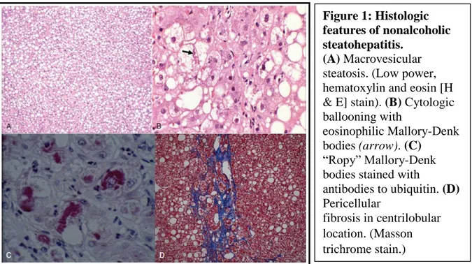
The characteristic histopathologic features of adult NAFLD include mainly zone 3 macrovesicular steatosis variably associated with lobular inflammation, cellular injury represented by cytologic ballooning, Mallory-Denk bodies (MDBs), or both and pericellular fibrosis. Based on these characteristics, NAFLD has two broad histologic patterns: hepatic steatosis, or NAFL, and Steatohepatitis (NASH) (Brunt and others 2011; Brunt and others 2003). At difference to its literal meaning, Steatohepatitis does not simply represent the presence of steatosis and inflammation. Steatohepatitis is defined by the presence of hepatic steatosis with varying degrees of inflammation along with evidence of cell injury, usually in the form of cytologic ballooning (Figure 1) (Charlton and others 2011; Cotrim and others 2004). Fibrosis is not required for the diagnosis of steatohepatitis (Bahrami and others 2003; Brunt and others 2011; Burt and others 1998). The inflammation associated with steatohepatitis is generally modest and has mainly lobular distribution. However, variable degree of portal
inflammation can be detected in specific individuals. Portal fibrosis may be associated with NAFLD, particularly in pediatric subjects and in those who are morbidly obese (Gramlich and others 2004).
2.1 Grading and Staging
To improve the prognostic assessement of NAFLD/NASH several systems for grading and staging NAFLD have been proposed, but only two systems have been validated to any
Figure 1: Histologic features of nonalcoholic steatohepatitis.
(A) Macrovesicular
steatosis. (Low power, hematoxylin and eosin [H & E] stain). (B) Cytologic ballooning with
eosinophilic Mallory-Denk bodies (arrow). (C)
“Ropy” Mallory-Denk bodies stained with
antibodies to ubiquitin. (D) Pericellular
fibrosis in centrilobular location. (Masson trichrome stain.)
6
degree. A landmark study by Brunt and colleagues examined 10 separate histologic parameters. Based on these parameters, a three-grade, four-stage system of classifying NAFLD was developed. Significant histologic lesions included steatosis, ballooning, and inflammation. The necro-inflammatory grade correlated with alanine aminotransferase (ALT) activity. The staging score reflected both location and the extent of fibrosis. More recently, using the Brunt classification as a starting point, the NIH-sponsored NASH CRN proposed and validated a scoring system. Although 14 separate parameters were evaluated, 4 were scored semi-quantitatively, including steatosis (0 to 3), cytologic ballooning (0 to 2), lobular inflammation (0 to 2), and fibrosis (0 to 4). A NAFLD activity score (NAS) was then developed that included the un-weighted scores for steatosis, inflammation, and cytologic ballooning (Table 1). The NAS correlated well with the presence of steatohepatitis during a blinded validation process and was typically associated with a score of 5 or higher. Those with a score of 3 or less were not usually found to have steatohepatitis, whereas a score of 4 was associated with some divergence of opinion (Figure 2). It is, however, important to note that the NAS cannot be used to diagnose the presence of steatohepatitis, which is identified by the presence of steatosis, inflammation, and cytologic ballooning in a typical pattern. The NASH CRN staging system divides stage 1 into several subsets (Table 2), thereby improving its sensitivity to change in earlier stages of the disease. This system is highly valuable as a research tool for the design and analysis of clinical trials related to NASH. However, its role in routine clinical practice remains to be established.
A major limitation of any histologic scoring system is sampling variability. In one study, two biopsy specimens were obtained from the same site at the same time. About 20% of subjects had at least a one-stage variability between specimens, whereas 12% had a two-stage variation. Similar data have been obtained from studies in which biopsy specimens from the left and right lobes were compared.
7
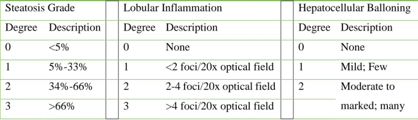
Table 1: NASH Clinical Research Network Scoring System: NAFLD Activity Score
Steatosis Grade Lobular Inflammation Hepatocellular Balloning
Degree Description Degree Description Degree Description
0 <5% 0 None 0 None
1 5%-33% 1 <2 foci/20x optical field 1 Mild; Few
2 34%-66% 2 2-4 foci/20x optical field 2 Moderate to
marked; many
3 >66% 3 >4 foci/20x optical field
Table 2: NASH Clinical Research Network Scoring System: Fibrosis Score Degree Description
0 None
1a Mild (delicate) zone 3 perisinusoidal fibrosis
1b Moderate (dense) zone 3 perisinusoidal fibrosis
1c Portal/periportal fibrosis only
2 Zone 3 perisinusoidal fibrosis with portal/
periportal fibrosis
3 Bridging fibrosis
8
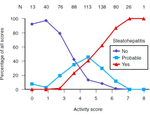
Figure 2: Relationship between the nonalcoholic fatty liver disease activity score (NAS) and the probability of having steatohepatitis.
A group of experienced pathologists diagnosed steatohepatitis to be present, absent, or probable and also independently scored the same biopsy specimens while unaware of their own interpretation to determine the NAS. A score higher than 4 was associated with a high probability of being considered to have steatohepatitis. (Adapted from Kleiner DE, et al.
Design and validation of a histological scoring system for nonalcoholic fatty liver disease. Hepatology 2005;41:1313–1321.)
9
3. Clinical Features
Most subjects with NAFLD are asymptomatic and in these individuals the diagnosis is often made incidentally because of either abnormal liver enzyme levels or features of a fatty liver on an imaging study when such tests are performed for unrelated reasons. In others, NAFLD may be diagnosed either as a result of an unusual appearance of the liver during abdominal surgery or because of persistent hepatomegaly. It is important to recognize that in only a minority of subjects has NAFLD been diagnosed and that it currently remains undiagnosed in the great majority of afflicted individuals.
3.1 Symptoms and Signs
Most patients have vague complaints of fatigue or malaise and a sensation of fullness or discomfort on the right side of the upper part of the abdomen without signs of chronic liver disease at the time of diagnosis. Such symptoms often antedate the diagnosis of NAFLD in a third of patients. Abdominal obesity and hepatomegaly are the most common physical findings. Obesity is present in 50% to 90% of subjects. About two thirds of subjects with NAFLD also have other features of metabolic syndrome. We recently confirmed abdominal obesity as a marker of both steatosis and grade of the disease. In addition, about 28% of subjects had increased dorso-cervical fat, which correlated strongly with histologic severity. Acanthosis nigricans may be found in individuals with NAFLD and is suggestive of an underlying insulin-resistant state.
Hepatomegaly is the most common liver-related physical finding in subjects with NAFLD. A minority of people have stigmata of chronic liver disease such as spider angiomas or palmar erythema. Jaundice and features of portal hypertension, such as ascites and variceal hemorrhage, are the initial findings in a small minority of subjects with advanced liver disease.
3.2 Laboratory Abnormalities
Suspicion for NAFLD is triggered by abnormal results on liver chemistry tests that are usually performed for non–liver related reasons. Approximately 7.9% of the U.S. population has persistently abnormal liver enzymes with negative tests for viral hepatitis and other common causes of liver diseases. The majority of these subjects could have NAFLD if they have risk factors associated with NAFLD, such as the presence of features of metabolic syndrome. It is also important to note that a large number of subjects with NAFLD have persistently normal liver enzyme levels, and the entire histologic spectrum of NAFLD can be
10
seen in such individuals. Mild to moderate elevation in serum aminotransferases (ALT) and aspartate aminotransferase (AST) is the most common and often the only laboratory abnormality found in patients with NAFLD. When these values exceed 300 IU/L, alternative causes of liver disease should be sought carefully. The AST/ALT ratio is usually less than 1 but can be reversed in those with advanced fibrosis or cirrhosis. A mild to modest increase in serum alkaline phosphatase and γ-glutamyltransferase (GGT) can be seen in patients with NAFLD, but the degree of elevation is less than that seen in those with alcoholic hepatitis, as is the case with an increased AST/ALT ratio.
Findings of chronic liver disease together with the presence of hypoalbuminemia, coagulopathy, hyperbilirubinemia, and thrombocytopenia suggest advanced liver disease with probable cirrhosis. Serum albumin and the prothrombin time become abnormal before bilirubin becomes elevated. In diabetic subjects, isolated hypoalbuminemia can result from diabetic nephropathy. Hematologic parameters are usually normal unless cirrhosis and portal hypertension lead to hypersplenism. In fact, a large proportion of patients with cryptogenic cirrhosis share many of the clinical and demographic features of patients with NAFLD, thus suggesting that cryptogenic cirrhosis is unrecognized NAFLD in an advanced stage.
About 30% to 50% of subjects with NAFLD have elevated blood glucose and about 60% also have associated hypertriglyceridemia, low HDL cholesterol, or both. An elevated ferritin level is also often seen in subjects with NAFLD. It is, however, not associated with iron overload in most cases and usually reflects an acute phase response. From a practical point of view, the laboratory evaluation of a subject with suspected NAFLD involves excluding alternative causes of liver enzyme abnormalities, documenting hepatic steatosis, making the distinction between hepatic steatosis and steatohepatitis, assessing the stage of the disease, and evaluating for the presence and severity of IR and other complications of metabolic syndrome. Each of these factors must be considered carefully when making a decision about the aggressiveness with which the answer to each of these is sought. It is important to remember that liver enzymes are notably poor predictors of steatosis and significant fibrosis. Therefore liver biopsy remains the gold standard for diagnosing steatohepatitis and for staging the liver disease, unless clinically evident cirrhosis is present.
4. Pathogenesis
Non-alcoholic fatty liver disease is a complex chronic condition caused by the occurrence of step-wise out of control metabolic, inflammatory and cellular activity. Different
11
factors contributing towards the development of NAFLD/NASH basically involves the influx of free fatty acids (FFA), formation of triglycerides, accumulation of fatty lipids or steatosis, onset of inflammation, mitochondrial dysfunction, oxidative stress and death of mature hepatocytes (Day 2006; Day and James 1998).
4.1 Mechanism of Steatosis
The accumulation of triglycerides originating from the esterification of free fatty acids (FFAs) and glycerol within the hepatocyte is an important aspect of NAFLD/NASH. The contributing factors for the accumulation of FFAs within the liver include enhanced lipolysis in the adipose tissue, dietary sources, and de novo lipogenesis (DNL) (Postic and Girard 2008). Liver has a central role in FFAs metabolisms as hepatocyte utilize FFAs as energy source degrading them in β-oxidation, but a large amount is re-esterificated to triglycerides and exported as very low density lipoprotein (VLDL). Liver VLDL particles are formed through the incorporation of triglyceride into apolipoprotein B (apoB) mediated by microsomal transfer protein (MTP) (Adams and others 2005). Alterations of MTP/apoB synthesis and secretion have been proposed as potential mechanisms underpinning the pathogenesis of NAFLD leading to a decreased capacity for lipid export (Lonardo and Loria 2002). Hence hepatic fat accumulation can occur as a result of increased fat synthesis, increased fat delivery, decreased fat export, and/or decreased fat oxidation (Postic and Girard 2008).
Lipid metabolism in the liver and adipose tissue is controlled by insulin-mediated signals. In healthy individuals, the binding of insulin to its receptor leads to phosphorylation of several substrates including insulin receptor substrates (IRS)-1, -2, -3 and -4, which propagate the insulin signal. Insulin stimulation of IRS-1 and -2 leads to activation of intracellular PI3K (phosphoinositide 3-kinase) and AKT/PKB (protein kinase B) pathways, which are intimately involved in mediating the metabolic effects of insulin (Bugianesi and others 2010). Ultimately, AKT/PKB activation results in translocation of glucose transporter, GLUT4 to the plasma membrane, thus facilitating glucose uptake. In addition, the expression of key lipogenic genes is increased, with a concomitant decrease in gluconeogenic gene expression via its regulation of forkhead (FOXO) transcription factor activity. Insulin has also a potent action in suppressing adipose tissue lipolysis.
Obesity and metabolic syndrome are characterized by an impaired transduction of insulin signals in peripheral tissues leading to a situation known as insulin resistance (IR). In situations of IR insulin-mediated suppression of lipolysis is impaired resulting in an increased
12
efflux of FFA from the adipose tissue. Hyper-insulinaemia associated with IR leads also to an up-regulation of the transcription factor sterol regulatory element binding protein-1c (SREBP-1c), which is a key transcriptional regulator of genes involved in DNL as well as to the
inhibition of β-oxidation of FFA thus further promoting hepatic lipid accumulation (Chitturi
and others 2002; Choudhury and Sanyal 2004). Furthermore, in NAFLD additional factors can interfere with insulin signalling cascade, and thus contribute to hepatic IR. These factors include tumour necrosis factor-alpha (TNF-a) signals and jun N-terminal kinase 1 (JNK1) and SOCS (suppressors of cytokine signalling) activation. Increased lipid metabolites such as diacylglycerol (DAG) have also been implicated in interfering with insulin signalling through the modulation of IRS-2 phosphorylation mediated by a protein kinase Ce (PKCe) (Capeau 2008; Choudhury and Sanyal 2005; Cusi 2009).
During the progression of NAFLD the worsening of steatosis is tightly associated with chronic hepatic inflammation, an effect in part mediated by activation of the Ikk-b/NF-kB signalling pathway. In murine models of high-fat diet (HFD)-induced steatosis, increased NF-kB activity is associated with elevated hepatic expression of inflammatory cytokines such as TNF-a, interleukin-6 (IL-6) and interleukin 1-beta (IL-1b), as well as with the activation of Kupffer cells. Accordingly, both serum and hepatic levels of TNF-α are elevated in patients with NASH, and levels correlate with histological severity. In addition to its proinflammatory effects, TNF-α promotes IR. Conversely, inhibition of TNF-α signalling improves IR and histological parameters of NASH. Similarly, serum IL-6 levels are also elevated in both animal and human models of IR and NAFLD, and levels correlate with increasing liver inflammation and fibrosis. Liver-specific NF-kB inhibition prevents HFD-induced inflammatory gene expression, whereas HFD-induced hyperglycaemia and IR can be reproduced by selective over-expression of constitutively active Ikk-b in hepatocytes. The Ikk-b/NF-kB pathway in hepatocytes can also be activated directly by FFA, providing a further mechanism by which central obesity with consequent increased hepatic FFA supply can contribute to inflammation. (Armutcu and others 2013; Baeck and others 2012; Hui and others 2004).
A further aspect in the mechanisms leading to hepatic steatosis directly involves the changes occurring in the adipose tissue. Adipose tissue is not just an inert site of energy storage, but an actively secreting endocrine organ. The functional role of adipocyte-derived cytokines (adipokines), is now increasingly recognized, with leptin and adiponectin amongst the best described. Leptin is a 16 kDa hormone produced mainly by mature adipocytes whose actions
13
include the regulation of energy intake and expenditure, regulation of the immune system, and promotion of inflammation and fibrogenesis. Conversely, adiponectin has anti-inflammatory activity and increases insulin sensitivity. In obesity adipocytes that accumulate triglycerides modify their pattern of adipokine secretion leading to higher leptin secretion at the expensed of adiponectin production (Marra and others 2005; Musso and others 2005). In fact, the circulating levels of adiponectin are inversely proportional to body fat content and are reduced in patients with NAFLD. Adiponectin antagonises the effects of TNF-α, which itself suppresses adiponectin production (Takei and Sato 2006). The importance of adiponectin in NAFLD is supported by studies showing that serum adiponectin levels can help to distinguish NASH from simple steatosis. Other adipose tissue derived factors found in excess in NAFLD include TNF-α, IL-6, angiotensinogen and resistin, all of which antagonise the lipogenic effects of insulin, but their precise role in the pathogenesis of NAFLD remains to be determined (Mirza 2011; Orlik and others 2010; Polyzos and others 2013).
4.2 Mechanism of Hepatocyte Injury
As previously mentioned, nonalcoholic steatohepatitis (NASH) is characterized by parenchymal injury involving hepatocyte ballooning, presence of Mallory-Denk bodies and extensive liver cell apoptosis (Duwaerts and Maher 2014). Accordingly, serum levels of of caspase-cleaved cytokeratins (CK) 8 and 18 have been recently proposed as specific markers
of hepatocyte death in NASH (Eguchi and others 2014). Several mechanisms have been
invoked to explain the proapoptotic state in NASH. Triglyceride accumulation itself increases apoptosis by the interaction of unoxidized palmitoyl CoA with serine to form dihydro-sphingosine, a precursor of ceramide. Ceramide is a potent inducer of apoptosis via an inducible nitric oxide synthetase (iNOS)-mediated pathway that requires the transcriptional factor NF-κB (Harbrecht and others 2012) (Alexander 1998; Harbrecht and others 2012; Ou and others 1997; Pinto and others 2000). It has also been shown that hepatocyte incapability to esterify the excess of FFAs secondary to increased peripheral lipolysis in the insulin-resistant state triggers apoptosis through a process known as produce lipotoxicity. Several mechanisms account for lipotoxicity. FFAs can directly induce translocation of Bax to lysosomes, where it causes the release of cathepsin. Cathepsin, acting via NF-κB, induces TNF-α and activation of TNF receptor–associated death pathways. Recently, the potential role of ER stress in the development of hepatocyte lipotoxicity in NASH has gained interest. The ER is the principal site for synthetic activity within cells. Such activity requires not only appropriate synthesis but
14
also local mechanisms to ensure that the proteins are correctly folded because this is essential for their recognition by appropriate receptors and trafficking to their final destination. Under conditions in which there is increased protein or lipid synthetic activity in the ER, depletion of ATP, depletion of calcium, or altered glucose homeostasis, the regulatory function of the ER in maintaining normal synthetic function is disrupted. This leads to the activation of an intracellular program called the unfolded protein response (UPR) (Henkel and Green 2013; Kapoor and Sanyal 2009; Malhi and Kaufman 2011; Zhang and others 2012a; Zheng and others 2011). Activation of the UPR initially leads to an adaptive response in which protein synthesis decreases and allows restoration of normal ER function. However, if the initiating factors are not corrected, alarm pathways are activated, including activation of a number of stress kinases, which eventually results in activation of homologous protein (CHOP), a potent apoptosis-inducing factor. Recent studies have reported that patients with NAFLD have a variable degree of UPR activation. Inositol requiring enzyme-1 (IRE-1) activation appears to play an important role in the genesis of cell injury in NASH via activation of JNK phosphorylation. Interestingly, there seems to be a close association of IRE-1 activation with the histologic activity of the disease. Failure to generate ER degradation-enhancing α-mannosidase like protein (EDEM) in response to spliced X box–binding protein (sXBP) in some subjects raises the possibility that patients with the lowest EDEM levels are at particular risk of progressing to cirrhosis because of insufficient degradation of unfolded proteins, thus perpetuating the ER stress. Despite increased phosphorylated eIF-2a, patients with NASH are apparently unable to up-regulate activating transcription factor 4 (ATF4), CHOP, and growth arrest and DNA damage- 34 (GADD34), which contributes to the failure to recover from ER stress (Cao and others 2012; Fang and others 2013; Lee and others 2012; Pagliassotti 2012; Zhang and others 2012b). Thus NASH is specifically associated with failure to generate sXBP1 and activation of c-jun N-terminal kinase (JNK) (Malhi and others 2006). Accordingly, JNK activation is evident in liver biopsies from NASH patients and pharmacological or genetic JNK inhibition prevents lipotoxicity “in vitro” and ameliorates steatohepatitis in rodent models of NASH (Cazanave and Gores 2010; Czaja 2010). Interestingly, the antiapoptotic B-cell lymphoma-2 (BCL2) protein appears to be strongly expressed in human steatohepatitis, probably representing an adaptive response. Thus, on the basis of the recognized significance of hepatocyte apoptosis in the pathogenesis of NAFLD, it has been proposed that the development of progressive NAFLD in some patients but not in others may be the result of increased susceptibility of steatotic hepatocytes to apoptosis arising from abnormal regulation of BCL2 proteins, alteration in JNK
15
activation, or preferential activation of ER stress (Li and others 2014; Malhi and others 2006; Panasiuk and others 2006).
Additional mechanisms of hepatocyte injury in NAFLD/NASH involve the effect of inflammatory cytokines and particularly TNF-α and oxidative stress. The key role of cytokine production in the progression of steatosis to NASH is supported by studies demonstrating that cytokines can replicate all of the histological features associated with NASH, including hepatocyte apoptosis/necrosis and Mallory body formation (Marra and others 2008).
4.3 Oxidative Stress
Oxidative stress is a result of an imbalance between prooxidant and antioxidant species. This could be due to either increased production of prooxidants reactive oxygen species (ROS) or reactive nitrogen species (RNS) or decreased antioxidant defenses Potential sources of ROS in the liver include the mitochondria, the peroxisomes, the microsomal oxidative system, and iron overload. (Alkhouri and McCullough 2012; Basaranoglu and others 2013; Baskol and others 2007). The mechanisms responsible for oxidative stress in NAFLD/NASH have been characterized to some extent showing that FFA and cholesterol accumulation within the mitochondria along with TNF-α cause mitochondrial dysfunction. These mitochondrial structural defects associated with impaired mitochondrial respiratory chain activity produce a state of uncoupled oxidation and phosphorylation that leads to increased ROS production. This concept is further supported by evidence of decreased ATP formation in the liver of subjects with NASH (Begriche and others 2006; Gambino and others 2011). Another possible source of ROS can be the cytochrome P-450 system. This system, particularly CYP2E1, is over-expressed in subjects with NAFLD along with cytochrome P4502E1 activity (Lieber 2004). CYP2E1 can be induced as a result of insulin-resistance as well as to cope with the increase in FFAs (Lieber 2004). In line with these findings, CYP2E1 deletion in mice results in less susceptibility to high fat diet induced NAFLD/NASH as well as in lower hepatic oxidative stress (Abdelmegeed and others 2012).
A growing body of evidence from the experimental models of NAFLD/NASH suggests that oxidative stress plays a key role in the mechanisms causing the death of fat-laden hepatocytes as well as contributes to the activation of hepatic stellate cells to matrix-producing myofibroblasts (Gambino and others 2011). Accordingly, antioxidant supplementation reduces liver injury in experimental rodent models of NASH (Laurent and other 2004). The relevance of these observations to humans is supported by several studies showing an increase in
16
oxidative stress markers, in the liver and in the serum of both adult paediatric patients with NAFLD/NASH (Chalasani and others 2004; Ikura and others 2006; Seki and others 2002). Furthermore, as compared to normal livers, liver biopsies from NASH patients display a lower mRNAs expression of different antioxidant enzymes (Sreekumar and others 2003). On the same vein, recent evidences indicates that antioxidant treatments might be effective in improving hepatic damage in NASH (Pacana and Sanyal 2012).
4.4 Mechanisms of Inflammation
Inflammation, along with hepatocyte damage, is the main feature of the progression from simple steatosis to NASH. In fact, the molecular mechanisms able to promote inflammation cross-talk with those responsible for hepatocellular damage and fibrosis. The precise mechanisms of inflammation in NASH have not yet been completely elucidated, but current evidence indicates both the innate and adaptive immunity have a role in the initiation and maintenance of lobular inflammation. The innate immune system is an important factor in the promotion and progression NASH. Hepatic infiltration of innate immune cells such as macrophages, granulocytes and natural killer (NK) has been commonly observed in early stages of the disease and they are considered as driving force of NASH progression.
The hepatic macrophage population is named after the scientist Karl Wilhelm von Kupffer. Kupffer cells are located predominantly in the periportal area. They originate from bone marrow and in healthy livers Kupffer cells are responsible for the phagocytosis of particulate matters, presentation of antigen accompanied by immune regulation, and the release of soluble mediators. Alike other macrophages, Kupffer cells and hepatic monocyte-derived macrophages are a highly plastic populations that may adopt various phenotypes ranging between the extreme states known as M1 and M2. Inflammation driven by M1 Kupffer cells is counterbalanced by alternatively-polarized M2 macrophages that produce anti-inflammatory cytokines such as IL-10, and promote resolution of inflammation (Murray and Wynn 2011; Sica and Mantovani 2012). Macrophage polarization into an M2 phenotype is promoted by Th2-derived cytokines (IL-4, IL-13) (Murray and Wynn 2011; Sica and Mantovani 2012), that may also originate from hepatocytes (Kang and others 2008), while such factors promoting M2 Kupffer cell polarization are poorly characterized.
Increasing evidence suggests that Kupffer cells critically contribute to the progression of NAFLD. In fact at the onset of NASH lipid accumulation in Kupffer cells significantly contribute to the production of pro-inflammatory cyto/chemochines, which, in turn, stimulate
17
the liver infiltration by circulating monocytes (Tosello-Trampont and others 2012; Leroux and others 2012). Hepatic monocyte infiltration is primarily promoted by CCL2, a chemokine up-regulated in the serum and in the liver of patients with NASH (Haukeland and others 2006), that drives the recruitment of inflammatory Ly6C-positive monocytes through the interaction with C–C chemokine receptor 2 (CCR2). According, pharmacological inhibition of genetic deficiency of CCL2/CCR2 dyad reduces macrophage infiltration and ameliorated steatohepatitis in experimental mice model of NASH (Baeck and others 2012; Miura and others 2012). However, variable results have been obtained in mice with different genetic backgrounds (Galastri and others 2012). Other chemokines contributing to monocyte recruitment include CCL5 (RANTES) as interference with CCL5 functions ameliorates experimental NASH (Berres and others 2010).
Upon liver infiltration monocytes rapidly differentiates to M1 polarized macrophages and the extent of macrophage M1 responses appears to modulate NASH severity among different mice strains (Maina and others 2012). Furthermore, macrophage release of IL-15 and CXCL16 is important for stimulating the recruitment and the survival of T-lymphocytes and NKT cells (Locatelli and others 2013; Wehr and other 2013). Pro-inflammatory cytokines released from activated Kupffer cells also activate hepatic sinusoidal endothelial cells to upregulate adhesion molecules (ICAM1, VCAM-1) and in combination with the chemokines secreted from macrophages stimulate the recruitment of neutrophils to the liver (Tosello-Trampont and others 2012). Neutrophils, in turn secrete reactive oxygen species (ROS), oxidants, defensins, as well as chemokines to attract more neutrophils and monocytes (Rensen and others 2012). Following their recruitment to the liver macrophages release not only pro-inflammatory cytokines but also growth factors as G-CSF, and GM-CSF that can extend the lifespan of neutrophils thus sustaining their presence at the site of inflammation (Rensen and others 2012). The production of ROS, NO and cytokines by neutrophils and macrophages significantly contribute to promote hepatocyte cytotoxicity.
Lymphocytes are commonly found throughout the parenchyma and in the portal tracts of healthy livers and include T and B cells subpopulations as well as other subsets belonging to innate immunity as natural killer (NK) and natural killer T-cells (NKT) that shares features in common with both NK and T cells. NKT cells are particularly frequent in the liver because these cells express a specific chemokine receptor (CXCR6) that interacts with the chemokine CXCL16 abundantly produced by hepatic sinusoidal endothelia (Geissmann and others 2005; Swain 2010). Although the total number of hepatic CD3+ T lymphocytes is not appreciably
18
modified in NASH, an imbalance between CD8+/CD4+ CD3+ T-cell subtypes has been observed (Ferreyra Solari and coworkers 2012). CD4+ T helper cells are a sub-group of lymphocytes that are capable of switching B cell to antibody production, activating cytotoxic T cells and contributing to the phagocyte functions. Recent reports have shown that an increase in circulating IFN-γ-producing CD4+ T-cells characterizes NASH in both paediatric and adult patients in conjunction with an enhanced liver IFN-γ expression (Ferreyra Solari and coworkers 2012; Inzaugarat and others 2011), suggesting the possible relevance of Th-1 responses to the human disease. Furthermore, a higher number of Th-17 cells has been observed in mice with steatosis induced by feeding a high fat diet and in liver biopsies of NASH patients. Accordingly, the Th-17 related genes (ROR-γT, IL-17, IL-21, IL-23) are up-regulated in NASH patients as compared to healthy controls and neutralization of IL-17 in high fat diet fed mice ameliorates liver injury and inflammation (Tank and other 2011). In spite IL-17 has been involved in the pathogenesis of hepatic fibrosis (Meng and others 2012), the actual role of DC4+ Th-17 T cells in the pathogenesis of NASH is still poorly characterized.
Conversely, recent studies have pointed out a possible involvement of natural killer T (NKT) cells in the progression of NASH. NKT cells account for 20-35% of mouse and 10-15% of rat and human liver lymphocytes. They recognize lipid antigens presented by the non-classical MHC class I-like molecule CD1 and can be directly cytotoxic by Fas ligand (FasL), or perforin/granzyme-dependent mechanisms in addition to regulate innate and adaptive immunity through the production of IFN-γ and IL-4 (Seino and Taniguchi 2005; Exley and Koziel 2004). Beside classical cytokines, NKT cells also secrete osteopontin (OPN) (Syn and others 2012), a cytokine with both pro-inflammatory and pro-fibrogenic capacities and the fetal morphogen, sonic hedgehog (Shh), which activates hepatic stellate cells (HSC) into collagen secreting myofibroblasts and amplifies the repair-associated inflammatory response (Uede 2011; Gao and Radaeva 2013; Syn and others 2012). The role of NKT cells during the progression from NAFLD to NASH is complex as the development of steatosis in mice receiving a high fat diet is associated with a reduction of hepatic NKT cells. This reduction is due to a lowering of hepatic CD1d expression and an increase of NKT apoptosis due to an impaired production of IL-15 in a combination with an up-regulation in hepatic expression of IL-12 (Li and other 2005; Yang and other 2007). Indeed, Kupffer cell depletion lowers IL-12 expression and blocks the impairment of NKT cells (Li and other 2005; Kremer and others 2010). On the contrary, in mice with advanced NASH there is an increase in the number of NKT cells, which parallels the progression of the disease to fibrosis. NKT cell expansion
19
involves IL-15 production and is associated with an increase in liver IFN-γ and OPN production (Locatelli and others 2012). Accordingly, induction of NASH in NKT cell deficient mice is characterized by a blunted OPN expression and by an improvement in liver injury and collagen deposition (Syn and others 2010). In the setting of human NASH, advanced fibrosis is correlated with increased hepatic levels of OPN and Hh and elevated plasma OPN levels in comparison with early fibrosis (Syn and others 2012; Tajiri and other 2009).
4.5 Mechanisms of Fibrosis.
In many chronic liver diseases unresolved inflammation promotes pathologic repair leading to progressive fibrosis and cirrhosis (Friedman 2008). As mentioned above hepatic cirrhosis represent the final outcomes of NASH. In fact, within 8 years, 15% of NASH patients develop clinically and histologically evident cirrhosis. Death rate ascribed to NASH-related cirrhosis accounts for 12-25%, while end-stage NASH is responsible for about 4-10% of liver transplants (Neuschwander-Tetri and Cadwell 2003). This makes NASH an increasingly important cause of liver cirrhosis. NASH-related fibrosis develops primarily in the pericentral areas, where thin bundles of fibrotic tissue surround groups of hepatocytes and thicken the space of Disse, in a “chicken wire” fashion (Brunt 2010). The main cell type responsible for extracellular matrix deposition are hepatic stellate cells (HSCs), These cells respond to the
production of transforming growth factor 1 (TGF-1), platelet-derived growth factor (PDGF)
and fibroblast-derived growth factors produced by macrophages trans-differentiating into myofibroblast-like cells (HSC/MSs) that are responsible for the secretion of collagen and extracellular matrix components (Friedman 2008). Furthermore, decreased hepatic matrix degradation due to a reduced production of matrix metalloproteases (MMPs) and/or an increased production of matrix metalloprotease inhibitors might also contribute to collagen accumulation (Friedman 2008). Macrophage activation in response to chronic inflammatory stimuli is mostly responsible for the secretion of pro-fibrogenic cytokines (Friedman 2008). In addition, HSC proliferation and transformation to collagen-producing myofibroblasts is influenced by lymphocyte-derived cytokines and oxidative stress (Novo and others 2008). Although the development of fibrosis in NASH does not appears to be appreciably different from those in other liver diseases, alterations in adipokine secretion consequent to obesity might have a specific role for the induction of fibrogenesis in this condition. Activated HSCs selectively express leptin receptors and leptin stimulates HSC survival, the expression of pro-inflammatory and angiogenic cytokines (Wang and others 2008). The pro-fibrogenic action of
20
leptin might be enhanced by the combined lowering of adiponectin as adiponectin reduces proliferation and increases apoptosis of cultured HSC (Wang and others 2008).
5. Aims of the work
In spite of the growing number of studies investigating the pathogenesis of NAFLD/NASH, a number of issues concerning the mechanisms involved in promoting lobular inflammation and the evolution of NASH to fibrosis/cirrhosis are still unresolved. In particular, it is still unclear why only some NAFLD patients develop steatohepatitis. Furthermore, among the subjects with NASH there is a large inter-individual variability in the evolution to fibrosis. The factors responsible for such an inter-individual variability represent a particular challenge as multiple interaction occur between hepatocytes, inflammatory cells and hepatic stellate cells in the different phases of the disease progression.
In my doctoral project, I have addressed some of these issues by investigating the possible involvement of adaptive immune mechanisms in the evolution of NASH and characterizing morphological and functional modifications occurring in monocyte-derived cells during the disease progression.
21
6. Paper 1
Adaptive Immune Responses Triggered by Oxidative Stress Contribute to Hepatic Inflammation in NASH
Published on Hepatology 2014;59:886-897.
Background and Aims: The mechanisms responsible for the progression of simple steatosis to
steatohepatitis (NASH) are still incompletely characterized. Oxidative stress is one of the features of NAFLD/NASH and hepatic oxidative stress markers, such as 4-hydroxynonenal (4-HNE) and 8-hydroxydeoxyguanosine, correlate with the severity of necro-inflammation and fibrosis, suggesting that oxidative injury might be involved in triggering steatohepatitis. Studies in atherosclerosis have evidenced that lipid peroxidation products originating from oxidized LDL generate a variety of neo-antigens able to stimulate the innate immune system, which in turn promotes inflammation in atheroma. As previous data indicate that both adult and children with NAFLD/NASH have increased titres of IgG against oxidative stress derived antigens, in in the present study we have used the methionine-choline deficient (MCD) diet model of steatohepatitis to get insides in the involvement of immune responses in promoting hepatic inflammation in NASH.
Key results: In MCD diet fed mice, the development of liver injury and lobular inflammation
paralleled with the presence IgG against malonildialdehyde (MDA) and 4-HNE-derived antigens. Moreover, the hepatic recruitment of CD4+ and CD8+ T-lymphocytes responsive to the same antigens was also observed. Mice immunization with MDA-adducted bovine serum albumin (MDA-BSA) before feeding the MCD diet stimulated transaminase release, lobular inflammation, and the hepatic expression of proinflammatory cytokines. Hepatic inflammation was accompanied by a stimulation in the liver recruitment and Th-1 activation of CD4+ T cells
that further stimulated macrophage M1 responses. In this setting, depleting CD4+ T-cells in
MCD-fed immunized mice by using an anti-CD4 monoclonal IgG significantly lowered lobular inflammation and focal necrosis.
Conclusions: These results indicate that adaptive immune responses triggered by oxidative
stress-derived antigens contribute to hepatic inflammation in experimental NASH by promoting Th-1 responses by CD4+ T-lymphocytes.
22
ADAPTIVE IMMUNE RESPONSES TRIGGERED BY OXIDATIVE STRESS CONTRIBUTE TO HEPATIC INFLAMMATION IN NASH
Salvatore Sutti, Aastha Jindal, Irene Locatelli, Marco Vacchiano, Luca Gigliotti, Cristina Bozzola, Emanuele Albano.
Dept. of Health Sciences and Interdisciplinary Research Centre for Autoimmune Diseases, University “Amedeo Avogadro” of East Piedmont,
Via Solaroli 17, 28100 Novara.
Running title: Adaptive immunity in mice NASH.
Keywords: Nonalcoholic fatty liver disease, lipid peroxidation, NKT cells, Th-1 responses, antibodies.
Words: 4723 References: 49
Note: The authors have no conflict of interest
Corresponding Author:
Prof. Emanuele Albano, Department of Health Science, University “Amedeo Avogadro” of East Piedmont, Via Solaroli 17, 28100 Novara, Italy.
23
Abstract
Previous studies have shown that human non-alcoholic steatohepatitis (NASH) is often associated with the presence of circulating antibodies against proteins adducted by lipid peroxidation products. Here we used the methionine-choline deficient (MCD) model of NASH to characterize the possible involvement of adaptive immunity in NASH.
In mice fed with the MCD diet up to 8 weeks liver injury and lobular inflammation worsen in a time-dependent manner in parallel with the development of IgG against malonyldialdehyde
(MDA) and 4-hydroxynonenal (4-HNE)-derived antigens and the hepatic recruitment of CD4+
and CD8+ T-lymphocytes that were responsive to the same antigens. Moreover, in these
animals the individual IgG reactivity against MDA-adducts positively correlated with transaminase release and TNF-α expression. To substantiate the role of immune responses triggered by oxidative stress in the progression of NASH, mice were immunized with MDA-adducted bovine serum albumin (MDA-BSA) before feeding the MCD diet. MDA-BSA immunization did not affect the livers of control mice, but further stimulated transaminase release, lobular inflammation and the hepatic expression of pro-inflammatory cytokine in MCD-fed mice. The increased severity of NASH in immunized MCD-fed mice was associated with the liver recruitment and the Th-1 activation of CD4+ T-cells that accounted for an increased M1 activation of hepatic macrophages. Moreover, hepatic fibrosis was also evident in these animals in concomitance with an IL-15-mediated increase of hepatic natural killer T-cells (NKT) and the up-regulation in liver osteopontin production by NKT T-cells and hepatic macrophage.
Conclusions: These results indicate that oxidative stress can contribute to the progression of NASH by stimulating both humoral and cellular responses, pointing to the possible contribution of adaptive immunity to the pathogenesis of the disease.
Abbeviations:
4-HNE 4-hydroxynonenal; MCD methionine-choline deficient; MDA malonildialdehyde; NAFLD NonAlcoholic Fatty Liver Disease; NASH NonAlcoholic SteatoHepatitis; NK natural killer; NKT natural killer T cells:OPN osteopontin
24
Introduction
A key issue in understanding the pathogenesis non-alcoholic fatty liver disease (NAFLD) concerns the identification of the mechanisms responsible for switching from simple steatosis to steatohepatitis (NASH). This aspect is clinically relevant because steatosis does not appear to adversely affect the long-term outcome of NAFLD [1], whereas parenchimal injury and inflammation are the driving forces for the disease evolution to fibrosis/cirrhosis [2,3]. Oxidative stress is one of the features of NAFLD/NASH [4] and oxidative stress markers, such as the hepatic content of 4-hydroxynonenal (4-HNE) and 8-hydroxydeoxyguanosine, correlate with the severity of necro-inflammation and fibrosis [5,6], suggesting that oxidative injury might be involved in triggering steatohepatitis. In this scenario, recent evidence indicates that lipid peroxidation products originating from phospholipid oxidation can act as damage-associated molecular patterns (DAMPs) and promote inflammation through the interaction with both soluble and cell-associated pattern recognition receptors [7,8]. A further mechanism by which oxidative stress can stimulate inflammation involves adaptive immunity. Indeed, in atherosclerosis as well as in several auto-immune diseases the interaction of lipid peroxidation products with cellular proteins leads to the formation of immunogenic adducts that induce both humoral and cellular immune responses [9,10].
Previous studies from our laboratory have shown that high titres of IgG against some of the antigens originating from oxidative stress, namely malondialdehyde- (MDA) derived adducts, are detectable in about 40% of adult NAFLD/NASH patients and in 60% of children with NASH [11,12]. In these latter, high antibody titres associated with more severe lobular inflammation and 13 fold increased risk of a NAFLD Activity Score ≥5 [12], while in adults anti-MDA IgG are an independent predictor of fibrosis [11]. From this background, we sought to investigate the possible contribution of immune reactions triggered by oxidative stress in modulating hepatic inflammation in NASH. For the experiments we relayed on a rodent model of NASH based on mice feeding with a methionine-choline deficient (MCD) diet that, despite it does not reproduce key features of human NAFLD/NASH such as obesity and insulin resistance, causes well evident oxidative stress and a rapidly progressing steato-hepatitis [13].
Material and Methods
Animal and Experimental protocol. Eight weeks old male C57BL/6 mice were purchased
from Harlan-Nossan (Corezzana, Italy) and fed for 4 or 8 weeks with either methionine-choline deficient (MCD) or control diets (Laboratorio Dottori Piccioni, Gessate, Italy). For
25
immunization experiments mice were injected subcutaneously with 100μg of MDA-adducted bovine serum albumin (MDA-BSA) in incomplete Freud’s adjuvant and re-boosted after one week with the same antigen. The control groups received either saline or incomplete Freud’s adjuvant injections. MCD diet feeding was started 2 weeks after the second injection. In some experiments immunized mice were treated with 400μg of the anti-CD4 monoclonal antibody GK1.5 (BioXCell, West Lebanon, NH, USA) while receiving the MCD diet (See
supplementary materials for further details) to deplete hepatic CD4+ T cells. The efficiency of
cell depletion was preliminary evaluated by flow cytometry and was >97% in both the liver and the spleen. All the experiments were approved by the Italian Ministry of Health and by the University Commission for Animal Care following the criteria of the Italian National Research Council.
Antigen preparation and antibody measurement. Protein adducts with lipid peroxidation
products were prepared as in [12,14] and used to coat polystyrene microwell ELISA plates (Nunc, S/A, Roskilde, Denmark). Mouse sera (0.20 ml, 1:50 dilution) were added in duplicate and the antibody binding was revealed using peroxidase-linked goat anti-mouse IgG or IgM sera as previously described [12,14]. The results were expressed as optical density following the subtraction of background reactivity.
mRNA extraction and Real time PCR. Liver RNA was retro-transcripted with High Capacity
cDNA Reverse Transcription Kit (Applied Biosystems Italia, Monza, Italy). RT-PCR was performed in a Techne TC-312 termalcycler (TecneInc, Burlington NJ, USA) using TaqMan Gene Expression Master Mix and TaqMan Gene Expression probes for mouse TNF-α, IL-12p40, IL-17a, IFN-γ CCL2, iNOS, CD40, CD40L, osteopontin, α1-procollagen, T-bet, RORγT and beta-actin (Applied Biosystems Italia, Monza, Italy). All samples were run in
duplicate and the relative gene expression calculated as 2-ΔCt was expressed as fold increase
over control samples.
Histology and immunohistochemistry. Steatosis and lobular inflammation were scored blind
according to Kleiner et al. [15] in hematoxilin/eosin stained sections. Hepatocyte apoptosis was detected by terminal deoxyribonucleotide transferase (TdT)-mediated dUTP nick-end labeling (TUNEL) using Apoptags Kit (Intergen Company, New York, USA). Liver infiltrating T- and B-cells were evidenced in frozen sections using, respectively, anti-mouse CD3, anti-mouse B220 rat monoclonal antibodies (R&D System Europe Ltd, Abingdon, UK) and a horse-radish peroxidase polymer kit (Biocare Medical, Concord, CA, USA). Polyclonal
26
antibodies against α- -SMA) (Labvision, Bio-Optica, Milan, Italy) were
used to detect activated hepatic stellate cells in formalin-fixed sections.
Intrahepatic lymphocyte isolation and flow cytometry analysis. Hepatic mononucleated cells
were isolated and purified on a density gradient as in [16]. The cells were stained with fluorochrome-conjugated antibodies for CD45, CD3, CD4, CD8, NK1.1, F4/80, CD69, CD107a, IL-2 and IFN-γ (eBiosciences, San Diego CA, USA) and analyzed with a FACScalibur (Becton Dikinson) flow cytometer. De-complemented mouse serum was used to block unspecific immunoglobulin binding. A polyclonal anti-osteopontin rabbit antiserum (Millipore, Temecula, CA, USA) and phycoerythrin-conjugated anti-rabbit IgG (Sigma-Aldrich, Milan, Italy) were used for detecting ostepontin producing cells. Intrahepatic lymphocyte response to MDA adducts was investigated by flow cytometry analysis of IL-2 production following overnight incubation with 10µg/mL MDA adducted or native murine albumin in the presence of brefeldin A (3µg/ml), anti-CD3e and CD28 antibodies (1µg/ml) according to [17].
Additional methods are described in the supplementary materials.
Data analysis and statistical calculations. Statistical analyses were performed by SPSS
statistical software (SPSS Inc. Chicago IL, USA) using one-way ANOVA test with Tukey’s correction for multiple comparisons or Kruskal-Wallis test for non-parametric values. Significance was taken at the 5% level. Normality distribution was preliminary assessed by the Kolmogorov-Smirnov.
Results
Immune responses against oxidative stress related-antigens associates with NASH progression.
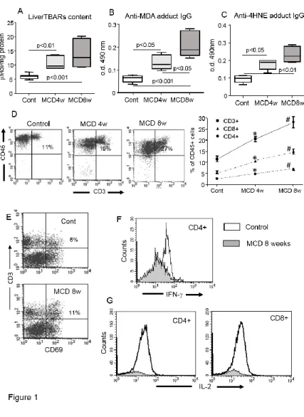
NASH induced by feeding mice with a methionine/choline deficient (MCD) diet is characterized by a time-dependent progression of liver injury (13). Accordingly, in C57BL/6 mice receiving the MCD diet for up to eight weeks we observed a progressive increase in hepatic triglyceride content, transaminase release and circulating TNF-α levels that paralleled with the histological severity of hepatic inflammation (Supplementary figure 1). Liver oxidative stress, as measured by thiobarbituric acid reactive compounds (TBARs) was also well evident at an early time point in the animals with NASH (Fig.1). As observed in humans, oxidative stress in the mice with NASH associated with the development of IgG against adducts originating from lipid peroxidation products such as malonyldialdehyde (MDA) and
27
4-hydroxynonenal (4-HNE) and the IgG titres increased in parallel with the disease progression (Fig. 1). Moreover, in these animals the individual IgG reactivity against MDA-adducts, but not liver TBARs, positively correlated with ALT and TNF-α mRNA values (r=0.61, p=0.04; r=0.66, p=0.03, respectively). Immunohistochemistry revealed that the hepatic inflammatory infiltrates were also enriched by T- and B-lymphocytes (Supplementary figure 1), the number of which positively correlated with the individual IgG reactivity against MDA-adducts (r=0.68, p=0.02; r=0.75, p=0.006, respectively). Flow cytometry analysis of hepatic mononucleated cells from control and NASH livers confirmed a progressive recruitment of T-lymphocytes
that involved both effector CD8+ T-cells and CD4+ helper T-cells (Fig. 1). Furthermore, the
proportion of CD3+ T-cells expressing the CD69 activation marker was increased in the livers
MCD-fed mice as compared to controls (Fig. 1). Intra-hepatic CD4+ T-cells from mice with
NASH also showed an enhanced interferon-γ (IFN-γ) expression (Fig.1), suggesting that lipid peroxidation-derived antigens might contribute to the development of cell-mediated immune responses. Supporting this view, we observed that CD8+ and CD4+ T-cells obtained from NASH, but not from healthy livers, produce IL-2 when incubated “in vitro” with MDA-modified murine albumin (Fig. 1).
The induction of immunity against MDA-adducts enhances NASH severity in mice.
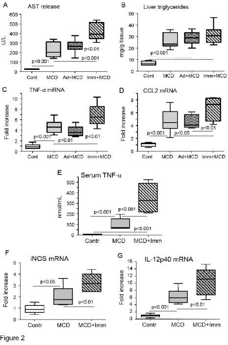
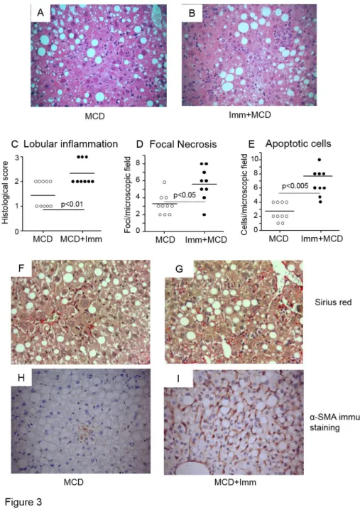
To substantiate the possible role of adaptive immunity triggered by lipid peroxidation-derived antigens in promoting hepatic inflammation in NASH, we induced immune reactions against MDA adducts by immunizing C57BL/6 mice with MDA-modified bovine serum albumin (MDA-BSA) in incomplete Freund’s adjuvant before the administration of the MCD diet. In preliminary experiments, this immunization protocol led to appreciable humoral and cellular reactivity against MDA adducts (not shown). In the animals receiving the control diet MDA-BSA immunization did not affect liver histology and ALT release neither significantly modified the hepatic expression of pro-inflammatory markers (Supplementary Fig. 2). However, following four weeks on the MCD diet ALT release and the hepatic mRNA expression of the inflammatory mediators, TNF-α and CCL2, were higher in MDA-BSA-immunized than in naïve mice (Fig. 2). No appreciable changes in liver injury and inflammation were observed in mice injected with incomplete Freund’s adjuvant before receiving the MCD diet (Fig 2). The enhanced severity of NASH was further supported by histology that showed higher scores for lobular inflammation and increased frequency of necro-inflammatory foci and apoptotic cells in MCD-fed immunized mice (Fig. 3).
28
Furthermore, circulating TNF-α levels were three fold higher in these latters as compared to similarly treated naïve mice (Fig. 2). In a recent report, Bieghs and co-workers demonstrated that the induction of IgM antibodies cross-reacting with oxidized phosphatidylcholine ameliorated NASH caused by feeding a high-fat/cholesterol diet to LDL receptor-deficient C57BL/6 mice [19]. In our hands, the immunization with MDA-BSA adducts did not influence the IgM reactivity towards MDA-derived antigens and moderately stimulated that against oxidized phosphatidylcholine (Supplementary Fig. 3), indicating that different mechanisms were involved. Thus, we sought to investigate further the role of oxidative stress-driven immunity in promoting liver inflammation in NASH.
Characterization of immune response associated with the development of NASH in immunized mice.
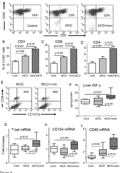
Flow cytometry of intrahepatic lymphocytes showed that MDA-BSA immunization did not modify liver T-cells profile in mice fed the control diet (Supplementary Fig. 3). However,
immunization further promoted CD3+ T-cell recruitment in MCD-fed mice increasing both the
CD8+ and CD4+ pools (Fig. 4). However, the proportion of CD8+ T-cells expressing the
CD107a activation marker was unchanged (Fig. 4). Th-1 and Th-17 activation of CD4+
T-lymphocytes are important pro-inflammatory stimuli. We observed that the expression of the Th-1 transcription factor T-cell T-box transcription factor (T-bet) as well as liver IFN-γ content were selectively increased in MCD-fed immunized animals (Fig. 4), in parallel with a stimulation of macrophages M1 activation markers IL-12p40 and inducible NO synthase (iNOS) (Fig. 2). Among immunized MCD-fed mice there was also a positive correlation between the individual IFN-γ expression and that of TNF-α, IL-12p40 and iNOS (r=0.82, 0.75 and 0.88 respectively; p<0.02). No changes were instead evident in the hepatic mRNAs for the Th-17 transcription factor retinoic acid-related orphan receptor-γt (ROR-γt) and of IL-17a (not shown). Furthermore, MCD-fed immunized mice also showed an up-regulation in the mRNAs of CD40 ligand (CD40L; CD154) and of its receptor CD40, a pair of co-stimulatory
receptor-ligand molecules involved in macrophage activation by CD4+ T-cells [19]. Consistently,
depleting CD4+ T-cells in MCD-fed immunized mice by using an anti-CD4 monoclonal IgG
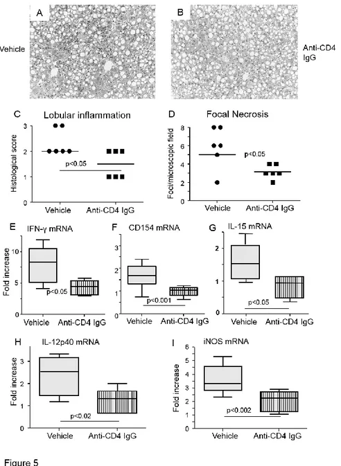
significantly lowered the hepatic mRNA expression of IFN-γ and CD40L as well as that of macrophage M1 markers iNOS and IL-12p40 (Fig. 5). In these animals, histology also showed
29
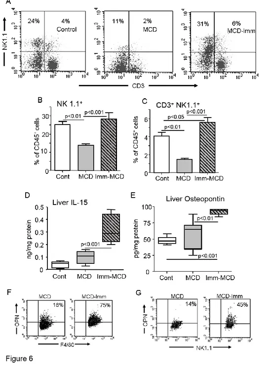
The progression of NASH in immunized mice also involves NKT cells.
Unexpectedly, changes in liver natural killer T (NKT) cells associated with the worsening of NASH occurring in immunized mice. According to previous observations [20,21], the development of NASH in naïve mice was characterized by the lowering of liver natural killer
(NK) (CD3-, NK1.1+) and NKT (CD3+, NK1.1+) pools. On the contrary, MCD-fed immunized
animals did not show NK cell depletion, while the NKT fraction was significantly higher than in controls (Fig. 6). Recent findings in animal models of NAFLD/NASH have implicated macrophages production of IL-15 in controlling liver NKT cell differentiation and survival [22]. In turn, NKT cells have been proposed to contribute to the progression of NASH to fibrosis through the production of osteopontin (OPN) [16,23]. In hour hands, the expansion of NKT cells observed in MCD-fed immunized mice paralleled with an increase of hepatic IL-15
content (Fig. 6). Such an effect was likely mediated by CD4+ T-cell activation, as depleting
immunized mice of CD4+ T-cells significantly lowered the intrahepatic IL-15 mRNA (Fig. 5).
We also observed that, while the development of NASH in naïve mice did not affect liver OPN, OPN levels were significantly up-regulated in MCD-feed immunized animals and such an increase involved an expansion of OPN-expressing NKT cells and F4/80-positive hepatic macrophages (Fig. 6). Furthermore, in line with OPN capacity to stimulate hepatic stellate cell
(HSC) activation, Sirius Red staining for collagen and α- -SMA)
positive HSCs were more evident in the livers of immunized as compare to naïve MCD-fed mice (Fig. 2).
Discussion
Recent studies have implicated the contribution of adaptive immunity to fat inflammation in
obesity, as CD4+/CD8+ T-cells are recruited to the adipose tissue and provide stimulation for
the macrophage production of pro-inflammatory mediators [24,25]. Lymphocytes are often detected in the lobular infiltrates of NASH [26], but the actual role of adaptive immunity in the disease pathogenesis is still poorly understood. We previously reported that sub-sets of adults and paediatric NAFLD/NASH patients show antibody responses against oxidative stress-related antigens, such as MDA-derived adducts, that associate with an increased severity of lobular inflammation or fibrosis [10,11]. Similar antibodies are also detected along with hepatic injury and inflammation in rats with NASH induced by the enteral nutrition with a high fat diet, while preventing oxidative stress with N-acetylcysteine attenuates both IgG formation and the severity of steatohepatitis [27]. In the present study, IgG against lipid peroxidation-derived
30
adducts are evident during the progression of NASH caused a methionine/choline deficient (MCD) diet in parallel with the liver recruitment of both CD4+ and CD8+ T-lymphocytes recognizing the same antigens and positively correlate with the severity liver injury. The possible contribution of such immune responses to the progression of experimental NASH is further substantiated by the observation that stimulating immune responses against MDA-protein adducts, one of the antigens recognized by the antibodies associated with both human and rodent NASH, promotes parenchymal injury and inflammation in mice fed with the MCD diet. Our data are not in contrast with a recent report Bieghs and co-workers showing that IgM targeting oxidized low density lipoproteins (LDLs) reduces NASH in LDL receptor-deficient mice receiving a high-fat/cholesterol diet [18]. These discrepancies, in fact, can be explained considering that in the two experimental settings the immune responses involved and the mechanisms leading to NASH are quite different. In Bieghs’s work mice were immunized with heat-inactivated pneumococci that lead to the production of natural IgM against bacterial antigens and cross-react with oxidized phosphatidylcholine in LDLs [18]. Feeding a high-fat/cholesterol diet to LDL receptor-deficient mice causes Kupffer cell engulfment by oxidized LDLs that, in turn, promotes Kupffer cell activation and hepatic inflammation [28]. In this scenario, IgM interaction with oxidized LDLs reduces their uptake by Kupffer cells, lowering the pro-inflammatory stimuli [19]. These conditions are quite different from those occurring in MCD-induced NASH, where parenchymal injury, oxidative stress and inflammation result from the impairment of hepatocyte lipid secretion [13]. Furthermore, our data indicate that the immunization with MDA-adducts mainly stimulates IgG production and T-cell responses and that these latter are responsible for stimulating inflammation.
Concerning the mechanisms by which adaptive immunity promotes the evolution of NASH, we have observed that a stimulation in liver IFN-γ production and CD40L (CD154) expression
parallels with the increase of hepatic CD4+ T-cells in MCD-fed immunized mice. CD40L is a
co-stimulatory molecule predominantly expressed by CD4+ T-cells and activated platelets that,
by the interacting with its receptor CD40 on macrophages and lymphocytes, has a key role in orchestrating inflammation and immunity in several diseases, including atherosclerosis and
obesity [29,30]. Conversely, CD4+ T-cell depletion prevents the up-regulation of IFN-γ and
ameliorates lobular inflammation, indicating that a Th-1 activation of CD4+ T-lymphocytes
plays a major role in promoting NASH. A Th-1 activation is also a feature of CD4+ T-cell
responses to LDL oxidation antigens in atherosclerotic plaques [29]. Interestingly, an increase