Short report
Gastric polyposis and desmoid tumours as a new
familial adenomatous polyposis clinical variant
associated with APC mutation at the extreme 3′-end
Vittoria Disciglio,
1Candida Fasano,
1Filomena Cariola,
1Giovanna Forte,
1Valentina Grossi,
1paola Sanese,
2Martina Lepore Signorile,
1,3Nicoletta resta,
2Claudio Lotesoriere,
4Alessandro Stella ,
2Ivan Lolli,
4Cristiano Simone
1,2To cite: Disciglio V, Fasano C, Cariola F, et al. J Med Genet epub ahead of print: [please include Day Month Year]. doi:10.1136/
jmedgenet-2019-106299 1Medical Genetics, National Institute of Gastroenterology "S. de Bellis", research hospital, Castellana Grotte, Italy 2Medical Genetics, Department of Biomedical Sciences and human oncology (DIMo), University of Bari "Aldo Moro", Bari, Italy
3Department of Molecular Medicine, Sapienza University of rome, rome, Italy
4Department of oncology, National Institute of
Gastroenterology "S. de Bellis", research hospital, Castellana Grotte, Italy
Correspondence to professor Cristiano Simone, Medical Genetics, National Institute of Gastroenterology "S. de Bellis", research hospital, Castellana Grotte, Italy; cristianosimone73@ gmail. com and Dr Ivan Lolli, Department of oncology, National Institute of Gastroenterology "S. de Bellis", research hospital, Castellana Grotte, Italy;
ivan. lolli@ irccsdebellis. it received 22 May 2019 revised 4 September 2019 Accepted 7 September 2019
© Author(s) (or their employer(s)) 2019. re-use permitted under CC BY-NC. No commercial re-use. See rights and permissions. published by BMJ.
AbsTrACT
Germline mutations of the ApC gene, which encodes a multidomain protein of 2843 amino acid residues, cause familial adenomatous polyposis (FAp). three FAp clinical variants are correlated with the location of APC mutations: (1) classic FAp with profuse polyposis (>1000 adenomas), associated with mutations from codon 1250 to 1424; (2) attenuated FAp (<100 adenomas), associated with mutations at APC extremities (before codon 157 and after codon 1595); (3) classic FAp with intermediate colonic polyposis (100–1000 adenomas), associated with mutations located in the remaining part of APC. In an effort to decipher the clinical phenotype associated with ApC C-terminal germline truncating mutations in patients with FAp, after screening APC mutations in one family whose members (n=4) developed gastric polyposis, colon oligo-polyposis and desmoid tumours, we performed a literature meta-analysis of clinically characterised patients (n=97) harbouring truncating mutations in ApC C-terminus. the APC distal mutations identified in this study cluster with a phenotype characterised by colon oligo-polyposis, diffuse gastric polyposis and desmoid tumours. In conclusion, we describe a novel FAp clinical variant, which we propose to refer to as Gastric polyposis and Desmoid FAp, that may require tailored management.
InTroduCTIon
The Adenomatous Polyposis Coli (APC) gene, located on chromosome 5q21–q22, consists of 15 coding exons, which translate into a 2843 amino acid multifunctional protein. Its various motifs and domains allow binding to key players of the Wnt pathway and cytoskeleton components.1 APC
germ-line mutations cause autosomal dominant colon cancer predisposition, known as familial adeno-matous polyposis (FAP), which is characterised by hundreds to thousands of colorectal adenomatous polyps. Three FAP clinical variants are correlated with the location of APC mutations, the large majority of which spans the 5′-half of the gene: (1) classic FAP with profuse polyposis (>1000 adenomas), associated with mutations from codons 1250 to 1424; (2) attenuated FAP (AFAP, <100 adenomas), associated with mutations at APC extremities (before codon 157 and after codon
1595); (3) classic FAP with intermediate colonic polyposis (100–1000 adenomas).2
A variable range of extracolonic manifesta-tions, including desmoid tumours (DTs) and upper gastrointestinal (GI) polyps, have been associated with mutations in APC specific regions.2 3
Specifi-cally, DT occurrence in patients with FAP has been linked with mutations at codons 1445–15783 and,
less frequently, beyond codon 1444.4 Although
still controversial, associations between upper GI polyposis phenotype and germline APC mutations have been described: gastric/duodenal adenomas have been associated with APC mutations at codons 564–14655 and codons 976–1067.6 Moreover,
germline mutations in APC promoter 1B have been identified in patients with hereditary gastric adeno-carcinoma and proximal polyposis of the stomach,7
which is characterised by a carpeting of more than 100 fundic gland polyps (FGPs). Overall, trunca-tions at APC extreme C-terminal have been rarely reported and have not been clearly associated with a specific phenotype.
Here, we report that mutations affecting APC distal portion identify a novel FAP clinical pheno-type, which we termed Gastric Polyposis and Desmoid FAP (GD-FAP).
MeThods Patients
We identified one family whose members share clin-ical findings of profuse gastric polyposis, restricted to the stomach body and fundus, while presenting few colon polyps along with DTs. All subjects included in this analysis provided written informed consent for genetic analysis. All investigations were performed in accordance with the World Medical Association’s Declaration of Helsinki.
Mutation analysis
Genomic DNA was purified from peripheral blood lymphocytes according to manufacturer’s instruc-tions (QIAamp DNA Blood Mini Kit; Qiagen, Carlsbad, California, USA). The APC complete coding region was screened for mutations as previ-ously described.8 APC promoter 1B was analysed for
mutations using a PCR-direct sequencing method as previously reported.7 Sequencing and capillary
electrophoresis were performed on the Applied Biosystems 3130 Genetic Analyzer (ThermoFisher
on October 11, 2019 at University of Bari. Protected by copyright.
Genotype-phenotype correlations
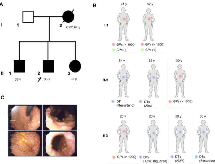
Figure 1 Family pedigree and relevant clinical findings in individual patients. (A) Squares indicate men, circles represent women. Unfilled symbols indicate
unaffected individuals. A slashed symbol indicates that an individual is deceased. Black filled symbols represent individuals carrying the ApC mutation. y indicates the age at latest follow-up. For patient I-2, the age at diagnosis (years) of colorectal cancer (CrC) is also indicated. (B) relevant clinical findings in individual family members (II-1, II-2, II-3) are depicted as filled circles (red circles indicate gastric polyps (Gps); green circles indicate colon polyps (Cps); blue circles indicate desmoid tumours (Dts)); y represents the age at diagnosis of the reported clinical observations. the number of Gps and Cps is indicated. predominant siding of Dts is also indicated and mapped according to age at diagnosis (years): Abs, abdominal rectal muscle; AbW, abdominal wall; Ing. Area, inguinal area. (C) endoscopic view showing Gps of affected family member II-3.
Scientific, Waltham, Massachusetts, USA). Mutations and poly-morphisms were confirmed in independently amplified PCR products. APC deletions were identified by Multiplex Liga-tion-dependent Probe Amplification (MLPA) analysis, using the SALSA P043 Multiplex Ligation-dependent Probe Ampli-fication (MLPA) kit (MRC-Holland, Amsterdam, The Neth-erlands) according to the manufacturer’s instructions. MLPA products were run on the Applied Biosystems 3130 Genetic Analyzer. Results were then processed with the Coffalyser software (MRC-Holland). Probes with a dosage ratio <0.7 or >1.3 revealed deletions or duplications in the corresponding sequences, respectively.
Meta-analysis
The meta-analysis was performed on the Human Gene Mutation Database Professional (HGMD Professional; Qiagen), a compre-hensive collection of germline mutations in nuclear genes that are associated with human-inherited diseases.9 The main
β-cat-enin-downregulating APC domains are located before codon 2051. Therefore, we considered the region beyond codon 2051 and the region coding for the last β-catenin-regulating domain
of APC (codons 2031–2051) to better define the phenotypes associated with germline truncating mutations at APC 3′-end. We reviewed all the papers identified in the aforementioned database and collected clinical information (i.e., gender, age of diagnosis, gastric or colon polyposis, DTs and specific APC muta-tions) concerning patients with truncating mutations at APC 3′-end. Studies including patients without clinical information were excluded.
resulTs
Family clinical findings
Here we report an Italian family in which a woman was diag-nosed with rectal cancer (at the age of 64) and her three chil-dren developed multiple DTs and profuse gastric polyposis at an early age (second and third decade) in the absence or paucity of colonic polyposis (figure 1A,B). The mother underwent surgical resection of rectal cancer and died one month later due to septic fever secondary to anastomotic dehiscence; thus, no upper gastrointestinal endoscopy (UGE) was performed. Preop-erative colonoscopy did not reveal colon polyps. Affected family
on October 11, 2019 at University of Bari. Protected by copyright.
member II-1 was diagnosed with numerous small sessile polyps in the stomach fundus and body and two tubular adenomas of the colon at the age of 31. One year later, a small sessile polyp of the sigmoid colon was detected by colonoscopy and surgically removed. UGE revealed multiple gastric polyps in the fundus and body, two of which were histologically characterised as FGPs. Affected family member II-2 was diagnosed with a localised tumour composed of cells with tapering nuclei and extracellular collagen in the stroma (DT) by abdominal CT at the age of 24. Two years after surgical intervention, he developed four novel abdominal DTs, which were surgically resected. The patient underwent regular follow-up with annual colonoscopies, which showed no evidence of polyposis. At the age of 28, the patient was diagnosed with tumour recurrence by MRI and subse-quently underwent combined chemotherapy (vinorelbine and methotrexate), which produced significant DT regression. At the age of 30, the patient was diagnosed with a squamous papilloma of the esophagus, which was surgically removed, and multiple gastric sessile polyps, some of which were histologically charac-terised as FGPs. At the age of 26, the affected family member II-3 underwent UGE, which showed multiple gastric polyps (0.2–0.8 cm) in the stomach fundus and body. After histological analysis, four gastric polyps were classified as FGPs (figure 1C). More-over, the patient developed several small duodenal polyps and duodenal adenomas with low grade of dysplasia. Between the age of 26 and 30, the patient was diagnosed with multiple DTs at different sites, including the hypogastric and epigastric region of the abdominal wall, the inguinal area and the pancreas.
Characterisation of APC germline mutation
The APC mutation search performed on the index case’s DNA revealed a nonsense mutation in APC exon 15 (NM_000038.5:c.7709C>G; p.Ser2570Stop). The proband’s mutation was inherited from the mother and was also present in one brother and one sister (figure 1A). Sequencing of
APC promoter 1B excluded the presence of three mutations
(chr5:112,707,523 A/C; c.-195A>C; and chr5:112,707,527 T/C; c.-191T>C; chr5:112,707,593 A/_; c.-125delA) — previ-ously identified in patients with hereditary gastric adenocarci-noma and proximal polyposis of the stomach 7 — in the DNA
of all analysed family members but revealed a single nucleotide variant (chr5:112,707,473 A/C; c.-245A/C; rs75580617) in the DNA of family member I-2. Analysis using the 1000 Genomes database (http://www. internationalgenome. org) revealed that the rs75580617 variant had a minor allele frequency >5%, indicating that it could be considered a common variant. MLPA analysis excluded deletions and duplications involving APC in all family members.
Meta-analysis results
Our meta-analysis, aimed at identifying clinical phenotypes specifically associated with germline truncating mutations in APC extreme 3′-end, revealed that a very small proportion (n=21/1418, 1.5%) of APC truncating mutations disrupts the protein C-terminal region. This region, starting at amino acid (aa) 2052, does not include motifs and domains involved in β-catenin downregulation, while it encompasses the basic domain (aa 2224–2575) and binding sites for both EB1 (aa 2670–2843) and DLG (aa 2841–2843) proteins. The remaining 1397 truncating mutations represent the majority (72%) of total APC germline alteration burden (n=1951) and cluster 5′ to codon 2052, leading to partial or total removal of domains involved in β-catenin regulation (figure 2A). Thus, we sought to
compare the phenotypic consequences of truncating mutations falling within the third SAMP repeat — APC last β-catenin-reg-ulating domain (region A: aa 2031–2051) — to those harboured in APC extreme C-terminal region, distal to SAMP3, which does not contain β-catenin-regulating domains (region B: aa 2052– 2843). A total of 35 unique alterations localising to these two different APC distal regions were recorded: (1) 14 were located in the third SAMP repeat domain, which is implicated in Axin binding (APC region A),10 and (2) 21 created a stop codon
in the distal part of the protein, which includes the binding domains for EB1 and DLG (APC region B).11–13 Among this
subset of 35 terminating mutations, only 14 were identified in patients (n=101) with data on clinical features (polyps, tumours, desmoids); of these, 6 (77 patients) were located in APC region A and 8 (24 patients) in APC region B, which also harbours the mutation characterised in the present study (figure 2B). The clinical features of these patients and of the family we identi-fied are detailed in online supplementary table 1. In this cohort, the percentage of patients with a lower count of colon polyps (<50) was significantly higher among patients with mutations in APC region B (p=0.0151). Moreover, the frequency of profuse gastric polyposis or adenomas was higher in patients with APC truncated in region B than in region A (p=0.0461). Finally, DTs were over-represented in patients with truncating mutations in APC region B (77.8%) compared to patients harbouring muta-tions in APC region A (25%) (table 1).
dIsCussIon
The location of APC mutations influences FAP phenotype and surveillance/treatment.1 To date, over 1900 APC mutations
have been described in FAP; of these, more than 70% occur 5′ to codon 1600, thus leading to the loss of β-catenin-regulating APC domains. Mutations truncating APC C-terminus cause the loss of domains required for microtubule binding and EB1/DLG interaction, potentially triggering loss of polarisation, inhibition of differentiation and chromosomal instability.11–13 APC
muta-tions in this specific region are extremely rare and have not been obviously associated with a distinct phenotype. In this study, we sought to elucidate the correlation between mutations located at
APC extreme 3′-end and clinical symptoms. Our results
demon-strate a trend towards a lower susceptibility to developing severe colonic polyposis (>50) in patients with mutations in APC region B (aa 2052–2843) compared to patients with mutations in APC region A (aa 2031–2051). Where mutations do not affect the Axin binding site (SAMP3 domain) in APC, its β-catenin-de-grading activity is preserved, which may explain the association with the oligo-polyposis phenotype. It is also noteworthy that patients with mutations in APC region B appear to have a higher risk of developing DTs and a higher susceptibility to developing profuse gastric polyposis or adenomas. Thus, it is tempting to speculate that truncating mutations in APC region A trigger Wnt-dependent oncogenic proliferation, which may drive substantial catastrophic consequences on a constantly renewing tissue such as the colon epithelium (short lifespan: 3–5 days).14
Conversely, truncating mutations in APC region B likely cause the loss of cell/tissue polarisation, which is a tumour suppressor mechanism. This could subvert the architectural structure of the gastric tissue, in which parietal cells have an extended lifespan of 54 days, and/or lead to deregulation of mesenchymal prolif-eration.15 Importantly, gastric adenomas in the gastric body/
fundus may arise in a background of numerous (carpet-like) FGPs. Consistently, previous studies have shown adenomatous dysplastic changes within FGPs in about 40% of FAP patients
on October 11, 2019 at University of Bari. Protected by copyright.
Genotype-phenotype correlations
Figure 2 APC coding region and genotype–phenotype correlation (A) Distribution of truncating mutations throughout APC coding region and genotype–
phenotype correlation. Conserved regions and domains that interact with other proteins are shown. (B) Distribution of mutations in the extreme 3′-end of APC coding region in patients with clinical information identified in our literature meta-analysis and in the present study. Light blue and yellow boxes with arrows indicate the location of premature stop codons of already described truncating mutations in ApC regions A and B, respectively. the nonsense mutation identified in our patients, which causes a stop at codon 2570, is indicated with a yellow arrow and circle.
Table 1 Relevant clinical features of patients harbouring truncating
variants in APC region A and APC region B
Phenotype
APC region A n (%)
APC region b n (%)
Fisher exact test p-value CP >50 47/66 (71.2) 3/22 (13.6) 0.0151 CP <50 19/66 (28.8) 19/22 (86.4) GPP/Ad+ 15/62 (24.2) 7/13 (53.84) 0.0461 GPP/Ad− 47/62 (75.8) 6/13 (46.15) DT+ 19/76 (25) 14/18 (77.8) <0.0001 DT− 57/76 (75) 4/18 (22.2)
CP, colon polyp; DT, desmoid tumour; GPP/Ad, gastric profuse polyposis/adenoma. with FGPs.16 In our study, among 13 clinically characterised
patients with truncating mutations in APC region B, five out of seven have developed adenomas in association with FGPs. These findings may have important consequences on surveil-lance, tailored management and overall quality of life of FAP patients. We are aware that our study has some limitations, the major one being the number of patients without available clinical
information. Also, it must be noted that DTs can remain unde-tected on conventional radiograph,17 thus we cannot exclude the
presence of these tumours in patients for which no data were reported about specific investigations. In conclusion, we report novel insights on FAP presentation when it is associated with mutations lying in APC extreme C-terminal region, supporting an emerging FAP clinical phenotype, which we termed Gastric Polyposis and Desmoid FAP (GD-FAP). Based on the National Comprehensive Cancer Network guidelines18 for AFAP patients
and on the higher risk of developing adenomas that has been observed in FAP patients with FGPs,19 20 in our view, GD-FAP
patients should start follow-up at the age of 20 by performing colonoscopy every 2–3 years, esophagogastroduodenoscopy and abdominal ultrasound annually.
Acknowledgements We thank Dr Francesco paolo Jori for his helpful discussion during the preparation of the manuscript and editorial assistance.
Contributors VD, IL and CS conceived and designed the study, performed data analysis, wrote and revised the manuscript for important intellectual content. CF performed statistical analysis, reviewed the data and helped to write the manuscript. FC, GF, VG, pS and MLS acquired the literature data and analysed and interpreted the
on October 11, 2019 at University of Bari. Protected by copyright.
data. Nr and AS performed genetic analysis and reviewed the data and manuscript for important intellectual content. CL provided clinical data. All the authors reviewed the manuscript.
Funding this work was supported by the Italian Ministry of health ’ricerca Corrente 2017-2019’ to IL, ’ricerca Corrente 2018–2020; 2019–2021’ to CS; by prIN - research projects of National relevance (prIN 2017, n. 2017WNKSLr-LS4) from the Italian MIUr to CS and by Fondazione puglia (grant ’ricerca di nuovi geni di predisposizione e di markers predittivi di neoplasia nelle sindromi di predisposizione ereditaria al cancro del colon retto’) to AS.
Competing interests None declared. Patient consent for publication obtained.
Provenance and peer review Not commissioned; externally peer reviewed. open access this is an open access article distributed in accordance with the Creative Commons Attribution Non Commercial (CC BY-NC 4.0) license, which permits others to distribute, remix, adapt, build upon this work non-commercially, and license their derivative works on different terms, provided the original work is properly cited, appropriate credit is given, any changes made indicated, and the use is non-commercial. See: http:// creativecommons. org/ licenses/ by- nc/ 4. 0/.
orCId ids
Alessandro Stella http:// orcid. org/ 0000- 0002- 9035- 6267
Cristiano Simone http:// orcid. org/ 0000- 0002- 2628- 7658
reFerenCes
1 Zhang L, Shay JW. Multiple roles of ApC and its therapeutic implications in colorectal cancer. J Natl Cancer Inst 2017;109.
2 Nieuwenhuis Mh, Vasen hFA. Correlations between mutation site in ApC and phenotype of familial adenomatous polyposis (FAp): a review of the literature. Crit Rev Oncol Hematol 2007;61:153–61.
3 Caspari r, olschwang S, Friedl W, Mandl M, Boisson C, Böker t, Augustin A, Kadmon M, Möslein G, thomas G, propping p. Familial adenomatous polyposis: desmoid tumours and lack of ophthalmic lesions (Chrpe) associated with ApC mutations beyond codon 1444. Hum Mol Genet 1995;4:337–40.
4 Friedl W, Caspari r, Sengteller M, Uhlhaas S, Lamberti C, Jungck M, Kadmon M, Wolf M, Fahnenstich J, Gebert J, Möslein G, Mangold e, propping p. Can ApC mutation analysis contribute to therapeutic decisions in familial adenomatous polyposis? experience from 680 FAp families. Gut 2001;48:515–21.
5 enomoto M, Konishi M, Iwama t, Utsunomiya J, Sugihara KI, Miyaki M. the relationship between frequencies of extracolonic manifestations and the position of ApC germline mutation in patients with familial adenomatous polyposis. Jpn J Clin Oncol 2000;30:82–8.
6 Bertario L, russo A, Sala p, Varesco L, Giarola M, Mondini p, pierotti M, Spinelli p, radice p. Multiple approach to the exploration of genotype–phenotype correlations in familial adenomatous polyposis. JCO 2003;21:1698–707.
7 Li J, Woods SL, healey S, Beesley J, Chen X, Lee JS, Sivakumaran h, Wayte N, Nones K, Waterfall JJ, pearson J, patch A-M, Senz J, Ferreira MA, Kaurah p, Mackenzie r, heravi-Moussavi A, hansford S, Lannagan trM, Spurdle AB, Simpson pt, da Silva L, Lakhani
Sr, Clouston AD, Bettington M, Grimpen F, Busuttil rA, Di Costanzo N, Boussioutas A, Jeanjean M, Chong G, Fabre A, olschwang S, Faulkner GJ, Bellos e, Coin L, rioux K, Bathe oF, Wen X, Martin hC, Neklason DW, Davis Sr, Walker rL, Calzone KA, Avital I, heller t, Koh C, pineda M, rudloff U, Quezado M, pichurin pN, hulick pJ, Weissman SM, Newlin A, rubinstein WS, Sampson Je, hamman K, Goldgar D, poplawski N, phillips K, Schofield L, Armstrong J, Kiraly-Borri C, Suthers GK, huntsman DG, Foulkes WD, Carneiro F, Lindor NM, edwards SL, French JD, Waddell N, Meltzer pS, Worthley DL, Schrader KA, Chenevix-trench G. point mutations in exon 1B of ApC reveal gastric adenocarcinoma and proximal polyposis of the stomach as a familial adenomatous polyposis variant. Am J Hum Genet 2016;98:830–42.
8 resta N, Stella A, Susca F, Montera M, Gentile M, Cariola F, prete F, tenconi r, tibiletti MG, Logrieco G, Mattina t, Andriulli G, Caruso ML, Fiorente p, russo S, Caputi-Jambrenghi o, Mareni C, Guanti G. Nine novel ApC mutations in Italian FAp patients.
Hum Mutat 2001;17:434–5.
9 Stenson pD, Mort M, Ball eV, evans K, hayden M, heywood S, hussain M, phillips AD, Cooper DN. the human gene mutation database: towards a comprehensive repository of inherited mutation data for medical research, genetic diagnosis and next-generation sequencing studies. Hum Genet 2017;136:665–77.
10 Spink Ke, polakis p, Weis WI. Structural basis of the Axin–adenomatous polyposis coli interaction. Embo J 2000;19:2270–9.
11 Wen Y, eng Ch, Schmoranzer J, Cabrera-poch N, Morris eJS, Chen M, Wallar BJ, Alberts AS, Gundersen GG. eb1 and ApC bind to mDia to stabilize microtubules downstream of rho and promote cell migration. Nat Cell Biol 2004;6:820–30.
12 Green rA, Wollman r, Kaplan KB. Apc and eB1 function together in mitosis to regulate spindle dynamics and chromosome alignment. Mol Biol Cell
2005;16:4609–22.
13 Ishidate t, Matsumine A, toyoshima K, Akiyama t. the ApC-hDLG complex negatively regulates cell cycle progression from the G0/G1 to S phase. Oncogene
2000;19:365–72.
14 Fatehullah A, Appleton pL, Näthke IS. Cell and tissue polarity in the intestinal tract during tumourigenesis: cells still know the right way up, but tissue organization is lost. Philos Trans R Soc Lond B Biol Sci 2013;368. 20130014.
15 Karam SM. A focus on parietal cells as a renewing cell population. World J Gastroenterol 2010;16:538–46.
16 Bianchi LK, Burke CA, Bennett Ae, Lopez r, hasson h, Church JM. Fundic gland polyp dysplasia is common in familial adenomatous polyposis. Clin Gastroenterol Hepatol
2008;6:180–5.
17 Castellazzi G, Vanel D, Le Cesne A, Le pechoux C, Caillet h, perona F, Bonvalot S. Can the MrI signal of aggressive fibromatosis be used to predict its behavior? Eur J Radiol
2009;69:222–9.
18 National Comprehensive Cancer Center (NCCN). NCCN guidelines version 1. Genetic/ familial high-risk assessment: colorectal2019. https://www. nccn. org/ professionals/ physician_ gls/ default. aspx
19 ezzedine S, Dumas r, Gonzalez JM, Vitton V, Barthet M, rampal p, Grimaud JC, Garcia S. premalignant potential of fundic gland polyps-associated familial polyposis syndromes. JSM Gastroenterol Hepatol 2014;1035.
20 Straub SF, Drage MG, Gonzalez rS. Comparison of dysplastic fundic gland polyps in patients with and without familial adenomatous polyposis. Histopathology
2018;72:1172–9.
on October 11, 2019 at University of Bari. Protected by copyright.