Original article
The reaction to nailing or cementing of the femur in rats
A microangiographic and fluorescence study
U. E. Pazzaglia
1, L. Andrini
2, A. Di Nucci
31Facolta` di Medicina e Chirurgia, Universita` di Brescia, Brescia, Italy 22a Facolta` di Medicina e Chirurgia, Universita` di Pavia, Pavia, Italy 3Istituto di Farmacologia 2, Universita` di Pavia, Pavia, Italy
Accepted: 29 January 1997
Summary. Bone reaction to cement and to a
ce-mentless stem was studied in the rat femur with
histological fluorescence and microangiographic
techniques. Periosteal and endosteal apposition,
and consequent remodelling, appeared as a
reac-tion to reaming rather than caused by cement or a
cementless stem. Every change in bone began with
proliferation, progression and orientation of the
vessels. Endosteal apposition was absent in
ce-mented femurs because the entire medulla was
occupied by the acrylic cement, but remodelling of
the subendosteal cortex followed medullary
re-vascularisation which was far advanced after
90 days. In cementless stems, endosteal apposition
of primary woven bone and remodelling was the
basis for bony ingrowth and anchorage through
bony bridges. Our results suggest that the pattern
of blood supply is relevant to the structural
orga-nisation of mature lamellar bone around the
im-plant. Cemented stems have maximum anchorage
and stability as soon as they are inserted, but this
decreases with time as revascularisation occurs.
Cementless stems can reach maximum integration
later after insertion, and revascularisation is less
critical because they usually do not fill the canal
completely.
Re´sume´. A l’aide des techniques histologiques
microangiographiques et a` fluorescence, la
re´ac-tion osseuse lors de l’introducre´ac-tion endome´dullaire
dans le fe´mur d’un rat de ciment acrylique et de
tiges non cimente`es a e´te´ e´tudie´e. La re´action
os-seuse du pe´rioste interne et externe et la
re-production osseuse ou le remodelage osseux
pa-raissent eˆtre des phe´nome`nes duˆs au procede´ de
fraisage du canal me´dullaire plutot qu’a` la
re´ac-tion spe´cifique de la cimentare´ac-tion ou a`
l’introduc-tion d’une tigue non cimente´e. Tout changement de
l’os, aussi bien en apposition qu’en remodelage
implique une prolife´ration, une progression et une
nouvelle orientation des vaisseaux. L’apposition
du pe´rioste interne n’apparait pas pour les fe´murs
cimente´s et on observe un remodelage de l’endoste
duˆ au phe´nome`ne de revascularisation me´dullaire;
ce phe´nome`ne est tre´s evident apre´s 90 jours. Pour
les tiges non cimente´es, par contre, l’apposition du
perioste interne de la matrice osseuse primaire et
le successif remodelage permettent la repousse
osseuse sur la surface de l’implant, en le soudant a`
l’aide de ponts osseux a` la superficie du pe´rioste
interne. Nous pouvons donc dire que d’apre´s nos
e´tudes, les tiges cimente´es garantissent un ancrage
optimal de´s son application mais il est e´vident
qu’au cours du temps, a` cause du phe´nome`ne de
revascularisation l’ancrage diminue. Les tiges non
cimente´es atteignent le maximum d’ancrage
long-temps apre´s l’implantation et le procede´ de
re-vascularisation est beaucoup moins critique vu que
l’implant, habituellement ne remplit pas
comple´-tement le canal me´dullaire.
Introduction
The main arterial supply to the diaphysis of long
bones is provided by the marrow arteries which
pass outwards from the endosteal surface into the
Reprint requests to: U. E. Pazzaglia, Clinica Ortopedica
Universita` di Brescia Spedali Civili, I-25100 Brescia, Italy International Orthopaedics (SICOT) (1997) 21: 267 – 273
Orthopaedics
International
from the diaphysis [14] and medullary arterial
re-vascularisation are obstructed with changes to the
pattern of the blood supply [4, 7, 11, 21, 24].
Anatomical and mechanical factors affect the
type of bonding and the structure of the interface.
In this study, we have reproduced the conditions of
cemented and cementless implants in the rat’s
femur and compared them.
Materials and methods
Twenty-four Wistar white rats (Stefano Morini, S Polo d’Enza, Reggio Emilia, Italy) weighing about 400 g were used. Under barbiturate anaesthesia, both knees were opened through a lateral parapatellar incision and a 3 mm drill hole was made in the intercondylar notch. The canal was then reamed by hand with a specially designed reamer from 1.5 to 3 mm diameter depending on the individual’s size. The canal was then re-peatedly irrigated with sterile saline under pressure to remove debris and fat. In group A, the right femur was filled with Sulfix cement (Howmedica, Rutherford, USA) through a 3 mm catheter and a specially designed syringe, a miniature version of a cement gun. In group B, a stainless rod was implanted in the left femur, 2 cm long and the same diameter as the reamer. The rod was introduced with gentle pressure as far as it would go.
In the control rats, the right femur was reamed, but no rod or cement inserted (Group C); the left femur was not touched (Group D).
with a cutting-grinding device (Exact Apparatebau, Norder-stadt, Germany).
Unstained sections, 50µthick, were examined in a bright field and in incident fluorescent light with a Leitz Aristophot microscope. Selected sections were thinned to 10 µ, stained with haematoxylin-eosin (HE) and examined in a bright field. Continuous labelling with tetracycline for the first 30 days allowed a semi-quantitative evaluation of fluorescence at 7, 15 and 30 days (Table 1) with regard to the following features:
(a) Metaphyseal appositional activity, and endosteal and periosteal apposition, in groups A, B and C were graded 0 when there was no increase in the width of the fluorescent bands compared to the controls (group D) for 7 days, or to the previous time interval in the same group. The width of the bands was assessed with a reticule in the eyepiece of the microscope. The value for each section was the mean of 4 measurements performed on 2 orthogonal axes of the femoral section (anterior/posterior and lateral/lateral). Apposition was graded + in the opposite case. Only differences above 10% were considered significant.
(b) Periosteal remodelling was graded according to the percentage of dark zones inside the fluorescent band: 0 = less than 10%; * = 10 to 30%; ** = more than 30%. The images were digitalised on a graphics tablet (Summasketch Plus, Summagraphic, Fairfield, CT, USA) coupled with an IBM AT personal computer. We developed a programme to calcu-late the percentage of dark areas.
(c) Cortical remodelling was graded 0 when fewer than 5 osteones were labelled, and @ when more than 5 were labelled.
No quantification was possible at 90 days because of the advanced stage of remodelling.
Table 1. Semiquantitative evaluation of fluorescence in the groups at intervals of 7, 15 and 30 days after continuous labelling with Tet-racycline (30 mg/kg/day) intraperitoneally
Groups Metaphyseal appositional activity Endosteal apposition Periosteal apposition Periosteal remodelling Cortical remodelling Days 7 15 30 7 15 30 7 15 30 7 15 30 7 15 30 A) Cemented + + + 0 0 0 + + 0 0 ** * 0 0 @ B) Cementless + + + + + + + + 0 0 ** * 0 0 @ C) Reaming alone + + + + + + + + 0 0 ** * 0 0 0 D) Normal 0 0 0 0 0 0 0 0 0 0 0 0 0 0 0
Results
Control unreamed femurs (group D)
These did not show a band of fluorescence on the
endosteal surface of the canal. A complete thin
band was present on the periosteal contour of the
diaphysis corresponding to normal bone growth in
rats of this age. Vessels reached the outer surface
of the cortex and had a regular radial pattern from
muscles to periosteum. The larger vessels were
straight or slightly wavy and ended in the
perios-teum. Smaller straight branches penetrated the
bone through the Volkmann and Haversian canals
(Fig. 1 a). A row of osteoblasts was laid down on
circumferential and concentric lamellae on the
outer cortex (Fig. 1 b).
A similar radial pattern from branches of the
medullary arteries was present on the endosteal
surface (Fig. 2 a, b). Labelled osteones were rarely
seen in the thickness of the cortex.
In the distal femur, a larger number of
epiphy-seal and metaphyepiphy-seal trabeculae had labelled
bor-ders because of the greater turn-over of cancellous
bone.
Periosteal reaction of implanted femurs
(groups A and B) and control reamed femurs
(group C)
At 7, 15 and 30 days, similar periosteal reaction
was found in groups A, B and C, so they are
considered together:
(a) At 7 days
A thick layer of periosteal bone was present
quantitatively
and
qualitatively
compared
to
group D femurs, the width of the former being
15 times the latter. Groups A and B showed the
same radial pattern of periosteal vessels, but they
were enlarged, congested and twisted. A large
amount of primary woven bone was laid down
between the vessels; labelling was not distributed
uniformly and there was a shaded irregular contour
269
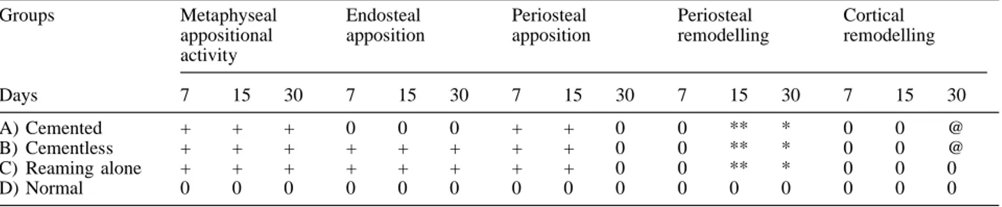
Fig. 1. Unreamed control femur (group D). a The normal
pattern of periosteal vessels; the thin fluorescent band repre-sents normal periosteal apposition and there is no activity on the endosteum (×100, fluorescence). b The periosteal apposi-tion is lamellar and concentric to the diaphyseal circumfer-ence; remodelling is the next step (×100, phase contrast)
Fig. 2 A, B. Unreamed control femur (group D). A Endosteal
supply from the medullary artery (×12, unstained, vascular tree injected). B Pattern of the distribution of vessels to the endosteal surface; injected vessels are present in the Volkmann canals of the cortex (×100, vascular tree injected)
because the outer layer was not labelled at all
(Fig. 3 a). In contrast, periosteal apposition in
group D showed a more homogeneous compact
and demarcated band of fluorescence.
(b) At 15 days
The rapid apposition of woven bone in groups A
and B had diminished; the outer layer of periosteal
apposition is smooth and well demarcated, the
periosteal vessels are normal, and fluorescence is
more evenly distributed. The width of periosteal
apposition is greater than in group C, but the main
difference is the presence of resorption lacunae
appearing as spherical unlabelled holes with an
injected vessel in the centre (Fig. 3 b). Turnover is
faster in woven compared to lamellar periosteal
bone.
(c) At 30 days
Periosteal appositional bone in groups A, B and C
presented a thick band of homogeneous densely
labelled bone because remodelled lacunae had
been filled by labelled concentric lamellae of new
osteones (Fig. 3 c).
(d) At 90 days
The periosteal activity of groups A and B did not
differ from group C.
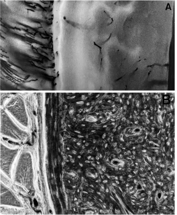
Fig. 3 A– C Reamed cemented femur (group A). A At 7 days,
the periosteal vessels are hypertrophied and congested and the outer layer of periosteal apposition is 15 times thicker than in unreamed controls (D). The outer part of the periosteal woven is not labelled by tetracycline because the bone is not yet calcified due to the rapid apposition (×100, fluorescence). B At 15 days, the rapid periosteal apposition has ceased; all the new bone is labelled and vessels have penetrated the woven bone forming lacunae (×80, fluorescence). C At 30 days, there is remodelling of periosteal apposition with injected vessels in the centre of new osteones (×80, fluorescence)
Fig. 4. Reamed cementless implant (group B). At 30 days,
labelled woven bone fills the medullary cavity between im-plant and endosteal surface; it is undergoing remodelling and a network of injected material is present inside the labelled bone.
Arrows show a band of periosteal apposition (×40, fluores-cence)
Endosteal reaction of implanted femurs
(groups A and B) and control reamed femurs
(group C)
(a) At 7, 15 and 30 days
Endosteal reactive woven bone was present in both
group C and group B, labelled woven bone filling
the medullary canal or the space between the
en-dosteal surface and the implanted cementless stem
(Fig. 4).
(b) At 90 days
Group B showed remodelling of primary endosteal
bone in the form of a thin lamellar capsule
con-nected to the surface by a few radial bridges of
differing consistency. A well developed network of
injected vessels was present between the
en-dosteum and the implant (Fig. 5).
Reactive endosteal bone had completely
re-sorbed in group C.
Reaction in cemented femurs (group A)
(a) At 7 days
There was no reactive endosteal bone.
(b) At 15 and 30 days
Injected vessels had appeared near the
bone-ce-ment interface and signs of subendosteal
re-modelling were more obvious at 30 days.
271
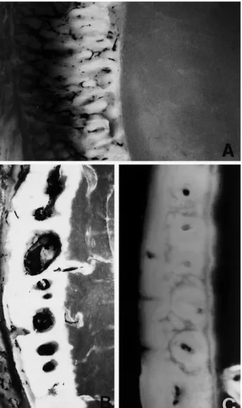
Fig. 5. Reamed cementless implant (group B). At 90 days,
most of the endosteal reactive bone has remodelled, the implant is connected to the endosteal surface by bony bridges and medullary vascularisation is restored around it (×10, HE, vascular tree injected)
Fig. 6 A, B. Reamed cemented femur (group A). A At 30 days,
new vessels penetrate the subendosteal area near the cement (CE = bone cement) (×12, fluorescence). B Remodelling of endosteal bone corresponding with the cement. Arrows indi-cate a band of labelled periosteal apposition (×80, fluores-cence)
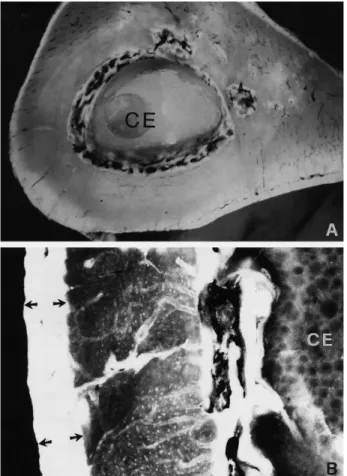
Fig. 7. Reamed cemented femur (group A). At 90 days,
revascularisation of the cancellous metaphysis has demarcated the cement, incorporating necrotic trabeculae which are en-gulfed in the cement (×12, fluorescence)
osteones were seen in the subperiosteal cortex in
groups A and B at 15 days. Extensive remodelling
involving the whole cortex was present in both
these groups at 30 days.
Discussion
Our experimental model reproduces the insertion
of a cemented and a cementless stem as used in
orthopaedic surgery. The miniaturised technique
was satisfactory and completely filled the canal
with acrylic cement with good penetration of
tra-becular bone. Bone metabolism is more rapid in
rats compared with man, but we consider it
justi-fiable to assume that the changes are similar to
those occurring in patients in whom cemented or
cementless prosthesis are inserted, and when load
bearing is avoided. The periosteal and endosteal
reaction, and the revascularisation seen after
reaming, have been recorded in fractures of human
long bones and do not differ quantitatively from
what is seen in the experimental model [18, 26].
However, what occurs in rats in 90 days would
take several years in man.
The first response of rapid periosteal apposition
appeared to be a general reaction to reaming rather
than to cement or nailing since it is present to the
same degree in the control reamed femur (group C).
Within 2 weeks this activity ceased and the only
change was remodelling of new bone. Although
the width of the fluorescent band increased
be-tween 7 and 15 days, this represented calcification
of already deposited bone, so the periosteal
reac-tion only occured in the first 7 days.
The irregular distribution of fluorescence in
reamed groups A and B shows a circumferential
lamellar pattern of appositional bone, in contrast
with woven primary bone in the unreamed group D
where osteoblasts lay down collagen fibres without
any spatial orientation. Apposition is also faster in
suggests that the flow is centripetal. The
enlarge-ment of the periosteal vessels is associated with an
increase of osteoblasts with rapid production of
woven bone during the first week. The bone is
remodelled to osteonic lamellar type with an
in-crease in the circumference of the femur. This
contradicts Grundes and Reikeras who did not
observe this increase in circumference, perhaps
due to insufficient reaming which they describe as
‘modest’ [9].
Remodelling of the old cortex takes place at
about 30 days and is mainly subendosteal,
in-creasing the inner circumference and depending on
revascularisation by the medullary vessels. Both
factors affect fixation of the implant because the fit
can loosen as the diameters increase.
In the conditions of our study, every change in
the bone, whether apposition or remodelling is
preceded by proliferation, progression and
orien-tation of the vessels. Mechanical stability,
condi-tions of loading, surface porosity and chemistry are
thought to affect anchorage of implants to bone,
but the pattern of blood supply, which we consider
relevant in determining the organisation of
la-mellar bone around the implant, has so far been
neglected.
Endosteal apposition is nonspecific reaction to
reaming since it was present in both the implanted
cementless group (B) and the control reamed
group (C). It does not occur in cemented femurs
possibly because the entire canal is occupied by
acrylic so that stability is by interlocking of cement
with the irregular bony surface. This is greatest
soon after implantation and decreases as
modelling of the inner cortex with medullary
re-vascularisation occurs [6, 10]. The process is far
advanced in rats after 90 days, but is much slower
in man where in retrieved stable cemented stems
interlocking is seen to be reduced to a few points
and most contact is with the connective tissue
which has replaced bone. This occurs in the
ab-sence of foreign body reaction which would hasten
the progressive reduction of the bonding area [15].
In cementless stems, the endosteal primary
woven bone leads to ingrowth on the surface of the
implant [1, 22], and remodelling forms a lamellar
capsule connected by radial bridges to the
en-dosteal surface [16]. The initial stability is assured
by the primary fit of the implant, and evolution of
the capsule increases fixation related to the number
and thickness of bony bridges. Remodelling also
occurred in this group (B), but has less influence
on the stability of the implant.
References
1. Bobyn JD, Engh CA (1985) Biological fixation in total hip arthroplasties. Slack Inc., Thorofare NJ
2. Bragdon JA, Foster L, Sosman MC (1949) Experimental Infarction of Bone and Bone Marrow. Am J Pathol 25: 709 – 723
3. Brookes M, Harrison RG (1957) The vascularization of the rabbit femur and tibia-fibula. J Anat 91: 61 – 72 4. Brookes M (1971) The blood supply of bone.
Butter-worths, London, 1 – 338
5. Danckwardt-Lillienstrom G (1968) Reaming of the me-dullary cavity and its effect on diaphyseal bone. Acta Orthop Scand [Suppl] 128: 1 – 153
6. Danckwardt-Lillienstrom G, Lorenzi GL, Olerud S (1970) Intracortical circulation after intramedullary reaming with reduction of pressure in the medullary cavity. A micro-angiographic study in the rabbit tibia. J Bone Joint Surg [Am] 52: 1390 – 1394
7. Draenert K (1981) Histomorphology of the bone-to-ce-ment interface: remodelling of the cortex and revascular-ization of the medullary canal in animal experiments. In: Salvati EA (ed) The hip proceedings of the ninth open Scientific Meeting of the hip society. Mosby, St Louis Toronto London
8. Feith R (1975) Side-effects of acrylic cement impianted into bone. Acta Orthop Scand [Suppl] 161: 2 – 136 9. Gothmann L (1960) The normal arterial pattern of the
rabbit tibia after the application of an intramedullary nail. A microangiographic study. Acta Chir Scand 120: 201 – 210
10. Grundes O, Reikeras O (1994) Nailing and occlusion of the medullary cavity. Flow and mechanical changes in rat femura. Acta Orthop Scand 65: 175 – 178
11. Gustilo RB, Nelson GE, Hamel A, Moe JH (1964) The effect of intramedullary nailing on the blood supply of the diaphysis of long bones in mature dogs. J Bone Joint Surg [Am] 46: 1362 – 1363
12. Harms J, Van De Berg PA, Mertz L (1974) Knochen-re-vascularization nach Mefobaine-Polacos-Fullung. Eine experimentelle untersuchung an der hund tibia. Arch Orthop Unfall-Chir 80: 71 – 78
13. Koekenberg LJL (1963) Vascularization in the healing of fractures. Thomas, Springfield
14. Nelson GG, Kelly PJ, Lowell FA, Peterson LFA, James JM (1961) Blood supply of the human tibia. J Bone Joint Surg [Am] 42: 625 – 635
15. Oni OOA, Stafford H, Gregg PJ (1987) An investigation of the routes of venous escape from the diaphyseal marrow of canine and rabbit long bones. Int J Microcirc Clin Exp 7: 31 – 41
16. Pazzaglia UE (1990) Pathology of the bone-cement in-terface in loosening of total hip repleacement. Arch Ortop Traum Surg 108: 83 – 88
17. Pazzaglia UE, Zatti G, Cherubino P, Frick W, Koch R (1991) Bone reaction to the implant of intramedullary pins of three different metals in the rats femur. Mater Med 2: 77 – 81
18. Reichert ILH, Mc Carthy ID, Hughes SPF (1995) The acute vascular response to intramedullary reaming. Mi-crosphere estimation of blood flow in the intact ovine tibia. J Bone Joint Surg [Br] 77: 490 – 493
19. Richany SF, Sprinz H, Kramer K, Ashby J, Mevrill TG (1965) The role of the diaphyseal medulla in the repair and regeneration of the femoral shaft in the adult cat. J Bone Joint Surg [Am] 47: 1565 – 1584
20. Rhinelander FW, Granilla RW, Phillips RS, Steel WM (1967) Microangigraphy in bone healing III. Osteotomies with internal fixation. J Bone Joint Surg [Am] 49: 1006 – 1009
21. Rhinelander FW (1968) The normal microcirculation of diaphyseal cortex and its response to fracture. J Bone Joint Surg [Am] 50: 1273 – 1298
22. Rhinelander FW, Nelson CL, Stewart RD, Steward CL (1979) Experimental reaming of the proximal femur and acrylic cement implantation. Clin Orthop 141: 74 – 89 23. Schenk RK, Herrmann W (1984) Histologic studies on the
incorporation of uncemented implants. Cementless fixa-tion of the hip endoprostheses. In: Morscher E (ed), Springer, Heidelberg, pp 52 – 58
24. Shim SS, Copp DH, Patterson FP (1968) Measurement of the rate and distribution of the nutrient and other arterial blood supply in long bones of the rabbit. A study of the relative contribution of the three arterial systems. J Bone Joint Surg [Br] 50: 178 – 183
25. Sund G, Rosenquist J (1983) Morphological changes in bone following intramedullary implantation of methyl-metacrylate. Effects of medullary occlusion: a morpho-metrical study. Acta Orthop Scand 54: 148 – 156
26. Trias A, Fery A (1979) Cortical circulation of long bones. J Bone Joint Surg [Am] 61: 1052 – 1059
27. Trueta J, Cavadias AX (1955) Vascular changes caused by the Kuntscher type of nailing. J Bone Joint Surg [Br] 37: 492 – 505
28. Trueta J (1963) The role of the vessels in osteogenesis. J Bone Joint Surg [Br] 45: 402 – 418