Research Article
Radioguided Parathyroidectomy with
Portable Mini Gamma-Camera for the Treatment of
Primary Hyperparathyroidism
Claudio Casella,
1Pierluigi Rossini,
2Carlo Cappelli,
3Chiara Nessi,
1Riccardo Nascimbeni,
1and Nazario Portolani
11Department of Molecular and Translational Medicine, Spedali Civili, 3rd Division of General Surgery, University of Brescia,
25123 Brescia, Italy
2Nuclear Medicine, A. O. Carlo Poma, 46100 Mantova, Italy
3Department of Medical and Surgical Sciences, Spedali Civili, 2nd Division of Internal Medicine, University of Brescia,
25123 Brescia, Italy
Correspondence should be addressed to Claudio Casella; [email protected] Received 9 July 2015; Accepted 3 September 2015
Academic Editor: John Ayuk
Copyright © 2015 Claudio Casella et al. This is an open access article distributed under the Creative Commons Attribution License, which permits unrestricted use, distribution, and reproduction in any medium, provided the original work is properly cited.
Background. A proper localisation of pathological parathyroid glands is essential for a minimally invasive approach in the surgical
treatment of primary hyperparathyroidism (PHP). The recent introduction of portable mini gamma-cameras (pMGCs) enabled intraoperative scintigraphic scanning. The aim of our study is to evaluate the efficacy of this new method and compare it with the preoperative localisation surveys. Methods. 20 patients were studied; they were evaluated preoperatively by neck ultrasound
and99mmTc-sestaMIBI-scintigraphy and intraoperatively with the pMGC IP Guardian 2. The results obtained from the three
evaluations were compared. Results. The pMGC presented a sensitivity of 95%, a specificity of 98.89%, and a diagnostic accuracy of 98.18%, which were higher than those of preoperative ultrasound (sensitivity 55%; specificity 95%; diagnostic accuracy 87%) and
scintigraphy with99mmTc-sestaMIBI (sensitivity 73.68%; specificity 96.05%; diagnostic accuracy 91.58%). Conclusions. The pMGC
can be used effectively as an intraoperative method to find the correct location of the pathological parathyroid glands. The pMGC is more reliable than the currently used preoperative and intraoperative localisation techniques.
1. Introduction
PHP is an increasingly more frequent endocrine disorder, being third after diabetes and thyroid disease [1].
Surgical therapy is indicated in patients with symptomatic PHP [2] whereas guidelines for surgical indications in patients with asymptomatic PHP were recently published [3]. PHP treatment has developed greatly over time, going from bilateral cervical exploration to minimally invasive surgical techniques [4]. This was made possible by the introduction of, both preoperative and intraoperative, localisation tech-niques investigations that allow the precise identification of pathological parathyroid glands and the introduction of the intraoperative dosage of parathyroid hormone (ioPTH) [5,6].
Ultrasound and scintigraphy with 99mTc-sestaMIBI are most frequently used, but their sensitivity and specificity do not always enable the correct localisation of parathyroid lesions, especially in cases of multiglandular hyperplasia, or ectopic glands, or in patients with concomitant thyroid disease [7].
This fact led to the development of technologies or devices in order to localise lesions intraoperatively [8,9]. The recent introduction of pMGCs enables intraoperative scintigraphic scanning of parathyroid glands [10,11], being an alternative to other intraoperative investigations, such as radioguided surgery [8] or methylene blue [12], which have been used so far.
There are few reports in literature about the use of pMGC in PHP treatment [10,11].
Volume 2015, Article ID 134731, 6 pages http://dx.doi.org/10.1155/2015/134731
In our study, we evaluated the effectiveness of using the pMGC in a group of patients undergoing parathyroidectomy for PHP, comparing the results obtained by this method with the investigation of preoperative localisation (ultrasound and scintigraphy).
2. Materials and Methods
We analysed data from 20 patients undergoing parathyroid surgery for PHP in the period between January 2011 and April 2013.
Demographic features, clinical symptoms of PHP, and preoperative laboratory tests, with particular reference to serum calcium and parathyroid hormone (PTH) levels, were assessed in all these patients. The data obtained on preop-erative imaging studies (ultrasound of the cervical region and scintigraphy with99mTc-sestaMIBI) were recorded in all patients.
All patients underwent surgery and in all cases the pMGC IP Guardian 2 (Li-Tech, Rome, Italy) was used.
The pMGC used in the study had the following technical features:
(i) Detection head of size 7 cm (𝐻) × 7 cm (𝐿) × 27 cm (𝑃), detection area: 44.1 mm × 44.1 mm, and weight: 1.2 kg.
(ii) Crystal: Csl(T1) matrix 18× 18 elements.
(iii) Collimator: tungsten, square holes, and length 24 mm.
(iv) Energy resolution: 20% at 141 keV (99mmTc).
(v) Control unit: 10.4-inch screen (1924× 768), Microsoft Windows XP Tablet PC Edition operating system; processor: Intel Centrino Mobile 2 MB L2, 1,20 GHz; size: 25.6 cm× 25.6 cm × 2.43 cm (depth); weight: 1.5 kg.
According to the intraoperative image acquisition proto-col, we intravenously infused 185 MBq of99mTc-sestaMIBI immediately after the induction of general anaesthesia. Sub-sequently, the patient was placed on the operating table and the operating field was prepared, so that enough time would pass for the isotope to concentrate in the pathological parathyroid tissue (about 15–20 minutes). Scintigraphic scans were then acquired, positioning the sensing head above the neck (Figures1and2).
After proper localisation, a minimally invasive parathy-roidectomy with minicervicotomy was started.
After removing the pathological gland, further images of the removed material and the operating field were acquired in order to confirm the radioactivity of the removed material and the absence of other sources of uptake in the neck.
The levels of ioPTH were recorded in all cases, according to Miami criteria [13].
We then compared the ability of the preoperative and intraoperative imaging techniques to correctly locate the parathyroid lesion not only in terms of right or left laterality, but also in terms of upper or lower quadrant with respect to thyroid isthmus. The results provided by the different
Figure 1: Intraoperative scans acquisition.
Figure 2: Lower left adenoma (white arrow).
methods of localisation were compared to the surgical and pathological findings.
We also recorded the operative times (from skin to skin) of the patients examined, comparing them with the operative time of a homogenous (observation period, number, age, sex, symptoms, preoperative investigations performed, and sur-geon) group of patients previously treated surgically without using the pMGC.
In the long term, all patients were monitored with clinical outpatient visits and laboratory tests (PTH and serum calcium) 1, 6, and 12 months after surgery.
3. Results
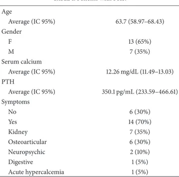
Of 20 accrued patients, 13 (65%) were females and 7 (35%) were males. The average age was 63.7 years, with a range from 40 to 80 years (average: 65 years).
No patient was affected by or had a documented family history of multiple endocrine neoplasia (MEN 1 or MEN 2A). Clinically 6 patients (30%) were PHP asymptomatic and 14 (70%) were PHP symptomatic (symptoms shown in
Table 1).
None of the treated patients had a concomitant thyroid disease, and none had previously undergone surgery on the cervical region.
Preoperatively, all patients had hypercalcemia with an average serum calcium of 12.26 mg/dL (IC 95%: 11.49–13.03).
Table 1: Patients with PHP. Age Average (IC 95%) 63.7 (58.97–68.43) Gender F 13 (65%) M 7 (35%) Serum calcium Average (IC 95%) 12.26 mg/dL (11.49–13.03) PTH Average (IC 95%) 350.1 pg/mL (233.59–466.61) Symptoms No 6 (30%) Yes 14 (70%) Kidney 7 (35%) Osteoarticular 6 (30%) Neuropsychic 2 (10%) Digestive 1 (5%) Acute hypercalcemia 1 (5%)
All cases showed an elevated PTH with a mean value of 350.1 pg/mL (IC 95%: 233.59–466.61) (Table 1).
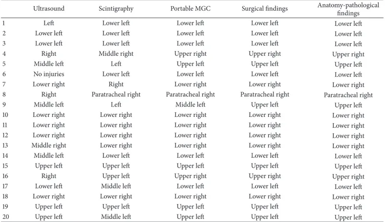
The data obtained from preoperative imaging studies (ultrasound and scintigraphy with 99mTc-sestaMIBI), per-formed in all patients, are depicted inTable 2.
All patients underwent surgery, according to the indica-tions provided by the latest available guidelines [3] for surgi-cal treatment of patients with symptomatic or asymptomatic PHP.
All surgical findings were confirmed by frozen sec-tion examinasec-tion at the time of surgery and by definitive histopathology of the excised tissue. The details of preopera-tive and intraoperapreopera-tive localisation data, as well as the surgical and pathologic findings, are given in Tables2and3.
Having considered the results obtained with the ultra-sound, the preoperative scintigraphy, and the pMGC, we evaluated the ability of the three methods to properly locate the pathological glands on the basis of the side and of the quadrant of the neck. As regards side localisation, neck ultrasound showed a sensitivity of 95% and a specificity of 100% with a diagnostic accuracy of 99%, a positive predictive value (PPV) of 100%, and a negative predictive value (NPV) of 98.76%. When considering quadrant localisation, the values decreased substantially with a sensitivity of 55%, a specificity of 95%, a diagnostic accuracy of 87%, PPV of 73.33%, and NPV of 89.41%.
The preoperative scintigraphy with 99mTc-sestaMIBI showed sensitivity, specificity, diagnostic accuracy, PPV, and NPV results of 100% with regard to side localisation, whereas quadrant location showed values of 73.68% and 96.05%, respectively, for sensitivity and specificity, with a diagnostic accuracy of 91.58%, PPV of 82.35%, and NPV of 93.59%.
With side localisation the pMGC showed sensitivity, specificity, diagnostic accuracy, PPV, and NPV values of 100%, while with quadrant localisation it showed sensitivity
and specificity levels of 95% and 98.89%, respectively, a diagnostic accuracy of 98.18%, PPV of 95%, and NPV of 98.88% (Tables4and5).
In 7 cases (35%) the pMGC gave more accurate informa-tion than the neck ultrasound and in 5 cases (25%) more than the preoperative scintigraphic scans.
After removal of parathyroid lesions, MGC scanning demonstrated no sources of residual radioactivity in the operating field of any patient.
The ioPTH according to Miami criteria [13] showed a significant decrease (over 50%) in all patients (100%).
The average operative time was 37.75 minutes (IC 95%): 31.06–44.44 with a median time of 32 minutes. The compar-ison with a previous group of patients undergoing parathy-roidectomy showed no significant differences in terms of average and median duration (39.2 minutes (IC 95%: 28.22– 50.17) and 30 minutes, resp.).
There was no mortality or morbidity in the study group. Postoperative 3rd-day monitoring of PTH and serum calcium demonstrated the following mean values: PTH 28.64 pg/mL (IC 95%: 22.48–38.4), with a decrease of 328.29 pg/mL from preoperative data, and serum calcium 9.22 mg/dL (IC 95%: 7.64–10.8) with a decrease of 3.69 mg/dL from preoperative data. At the clinical and laboratory follow-up after 1, 6, and 12 months, no persistent or recurrent PHP was detected.
4. Discussion
The traditional surgical approach to PHP treatment consisted of bilateral cervical exploration [14,15]: this technique led to a success rate of up to 95%, when performed by dedicated surgeons [16]. As 85% of PHP cases are supported by a single parathyroid adenoma [17], minimally invasive surgery was introduced, with a focal approach to parathyroid lesions, made possible thanks to the improvements in preoperative localisation and to the introduction of ioPTH [4].
The most frequently used preoperative localisation meth-ods are neck ultrasound and scintigraphy with 99m Tc-sestaMIBI [18]. The ultrasound has a sensitivity of 76.1% (range 30%–88%) [19, 20], which reaches a value of 97% [21], and a specificity of 40% [20] in case of typical cervical localisation.
The scintigraphy with 99mTc-sestaMIBI provides infor-mation of functional type [22]. Its sensitivity for detecting single adenomas varies between 68 and 95%, which decreases to 44% in case of hyperplasia and 30% in case of double ade-nomas [18]. The SPECT (Single Photo Emission Computed Tomography) technique associated with CT (SPECT/CT) is useful in case of concomitant nodular thyroid disease or ectopic parathyroid glands (sensitivity over 90%) [23].
The CT and RMN are usually reserved for persistent or recurrent cases of PHP or ectopic adenomas localisation or when the ultrasound and the scintigraphy do not identify the same localisation [24,25].
Intraoperative localisation techniques have been intro-duced in order to guide the surgeon during the parathy-roidectomy, overcoming the limitations of preoperative imag-ing [12,26].
Table 2: Results of preoperative and intraoperative surgical findings and pathological anatomical study locators.
Ultrasound Scintigraphy Portable MGC Surgical findings Anatomy-pathological
findings
1 Left Lower left Lower left Lower left Lower left
2 Lower left Lower left Lower left Lower left Lower left
3 Lower left Lower left Lower left Lower left Lower left
4 Right Middle right Upper right Upper right Upper right
5 Middle left Left Upper left Upper left Upper left
6 No injuries Lower left Lower left Lower left Lower left
7 Lower right Right Lower right Lower right Lower right
8 Right Paratracheal right Paratracheal right Paratracheal right Paratracheal right
9 Middle left Left Middle left Upper left Upper left
10 Lower right Lower right Lower right Lower right Lower right
11 Lower right Lower right Lower right Lower right Lower right
12 Lower right Lower right Lower right Lower right Lower right
13 Middle right Lower right Lower right Lower right Lower right
14 Middle left Lower left Lower left Lower left Lower left
15 Upper left Upper left Upper left Upper left Upper left
16 Right Upper left Upper right Upper right Upper right
17 Lower left Middle left Lower left Lower left Lower left
18 Lower right Lower right Lower right Lower right Lower right
19 Upper left Upper left Upper left Upper left Upper left
20 Upper left Middle left Upper left Upper left Upper left
Table 3: Surgical findings.
Lower left adenoma 6 (30%)
Lower right adenoma 6 (30%)
Upper left adenoma 5 (25%)
Upper right adenoma 2 (10%)
Ectopic adenoma 1 (5%) paratracheal right
Table 4: Sensitivity, specificity, diagnostic accuracy, PPV, and NPV of the localisation investigations used according to the side of the neck.
Sensitivity Specificity Diagnostic
accuracy PPV NPV
Ultrasound 95% 100% 99% 100% 98.76%
Preoperative
scintigraphy 100% 100% 100% 100% 100%
pMGC 100% 100% 100% 100% 100%
In 1971 Dudley [12] was the first to report the intra-operative use of methylene blue: this method, however, has limitations, arising from the fact that even the normal parathyroid glands may be discoloured, just like thyroid nodules and lymph nodes [9].
The use of a gamma probe is the basis of minimally invasive radioguided parathyroidectomy (MIRP), proposed for the first time by Norman and Chheda in 1997 [26].
Table 5: Sensitivity, specificity, diagnostic accuracy, PPV, and NPV of the localisation used according to the quadrant of the neck.
Sensitivity Specificity Diagnostic
accuracy PPV NPV
Ultrasound 55% 95% 87% 73.33% 89.41%
Preoperative
scintigraphy 73.68% 96.05% 91.58% 82.35% 93.59%
pMGC 95% 98.89% 98.18% 95% 98.88%
The use of gamma probes allows the detection of sources of radiotracers administered at the time of induction of anaesthesia [27].
Gamma probes, however, translate focus intensity into count rate and audio signalling and as such do not guarantee the more precise localisation given by imaging [28].
A further improvement was provided by the introduction of the pMGC, small in size and lightweight, which can provide scintigraphic images during surgery [10,29,30]. The use of the pMGC involves 99mTc-sestaMIBI being injected before surgery and the acquisition of scans before the lesion is removed, in order to locate the lesion itself, and also after it has been removed, to confirm the excision of the whole pathological tissue, along with the scanning of the specimens [10]. The first clinical application of this method appeared in 2007 [10].
The pMGC presented high levels of sensitivity and specificity, higher than those of the preoperative ultrasound
and scintigraphy [10,11], and appears to be useful in cases of concomitant thyroid disease and in cases with negative preoperative studies [11].
Estrems and colleagues [11] evaluated the feasibility of this method in a group of 29 patients: side localisation with pMGC showed a sensitivity of 86.6% and a specificity of 90.9% compared to the 79.3% and 92.5%, respectively, of preoperative investigations (ultrasound + scintigraphy) while quadrant localisation showed a sensitivity of 83.3% and a specificity of 90.9%, when compared to 48.35% and 72.7%, respectively, reported in the preoperative surveys.
In our experience the pMGC properly localised all lesions by side (diagnostic accuracy 100%) with both a sensitivity and a specificity of 100%, while as far as quadrant was considered pMGC showed a diagnostic accuracy of 98.1%, a sensitivity of 95.0%, and a specificity of 98.8% (Tables4and5).
In particular, its use guaranteed a more precise localisa-tion of lesions in 7 cases (35%) with respect to ultrasound scan and in 5 cases (25%) with respect to scintigraphy with 99mTc-sestaMIBI. The data obtained from our experience are comparable to those reported previously [10,11,31].
This technology appears to be a potential alternative to ioPTH measurement allowing obtaining and comparing easy-to-read images before and after excision of the parathy-roid lesions [31,32]. The absence of radiotracer uptake sources in the postexcision images confirms the completeness of the parathyroidectomy, completeness that is usually confirmed by the significant fall in PTH, also.
Despite the limited number of patients studied, our study has confirmed the possibility of replacing the ioPTH measurement with the intraoperative use of the pMGC, because in all cases the images obtained after removal of the 12 parathyroid lesions were comparable to the fall in ioPTH levels. The pMGC may also be a possible alternative to preoperative scintigraphy with99mTc-sestaMIBI.
As far as operative time is concerned, there was no statistical difference between the duration of the mini-invasive radioguided parathyroidectomy with pMGC and the duration of a parathyroidectomy guided only by preoperative studies (𝑝 > 0.05). This may lead to the inference that the additional time used to acquire the images was offset by the easier retrieval of surgical lesions, made possible by the more precise information provided by the pMGC.
5. Conclusions
We believe that the pMGC may be used as an intraoperative method to locate the correct position of the pathological parathyroid glands. The pMGC is more reliable than the preoperative and intraoperative localisation techniques used so far. Our study has also confirmed that the pMGC could replace the ioPTH measuring, by comparing the images obtained before and after the excision of the parathyroid lesions.
Conflict of Interests
The authors declare no conflict of interests regarding the publication of this paper.
References
[1] B. C. Stack Jr., “Minimally invasive radioguided parathyroidec-tomy,” Operative Techniques in Otolaryngology, vol. 20, no. 1, pp. 54–59, 2009.
[2] R. Mihai, D. Simon, and P. Hellman, “Imaging for pri-mary hyperparathyroidism–an evidence-based analysis,”
Lan-genbeck’s Archives of Surgery, vol. 394, no. 5, pp. 765–784, 2009.
[3] J. P. Bilezikian, A. A. Khan, and J. T. Potts Jr., “On behalf of the Third International Workshop on the Management of Asymp-tomatic Primary Hyperparathyroidism. Guidelines for the management of asymptomatic primary hyperparathyroidism: summary statement from the third international workshop,”
The Journal of Clinical Endocrinology & Metabolism, vol. 94, pp.
335–339, 2009.
[4] K. Lorenz, P. Nguyen-Thanh, and H. Dralle, “Unilateral open and minimally invasive procedures for primary hyperparathy-roidism: a review of selective approaches,” Langenbeck’s Archives
of Surgery, vol. 385, no. 2, pp. 106–117, 2000.
[5] P. Miccoli, P. Berti, G. Materazzi, C. E. Ambrosini, L. Fre-goli, and G. Donatini, “Endoscopic bilateral neck exploration versus quick intraoperative parathormone assay (qPTHa) dur-ing endoscopic parathyroidectomy: a prospective randomized trial,” Surgical Endoscopy and Other Interventional Techniques, vol. 22, no. 2, pp. 398–400, 2008.
[6] D. Rubello, D. Casara, and M. R. Pelizzo, “Symposium on parathyroid localization. Optimization of peroperative proce-dures,” Nuclear Medicine Communications, vol. 24, no. 2, pp. 133–140, 2003.
[7] C. D. Phillips and D. R. Shatzkes, “Imaging of the parathyroid glands,” Seminars in Ultrasound, CT and MRI, vol. 33, no. 2, pp. 123–129, 2012.
[8] M. Shabtai, M. Ben-Haim, Y. Muntz et al., “140 Consecutive cases of minimally invasive, radio-guided parathyroidectomy: lessons learned and long-term results,” Surgical Endoscopy and
Other Interventional Techniques, vol. 17, no. 5, pp. 688–691, 2003.
[9] D. B. Kuriloff and K. V. Sanborn, “Rapid intraoperative local-ization of parathyroid glands utilizing methylene blue infusion,”
Otolaryngology—Head and Neck Surgery, vol. 131, no. 5, pp. 616–
622, 2004.
[10] J. Ortega, J. Ferrer-Rebolleda, N. Cassinello, and S. Lledo, “Potential role of a new hand-held miniature gamma camera in performing minimally invasive parathyroidectomy,” European
Journal of Nuclear Medicine and Molecular Imaging, vol. 34, no.
2, pp. 165–169, 2007.
[11] P. Estrems, F. Guallart, P. Abreu, P. Sopena, J. Dalmau, and R. Sopena, “The intraoperative mini gamma camera in pri-mary hyperparathyroidism surgery,” Acta Otorrinolaringologica
Espanola, vol. 63, no. 6, pp. 450–457, 2012.
[12] N. E. Dudley, “Methylene blue for rapid identification of the parathyroids,” British Medical Journal, vol. 3, no. 776, pp. 680– 681, 1971.
[13] M. Barczynski, A. Konturek, A. Hubalewska-Dydejczyk, S. Cichon, and W. Nowak, “Evaluation of Halle, Miami, Rome, and Vienna intraoperative iPTH assay criteria in guiding minimally invasive parathyroidectomy,” Langenbeck’s Archives of Surgery, vol. 394, no. 5, pp. 843–849, 2009.
[14] Consensus Development Conference Panel, “Diagnosis and management of asymptomatic primary hyperparathyroidism: consensus development conference statement,” Annals of
[15] J. L. Doppman, “Preoperative localization of parathyroid tissue in primary hyperparathyroidism,” in The Parathyroids, p. 475, Academic Press, San Diego, Calif, USA, 2nd edition, 2001. [16] R. Harris, H. Ryu, T. Vu et al., “Modern approach to surgical
intervention of the thyroid and parathyroid glands,” Seminars
in Ultrasound, CT and MRI, vol. 33, no. 2, pp. 115–122, 2012.
[17] W. D. Fraser, “Hyperparathyroidism,” The Lancet, vol. 374, no. 9684, pp. 145–158, 2009.
[18] N. A. Johnson, S. E. Carty, and M. E. Tublin, “Parathyroid imaging,” Radiologic Clinics of North America, vol. 49, no. 3, pp. 489–509, 2011.
[19] C. C. Solorzano, D. M. Carneiro-Pla, and G. L. Irvin III, “Sur-geon-performed ultrasonography as the initial and only local-izing study in sporadic primary hyperparathyroidism,” Journal
of the American College of Surgeons, vol. 202, no. 1, pp. 18–24,
2006.
[20] B. J. Ammori, M. Madan, T. D. Gopichandran et al., “Ultra-sound-guided unilateral neck exploration for sporadic primary hyperparathyroidism: is it worthwhile?” Annals of the Royal
College of Surgeons of England, vol. 80, no. 6, pp. 433–437, 1998.
[21] D. Hajioff, T. Iyngkaran, C. Panagamuwa, D. Hill, and M. P. Stearns, “Preoperative localization of parathyroid adenomas: ultrasonography, sestamibi scintigraphy, or both?” Clinical
Oto-laryngology & Allied Sciences, vol. 29, no. 5, pp. 549–552, 2004.
[22] D. Rubello, G. Mariani, and M. R. Pelizzo, “Minimally invasive radio-guided parathyroidectomy on a group of 452 primary hyperparathyroid patients,” NuklearMedizin, vol. 46, no. 3, pp. 85–92, 2007.
[23] S. Hassler, D. Ben-Sellem, F. Hubele, A. Constantinesco, and C. Goetz, “Dual-isotope 99mTc-MIBI/123I parathyroid scintig-raphy in primary hyperparathyroidism: comparison of sub-traction SPECT/CT and pinhole planar scan,” Clinical Nuclear
Medicine, vol. 39, no. 1, pp. 32–36, 2014.
[24] N. D. Gross, J. L. Weissman, E. Veenker, and J. I. Cohen, “The diagnostic utility of computed tomography for preoperative localization in surgery for hyperparathyroidism,” Laryngoscope, vol. 114, no. 2, pp. 227–231, 2004.
[25] E. L. H¨anninen, T. J. Vogl, T. Steinm¨uller, J. Ricke, P. Neuhaus, and R. Felix, “Preoperative contrast-enhanced MRI of the parathyroid glands in hyperparathyroidism,” Investigative
Radi-ology, vol. 35, no. 7, pp. 426–430, 2000.
[26] J. Norman and H. Chheda, “Minimally invasive parathyroidec-tomy facilitated by intraoperative nuclear mapping,” Surgery, vol. 122, no. 6, pp. 998–1004, 1997.
[27] D. Rubello, A. Al-Nahhas, G. Mariani, M. D. Gross, L. Rampin, and M. R. Pelizzo, “Feasibility and long-term results of focused radioguided parathyroidectomy using a ‘low’ 37 MBq (1 mCi) 99mTc-sestamibi protocol,” International Seminars in Surgical
Oncology, vol. 3, article 30, 2006.
[28] M. Tsuchimochi and K. Hayama, “Intraoperative gamma cam-eras for radioguided surgery: technical characteristics, perfor-mance parameters, and clinical applications,” Physica Medica, vol. 29, no. 2, pp. 126–138, 2013.
[29] L. Menard, Y. Charon, M. Solal et al., “POCI: a compact high
resolution 𝛾 camera for intra-operative surgical use,” IEEE
Transactions on Nuclear Science, vol. 45, no. 3, pp. 1293–1297,
1998.
[30] A. Ferretti, S. Chondrogiannis, A. Marcolongo, and D. Rubello,
“Phantom study of a new hand-held𝛾-imaging probe for
radio-guided surgery,” Nuclear Medicine Communications, vol. 34, no. 1, pp. 86–90, 2013.
[31] N. Cassinello, J. Ortega, and S. Lledo, “Intraoperative real-time 99mTc-sestamibi scintigraphy with miniature gamma camera allows minimally invasive parathyroidectomy without ioPTH determination in primary hyperparathyroidism,” Langenbeck’s
Archives of Surgery, vol. 394, no. 5, pp. 869–874, 2009.
[32] T. Fujii, R. Yajima, S. Yamaguchi, S. Tsutsumi, T. Asao, and H. Kuwano, “Could the eZ-SCOPE AN gamma camera replace intraoperative measurement of iPTH for PHPT?” International
Submit your manuscripts at
http://www.hindawi.com
Stem Cells
International
Hindawi Publishing Corporationhttp://www.hindawi.com Volume 2014
Hindawi Publishing Corporation
http://www.hindawi.com Volume 2014
INFLAMMATION
Hindawi Publishing Corporation
http://www.hindawi.com Volume 2014
Behavioural
Neurology
Endocrinology
International Journal of Hindawi Publishing Corporationhttp://www.hindawi.com Volume 2014
Hindawi Publishing Corporation
http://www.hindawi.com Volume 2014
Disease Markers
Hindawi Publishing Corporation
http://www.hindawi.com Volume 2014
BioMed
Research International
Oncology
Journal ofHindawi Publishing Corporation
http://www.hindawi.com Volume 2014
Hindawi Publishing Corporation
http://www.hindawi.com Volume 2014
Oxidative Medicine and Cellular Longevity
Hindawi Publishing Corporation
http://www.hindawi.com Volume 2014
PPAR Research
The Scientific
World Journal
Hindawi Publishing Corporation
http://www.hindawi.com Volume 2014
Immunology Research
Hindawi Publishing Corporation
http://www.hindawi.com Volume 2014
Journal of
Obesity
Journal ofHindawi Publishing Corporation
http://www.hindawi.com Volume 2014
Hindawi Publishing Corporation
http://www.hindawi.com Volume 2014
Computational and Mathematical Methods in Medicine
Ophthalmology
Journal ofHindawi Publishing Corporation
http://www.hindawi.com Volume 2014
Diabetes Research
Journal ofHindawi Publishing Corporation
http://www.hindawi.com Volume 2014
Hindawi Publishing Corporation
http://www.hindawi.com Volume 2014
Research and Treatment
AIDS
Hindawi Publishing Corporation
http://www.hindawi.com Volume 2014
Gastroenterology Research and Practice
Hindawi Publishing Corporation
http://www.hindawi.com Volume 2014