Adhesive restoration of endodontically treated premolars: influence of posts on cuspal deflection
8
0
0
Testo completo
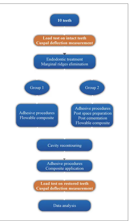
(2) n. 10 teeth. ot. Q ui. by N ht. pyrig No Co t fo rP ub lica tio n te ss e n c e. Acquaviva et al. fo r. Load test on intact teeth Cuspal deflection measurement. Endodontic treatment Marginal ridges elimination. Group 1. Group 2. Adhesive procedures Flowable composite. Adhesive procedures Post space preparation Post cementation Flowable composite. Cavity recontouring. Adhesive procedures Composite application. Load test on restored teeth Cuspal deflection measurement. Data analysis. Fig 1 Study design.. Other authors focused their attention on the role of fiberreinforced composite (FRC) posts. A study by Salameh et al44 observed that in endodontically treated mandibular molars restored with composite resins and loaded to fracture, the resistance is influenced by the number of residual walls, and that fiber-reinforced posts optimized fracture patterns. A similar conclusion was drawn by Akkayan et al,2 who reported that the use of a quartz fiber post was associated with the highest fracture load if compared to titanium, glass 280. fiber, or zirconia posts; in addition, fractures of quartz or glass fiber posts were more likely to be reparable. Courmier et al15 compared the fracture resistance of different post systems and found a lower incidence of unrestorable fractures in fiber posts compared to conventional posts. Although post-and-core full crowns or cuspal coverage onlays should still be considered the gold standard in post endodontic restorations,17 an in vivo investigation carried out by Mannocci et al34 reported no significant difference in The Journal of Adhesive Dentistry.
(3) n. ot. Q ui. by N ht. opyrig et al No CAcquaviva t fo rP ub lica tio n te ss e n c e. fo r. Fig 2 Load was applied to the free end of the lever arm (not visible in the picture), and was transmitted to the tooth sample via a 6.3-mm steel cylinder making contact with the upper halves of the occlusal cuspal stopes.. success rate between teeth restored with fiber posts and direct composite restorations compared to post-and-core with full crown coverage after 3 years. Another in vivo study by Ferrari et al20 reported that, in a 2-year time frame, teeth with full crown coverage and a postand-core buildup showed a higher success rate compared to the same restorations without posts. Little information8 can be found on the comparison of adhesive direct resin composite restorations with or without fiber posts. The purpose of this study was thus to evaluate the influence of FRC posts in endodontically treated maxillary premolars on cuspal deflection. The null hypothesis was that no difference exists in elastic behavior under different axial loads between resin composite only and resin composite with fiber post MOD restorations.. MATERIALS AND METHODS Tooth Selection and Preparation Ten noncarious human maxillary premolars, freshly extracted for orthodontic reasons from patients ranging in age from 15 to 29 years, were selected. Each tooth was carefully examined visually, radiographically, and by means of transillumination in order to discard the ones with structural defects (cracks on enamel surface). Teeth were stored in standard saline solution (0.9% NaCl) at 20°C during trials; during preparation and testing procedures, care was taken to prevent dehydration. Each tooth was embedded in self-curing polymethyl methacrylate (PMM) resin cylinders (18 mm in diameter), maintaining the direction of the main vertical axis, up to 2 mm below the cementoenamel junction so as to simulate the alveolar bone support in natural teeth. In order to prevent the tooth from sinking into the resin cylinder under Vol 13, No 3, 2011. load, direct contact of the apex with the plane of the loading device was obtained. Small, smooth vertical surfaces (about 1 mm in diameter) were created on the buccal and oral aspects of the teeth by means of abrasive disks (Sof-lex, 3M ESPE; St Paul, MN, USA). Thus, points of reference to allow sample placement in the same position throughout the whole trial were obtained. Loading Device The loading device (Fig 2) consisted of a steel lever arm with the fulcrum at one end, the sample placed in a clamp at a 10 cm distance from the fulcrum, and at the other end (50 cm from the fulcrum) a part specially prepared to hold the load. Load was applied by adding several 1 kg metal disks; thanks to the lever principle, the actual force applied on the sample (that could be reproduced throughout the trial) were 0, 98, 147, 196, 245 and 294 N. A 6.3-mm-diameter steel cylinder, connected to the lever arm by means of a custom-made joint, was placed between the lever arm and the occlusal surface of samples, thus applying a balanced load along the tooth’s major axis only. The contact points between cylinder and cuspal slopes lay in the higher halves of internal cuspal sides, not interfering with the following endodontic access and restorative procedures. Load Test on Sound Teeth The cuspal deflection induced with the equipment described above was measured by means of two laser sensors systems: Laser Twin Sensor (LMI Technologies; Heerlen, The Netherlands). Laser beams were directed onto the vertical areas created on samples sides, and treated beforehand with a thin coat of opaque varnish. Such coating was meant to avoid instrument reading errors, due to the fact that laser beams can penetrate the opalescent enamel. 281.
(4) Q ui. by N ht. pyrig No Co t In group 1, cavities were etched withfo 37% r P H3PO4 (Total ub20 s, startEtch, Ivoclar Vivadent; Schaan, Liechtenstein) for licawater ing from the enamel; acid was thoroughly rinsed with ti n spray and water excess was removed without te overdryingothe e ss e n cbonding surface. The one-bottle dual-curing etch-and-rinse n. fo r. Fig 3 Standardized endodontic access cavity with marginal ridges removed.. Cuspal deflection value was measured (tolerance ± 1 μm) for each load increase. Since load was applied on both cusps simultaneously, the cuspal displacement was calculated by the sum of the deflection of each cusp. Deflection values were recorded about 10 s after the load was applied, to allow stabilization of deformation values. Cuspal deflection going back to no-load values at the end of each load cycle was checked, in order to avoid a permanent deformation of the dental structure. Endodontic and Restorative Treatments Endodontic access cavities were performed with standard contour shapes (ie, oval for maxillary premolars), wide enough to guarantee the preparation of root canals without coronal interference but preventing any extension beyond the two contact spots with the load cylinder. Root canal therapy was performed with NiTi endodontic instruments (Profile, Maillefer-Dentsply; Ballaigues, Switzerland) and NaOCl irrigation. Root canal system obturation was performed with vertically condensed warm gutta-percha and Pulp Canal Sealer (Kerr; Romulus, MI, USA). After endodontic procedures, the marginal ridges of all samples were eliminated using a cylindrical diamond bur (Fig 3). The width of the isthmus of the occlusal preparation was equal to one-third of the intercuspal distance; the bucco-lingual width of each proximal box was equal to one-third of the tooth width. The height of the box was made so that the cervical edge of the preparation was 1 mm above the cementoenamel junction. The internal edges of the box were then rounded and no bevel was made on the outer edges of the preparation. The ten available premolars were randomly split into two experimental groups of five teeth each. 282. ot. Acquaviva et al. system Excite DSC Single Dose (Ivoclar Vivadent) was applied and light cured for 40 s with a Swiss Master light curing lamp (EMS; Zurich, Switzerland) with an energy output set at 600 mW/cm2. A thin layer of Tetric flow was used on the cavity floor as a buildup material (Table 1). In group 2, post space was prepared in the palatal canal of each tooth with a Largo drill ISO no. 1-2 (Maillefer) and Postec no. 1 (Ivoclar Vivadent) calibrated burs down to a depth of 7 mm, then accurately cleaned with endodontic brushes. Posts were tried in the canal, then cut with a separator disk in order to have a 1-mm space between the post head and occlusal surface. The cavity and root canal were etched, the Excite DSC Single Dose was applied, its excess was removed with air blow and paper cones, and light curing was performed. The same adhesive was applied on the post after cleansing with alcohol. Variolink II (Ivoclar Vivadent) was mixed and placed into the canal with AccuDose NeedleTubes (Centrix; Shelton, CT, USA) in order to avoid macro- and microbubble inclusion. The post was inserted and the excess of cement was removed with a probe and a microbrush. Light curing was performed in the same way mentioned above and the cement was allowed to set for 7 min. A thin layer of Tetric flow material was positioned on the pulp chamber floor as a buildup material. In both groups, the cavity margins were refined with fine grit burs in order to eliminate any excess of flowable composite. The adhesive procedure was repeated and all samples had a self-locking matrix (Automatrix, Dentsply; Konstanz, Germany) placed in order to restore the proximal walls. Cavities were filled with Artemis composite resin (Ivoclar Vivadent) using an anatomical layering technique, starting from the marginal ridges. Each layer of material, not thicker than 2 mm, was light cured with the same curing device for 40 s. Restorations were finished and polished with Astropol polishers (Ivoclar Vivadent). Load Test on Restored Teeth After 24-h storage in physiological saline solution, each sample underwent the cuspal deflection trial under the same experimental conditions described above. Statistical Analysis Skewness and kurtosis of pooled data were assessed and ANOVA was selected. Results for all experimental groups were evaluated with one-way ANOVA for repeated measures. Differences at the 5% level (p < 0.05) were considered statistically significant.. RESULTS The mean cuspal deflection values for each group are shown in Table 2. A different stress-deformation pattern was observed in intact vs treated teeth. In group 2 (composite with fiber post), a smaller increase in cuspal deflection was observed than in group 1 (no post). The Journal of Adhesive Dentistry.
(5) Group 2. 37% H3PO4 Excite DSC (dual-curing adhesive system). 37% H3PO4 Excite DSC (dual-curing adhesive system). Tetric flow (flowable composite). Variolink II (composite cement) Postec FRC (fiber-reinforced composite post) Tetric flow (flowable composite) Artemis (microhybrid composite). n. Group 1. fo r. Artemis (microhybrid composite). ot. Q ui. by N ht. Table 1 Materials for coronal restoration. opyrig et al No CAcquaviva t fo rP ub lica tio n te ss e n c e. Table 2 Mean deflection values. Load (N). 0 98 147 196 245 294. Mean cuspal deflection (μm) Intact (n = 10) Restored Mean ± SD Group 1 (n = 5) Group 2 (n = 5) Mean ± SD Mean ± SD 0 3.43±2.90 4.95±3.49 6.16±4.37 8.39±7.64 12.17±14.88. 0 14.42±9 18.11±10.12 21.65±12.07 25.7±16.43 26.93±16.43. 0 15.35±5.17 16.27±5.28 17.39±5.45 19.27±5.57 20.39±6. ANOVA for repeated measures yielded p = 0.0019 (among) and p = 0.02 (between). Power of ANOVA was 0.976.. DISCUSSION Posterior teeth deflect under load as a function of their structural design. Endodontic access cavities and loss of proximal walls due to caries, especially in MOD cavities,26,40 may increase their proneness to deformation under mechanical forces. Coronal restorations that protect residual tooth structure from these stresses are thus highly recommended. In the past, the gold standard in post-endodontic restoration was full crown coverage.1,17 Using nonbonded materials, both for direct and indirect reconstructions, cuspal coverage was required, but adhesive restorations are now increasingly popular among dentists,7 and some authors18 suggested using these kinds of reconstructions in association with root canal fiber posts. Fiber-reinforced composite post systems were introduced to prevent root fractures and, thanks to a modulus of elasticity similar to that of dentin, yield better results than cast posts.4 In recent years, destructive methods were utilized6,10,50 to evaluate the resistance of the tooth-restoration complex. The results of this type of investigation were strongly influenced by the size and morphology of teeth, which vary greatly. In vitro, fractures due to peak of load may occur with valVol 13, No 3, 2011. ues ranging from 302 to 502 N.48 Such fracture loads due to compression are much higher than those reached within the oral cavity, even in maximum chewing mode; forces during chewing have been reported between 13 and 18 N16 up to a maximum of 147 to 261 N.3,24 Some authors18,22 have observed that tooth fracture seems to occur mostly due to a fatigue phenomenon: over time, repeated stress can greatly reduce the resistance to fracture, even under forces far below the loading force needed to break a healthy tooth.31 Several in vitro studies on post-endodontic restorations are available in the literature, both with destructive2,15,44 and nondestructive13,40,41 techniques. Some interesting clinical trials20,34 have also been carried out to assess the reliability of adhesive techniques and materials compared to the traditional prosthetic procedures. The measurement of cuspal deflection under load has been used in order to investigate both polymerization shrinkage11 and the mechanical properties of the tooth-restoration complex.13,25,40-42 In particular, the nondestructive evaluation of tooth deformation in terms of cuspal deflection under axial load seems to be a valuable means of predicting the ability of the tooth-restoration complex to withstand stress in the oral cavity. The rationale for this approach is that, since there is a linear relationship between fatigue and static loading,22 283.
(6) by N ht. n. fo r. 284. Q ui. the lower the amount of deflection, the lower the fatigue of the tooth-restoration complex and the better the prognosis.51 The aim of this paper was to focus on this last aspect in order to assess differences in reconstructions made with or without the use of fiber resin root canal posts. The original nondestructive protocol employed in this study adopted a load range similar to or just higher (0 to 294 N) than that normally registered under physiological conditions for the type of teeth examined, ie, maxillary premolars. Maxillary premolars were chosen because they have the highest risk of fracture, as reported in the literature.42 Since cuspal deflection was tested on the same tooth samples when they were intact and following endodontic treatment/coronal restoration, the variability bias from tooth to tooth was eliminated or strongly reduced.11,13,40,41 As mentioned above, the influence of fiber post use in direct bonded composite restorations is described in the literature in destructive in vitro tests and a few clinical trials. The present study focussed on the cuspal deflection of intact vs adhesively restored premolars, with or without fiber posts, thus determining the contribution of the post in limiting the cuspal deflection under load. Several papers15,25,27,29,37,40,42 reported a remarkable difference in static load resistance between sound teeth and restored ones, so a restricted number of samples was considered sufficient to test the null hypothesis. According to the measurement method chosen, small flat surfaces were created on the buccal and palatal sides of each sample to allow the laser beams to detect more accurately each cuspal displacement without being misled by tooth anatomy. Otherwise, the convex shape of tooth surfaces could have caused unreliable data following a possible vertical micromovement of the tooth during loading. Total deflection was recorded as the sum of the deflection of both cusps, not considering the concept of “cuspal independence” reported by Sakaguchi et al.43 The creation of endodontic access cavities with removal of marginal ridges was meant to simulate the worst condition for the prognosis of the tooth. However, no deflection test was performed on open MOD cavities, as the weakening of cusps was reported several times in the literature.27,37,40,42,50,51 Such testing could have led to a great loss of samples without being really relevant to the purpose of comparing the two restoration techniques. The materials chosen for the restorations were all by the same manufacturer, in order to ensure maximum compatibility among adhesive system, luting agent, and fiber post. According to the literature,9,33,38 glass fiber posts were preferred to cast posts and carbon posts because of their better performance characteristics and light transmission capability. A further advantage of Postec FRC is that its resin matrix is made of UDMA, more compatible with adhesive systems than the epoxy resin of which other posts are made. Thus, adhesion between resinous cement and post is made possible by mechanical interlocking and by the unreacted carbon double bonds on the post surface, even if the resin matrix is highly cross linked.35 Post insertion depth was chosen at 7 mm,34 as no updated guidelines are available in the literature regarding this aspect.7 Standardization of inser-. pyrig No Co t fo tion depth was preferred to individualization rinPorder to elimubwas used inate a source of variability. A flowable composite lica11 to reduce polymerization stress on the residual cusps. tio n The 24-h delay in deflection testing after te endodontic and ss e n c e comrestorative treatment was meant to let the restorations. ot. Acquaviva et al. plete their polymerization reactions and settle the stress of composite contraction. Load application followed the axis of the tooth, in order to simulate the normal occlusal relationship of maxillary first premolars with their antagonist teeth, and to standardize the test conditions as much as possible. In addition, since deflection was considered as an aspect to prevent fatigue, angulated load direction was avoided, being less likely to occur in the oral cavity than the axial direction. The results obtained confirm an increase in deformation under load in all endodontically treated and adhesively restored teeth compared to intact ones. Standard deviation values were quite high compared to the mean deflection values, as a consequence of anatomical variability: each tooth reacts differently under load, depending on its size, morphology, and age. All restored teeth deflected more than the intact ones; depending on the material employed, bonded coronal restorations then contribute in different ways to recovering the initial properties. Such an increase in cuspal deflection was smaller in group 2, and the differences between the groups are greater at higher load values. Significant differences (p = 0.02) were found between the two experimental groups: since the conditioning and adhesive agents and the composite resin were the same in both groups, the different results can be related to the different mechanical properties (eg, modulus of elasticity) of the whole reconstruction with the fiber post inserted into the root canal. Although the literature reports that posts only provide retention for the core and the coronal restoration and do not strengthen the root,46 a fiber post might improve the ability of the toothrestoration complex to absorb the occlusal loads by distributing stresses along the major axis of the tooth. Comparing the results collected to those of similar studies,27,40,41,51 slight differences in deflection values can be found. A number of factors can be involved in this, such as different types of deflection sensors (strain gauges or differential transformers vs laser sensors), load range, load application mode, restorative material, and MOD preparation design. The results are comparable to those obtained in a similar study13 with different composite materials. While it is difficult to compare data with similar studies, it must be pointed out that several works report the advantages of fiber post application in post-endodontic restorations. A destructive study by Nothdurft et al39 found that the use of a post in premolars with Class II cavities significantly increased the resistance towards extra-axial forces. In addition, the use of a fiber post may optimise eventual crack patterns, making teeth more likely to be restorable2,15,44,47 should a fracture occur. An in vivo study20 also supports the use of fiber posts in post-and-core restorations with full crowns, reporting that over a two-year observation period, post placement resulted in a significant reduction of failure risk especially when three or more coronal walls have been lost. The Journal of Adhesive Dentistry.
(7) Vol 13, No 3, 2011. fo r. 1. Abou-Rass M. Post and core restoration of endodontically treated teeth. Curr Opin Dent 1992;2:99-107. 2. Akkayan B, Gulmez T. Resistance to fracture of endodontically treated teeth restored with different post systems. J Prosthet Dent 2002;87: 431-437. 3. Anderson DJ. Measurement of stress in mastication. I. J Dent Res 1956; 35:664-670. 4. Asmussen E, Peutzfeldt A, Heitmann T. Stiffness, elastic limit, and strength of newer types of endodontic posts. J Dent 1999;27:275-278. 5. Bell AM, Lassila LV, Kangasniemi I, Vallittu PK. Bonding of fibre-reinforced composite post to root canal dentin. J Dent 2005;33:533-539. 6. Belli S, Cobankara FK, Eraslan O, Eskitascioglu G, Karbhari V. The effect of fiber insertion on fracture resistance of endodontically treated molars with MOD cavity and reattached fractured lingual cusps. J Biomed Mater Res B Appl Biomater 2006;79:35-41. 7. Bitter K, Kielbassa AM. Post-endodontic restorations with adhesively luted fiber-reinforced composite post systems: a review. Am J Dent 2007;20:353-360.. ot. REFERENCES. by N ht. Bearing in mind the limits of laboratory research, represented by the need of standardization and elimination of variables, the cuspal deflection values obtained here in restored teeth indicate that the present adhesive systems and composite materials could be a valuable choice to avoid or delay prosthetic solutions, especially in borderline situations such as post-endodontic MOD cavities. The results of the present in vitro research should be confirmed clinically by monitoring the behavior of the tested materials in randomized clinical trials.. 9.. n. CONCLUSION. 8.. Q ui. On the other hand, partly different conclusions were drawn in a randomized clinical trial by Bitter et al:8 after a 32-month observation period, significant differences between post group and no-post group were found only when no coronal walls were present. The authors also recommended evaluating the need for posts when tissue loss is more limited. While the contribution of glass fiber posts appears significant, innovative materials have been introduced in dental practice in recent years. In semi-interpenetrating polymer network (IPN) posts,35 for example, fibers are embedded in a mixture of linear and cross-linked polymers which are not bonded together as a single network. This allows penetration of the bonding resin into the post material, resulting in interdiffusion and thus in a better bond between post and adhesive or luting material.5 Another alternative to the use of prefabricated FRC posts is represented by customized posts, which may be manufactured with either cross-linked glass fiber or IPN posts, with indirect or semi-direct procedures. Customized posts afford the advantage of totally filling the post space, yielding higher adaptation, reducing the quantity of luting agent, and providing more resistance to stress. A further advantage may be obtained when customized IPN posts are used, combining the advantages of better adaptation to that of interdiffusion of adhesive resin and core composite resin. Future developments of the present study might include this class of posts.. opyrig et al No CAcquaviva t foH, Neumann K, KielBitter K, Noetzel J, Stamm O, Vaudt J, Meyer-Lueckel r Pof post placebassa AM. Randomized clinical trial comparing the effects ubresults of ment on failure rate of postendodontic restorations: preliminary lica a mean period of 32 months. J Endod 2009;35:1477-1482. tio Bonfante G, Kaizer OB, Pegoraro LF, do Valle AL. Fracture strength of teeth n t with flared root canals restored with glass fibreeposts. Int Dent J e s s c en 2007;57:153-160.. 10. Burke FJ. Tooth fracture in vivo and in vitro. J Dent 1992;20:131-139. 11. Cara RR, Fleming GJ, Palin WM, Walmsley AD, Burke FJ. Cuspal deflection and microleakage in premolar teeth restored with resin-based composites with and without an intermediary flowable layer. J Dent 2007;35:482-489. 12. Cavel WT, Kelsey WP, Blankenau RJ. An in vivo study of cuspal fracture. J Prosthet Dent 1985;53:38-42. 13. Cerutti A, Flocchini P, Madini L, Mangani F, Putignano A, Docchio F. Effects of bonded composites vs. amalgam on resistance to cuspal deflection for endodontically-treated premolar teeth. Am J Dent 2004;17:295-300. 14. Cobankara FK, Unlu N, Catin AR, Ozkan HB. The effect of different restoration techniques on the fracture resistance of endodontically-treated molars. Oper Dent 2008;33:526-533. 15. Cormier CJ, Burns DR, Moon P. In vitro comparison of the fracture resistance and failure mode of fiber, ceramic, and conventional post systems at various stages of restoration. J Prosthodont 2001;10:26-36. 16. De Boever JA, McCall WD Jr, Holden S,Ash M Jr. Functional occlusal forces: An investigation by telemetry. J Prosthet Dent 1978;40:326-333. 17. Dietschi D, Duc O, Krejci I, Sadan A. Biomechanical considerations for the restoration of endodontically treated teeth: a systematic review of the literature, Part II (Evaluation of fatigue behavior, interfaces, and in vivo studies). Quintessence Int 2008;39:117-129. 18. Duret B, Reynaud M, Duret F. New concept of coronoradicular reconstruction: The Composipost (1) [in French]. Chirur Dent France 1990;60: 131-141. 19. Fennis WM, Kuijs RH, Kreulen CM, Verdonschot N, Creugers NH. Fatigue resistance of teeth restored with cuspal-coverage composite restorations. Int J Prosthodont 2004;17:313-317. 20. Ferrari M, Cagidiaco MC, Grandini S, De Sanctis M, Goracci C. Post placement affects survival of endodontically treated premolars. J Dent Res 2007;86:729-734. 21. Ferrari M, Mannocci F. A ‘one-bottle’ adhesive system for bonding a fibre post into a root canal: an SEM evaluation of the post–resin interface. Int Endod J 2000;33:397-400. 22. Garoushi S, Vallittu PK, Lassila LV. Continuous and short fiber reinforced composite in root post-core system of severely damaged incisors. Open Dent J 2009;3:36-41. 23. Gher ME, Dunlap RM, Anderson MH, Kuhl LV. Clinical survey of fractured teeth. J Am Dent Assoc 1987;114:174-177. 24. Gibbs CH, Mahan PE, Lundeen HC, Brehnan K, Walsh EK, Sinkewiz SL, Ginsberg SB. Occlusal forces during chewing—influences of biting strength and food consistency. J Prosthet Dent 1981;46:561-567. 25. Gonzalez-Lopez S, De Haro-Gasquet F, Vilchez-Diaz MA, Ceballos L, Bravo M. Effect of restorative procedures and occlusal loading on cuspal deflection. Oper Dent 2006;31:33-38. 26. González-López S, Vilchez Díaz MA, de Haro-Gasquet F, Ceballos L, de Haro-Muñoz C. Cuspal flexure of teeth with composite restorations subjected to occlusal loading. J Adhes Dent 2007;9:11-15. 27. Hansen EK. In vivo cusp fracture of endodontically treated premolars restored with MOD amalgam or MOD resin filling. Dent Mater 1988;4: 169-173. 28. Helfer AL, Melnik S, Schilder H. Determination of the moisture content of vital and pulpless teeth. Oral Surg Oral Med Oral Pathol 1972;34:661-670. 29. Howe CA, McKendry DJ. Effect of endodontic access preparation on resistance to crown- root fracture. J Am Dent Assoc 1990;121:712-715. 30. Huang TG, Schilder H, Nathanson D. Effect of moisture content and endodontic treatment on some mechanical properties of human dentin. J Endod 1992;18:209-215. 31. Jantarat J, Palamara JE, Messer HH. An investigation of cuspal deformation and delayed recovery after occlusal loading. J Dent 2001;29:363-370. 32. Lewinstein I, Grajower R. Root dentin hardness of endodontically treated teeth. J Endod 1981;7:421-422. 33. Maceri F, Martignoni M, Vairo G. Mechanical behaviour of endodontic restorations with multiple prefabricated posts: a finite-element approach. J Biomech 2007;40:2386-2398. 34. Mannocci F, Bertelli E, Sherriff M, Watson TF, Ford TR. Three-year clinical comparison of survival of endodontically treated teeth restored with either full cast coverage or with direct composite restoration. J Prosthet Dent 2002;88:297-301.. 285.
(8) 47.. by N ht. 46.. n. fo r. 286. 45.. Q ui. 35. Mannocci F, Machmouridou E, Watson TF, Sauro S, Sherriff M, Pilecki P, Pitt-Ford TR. Microtensile bond strength of resin-post interfaces created with interpenetrating polymer network posts or cross-linked posts. Med Oral Patol Oral Cir Bucal 2008;13:E745-752. 36. Mannocci F, Sherriff M, Watson TF, Vallittu PK. Penetration of bonding resins into fibre-reinforced composite posts: a confocal microscopic study. Int Endod J 2005;38:46-51. 37. Morin D, De Long R, Douglas WH. Cusp reinforcement by the acid-etch technique. J Dent Res 1984;63:1075-1078. 38. Nakamura T, Ohyama T, Waki T, Kinuta S, Wakabayashi K, Mutobe Y, Takano N, Yatani H. Stress analysis of endodontically treated anterior teeth restored with different types of post material. Dent Mater J 2006;25: 145-150. 39. Nothdurft FP, Seidel E, Gebhart F, Naumann M, Motter PJ, Pospiech PR.The fracture behavior of premolar teeth with class II cavities restored by both direct composite restorations and endodontic post systems. J Dent 2008;36:444-449. 40. Panitvisai P, Messer HH. Cuspal deflection in molars in relation to endodontic and restorative procedures. J Endod 1995;21:57-61. 41. Reeh ES, Douglas WH, Messer HH. Stiffness of endodontically-treated teeth related to restoration technique. J Dent Res 1989;68:1540-1544. 42. Reeh ES, Messer HH, Douglas WH. Reduction in tooth stiffness as a result of endodontic and restorative procedures. J Endod 1989;15:512-516. 43. Sakaguchi RL, Brust EW, Cross M, DeLong R, Douglas WH. Independent movement of cusps during occlusal loading. Dent Mater 1991;7:186-190. 44. Salameh Z, Sorrentino R, Papacchini F, Ounsi HF, Tashkandi E, Goracci C, Ferrari M. Fracture resistance and failure patterns of endodontically treated mandibular molars restored using resin composite with or without translucent glass fiber posts. J Endod 2006;32:752-755.. pyrig No Co t fo teeth more brittle? J Sedgley CM, Messer HH. Are endodontically treated rP Endod 1992;7:332-335. u fracture resisSorensen JA, Engelman MJ. Effect of post adaptation on b lica tance of endodontically treated teeth. J Prosthet Dent 1990;64:419-424. tio n Sorrentino R, Monticelli F, Goracci C, Zarone F, Tayt FR, García-Godoy F, Fere rari M. Effect of post-retained composite restorationssand amount of coroe s c en nal residual structure on the fracture resistance of endodontically-treated. ot. Acquaviva et al. teeth. Am J Dent 2007;20:269-274. 48. Sorrentino R, Salameh Z, Zarone F, Tay FR, Ferrari M. Effect of post-retained composite restoration of MOD preparations on the fracture resistance of endodontically treated teeth. J Adhes Dent 2007;9:49-56. 49. Vire DE. Failure of endodontically treated teeth: classification and evaluation. J Endod 1991;17:338-342 . 50. Wendt SL Jr, Harris BM, Hunt TE. Resistance to cusp fracture in endodontically treated teeth. Dent Mater 1987;3:232-235. 51. Zidan O, Abdel-Keriem U. The effect of amalgam bonding on the stiffness of teeth weakened by cavity preparation. Dent Mater 2003;19:680-685.. Clinical relevance: Adhesive restoration of endodontically treated teeth is an appropriate way to provide good resistance to occlusal loads. In this context, the use of a fiber post in post-endodontic MOD restorations might be recommended in order to improve the prognosis.. The Journal of Adhesive Dentistry.
(9)
Figura


Documenti correlati
Un deciso mutamento della situazione avviene nei primi anni sessanta, grazie tra l’altro ad un fatto di enorme importanza in una realtà come quella sarda, vale a dire il
Donatella Rita Fiorino (coord.); Giovanni Battista Cocco, Anna Maria Colavitti, Maurizio Memoli, Andrea Pirinu ed Emanuela Quaquero (Università degli Studi di Cagliari); Lisa
Studies meeting the following inclusion criteria were subsequently included in the scoping review: 1 study participants or population with learning disabilities, 2 aged 16 years
Ritgen ’s maneuver was defined as an upward pressure from the coccygeal region to extend the head during actual delivery, using one hand to pull the fetal chin between the maternal
Cigarette smoking and lung cancer - relative risk estimates for the major histological types from a pooled analysis of case-control studies... INTERNATIONAL JOURNAL
The aim of this study is to evaluate the safety and efficacy of the barbed auto-locking absorbable suture for the closure of an anastomotic stapler-access enterotomy during a
Già da diversi anni sono coordinatrice scientifica e didattica di corsi di perfezionamento sulla tematica della marginalità e dell’esclusione sociale, attivati presso la
