AMPA receptor GluA2 subunit defects are a cause
of neurodevelopmental disorders
Vincenzo Salpietro et al.
#AMPA receptors (AMPARs) are tetrameric ligand-gated channels made up of combinations
of GluA1-4 subunits encoded by GRIA1-4 genes. GluA2 has an especially important role
because, following post-transcriptional editing at the Q607 site, it renders heteromultimeric
AMPARs Ca
2+-impermeable, with a linear relationship between current and trans-membrane
voltage. Here, we report heterozygous de novo GRIA2 mutations in 28 unrelated patients with
intellectual disability (ID) and neurodevelopmental abnormalities including autism spectrum
disorder (ASD), Rett syndrome-like features, and seizures or developmental epileptic
ence-phalopathy (DEE). In functional expression studies, mutations lead to a decrease in
agonist-evoked current mediated by mutant subunits compared to wild-type channels. When
GluA2 subunits are co-expressed with GluA1, most GRIA2 mutations cause a decreased
current amplitude and some also affect voltage recti
fication. Our results show that de-novo
variants in GRIA2 can cause neurodevelopmental disorders, complementing evidence that
other genetic causes of ID, ASD and DEE also disrupt glutamatergic synaptic transmission.
https://doi.org/10.1038/s41467-019-10910-w
OPEN
Correspondence and requests for materials should be addressed to D.M.K. (email:[email protected]) or to H.H. (email:[email protected]).#A full list
of authors and their affiliations appears at the end of the paper.
123456789
A
bnormal glutamatergic synaptic transmission and
plasti-city has been implicated in some neurodevelopmental
disorders (NDDs) featuring intellectual disability (ID),
developmental delay (DD), and autism spectrum disorders
(ASDs)
1–5, as exemplified by the identification of rare de-novo
mutations in genes encoding ionotropic glutamate receptor
(iGluR) subunit genes,
6–13. iGluRs are the major mediators of fast
excitatory neurotransmission in the vertebrate brain
13–17. They
include N-methyl-
D-aspartate receptors (NMDARs), kainic acid
receptors (KARs), and
α-amino-3-hydroxy-5-methyl-4-isoxazole
propionic acid receptors (AMPARs)
14,18. AMPARs are assembled
from four subunits (GluA1-4), with GluA1/GluA2
hetero-tetramers being the most frequent combination in the forebrain
19.
Mutations in GRIA1, GRIA3, and GRIA4 (encoding GluA1,
GluA3, and GluA4) have been established as very rare causes of
NDDs
9,10,20,21. The GluA2 subunit, encoded by GRIA2, has a
major role in the regulation of AMPAR Ca
2+permeation and
voltage rectification, in large part mediated by an arginine residue
in the ion-selectivity
filter that results from post-transcriptional
editing of a codon for glutamine
22,23. Hitherto, evidence for a role
of GluA2 in NDDs has been sparse. A microdeletion case report
previously suggested a possible link between GRIA2
hap-loinsufficiency and ID
24, and an in-frame deletion of three amino
acids was identified in one individual recruited in the Deciphering
Developmental Disorders (DDD) study (
https://www.ddduk.org/
),
containing exome sequencing data from over 13,000 individuals
affected with developmental disorders
3. Furthermore, abnormal
translation of the AMPAR GluA2 subunit via changes in GRIA2
expression or alternative splicing has been implicated in the
pathophysiology and the neurological phenotype of a wide array
of NDDs, including Fragile X syndrome (FXS) and Rett syndrome
(RTT)
25–27. Further underlining the potential importance of
GluA2 for normal CNS development and function, Gria2
−/−mice have increased mortality, show enhanced
NMDAR-independent long-term potentiation, consistent with abnormal
Ca
2+permeation through GluA2-lacking AMPARs, and exhibit
impaired motor coordination and behavioral abnormalities
28.
Despite the hints from the above studies, GRIA2 mutations have
hitherto not been considered an important cause of human
dis-ease, and there is no established disease association for this gene
in the Online Mendelian Inheritance in Man (OMIM) database
(MIM #138247).
We report 28 unrelated individuals, affected with
neurodeve-lopmental abnormalities encompassing ID/DD, ASD, RTT-like
features and seizures or developmental epileptic encephalopathy
(DEE), in whom we have identified heterozygous de novo
var-iants in GRIA2. Functional analyses reveal loss of function for the
majority of the mutations, supporting GluA2 defects as a cause of
NDDs with variable associated neurological phenotypes.
Results
Identification of GRIA2 de-novo variants. The index case was a
4-year-old boy diagnosed with DEE (Patient 1, Supplementary
Table 1) who was found to carry a de-novo variant in GRIA2 by
trio whole exome sequencing (WES). We next screened exomes
and genomes (WGS) from the DDD Study and the SYNaPS Study
Group (
http://neurogenetics.co.uk/synaptopathies-synaps
) and
compared genetic datasets with collaborators and identified seven
individuals carrying GRIA2 de-novo intragenic variants
(Sup-plementary Table 1, Patients 2–8). Through further
collabora-tions and research networks we ascertained sixteen additional
individuals (Patients 9–25) carrying GRIA2 de-novo variants and
three individuals (Patients 26–28) with de-novo 4q32.1
micro-deletions leading to GRIA2 haploinsufficiency (Supplementary
Fig. 1). In total, we found 20 different GRIA2 de-novo intragenic
variants including missense (n
= 15), splice-site (n = 2), in-frame
deletion (n
= 1), stop-gain (n = 1) and frameshift (n = 2) variants
(Supplementary Tables 1–2, Fig.
1
a). Intragenic variants were
first
identified by WES, WGS or massively parallel targeted
sequen-cing and confirmed as de-novo by trio Sanger sequensequen-cing in all
patients (Methods, Supplementary Fig. 2). De-novo
microdele-tions were found by chromosomal microarray analysis (Patients
26–28) and validated using established laboratory protocols.
GRIA2 is constrained and intolerant to loss-of-function. In the
Exome Aggregation Consortium (ExAC) database (
http://exac.
broadinstitute.org
) GRIA2 is highly constrained for missense
variation (z-score: 4.43) and intolerant to loss-of-function (LoF,
intolerance score: 1.00)
29. Among the affected probands reported
here, we found several frameshift, stop-gain and splice-site
var-iants and 4q32.1 microdeletions predicted to lead to GRIA2
haploinsufficiency (Supplementary Table 2). All the identified
intragenic de novo variants were absent from Genome
Aggrega-tion Database (GnomAD,
http://gnomad.broadinstitute.org
) and
ExAC and displayed high conservation (Fig.
1
c, Supplementary
Fig. 3) with a mean: GERP
++score 5.51 and in-silico pathogenic
predictor scores (mean: CADD_Phred 28.924). In total, in our
cohort de-novo GRIA2 variants with predicted LoF were found in
8 out 28 patients. De novo frameshift deletions in Patients 10 and
12 lead to changes in the reading frame with the generation of a
premature stop at codon 37 and 14 amino acids downstream,
respectively. In Patient 19, a single-nucleotide substitution leads
to a stop-gain variant (p.R323ter). The three de novo 4q32.1
microdeletions
identified in patients 26–28 encompass
chr4:157,343,163–158,271,008 bp (GRCh37/hg19) as the smallest
overlapping deleted genomic region, and within this region
GRIA2 is the gene most intolerant to LoF (Supplementary
Tables 3–5). De novo splice-site variants in Patients 8 and 11 are
predicted to cause loss of donor splice sites at exons 1 and 11,
respectively, according to in-silico Alamut predictions
(Supple-mentary Figs. 4–5)
30. Of the other 19 patients, 18 harbored de
novo missense mutations, and one had a 9-bp deletion predicted
to lead to loss of amino acids 528–530.
Phenotypic spectrum associated with
GRIA2 de novo variants.
Consistent with the role of GluA2 channels in synapse
develop-ment and plasticity
19, phenotypic analysis of patients carrying
de-novo GRIA2 variants demonstrated an NDD spectrum including
ID/DD, developmental regression, ASD, speech impairment,
RTT-like features, and seizures or DEE (Fig.
1
b; Supplementary
Videos 1–5). Supplementary Table 1 summarizes the core
phe-notypic features of all 28 Patients, aged between 3 months and 31
years. In all cases, onset of GRIA2-related NDD occurred in
childhood. Several individuals had normal early developmental
milestones and started to exhibit variable impairment of motor
coordination, social interaction, and language abilities in infancy
(Supplementary Table 1, Supplementary Notes 2, 3, 4, and 16). In
some affected individuals, social or language regression was
reported (Patients 3 and 4). Between 2 and 6 years of age several
patients developed RTT-like features (Supplementary Table 1,
Supplementary Table 6), including stereotyped hand movements
(Supplementary Notes 2, 3, 4, and 7), screaming episodes
(Sup-plementary Notes 2, 3, 6, and 14), gait abnormalities including
ataxia and dyspraxia (Supplementary Notes 4 and 6), abnormal
sleep rhythm (Supplementary Notes 4 and 14), and irregular
breathing patterns with hyperventilation episodes
(Supplemen-tary Note 7). Progressive microcephaly was observed in 4 out of
28 individuals (Supplementary Notes 6, 13, 17, and 21) with a
deceleration of head growth usually occurring during infancy
(Supplementary Table 1). Several patients were diagnosed with
ASD (Supplementary Notes 2, 3, 9, 10, 11, 12, 15, 19, 22, 23, and
26), and some presented repetitive behavior patterns and
impaired social interaction (Supplementary Notes 8, 27, and 28).
Language impairment was present in all patients, with the
majority attaining no meaningful speech (Supplementary
Table 1). Twelve patients suffered from seizures (Supplementary
Notes 4 and 5) or DEE (Supplementary Notes 1, 7, 13, 16, 17, 18,
20, 21, 24, and 25) usually starting within the
first 6 months of
life, including infantile spasms, tonic-clonic, myoclonic and focal
seizures (Supplementary Table 7). EEG features included
poly-spikes, slow spike and wave, and bilateral temporal
non-synchronized epileptic activity. The clinical outcome was also
variable (Supplementary Table 1, Supplementary Notes 1, 7, 13,
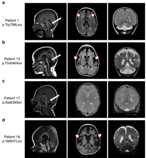
16, 17, 18, 20, 21, and 24). MRI scans in 7 DEE patients showed
progressive brain (mainly cerebellar) atrophy and white matter
abnormalities in some (Fig.
2
; Supplementary Table 1).
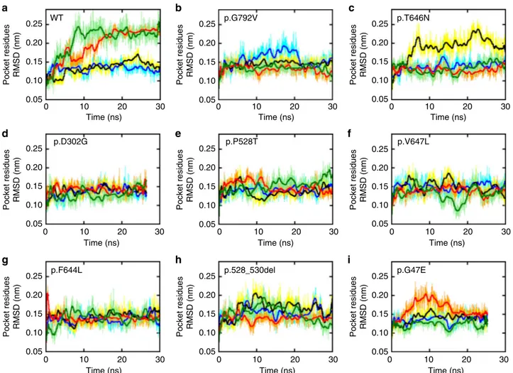
AMPAR Molecular dynamic stimulations. To compare the
structural mobility of GluA2 and its mutants we built a model of
each protein ectodomain including a ligand-binding domain
(LBD) and an amino-terminal domain (ATD; Methods) by
modeling mutations on the wild type and followed their behavior
along time by means of atomistic molecular dynamics simulations
in water solvent. Proteins with mutations in the pore region
where omitted as either the mutations were close to or included in
the transmembrane domains (TMDs) which were not modeled.
We aimed to ascertain the effect of the mutations on the
gluta-mate (GLU) binding-pocket. Interestingly, in the studied mutated
proteins this group of atoms appear to have a higher level of
rigidity compared to the wild-type protein (Figs.
3
and
4
).
Although the wild-type crystal structure is symmetric, after 10 ns
the conformations of pockets associated with chains C and D,
which are coupled in the binding site, diverge with respect to
those associated with A and B which do not change
conforma-tion. In the observed timeframe molecules underwent concerted
macroscopic movements and this is reflected by minor variations
in their backbone root mean squared deviation (RMSD,
Supple-mentary Fig. 9) and radius of gyration (SuppleSupple-mentary Fig. 10).
The RMSD, which is a measure of the average atoms
displace-ment from the starting configuration, clearly indicates that amino
acids in the GLU binding site are independently mobile at a
timescale consistent with our simulations with two pockets
reaching values larger than 0.27 nm. However, the same is not
true for most mutants: p.D302G, p.F644L, p.P528T, and p.V647L
whose RMSD do not exceed 0.22 nm. The remaining mutants
studied showed an intermediate behavior.
Functional analyses of the identi
fied GRIA2 variants. To assess
the functional consequences of GRIA2 missense mutations, we
synthesized cDNA encoding the human GluA2 wild-type and
mutant channels and transfected HEK293T cells together with the
auxiliary stargazin protein (Methods). Except for one mutation
(see below), amino acid position 607 was made to encode an
arginine residue in GluA2 to mimic post-transcriptional editing
of a genomic glutamine-encoding codon. Coding variants in the
N-terminal domain, linkers (including the three amino acid
c.88+2T>C p.G47E AT D LBD TMD CTD p.F595Lfs*37 c.1844+1G>A H. Sapiens M. Musculus B. Taurus G. Gallus D. Rerio D. Melanogaster GRIA1 GRIA2 GRIA3 p.Q/R607G/E (Q/R site) p.D611N COOH M3 M2 M1 M4 p.P528T NH2 p.P286Lfs*14 Patient 1 p.Trp788Leu Patient 2 p.Pro528_Lys530del Patient 3 p.Asp611Asn Patient 12 p.Pro286LeufsX14 Patient 7 p.Arg607Gly Patient 10 p.Phe595LeufsX37 p.D302G p.R323Ter p.V647L p.E776D p.W788L p.G792V p.A807V p.N812S p.G609R p.A639S p.F644L p.T646N S1 M3-S2 link er S2 p.P528_530del
a
b
c
Fig. 1 GRIA2 intragenic de-novo variants identified in this study. a Schematic of the human GluA2 protein (NP_000817.2) indicating the positions of twelve missense changes (dot arrows), two frameshift deletions (cross arrows), two splice-site variants (arrows) and an in-frame deletion (dot arrow). Glutamate binding regions are displayed in red,flip/flop alternatively spliced region is represented in green. b Left panel: Patient 1, carrying the de-novo p.W788L GluA2Flopvariant, at 3 years, exhibitinghypotonia and an oculogyric crisis; he is wheelchair dependent. Middle left panel: Patient 2 (top) carrying the de-novo p.P528_K530del in-frame deletion, at 12 years; Patient 3 (bottom) carrying the de-de-novo p.D611N variant, at 18 years, exhibiting hand-wringing suggestive of RTT. Middle right panel: Patient 7, carrying the de-novo p.Q607E/p.R607G heterozygous mutation (affecting the Q/R editing site) at 10 years, exhibiting hand-wringing as part of a RTT-like presentation. Right panel: Patient 10 (top) carrying the de-novo p.F595LfsX37 variant, at 5 years; Patient 12 (bottom) carrying the de-novo p.P286LfsX14 at 6 years.c Multiple alignment showing GluA2 protein complete conservation across species and inter AMPAR homolog subunits (GluA1, GluA3, and GluA4) alignment. Human GRIA2 (NP_000817.2), mouse GRIA2 (NP_001077275.1), bos taurus GRIA2 (NP_001069789.2), gallus gallus GRIA2 (NP_001001775.2), danio rerio (NP_571970.2), drosophila melanogaster (NP_476855.1), Human GRIA1 (NP_000818.2) Human GRIA3 (NP_015564.4), and Human GRIA4 (NP_000820.3)
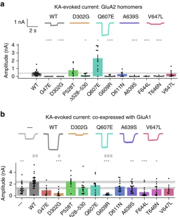
deletion) and pore were selected for functional analysis. Inward
currents evoked by the non-desensitizing agonist kainic acid (KA,
1 mM) on HEK cells expressing homomeric GluA2 were
sig-nificantly decreased for 7 out of the 11 variants tested, including
those associated with NDD or NDD and DEE (Fig.
5
a). Three
variants (p.P528T, p.D611N, and p.V647L) exhibited apparently
normal current amplitudes. In contrast, the p.Q607E variant
exhibited KA-evoked currents that were larger than the wild-type
control. This mutation affects the Q607 codon that is normally
edited to an arginine residue. The increase in current amplitude is
consistent with removal of a positively charged residue from the
ion conduction pathway.
GluA2 homomers are not thought to occur naturally. A
common stoichiometry in the forebrain is channels composed of
two GluA1 and two GluA2 subunits. Presence of the GluA2,
edited at the Q607 site, in the heteromeric channel reduces the
single channel conductance, confers Ca
2+-impermeability, and
results in a linear current-voltage relationship
22,23. In contrast,
homomeric
GluA1
channels
exhibit
a
larger
rectifying
conductance and are Ca
2+-permeable. We therefore repeated
the functional studies co-expressing wild type or mutant GluA2
together with GluA1 and stargazin. Wild-type GluA2
co-expressed with GluA1 yielded approximately two-fold larger
KA-evoked currents than GluA1 alone (Fig.
5
b). Five of the
mutants significantly decreased the KA-evoked current amplitude
relative to wild-type GluA2. Interestingly, two of these variants (p.
D302G and p.G609R) reduced the current below the level
obtained with GluA1 alone, suggesting a dominant negative effect
(Fig.
5
b). Of the remaining variants, all but one (p.P528T)
exhibited an apparent decrease relative to wild type, although
falling short of significance.
We complemented the KA-evoked current amplitude
measure-ments with assessment of rectification by ramping the holding
voltage between
−104 and +76 mV. We confirmed that GluA1
expressed alone yielded a doubly rectifying current-voltage
relationship. It was linear when wild-type GluA2 was
co-expressed (Fig.
6
). Seven of the mutants significantly increased
the degree of rectification compared to wild-type GluA2.
Patient 1 p.Trp788Leu Patient 13 p.Thr646Asn Patient 17 p.Ala639Ser Patient 18 p.Val647Leua
b
c
d
Fig. 2 Brain imaging in 4 individuals with GRIA2-related DEE and brain and cerebellar atrophy. a Sagittal T1 weighted (left panel), Axial T1 weighted (middle panel), andcoronal T2 weighted (right panel) images from Patient 1 (carrying the de-novo p.W788L mutation) scanned at the age of 3 years. There is reduction in the cerebral white matter (red and white arrows), brain volume and marked cerebellar atrophy with vermian deficiency (white arrows). b Sagittal T1 weighted (left panel) images from Patient 13 (carrying the de-novo p.T646N mutation) at the age of 2 months, and axial T1 weighted (middle panel) and coronal T2 weighted images (right panel) from the same Patient at the age of 11 months demonstrate white matter signal abnormality (white and red arrows) with generalized reduction in the cerebral white matter volume and cerebellar atrophy with vermian deficiency (white arrows). c Sagittal T1 weighted (left panel), Axial T2 weighted (middle panel) and coronal T2 weighted (right panel) images from Patient 17 (carrying the de-novo p. A639S mutation) at the age of 6 days; there is an underdeveloped/hypoplastic cerebellum (white arrows) and delayed maturation of myelin.d Sagittal T1 weighted (left panel), axial T1 weighted (middle panel) and coronal T2 weighted (right poanel) images from Patient 18 (carrying the de-novo p.V647L mutation) scanned at the age of 18 months; there is a global cerebral atrophy and white matter changes which suggest hypomyelination (white and red arrows). Although the volume of the cerebellar hemispheres is preserved, atrophy of the inferior cerebellar vermis and wide cerebellar sulci are seen
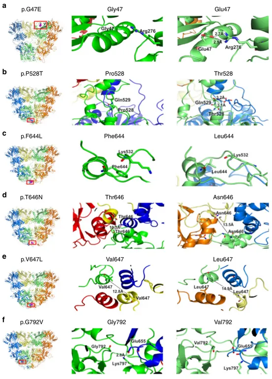
p.G47E p.P528T Pro528 Thr528 Leu644 Phe644 p.F644L p.T646N Thr646 Asn646 Leu647 Val647 p.V647L p.G792V Gly792 Val792 Gly47 Glu47
a
b
c
d
e
f
Fig. 3 Molecular modeling and predicted consequences of 6 GRIA2 de-novo missense variants Left panel: Six modeled GRIA2 de-novo missense variants (highlighted in purple within red rectangle) affecting the amino-terminal domain (ATD) and ligand-binding domain (LBD) of GluA2; Middle panel: particular of the wild-type GlUA2 residue; Right panel: particular of the mutated GluA2 residue.a For variant p.G47E, the presence of the glutamic acid residue (right panel) in place of the glycine residue (middle panel) is predicted to cause the formation of two hydrogen bonds with a neighboring arginine residue; hydrogen bond distances are shown for the mutant structure.b The presence of a Threonine residue in position 528 (right panel) is predicted to cause a slight change in the backbone conformation of the neighboring residues that, in turn, allows hydrogen bonding formation between the two chains forming the LBD domain.c Mutation p.F644L causes the loss of hydrophobic interactions between residue 644 and the side chain of Lys532. d For mutation p. T646N, the presence of a more hydrophilic Asparagine residue increases the distance between the distal chains by about 3 Å, at the interface of LBD and TMD (right panel).e For mutation p.V647L, increased hydrophobicity of Leucine (right panel) compared to Valine (middle panel) increases the separation of helices at the interface between LBD and TMD by about 2 Å.f For variant p.G792V, the presence of a more hydrophobic Valine residue in close proximity to the binding site is predicted to cause a sliding movement with respect to the neighboring chain, disrupting the interchain salt bridge between Glu655 and Lys797; hydrogen bond distance is shown in the wild-type GluA2 (middle panel)
Although consistent with partial or complete loss of GluA2
incorporation in surface-expressed channels, this does not explain
why some mutations that affected rectification, including p.
Q607E, did not significantly reduce the KA-evoked current
amplitude. Indeed, when voltage ramps were applied to cells
expressing p.Q607E without GluA1, the current-voltage
relation-ship was strongly rectifying (Fig.
7
), as expected from loss of a
polyamine-repelling positively charged residue. Overall, all but
one variant (p.P528T) affected either KA-evoked current
amplitude or rectification or both when co-expressed with
GluA1, although in some cases the effects fell short of significance
when unpaired t-tests included Holm-Bonferroni correction for
multiple comparisons (Fig.
8
, Supplementary Table 8).
To determine whether mutations affect channel synthesis or
trafficking, we used a biotinylation assay to probe surface
expression of selected mutants. When co-expressed with GluA1,
p.A639S exhibited a decrease in total expression of GluA2 (Fig.
9
,
Supplementary Fig. 6). As a fraction of total GluA2, protein at the
cell surface was decreased for p.A639S, but also for p.Q607E,
consistent with evidence that the arginine normally present at
codon 607 affects trafficking
31(Fig.
9
).
We modified the GluA2 sequence to examine the effect of two
further codon changes. p.I375V, which is found in 103 subjects
out of 60706 individuals from the ExAC database and is therefore
likely to be a low-frequency variant of uncertain significance, this
increased the current carried by homomeric channels, but not
when co-expressed with GluA1 (Supplementary Fig. 7). We also
introduced another codon change, p.A643T, that corresponds to
the Lurcher mutation in the related non-functional receptor
GluD2, because it is near a cluster of 4 mutations identified in the
cohort (p.A639S, p.F644L, p.T646N, and p.V647L). The Lurcher
mutation disrupts murine cerebellar development and function
by creating a leaky receptor that
fluxes cations in the absence of
ligand. When introduced in GluA2, p.A643T was non-functional,
and the holding current was no different from WT-expressing
cells (or indeed, cells expressing any of the other variants tested,
Supplementary Fig. 7). We conclude that loss of function is
caused by multiple molecular mechanisms involving both altered
channel surface expression and altered channel function, and that
the mechanisms do not involve a Lurcher-like leak conductance
(Supplementary Fig. 8).
Discussion
The genetic and functional expression data presented here
iden-tify de-novo mutations and microdeletions involving GRIA2 as a
cause of NDDs and DEE and underline the importance of the
GluA2 subunit in the regulation of Ca
2+permeation and voltage
rectification of AMPARs and therefore in human synaptic
plas-ticity and brain development and function
22,23,28.
0.25 WT p.D302G p.F644L p.528_530del p.G47E p.P528T p.V647L p.G792V p.T646N 0.20 Pocket residues RMSD (nm) 0.15 0.10 0.05 0.25 0.20 Pocket residues RMSD (nm) 0.15 0.10 0.05 0.25 0.20 Pocket residues RMSD (nm) 0.15 0.10 0.05 0 10 20 Time (ns) 30 0.25 0.20 Pocket residues RMSD (nm) 0.10 0.05 0 10 20 Time (ns) 30 0.25 0.20 Pocket residues RMSD (nm) 0.15 0.15 0.10 0.05 0 10 20 Time (ns) 30 0.25 0.20 Pocket residues RMSD (nm) 0.15 0.10 0.05 0 10 20 Time (ns) 30 0.25 0.20 Pocket residues RMSD (nm) 0.15 0.10 0.05 0 10 20 Time (ns) 30 0.25 0.20 Pocket residues RMSD (nm) 0.15 0.10 0.05 0 10 20 Time (ns) 30 0.25 0.20 Pocket residues RMSD (nm) 0.15 0.10 0.05 0 10 20 Time (ns) 30 0 10 20 Time (ns) 30 0 10 20 Time (ns) 30
a
b
c
d
e
f
g
h
i
Fig. 4 GluA2 Molecular dynamic stimulations predict reduced mobility at the agonist binding site. Root Mean Square Deviation (RMSD) of the GluA2 Glutamate binding pocket (GLU) amino acids along the simulated time for the wild-type protein (top left) and GRIA2 mutants (a–i). In all panels the color code is: chain A (blue/cyan), chain B (black/yellow), chain C (red/orange), and chain D (light/dark green). Thicker lines indicate running averages over 100 samples
The structure of each GluA2 subunit includes (i) a large ATD
(ii) a LBD formed by the proximal part of the N terminus (S1
lobe) and the large loop between transmembrane segments M3
and M4 (S2 lobe) (Fig.
1
), (iii) a TMD formed by hydrophobic
membrane-spanning helices M1, M3 and M4 and the M2 helix
and re-entrant loop, and (iv) a carboxy-terminal (CTD)
intra-cellular region involved in synaptic localization and receptor
regulation
30,32. The intracellular M2 loop together with the M3
helix form the ion-conducting pore
33. Receptor subunits
first
form dimers, then tetramers, and structural studies reveal 2-fold
symmetry of the extracellular domains which transitions to 4-fold
symmetry in the transmembrane domains
34. Three
flexible
stretches of amino acids link the ligand-binding domain to the
transmembrane helices and allow energy transfer from the
ago-nist binding site to the channel gate at the top of M3.
GluA2 subunits are post-transcriptionally edited at the Q/R site
at position 607, where M2 protrudes into the pore, rendering the
channel non-rectifying and calcium-impermeable. In adults,
nearly 100% of GluA2 subunits are in the edited R form. The p.
Q607E mutation identified in the present study is caused by a
cytosine to guanidine base change immediately 5′ to the
adeno-sine that is edited to inoadeno-sine by ADAR2. The normal editing
results in a glutamine (CAG) to arginine (CIG) change, because
the inosine base is read as a guanosine by the ribosome. ADAR2
recognition assays predict that the mutant codon (GAG) is edited
~90% less than the normal CAG
35, so we did not investigate the
effect of a glycine (GGG) residue at position 607. When GluA2
was expressed in HEK cells, we observed increased current for
Q607E homomers which also exhibited inward rectification.
During experiments, we also noticed fewer surviving HEK cells,
suggesting a possible toxic effect of this mutation. In contrast,
another disease-associated mutation two codons 3′ from codon
607, p.G609R, almost eliminated current in both homomeric and
co-expression experiments. Site-directed mutagenesis at or near
the Q/R site 607 was previously shown to cause misassembled
homomeric GluA2 channels which are retained in the ER
32. Our
data suggest that GluA2 subunits with the G609R mutation are
trafficked to the surface but are non-functional and cause
co-expressed subunits to also be non-functional. The p.D611N
var-iant had a milder effect, with a trend towards decreased function
when co-expressed with GluA1.
Importantly, de novo missense variants affecting the M3
channel gate or the M3-S2 linkers have been previously identified
in several iGluR subunit genes (e.g., GRIA1, GRIA3, and GRIA4);
phenotypes of these patients include ID, autism and epilepsy
7–10.
We identified four mutations associated with
neurodevelop-mental phenotypes in on near the SYTANLAAF motif, a highly
conserved nine-amino acid region at the top of the M3
trans-membrane helix, which forms the channel gate
36. Extensive prior
work demonstrates the sensitivity of this motif to mutation across
the iGluR superfamily. For example, a mutation associated with
NDD affecting the eighth residue in GluA1 (p.A636T;
SYTAN-LAAF) results in leaky channels
9, and the equivalent mutation (p.
A654T) in the GRID2 gene (encoding the GluD2 receptor) was
associated with human movement disorder
37as well as the
Lurcher mouse ataxic phenotype
38. We tested the analogous
GluA2 p.A643T as a positive control in our electrophysiology
experiments and observed loss of KA-evoked current but no
change in holding current that would suggest a leak. The p.A653T
mutation in GluA3, affecting the seventh residue (SYTANLAAF),
causes NDD and altered sleep and eliminates KA-evoked
cur-rents
20. Structural data in GluA2 homomers places this residue in
close proximity to A639 (SYTANLAAF) of the adjacent subunit
and suggests that A639 may act as a
‘hinge’ in the
M3 structure
39,40. In our study, p.A639S caused loss of
KA-evoked current in GluA2 homomers and a decrease in current
when GluA1was co-expressed, showing that this position is
highly sensitive to even a conservative amino acid change. The
p.F644L variant is the top of the SYTANLAAF motif, and p.
T646N and p.V647L map two and three amino acids downstream
of the SYTANLAAF motif, respectively. For p.V647L, an
inher-ited mutation at the equivalent site in GRIA1 (p.V640L) caused
ASD
9. For p.T646N, reduced current in GluA1-GluA2
co-expression and partial reduction in RI would be consistent with
reduced surface expression combined with impaired channel
gating (although a decrease in surface expression was observed it
did not reach significance). Molecular dynamic simulations
indicated a loss of symmetry between subunit pairs in the
pre-sence of p.T646N, with Chain B alone becoming more mobile, in
contrast with the WT channel where chains moved in pairs
(Fig.
4
). This suggests that p.T646N compromises tertiary
struc-ture stability. In contrast, p.F644L appeared to be robustly
expressed but caused reduction in currents to below the level of
GluA1 when co-expressed, demonstrating a gating deficiency
which impacts co-assembled WT subunits.
In summary, we noted that effects on currents greatly varied
among mutants, with striking differences even when comparing
nearby residues. Three mutations (p.D302G, p.G609R, p.F644L)
eliminated the GluA1 current when co-expressed, suggesting that
they lock other subunits into non-functional channels. They are
all located in different domains. Perhaps surprisingly, these
mutations are not clearly associated with a more severe
KA-evoked current: GluA2 homomers
KA-evoked current: co-expressed with GluA1
— WT D302G Q607E A639S V647L WT D302G Q607E A639S V647L 2 s 1 nA 0 2 4 Amplitude (nA) ** *** *** ** *** * ## # ### 0 1 2 3 4 Amplitude (nA) *** *** * * *** *** *** ***
a
b
WT G47ED302G P528T Δ528–530 Q607EG609R D611N A639S F644LT646N V647L WT G47ED302G P528T Δ528–530 Q607EG609R D611N A639S F644LT646N V647L —Fig. 5 Agonist-evoked currents in HEK cells. 1 mM KA was applied transiently to HEK cells expressing GluA2 and stargazin (a), or GluA1, GluA2 and stargazin (b). Amplitude was compared to WT (*p < 0.05, **p < 0.01, ***p < 0.001) and for co-expression also to the negative control (GluA1 without GluA2,—, #p < 0.05, ##p < 0.01, ###p < 0.001). Mean ± SEM. Data are from the following numbers of independent cells: (a) WT:25, G47E:7, D302G:6, P528T:7,Δ528–530:11, Q607E:10, G609R:13, D611N:8, A639S:10, F644L:6, T646N:10, V647L:10 (b)—:21, WT:28, G47E:7, D302G:10, P528T:9,Δ528–530:15, Q607E:12, G609R:11, D611N:15, A639S:19, F644L:14, T646N:12, and V647L:6
−70 40 −2 2 — −70 40 −2 2 WT −70 40 −2 2 G47E −70 40 −2 2 D302G −70 40 −2 2 P528T −70 40 −2 2 Δ528-530 −70 40 −2 2 Q607E −70 40 −2 2 G609R −70 40 −2 2 D611N −70 40 −2 2 A639S −70 40 −2 2 F644L −70 40 −2 2 T646N −70 40 −2 2 V647L 0 1 RI *** *** *** * *** *** ** * ### ### ### ### ### ### ### # ### nA mV WT G47E D302G P528T Δ528–530 Q607EG609R D611N A639S F644L T646N V647L —
Fig. 6 Rectification of GluA2 mutants when co-expressed with GluA1. Voltage was ramped from −104 mV to +76 in order to assess rectification of KA-evoked currents. Ramp currents recorded in the absence of KA were subtracted from ramp currents in 1 mM KA. Controls showed linear current–voltage (I–V) relations for cells co-expressing GluA1 and GluA2 WT, and clear rectification in cells transfected with GluA2 alone. The panels show average ramps ±SEM for each mutant (n ≥ 6 cells per mutant). Rectification index (RI) was quantified as (I+40/I−70) *(−7/4). RI was compared to WT (*p < 0.05, **p <
0.01, ***p < 0.001) and GluA1-negative control (#p < 0.05, ##p < 0.01, ###p < 0.001). Data are averaged from the following numbers of cells per mutant: —:19, WT:19, G47E:6, D302G:7, P528T:9, Δ528–530:14, Q607E:10, G609R:11, D611N:13, A639S:18, F644L:10, T646N:10, V647L:5
0.0 0.5 1.0 RI *** * −70 40 −2 2 WT −70 40 −2 2 Q607E −70 40 −2 2 D611N Q607 D611 M3 M2 M2 M3
b
WT Q607E D611Na
Fig. 7 Changes at the polyamine binding site alter rectification in GluA2 homomeric channels. a Pore detail of 2 opposing subunits in GluA2 tetramer (cryo-EM structure pdb 6dm0).b Ramps were applied to HEK cells expressing homomeric GluA2 channels and rectification quantified as for Fig.4(*p < 0.05, **p < 0.01, ***p < 0.001). Number of cells recorded per mutant: WT:6, Q607E:7, D611N:3
phenotype. For another three mutations (p.G47E, p.Q607E, and
p.A639S), GluA2 surface protein was significantly reduced in the
co-expression western blots, showing that defects in
hetero-merization and/or surface trafficking contribute to some
pheno-types. This mechanism also shows no correlation with structural
location, again highlighting the diversity among the tested
mutants. In general, we are unable to predict clinical severity on
the basis of receptor physiology. However, in the case of p.Q607E,
we speculate that gain-of-function in the homomeric channel
could explain the patient’s seizures. The most severe clinical cases
were associated with p.A639S, which caused epileptic
encepha-lopathy and death in infancy. This mutation caused a significant
reduction in total GluA2 protein, but our analysis was hampered
by a lack of transfected cells, suggesting that this mutation should
be investigated for cellular toxicity.
Further clues to the function of GluA2 come from genetically
engineered mice.
“Q/R” editing-deficient knock-in mice die of
sei-zures at 3 weeks
41, whilst Gria2
−/−mice have increased mortality,
impaired motor coordination and behavioral abnormalities
28.
Gria2
−/−mice also exhibit enhanced long-term potentiation in
hippocampal principal neurons that is resistant to blockade of
NMDA receptors, consistent with Ca
2+entry via AMPARs
trig-gering an increase in synaptic strength. A similar
NMDAR-independent form of LTP normally occurs in a subset of
inter-neurons with rectifying AMPARs, which are deficient in GluA2
42.
Although heterozygous Gria2
+/−mice are developmentally normal,
they also exhibit NMDAR-independent long-term potentiation in
principal neurons, albeit less than homozygous mice
43.
The difference between the severe neurodevelopmental
phe-notypes reported in the present study and the heterozygous mice
suggests that the human brain is more sensitive to AMPAR
dysfunction. For several mutations associated with either NDD or
DEE we observed increased rectification of AMPARs when
GluA1 and mutant GluA2 were co-expressed, consistent with
failure to incorporate mutant GluA2 in heteromeric channels.
Although no clear genotype-phenotype correlations emerge for
the majority of patients, we did observe a striking correspondence
between specific recurrent mutations and the individual
pheno-types. Specifically, the p.Val647Leu variant was associated with
DEE in 3 cases who showed overlapping electro-clinical features
(Supplementary Table 6). Moreover, two individuals (Patient 17,
20) who died with sudden unexplained death in epilepsy
(SUDEP) carried the same p.Ala639Ser mutation affecting a
conserved Alanine residue proximal to the SYTANLAAF domain.
The phenotypic differences in the remaining individuals may
arise from different effects of distinct mutations that the in vitro
experiments fail to capture. A potential limitation of the present
study is that kainate was used as a non-desensitizing agonist.
Rapid application of glutamate to outside-out membrane patches
may uncover alterations in kinetics that were not captured with
kainate application to HEK cells recorded in whole-cell mode.
Another possible area for study is the interaction with stargazing
and other auxiliary proteins. However, in pilot experiments
without stargazin co-expression, no kainate-evoked currents were
observed, limiting our ability to quantify this interaction. Further
possible contributions to the phenotypes are the effects of
mod-ifying genes and stochastic processes during development
44. The
broad range of phenotypes associated in GRIA2 mutations
identified here is reminiscent of the variable neurological
phe-notypes reported in association with mutations in other genes
encoding homologous AMPAR/NMDAR subunits
7–11,18,20,21.
These include the AMPAR subunit genes GRIA1, GRIA3, and
GRIA4, causing an NDD spectrum including ID, loss of speech,
epilepsy, gait abnormalities, and abnormal sleep patterns
9,10,20,21.
It is highly likely that dysregulation of a number of
tran-scriptional and post-trantran-scriptional modulations is implicated in
the GRIA2 neurodevelopmental disorders. Further studies will
determine whether the expression profiles of other genes or
proteins contribute to the phenotype associated to GRIA2 or
other AMPAR subunit gene mutations. In patients with
GRIA2-related disorders, NDD is often associated with a number of
GluA1 102 kDA 98 kDA 38 kDA 102 kDA 98 kDA 38 kDA 102 kDA 98 kDA 38 kDA WT WT R607G R607E R609R D611N WT A639S F644L T646N V647L G47E D302G P528T ΔPQK IN PD IN PD IN PD IN PD IN PD IN PD IN PD IN PD IN PD IN PD IN PD IN PD IN PD IN PD IN PD GluA2 GAPDH GluA1 GluA2 GAPDH GluA1 GluA2 GAPDH 0.8 0.6 Surf/total 0.4 0.2 0.0
GluA2 surface protein
WT
G47E D302G P528T
Δ528–530Q607ER607G G609R D611N A639SF644LT646N V647L
a
b
Fig. 9 Some GluA2 mutations disrupt surface expression. a Samples of transfected HEK cells biotinylated before (IN) and after purification by pull down with streptavidin beads (PD).b IN and PD were quantified relative to GADPH and then normalized to a WT control on the same gel. Surface protein was quantified as PD/IN. *p < 0.05, **p < 0.01 vs WT. N = 4 experiments from independent batches of transfected cells
0.5 1.0 1.5 2.0 2.5 KA max I (nA) −0.2 0.0 0.2 0.4 0.6 0.8 1.0 1.2 RI A639S D302G D611N F644L G47E G609R P528T Q607E T646N V647L WT Δ528–530 —
Fig. 8 Scatter plot summarizing changes in current amplitude and rectification when GluA2 is co-expressed with GluA1 in HEK cells. Relationship between RI and KA-evoked current amplitude for mutant GluA2, compared to WT and negative (GluA1) control (—). Error bars are SEM
additional clinical features, including neurological and psychiatric
comorbidity and other systemic signs, implying that affected
individuals should have regular neurological and
neuropsychia-tric assessments. In a proportion of cases, variants were identified
from independent groups of individuals affected with molecularly
undefined DEE or RTT-like syndrome (Supplementary notes,
Supplementary Tables 5–6). However, WES in our cohort did not
identify any pathogenic variants in known NDD- or
DEE-associated genes, including MeCP2 and CDKL5 which underlie
RTT and RTT-like (or DEE) phenotypes, respectively. For
patients 26–28, other genes within the 4q32.1 deletion may also
contribute to aspects of their clinical phenotype.
Genes associated with DEEs and NDDs that do not encode
glutamate receptors may also have an impact on the regulation of
excitatory synaptic strength through a variety of mechanisms
including GRIA2 translation and alternative
flip/flop splicing
26,27and abnormal editing at the Q/R site has been also implicated in
neurological disorders
45–47. Taken together, these studies
impli-cate GluA2 dysfunction as a point of convergence for multiple
genetic disorders underlying NDD and DEE. Further research is
needed to establish the full range of neurological disorders
relating to abnormal GluA2 expression and conductance, and to
establish whether drugs targeting AMPARs, such as AMPAkines,
would ameliorate clinical outcomes. At present, given the
evi-dence for both a decrease in AMPAR function and an increase in
Ca
2+permeability, we urge caution in extrapolating whether
AMPAR inhibitors or desensitization blockers are candidates.
Methods
Patients recruitment. For each affected individual, clinical data as well as brain imaging and EEG were reviewed by the clinicians (geneticists, neurologists, pediatricians) from the participating centers. Genomic DNA was extracted from the whole blood or saliva of the affected individuals and their parents. Informed consent for DNA analysis was obtained from study participants in line with local institutional review board requirements at the time of collection. The study was approved by the ethics committee of University College London (07/Q0512/26) and additional local ethics committees of the participating centers. We complied with all relevant ethical regulations for human patients and obtained informed consents from all the families involved in this study. Families from research par-ticipants provided informed consent for publication of the images in Fig.1b and for publication of videos in the Supplementary Information. Parents of the affected individuals (and when available unaffected siblings) were recruited for segregation analysis, which was carried out using Sanger sequencing. Individuals diagnosed with NDD (including ID, DD, ASD, RTT-like and DEE) were recruited in the different centers participating to the study. Based on the International League against epilepsy (ILAE) classification, a DEE was defined in the patients as refractory seizures and cognitive slowing or regression associated with frequent, ongoing epileptiform activity48. Based on the RTT diagnostic criteria49,50, the affected individuals from this cohort have at least 1/4 main RTT criteria and ≥4 supportive criteria51,52. ID was defined based on the presence of significant deficits in conceptual, social and/or practical skills associated with significant deficits in adaptive behavior53. Detailed epilepsy and medical histories were obtained together with the results of investigations including EEG and MRI studies. The 28 individuals carrying de-novo GRIA2 intragenic variants and 4q32.1 microdeletions (Supplementary Figs. 1 and 2, Supplementary Tables 1–4) were recruited from different research groups and consortia in the UK and inter-nationally. Individuals 1, 4, and 17 were studied as part of the SYNAPS Study Group initiative (http://neurogenetics.co.uk/synaptopathies-synaps/). Individuals 3 was initially referred for trio WES to diagnostic laboratories (GeneDX:https:// www.genedx.com) from their clinicians from different centers and and followed-up as also part of the SYNAPS Study Group cohort of patients. Also, individuals 6, 7, 8, 21, 22, 24, and 25 were sequenced by trio WES at GeneDX. Individual 2 was initially recruited as part of the DDD Study (DDD4K.03245) and also followed-up in the SYNAPS Study Group. Individuals 10, 11, 12, and 15 were recruited as part of the Autism Clinical and Genetic Resources in China (ACGC)54study in China, which consist of more than 4000 individuals affected with ASDs. Patient 19 was recruited in“The Autism Simplex Collection” cohort consisting in 1700 WES trios. Patient 14 was identified by analyzing the negative exome sequencing data from a published cohort of patients with RTT-like features or DEE51. Patient 9 was recruited as part of a project on the genetics of developmental disorders at Uni-versity Hospital Pitié- Salpêtrière in Paris. Individuals 13, 16, and 18 were recruited within single Institution Epilepsy research centers University Hospital d’ enfants Armand Trousseau, University Hospital of Angers, University Hospital of Mel-bourne) as part of genetic analysis for undiagnosed infantile-onset epileptic
encephalopathies. Individual 5 was recruited at the Leiden University Medical Center as part of a research project on ID and then was submitted to DECIPHER (DECIPHER ID: 322236). Patient 20 was recruited at University Hospital of Sao Paulo. Patient 21 was identified at University of Amsterdam. Patient 22 was identified and genetically investigated at the Center for Autism and Related Dis-orders in the Kennedy Krieger Institute. Patient 23 was recruited at the Child and Adolescent Psychiatry Unit of the Universidad Complutense in Madrid, Spain. Patient 24 was recruited at Mayo Clinic. Patient 25 was recruited as part of the Care for Rare Program at Ottawa University Children’s Hospital in Canada. Individuals 26, 27, and 28 were studied by micro-array analysis as part of single Institutions projects on copy number variants in neurodevelopmental phenotypes and also submitted to DECIPHER (DECIPHER IDs: 328135, 269176, and 296516, respec-tively). Initial diagnostic work-up (including genetic and metabolic investigations) was normal in all cases. All families gave written informed consent for inclusion in the study and consent for the publication of photographs was obtained for indi-viduals 1, 2, 3, 7, 10, and 12.
Genetic analyses. All research centers involved in this study followed a trio-based WES or targeted sequencing approach to identify the de novo GRIA2 variants as the cause of the neurodevelopmental phenotypes of the patients. The DDD Study analyzed more than 13,000 children with severe developmental disorders and their parents3, GeneDx laboratory analyzed over than 11,000 individuals affected with NDDs with at least 9000 of them being sequenced with both parents and following the method described above, the SYNAPS Study Group analyzed approximately 260 trios of children with NDDs and EE, the Leiden University Medical Center tested over than 500 ID trios55. Following their respective analysis pipelines, par-ticipating centers generated a list of candidate variantsfiltered against public database variants and according to modes of inheritance. All variants reported in the present study were determined independently by participating centers. Con-necting the different contributing centers was facilitated by the web-based tools GeneMatcher31and DECIPHER56. Variants of interest in GRIA2 gene were mostly identified by WES of trios (Individuals 1, 3, 4, 5, 6, 7, 8, 9, 13, 14, 16, 17, and 18) and targeted sequencing with Molecular Inversion Probes (MIPs, individuals 10, 11, 12, 15, and 19), or Microarray analysis (Individuals 26, 27, and 28). In indi-viduals 3, 6, 7, and 8, trio-based WES was performed at GeneDX using the Clinical Research Exome kit (Agilent Technologies, Santa Clara, CA). Massively parallel (NextGen) sequencing was done on an Illumina system with 100 bp or greater paired-end reads. Reads were aligned to human genome build GRCh37/UCSC hg19, and analyzed for sequence variants using a custom-developed analysis tool57. Individuals 10, 11, 12, 15, and 19 were studied through targeted sequencing of ASD candidate genes including GRIA2 from a cohort of 3910 ASD individuals recruited as part of the ACGC study using a single-molecule molecular inversion probes method54. Reads were aligned against hg19 with BWA-MEM (v0.7.13) after removing incorrect read pairs and low-quality reads and single-nucleotide variants and indels were called with Free Bayes (v0.9.14). For Individuals 1 and 17 Nextera Rapid Capture Enrichment kit (Illumina) was used according to the manufacturer instructions. Libraries were sequenced in an Illumina HiSeq3000 using a 100-bp paired-end reads protocol. Sequence alignment to the human reference genome (UCSC hg19), and variants call and annotation were performed using in-house pipelines39,41. Libraries were prepared from parent and patient DNA, and exomes were captured and sequenced on Illumina sequencers. Raw data were processed andfiltered with established pipelines at the academic or diagnostic
laboratories58–62. Variant (single nucleotide and indel) calling andfiltering was performed using the Genome Analysis Tool Kit (GATK; see URLs). Variants that did not adhere to the following criteria were excluded from further analysis: allele balance of >0.70, QUAL of <20, QD of <5, and coverage of <20×. Variants were annotated and the Exome variant server ESP6500 (see URLs) was used to assess variant frequency in the control population. In the index case (Individual 1) trio WES, the average sequencing depth of the on-target regions was 76.8 reads per nucleotide, with 96.8% of the regions covered at least 20×. In our analysis, we excluded non-exonic variants and exonic synonymous variants and prioritized rare variants (with a frequency <1% in ExAC and 1000 Genomes project). Traditional Sanger sequencing was used to validate the variants and to assess their segregation within the families (detailed conditions of the primers used, and sequencing methods are available upon request). In regard to variantsfiltering and inter-pretation, autosomal recessive and dominant de-novo mutations were prioritized in our analysis at the different centers. Variants were annotated using the Variant Effect Predictor (Ensembl release 75) based on Sequence Ontology nomenclature: missense variant, initiator codon variant, splice donor or acceptor variant, fra-meshift variant, stop lost, stop gained, in-frame insertion or deletion. We prior-itized annotations using the transcript associated with the most severe consequence for each variant and, in case of similar consequences, we prioritized theflip transcript (NM000826.3) being the one with largest base pairs length. To exclude likely benign amino acid changes, non-synonymous variants were further con-sidered if predicted damaging by at least 3 out of 5 in-silico methods among PolyPhen-2, SIFT, Mutation Taster, Condel and CADD (see URLs). Variants that were not present in both the mother and the father of the probands were con-sidered de-novo. In recessivefiltering, we included homozygous, hemizygous or compound heterozygous variants. Variants present in >1% of our internal exome dataset at the UCL Institute of Neurology (containing ~5000 exomes from
individuals affected with a range of neurological disorders) were excluded. Exome data were analyzed for variants in genes linked before to RTT or RTT-like syn-drome, epilepsy and NDDs, and for variants in other genes not linked to diseases. Genes involved in EE and RTT-like presentations were retrieved from the litera-ture58. Based on values from the ExAC database (containing 60,706 individuals), variants in genes with high probability of being LoF intolerant (i.e., ExAC pLI >0.9) and highly constrained for missense variations (Z-score >2) were prioritized. In the case of candidate genes, variants in genes whose homologs are known to be implicated in neurological and neurodevelopmental disorders were prioritized in the analysis. Patient was analyzed in the discovery phase of this study and found to carry a single de-novo exonic variant (with a MAF <0.001) in GRIA2
(NM001083619.1: c.2363G>T; p.Trp788Leu). This was confirmed by Sanger in the trio. Similarly to the index case (Patient 1), also in other research and diagnostic laboratories the identified variants in GRIA2 were prioritized and emerged as the most likely explanation for the individuals disease pathogenesis, as supported by (i) high conservation of the affected residue across species, as well as in-silico analysis and high pathogenic scores (Supplementary Table 1); (ii) biological importance of the residues affected by the mutations (the identified variants mostly affect con-served sites within the transmembrane domain known to be important in GluA2 and AMPAR function); (iii) crucial function of the gene and its encoded protein in synaptic plasticity and brain development and function; (iv) publications linking this gene homologs (GRIA1, GRIA3, GRIA4) to similar NDD phenotypes; (v) de-novo occurrence of the GRIA2 variants which was demonstrated in all the laboratories by trio-based traditional Sanger sequencing. The comparison of phe-notypes across the different GRIA2 mutated individuals identified within the dif-ferent centers involved the study confirmed the implication of GRIA2 de-novo variants in the observed spectrum of neurological abnormalities.
Functional characterization of the identified GRIA2 variants. Human GluA2 (flip, Q/R edited) plasmids were produced under contract by Genscript, USA. cDNA was synthesized and cloned into pcDNA3.1+ using HindIII and XhoI, prior to mutagenesis. pIRES2-GFP-Stargazin was a gift from Stuart Cull-Candy and Mark Farrant, University College London. HEK cells were cultured in DMEM with 10% FBS and passaged 2 times per week. Cell line verification was not carried out, however all experiments used the same frozen stocks and mutant experiments were interleaved with controls. Twenty-four hours prior to transfection, HEK cells were seeded in 6-well plates at a cell density of 300,000 cells/well in 2 mL media. For western blots the wells were pre-coated with poly-D-lysine. Cells were transfected
using TurboFect (ThermoFisher, UK) according to the manufacturer’s protocol. For experiments testing homomeric channels, 1.5 µg GluA2 (WT or mutant) plasmid was combined with 1 µg stargazin-GFP plasmid. Negative controls for western blots excluded GluA2. For experiments testing co-expressed GluA1 and GluA2 (WT or mutant), both plasmids and stargazin were co-transfected at 0.7 µg each. GluA1 without GluA2 acted as the negative control. Transfection proceeded for 4–6 h, and then cells were washed (for Western blots) or re-plated onto poly-D-lysine coated 13 mm round coverslips (for electrophysiology).
Cells were perfused with external solution composed of (mM): NaCl 140, KCl 2.4, CaCl22, MgCl21, HEPES 10, Glucose 10; pH 7.4 (NaOH). Internal solution
was composed of (mM): CsCl 145, CaCl22, MgCl22, EGTA 10, HEPES 10, Glucose
17.5; pH 7.4 (CsOH). Patch pipettes were typically of 4.5MΩ resistance after polishing. Cells were whole-cell voltage clamped at−74 mV (adjusted for a liquid junction potential of 4 mV). Series resistance did not exceed 25 MΩ and compensation was not applied. Data were acquired with a Multiclamp 700B (Molecular Devices, USA), logged at 1 kHz using a BNC-2090A (National Instruments, USA) and WinEDR version 3.8.0 (University of Strathclyde, UK). Cells on coverslips were secured in a custom-made bath and visualized by GFP fluorescence on an IX73 inverted microscope (Olympus). Kainic acid (Hello Bio, UK) was diluted freshly on the day of recording at 1 mM. Control and drug solutions were applied to a patch-clamped cell in parallel streams through a glass theta tube (TGC150–10, Harvard Apparatus), pulled with openings ~300 µm wide. We used a hand-operated piezo manipulator (Scientifica) to switch solutions, giving exchange times of ~100 ms. Where GluA1 and GluA2 were co-expressed, incorporation of both subunits was probed with voltage ramps from−104 to + 76 mV, over 1.8 s. Peak current amplitude was measured using WinEDR. Ramp currents during kainate application were analyzed using a custom Python script.
For the biotinylation assay, the cells were washed with buffer PBS containing 0.1 mM CaCl2and 1 mM MgCl2(PBS-CM) and incubated with 600μl of the same
buffer containing 0.5 g/ml of sulfo-NHS-Biotin (Thermo Fisher) for 30 min on ice. The cells were then washed with 100 mM glycine in PBS-CM and incubated in the same buffer 20 min on ice. After that, cells were washed twice with PBS and lysate with 300μl of lysis buffer composed of: 100 mM NaCl, 5 mM EDTA, 1% (v/v) Triton X-100, protease inhibitor cocktail (Sigma) and 50 mM HEPES, pH 7.4 (pH 7.4). Remained cells were scraped and then sonicated for 5 min in a 0.5 ml microcentrifuge tube and vortexed 15 min at RT. Then, the lysate was centrifuged 10 min at 20,000 × g 4 °C. Aliquots of 15μl of supernatant were stored as “input controls” and the remaining supernatant was added to 40 μl of Neutravidin-agarose beads (Thermo Fisher) previously washed twice with lysis buffer, and incubated on a rotor wheel for 1 h at RT. Samples were then eluted with 4x LDS sample buffer for 20 min at 76 °C. Input (lysate, 1.25% of total) and eluates (33% of total) were subjected to SDS–PAGE (Bis–Tris 4–12% gradient gels, Invitrogen) in MES buffer
(Invitrogen), and transferred to PVDF membranes. The membranes were immunoblotted whit primary antibodies anti-GluA1, anti-GluA2 (Alomone Labs, #cat: AGC-004 and AGC-005, concentration 1:500) and anti-GAPDH (Abcam, #cat: ab9483, concentration 1:5000) over night and secondary HRP antibody for 30 min. Quantification was performed using Bio-Rad Image Lab. Reproducibility of results was confirmed by performing three independent experiments.
Statistics. Statistics were performed in Python version 3.6.1 (Anaconda version 4.4.0) with scipy version 0.19.0 and statsmodels version 0.8.0. Data for mutant channels were compared with WT using independent t-tests in scipy.stats, without assuming equal variances (Welch’s correction). P-values were then adjusted for multiple comparisons using statsmodels.sandbox.stats.multicomp.multipletests to apply step-down Holm-Bonferroni p-value adjustments. The p-value adjustment was performed on the whole data set simultaneously, including the mutations presented in Supplementary Figs. 6–8. All figures show mean ± SEM. Molecular modeling and dynamic stimulations. The soluble WT AMPA frag-ment was built from structure PDB ID 3KG259. Missing atoms were added with DeepView - Swiss-PdbViewer 4.160and removed its transmembrane fragment 514–617 and 789–817 identified with PPM serverhttp://opm.phar.umich.edu. The resulting model (comprising fragments 10–513 and 618–788) was minimized, then placed in a cubic box with a water layer of 0.7 nm and Na+Cl−ions to neutralize the system, and a second minimization was performed. The resulting structure was then employed as a template ad was mutated to obtain all the constructs using the software Coot61for mutation, rotamer manual selection and regularization of the backbone. All models were subsequently minimized, placed once again in a cubic box with a water layer of 0.7 nm and Na+ Cl− ions to neutralize the system, and a second minimization was performed. In all cases we used AMBER99SB-ILDN62 forcefield and Simple Point Charge water. On all systems we performed NVP and NPT equilibrations for 100 ps, followed by 30 ns NPT production run at 300 K. The temperature was controlled with a modified Berendsen thermostat63, the pressure with an isotropic Parrinello-Rahman at 1 bar. The iteration time step was set to 2 fs with the Verlet integrator and LINCS constraint64. We used periodic boundary conditions. Configurations were sampled every 10 ps. All the simulations and their analysis were run as implemented in the GROMACS package65. Figures showing details of the molecular structures of AMPA GLU and its mutants were made with PyMOL (www.pymol.org). To compare the structural mobility of GluA2 and its mutants we built a model of each protein ectodomain including LBD and ATD by modeling mutations on the wild-type and followed their behavior along time by means of atomistic molecular dynamics simulations in water solvent. Proteins with mutations in the pore region where omitted as either the mutations were close to or included in the TMDs which were not modeled.
URLs. For Interactive bio-software, seehttps://www.interactive-biosoftware.com/ doc/alamut-visual; for CADD, seehttp://cadd.gs.washington.edu/; for ClustalX, see http://www.ebi.ac.uk/Tools/msa/clustalw2/; for Exome Variant Server of the National Heart, Lung, and Blood Institute Grand Opportunity (NHLBI GO) Exome Sequencing Project (accessed February 2014), seehttp://evs.gs.washington. edu/EVS/; for Genome Analysis Toolkit (GATK), seehttp://www.broadinstitute. org/gatk/; for GenotypeTissue Expression (GTEx) Project, seehttp://www. gtexportal.org/; for NCBI ClinVar database, seehttp://www.ncbi.nlm.nih.gov/ clinvar/; for Online Mendelian Inheritance in Man (OMIM), seehttp://omim.org/; for Picard, seehttp://broadinstitute.github.io/picard/; Primer-BLAST,http://www. ncbi.nlm.nih.gov/tools/primerblast/; for UCSC Genome Browser, seehttp:// genome.ucsc.edu/; for UniProt database, seehttp://www.uniprot.org/; for Exome Variant Server, see evs.gs.washington.edu/; for Ensembl, seehttps://www.ensembl. org/; for GnomAD, seehttp://gnomad.broadinstitute.org/; for GTEx, seehttps:// www.gtexportal.org/home/; for Exome Aggregation Consortium (ExAC), seewww. exac.broadinstitute.org; for LOVD, seehttps://www.lovd.nl.
Reporting summary. Further information on research design is available in the Nature Research Reporting Summary linked to this article.
Data availability
The authors declare that all the data supporting thefindings of this study are included in the article (or in the Supplementary material) and available from the corresponding author (H.H.). The source data underlying Figs. 3, 4, 5, 6, and 7 are provided as Source Datafiles (https://figshare.com/s/9bd6a3ebb2f304d31b59). Data of mutations reported within this study have been deposited in Leiden Open Variation Database (accession numbers for the DNA sequences: 00231337; 00231345; 00231346; 00231347; 00231348; 00231349; 00231388; 00231389; 00231356; 00231359; 00231362; 00231363; 00231365; 00231366; 00231368; 00231369; 00231371; 00231372; 00231376; 00231377; 00231378; 00231379; 00231380; 00231381).