DEPARTMENT OF BIOLOGICAL AND ECOLOGICAL SCIENCES (DEB)
XVII PhD COURSE IN GENETICS AND CELLULAR BIOLOGY
BIO/11
Importance of yeast metacaspase in metabolic
pathways and in programmed cell death:
an integrated “omics” approach
Tesi di dottorato di:
Dott.ssa Valentina Longo
Coordinatore del corso Tutore
Index
1
Index
Abstract……….. 4
Riassunto……… 5
Aim of the thesis ………... 7
Chapter 1. Introduction 1.1 Saccharomyces cerevisiae as a study model……….. 8
1.2 Apoptosis, caspases and cancer……….. 9
1.3 Yeast Programmed Cell Death ………... 10
1.4 Acetic acid-induced PCD in yeast……….. 14
1.5 Yeast metacaspase (Yca1p) ……...……….. 17
1.6 Importance of proteomics in yeast study……… 25
REFERENCES……… 28
Chapter 2. Non-death roles of Yca1p Differential proteome-metabolome profiling of YCA1-knock-out and wild type cells reveals novel metabolic pathways and cellular processes dependent on the yeast metacaspase 2.1 Introduction ……… 40
2.2 Materials and methods 2.2.1 Yeast strains, growth conditions and protein extraction………… 42
2.2.2 2D-SDS-PAGE……….. 43
2.2.3 Image analysis and statistics……….. 43
2.2.4 Tryptic digestion……… 44
2.2.5 LC–ESI–CID–MS/MS (proteomic analysis) ……… 44
Index
2
2.2.7 Rapid Resolution Reversed-Phase HPLC ...……… 45
2.2.8 Mass spectrometry: Q-TOF settings………... 46
2.2.9 Untargeted metabolomics analysis……….. 46
2.2.10 Bioinformatics analysis……… 47
2.3 Results and Discussion 2.3.1 Differential proteomic and metabolomic analysis of WT and Δyca1 W303-1B cells………... 47
2.3.2 Δyca1 cells have altered carbohydrate, amino acid and nucleotide metabolism……… 57
2.3.2.1 Carbohydrate metabolism………... 57
2.3.2.2 Amino acid metabolism………... 59
2.3.2.3 Nucleotide metabolism……… 60
2.3.3 Protein biosynthesis, transport and folding are down-regulated in cells lacking YCA1………. 61
2.3.4 Δyca1 cells are in oxidizing condition……… 63
2.3.5 Cell wall biosynthesis is altered in Δyca1 cells……….. 63
2.3.6 Other cellular processes affected by YCA1 deletion……… 64
2.4 Conclusions……… 65
REFERENCES………..……… 68
Chapter 3. Apoptotic role of Yca1p Roles of metacaspase in yeast acetic acid-induced programmed cell death 3.1 Introduction ……… 74
3.2 Materials and methods 3.2.1 Yeast strains, growth condition and protein extraction…………... 76
3.2.2 TUNEL assay……….. 77
3.2.3 2D-SDS-PAGE………...……… 77
3.2.4 Image analysis and statistics………... 78
3.2.5 Tryptic digestion………. 78
Index
3
3.2.7 Sample preparation for metabolomic analysis……… 80
3.2.8 Rapid resolution reversed-phase HPLC………. 80
3.2.9 Mass spectrometry: Q-TOF settings……….. 81
3.2.10 Data elaboration and statistical analysis………. 81
3.3 Results 3.3.1 Effect of acetic acid on apoptosis in WT and yca1 cells…… 82
3.3.2 Identification of differentially expressed proteins in WT and yca1 cells undergoing AA-PCD and related metabolites………. 84
3.3.3 Identification of differentially expressed proteins exclusively detected in WT cells undergoing AA-PCD and related metabolites………... 104
3.3.4 Identification of differentially expressed proteins exclusively detected in yca1 cells undergoing AA-PCD and related metabolites………..………..……….. 106
3.3.5 Comparative metabolomic analysis of WT and yca1 cells undergoing AA-PCD………... 108
3.4 Discussion………..………..………..………..……… 111
3.4.1 Analysis of differential proteins in AA-PCD of WT and yca1 cells………..………..………..………..……. 112
3.4.2 Analysis of differential proteins exclusively in wild type AA-PCD………...……..………..…………..………..…… 113
3.4.3 Analysis of differential proteins exclusively in yca1 AA-PCD………..………..…………..……….. 115
3.5 Conclusion……….. 117
REFERENCES……….. 119
Supporting Information ……… 127
Abstract
4
ABSTRACT
This PhD work focuses on the effect of metacaspase gene deletion on W303-1B Saccharomyces cerevisiae metabolism and programmed cell death.
In the first section, we have analyzed the proteome and metabolome of wild type and yca1 cells to better understanding physiological role of YCA1. Our results increased the knowledge about cellular process and proteins whose roles and function depend on YCA1 in yeast. The data obtained show a role of YCA1 in the modulation of central carbon metabolism as well as amino acid and nucleotide metabolism. YCA1 deletion appears to down-regulate glycolysis, TCA cycle and alcoholic fermentation as compared with WT cells. Δyca1 cells also showed a down-regulation of PPP and an accumulation of pyruvate, correlated with higher levels of certain amino acids found in these cells. Accordingly, there is a decrease in protein biosynthesis and protein transport/folding, and accumulation of various stress response proteins like Ahp1p, which possibly provides these cells with a better protection against stress.
In the second part of this work, we have focused our attention on the apoptotic role of metacaspase, studying proteomic and metabolomic state during acetic acid-induced programmed cell death in wild type and yca1 cells.
Although a common decrease in glycolytic cycle enzymes and shift towards the pentose phosphate pathway happened in both cases, WT cells seem to be more affected. Moreover, it could be in relation to the presence of metacaspases and its biochemical function, this allowed us to conclude that metacaspase could has a key role for the shift from glycolysis to pentose phosphate pathway during death.
We have also discussed a pro-apoptotic mechanism YCA1-indipendent that involve FAS2 protein. The activation of Ceramides (C18, C16, and 20), in our opinion, can trigger apoptosis. It is likely that the gene has a role on FAS2 regulation.
Furthermore, this study emphasized the central role of metacaspase gene in proteolysis, because its presence or absence influence the expression of ubiquitin proteasome pathway through the modulation of crucial proteins.
Riassunto
5
RIASSUNTO
Questo lavoro di dottorato si concentra sullo studio degli effetti della delezione del gene della metacaspase di lievito sui processi metabolici e sulla morte cellulare programmata di Saccharomyces cerevisiae W303-1B.
Nella prima sezione, abbiamo analizzato il proteoma e il metaboloma di cellule wild type e cellule yca1 per comprendere meglio il ruolo fisiologico di YCA1. I nostri risultati hanno aumentato la conoscenza dei processi e delle proteine i cui ruoli e funzioni dipendono YCA1. I dati ottenuti mostrano un ruolo YCA1 nella modulazione del metabolismo centrale del carbonio, così come del metabolismo aminoacidico e nucleotidico. La delezione di YCA1 sembra down-regolare la glicolisi, il ciclo di Krebs e la fermentazione alcolica rispetto a quello che avviene nelle cellule wild type. Le cellule Δyca1 hanno mostrato un down-regulazione del ciclo dei pentosi fosfato e un accumulo di piruvato, correlata con livelli più elevati di alcuni aminoacidi presenti in queste cellule. Di conseguenza, vi è una diminuzione della biosintesi delle proteine e di proteine di trasporto/folding, e l'accumulo di proteine di risposta allo stress come Ahp1p, che fornisce a queste cellule una migliore protezione contro lo stress.
Nella seconda parte di questo lavoro, abbiamo focalizzato la nostra attenzione sul ruolo apoptotico della metacaspase, studiando lo stato proteomico e metabolomico durante la morte cellulare programmata indotta da acido acetico cellule wild type e cellule yca1.
Sebbene si abbia una diminuzione degli enzimi del ciclo glicolitico e uno spostamento verso la via dei pentosi fosfati in entrambi i casi, le cellule WT sembrano essere più proni a questo cambiamento. Siccome questo evento potrebbe essere in relazione con la presenza della metacaspasi e la sua funzione biochimica, questo permette di concludere che la metacaspase potrebbe ha un ruolo chiave per il passaggio dalla glicolisi alla via dei pentosi fosfati durante la morte.
Abbiamo anche ipotizzato un meccanismo pro-apoptotico YCA1-indipendente che coinvolge la proteina FAS2. L'attivazione di ceramidi (C18, C16, e 20), a nostro avviso, può innescare l'apoptosi. È probabile che il gene abbia un ruolo nella regolazione di FAS2.
Riassunto
6
Inoltre, questo studio ha sottolineato il ruolo centrale del gene della metacaspase nella proteolisi, perché la sua presenza o assenza influenza l'espressione del pathway di ubiquitinazione attraverso la modulazione di proteine chiave di questo meccanismo.
Aim of the thesis
7
Aim of the thesis
The yeast Saccharomyces cerevisiae, unlike mammalian, expresses only one member of the caspase-like proteins, called metacaspase, encoded by the YCA1 gene.
Deletion of this gene imply several change in cell metabolism and in death pathways, suggesting that this protein have both non-death roles and apoptotic function.
During programmed death cell (PCD) , the rate of survival of knock-out cells are higher of wild type cells. When metacaspase is not presence, a different PCD occurs. These pathway is cytochrome c independent.
Is not clear, what happen in cell as a result of YCA1 gene deletion.
In my PhD thesis, we have combined proteomics and metabolomics data to value:
1. In the first section, the implication of metacaspase in carbohydrate, amino acidic and nucleotide metabolism, in stress response and in protein biosynthesis, analyzing wild type cells and YCA1-knock-out cells.
2. In the second part, the role of Yca1p in yeast programmed death cell induced by adding of acetic acid. We have studies proteome and metabolome of two cell genotype, before and after the induction of acetic acid.
Chapter 1 Introduction
8
INTRODUCTION
1.1 Saccharomyces cerevisiae as a study model
Saccharomyces cerevisiae represent a valid model in several studies focused on superior eukaryote physiology and pathology. This is true in particular for the study of human diseases. In fact, the use of yeast for human physiopathology studies has various benefit:
a. It has a structure more similar to superior eukaryote organism: it hold all intracellular organelles like the other eukaryote cells, including mitochondria (Botstein, 1991).
b. Despite this important aspect, it has some advantage typical of unicellular organism. First of all, it grows very quickly in minimal medium, so its study is economic and rapid. Furthermore, there are two forms in which S.cerevisiae can survive and grow: haploid and diploid. Because of this, it’s possible to study both dominant and recessive mutations.
c. Yeast is first eukaryotic organism to have its genome completely sequenced and published (Goffeau et al., 1996). Genome project have identified 6300 genes in S.cereviasiae: this number is 1/5 of human genes (Venter et al., 2001). Yeast genome is very compact with few introns. Comparative analysis of amino acidic sequences obtained by S.cerevisiae genome sequencing have suggested that more basic function of eukaryote are performed by ortholog proteins. For this reason, protein analysis conducted on yeast can be useful to better understand superior eukaryote. In particular, is more interesting that 46% of human proteins have homolog proteins in yeast. There are proteins involved in replication, DNA repair, transcription and traslation, more metabolic enzyme, transport proteins and proteins of mitochondrial biogenesis (Venter et al., 2001). The knowledge of genome and genetic engineering have allowed to complete large-scale analysis on yeast genome and proteome (Foury and Kucej, 2001). Thanks to this, an international
Chapter 1 Introduction
9
consortium have constructed 5943 strains knock-out for a specific gene (Winzeler et al., 1999).
Yeast has been widely used to study biological processes involved in cell stress, aging and cell death. In particular, after the discovery of yeast apoptosis, in 1997, by Madeo and coworkers (Madeo et al., 1997) several studies has led to identification multiple orthologs of crucial mammalian apoptotic proteins, delineating conserved cell death pathaways (Carmona-Gutierrez et al., 2010).
S.cerevisiae is also the only known organism in which mitochondrial genetic transformation is possible (Tuppen et al., 2010). This is important to evaluate the role of mitochondria in different cell processes and makes yeast a valid experimental platform for analyzing both cell response to mitochondrial dysfunction and mitochondrial role in pro-death and pro-survival signaling pathways. The best known intracellular pathway to mitochondrial dysfunctions are programmed cell death (PCD).
For all these reasons, S.cerevisiae was named “honorary mammal” (Resnick et al., 2000).
1.2 Apoptosis, caspases and cancer
Mammalian apoptosis and yeast PCD share a variety of features including reactive oxygen species (ROS) production, protease activity and a major role played by mitochondria (Guaragnella et al., 2012).
Alterations in mitochondrial structure and function, during PCD, depend on a variety of specific triggers, respiratory or fermentative growth conditions, and on overall cell metabolism.
In addition to their role as cell powerhouse mitochondria are key organelles in the processes deciding about cell life or death that are crucial for tumor cell growth and survival, as well as for tumor cell ability to metastasize. In fact, alterations in mitochondrial structure and functions have long been observed in cancer cells. Thus targeting mitochondria as an anticancer therapeutic strategy has gained momentum recently (Giannattasio et al., 2013).
Since yeast shares with cancer cells the metabolic features identified as the underlying causes of the Warburg effect (shift from aerobic respiration to glycolysis and
Chapter 1 Introduction
10
lactic acid fermentation) (Ruckenstuhl et al., 2009; Diaz-Ruizetal et al., 2010), it is a suitable model organism to identify cell compounds responsible for tumorigenesis for development of targeted cancer drugs.
Chromatin condensation, nuclear DNA fragmentation and phosphatidylserine externalization onto the cell surface are general markers of both mammalian and yeast PCD cells. A characteristic feature of mammalian apoptosis is the activation of caspases, proteases that initiate and execute cell death through degradation of cell components. Deregulations in the expression or activity of these proteases can lead to the development of several human apoptotic diseases, including cancer and neurodegenerative disorders. The high complexity of mammalian caspase-signalling pathways led several research groups to investigate simpler eukaryotic systems as complementary cell models (Pereira et al., 2012). Yeast contains only one gene homolog of caspases, named YCA1, encoding for yeast metacaspase (Madeo et al., 2002) which has substrate specificity different from caspases (Wilkinson and Ramsdale, 2011). YCA1 shares structural homology and mechanistic features with mammalian caspases, but major differences in the primary cleavage specificity have lead to the questioning of its classification as a ‘true’ caspase. However, although mammalian caspases specifically cleave their substrates after aspartic acid residues, metacaspases specifically cleave substrates after an arginine or lysine (basic residues) (Vercammen et al., 2004). Furthermore, yeast PCD mechanisms occurring both in YCA1-dependent and –independent manner as well as the role of other proteases in yeast PCD remain to be established (Madeo et al., 2009; Wilkinson and Ramsdale et al., 2011).
The study of yeast metacaspase and the role of mitochondria in S.cerevisiae PCD pathway can be help to development of targeted cancer drugs.
1.3 Yeast Programmed Cell Death
In yeast, like in mammal cells, all three major PCD (apoptosis, autophagy and necrosis) happen: these can be activated in response to different intra- and extracellular stress.
Autophagy refers to a group of processes that involve degradation of cytoplasmic components including cytosol, macromolecular complexes, and organelles, within the vacuole or the lysosome of higher eukaryotes (Reggiori et al., 2013). Many
Chapter 1 Introduction
11
aspects of autophagy are conserved from yeast to human; in particular, this applies to the gene products mediating these pathways as well as some of the signaling cascades regulating it, so that the information we relate is relevant to higher eukaryotes.
Necrosis was long regarded as an accidental cell death process resulting from overwhelming cellular injury such as chemical or physical disruption of the plasma membrane. Really, mitochondria, aging and a low pH are positive regulators of this process while cellular polyamines (e.g. spermidine) and endonuclease G as well as homeostatic organelles like the vacuole or peroxisomes are potent inhibitors of necrosis. Physiological necrosis may stimulate intercellular signaling via the release of necrotic factors that promote viability of healthy cells and, thus, assure survival of the clone. Together, the data obtained in yeast argue for the existence of a necrotic program, which controls longevity and whose physiological function may thus be aging ( Einsenberg et al., 2010).
In the last decade, yeast apoptosis has been more investigated than the other two programmed deaths. It has been shown most of morphological and biochemical hallmarks of mammalian apoptosis, such as phosphatidylserine externalization to the outer layer of the plasma membrane, DNA fragmentation, chromatin condensation, ROS production and involvement of specific pro-apoptotic proteins, including cytochrome c, Aif1p and BH3-containing protein (Ludovico et al., 2002; Wissing et al., 2004; Buttner et al., 2011).
These similar events between unicellular yeast and mammalians are due to conserved suicide programme during evolution in which there are an high coordination. In fact, yeast population can be seen like a multicellular community of interacting individuals, rather than a group on single individual that do not communicate among each other (Carmona-Gutierrez et al., 2010).
It’s known that the altruistic death of single cell in promoting the long-term survival of population is very common in older and damaged cells during aging (Laun et al., 2001; Herker et al., 2004; Vachova and Palkova, 2005), in infertile or damaged cells during failed mating (Severin and Hyman, 2002) and in diploid cells during meiosis (Ahn et al., 2005, Knorre et al., 2005).
Chapter 1 Introduction
12
For individual cells or uniform cell suspension can be used the term “phenoptosis”, but also the term “apoptosis” is generally related both phenoptosis and death of individual cells in biofilms and cell colonies (Sukhanova et al., 2012).
Numerous stimuli can induce yeast apoptosis (Figure 1). A wild-type yeast population promotes its own long-term survival and spreading of the clone by eliminating infertile or otherwise damaged cells (failed mating), or genetic recombinants not adapted to the surroundings (meiosis and sporulation). In addition, death of old cells within the colony center feeds the young cells at the colony margin (colony differentiation). The death of chronologically old cells preserves resources, releases nutrients, and allows adaptive regrowth (chronological aging), whereas replicatively old cells die for the good of young cells, which inherit the undamaged cellular material upon cellular division (replicative aging). However, death in the population may also be triggered by toxins from either non-clonal enemy strains in competition for nutrients (killer strain attack), or higher eukaryotes in their defense against pathogenic fungi (plant or animal attack). In these cases of external cell death induction, the endogenous apoptotic machinery is hijacked (Carmona-Gutierrez et al., 2010).
Such stimuli can be provided externally in the form of chemical or physical stress, via heterologous expression of human proapoptotic proteins (exogenous triggers)
Chapter 1 Introduction
13
or by the yeast cells themselves, as part of lethal signal transduction pathways (endogenous triggers) (Carmona-Gutierrez et al., 2010).
External stresses reported to induce apoptosis in yeast can be physical agents like UV radiation or heat or chemical agents like ethanol, hypochlorous acid, high concentration of salt and the most commonly used triggers hydrogen perodixe (H2O2)
and acetic acid (AA).
Yeast PCD can also be caused by natural signal substance such as α-factor pheromone produced by α-type haploid cells of S.cerevisiae (Severin and Hyman et al., 2002).
Instead, endogenous trigger of cell death in yeast include defects in chromatin cohesion, mRNA stability and ubiquitination (Mazzoni et al., 2005; Ren et al., 2005; Bettiga et al., 2006). Furthermore, several cellular processes like inositol starvation, lipid toxicity and the inhibition of N-glycosylation have been connected to ER-stress associated cell death (Austriaco, 2012). Yeast secretes toxins that induce apoptosis in competing yeast cells in the fight for nutrients (Reiter et al., 2005).
Finally, the yeast death can be provoked by superior eukaryotes (plants and animals) as a result of activation of immunological defense against pathogenic fungi (Narasimhan et al., 2005; Morton et al., 2007).
Several mitochondrial proteins are involved in yeast PCD and in its regulation. In particular, in this complex mechanism play an important role proteins involved in electronic transfer along the respiratory chain and oxidative phosphorylation, in mitochondrial dynamics and permeabilization and in trafficking from mitochondria to cytosol and vice versa.
Yeast were reported to externalize phosphatidylserine on the outer leaflet of their plasma membrane (Martin et al., 1995), and to undergo DNA degradation and chromatin condensation (Clifford et al., 1996), all characteristics of apoptotic mammalian cells (Madeo et al. 1997; Madeo et al., 1999). Yeast also encode orthologs of the DNA endonuclease EndoG that is involved in cell death of mammals and Caenorhabditis elegans (Ikeda and Kawasaki, 2001; Li et al., 2001; Parrish et al., 2001; Wang et al., 2002), and mammalian Beclin, a Bcl-2-interacting factor that is homologous to the autophagy regulator Apg6/Vps30 in yeast (Liang et al., 1999). Despite these analogies, yeast appear to lack the Bcl-2 family proteins and caspases that
Chapter 1 Introduction
14
constitute the core cell death machinery in mammals. However, the cell death-promoting function of plant and yeast metacaspases fuel the idea that yeast possess a protease-mediated death pathway analogous to mammals (Madeo et al., 2002b; Hoeberichts et al., 2003; Suarez et al.,2004).
Yeast apoptosis shares several processes with mammalian intrinsic apoptotic pathway, such as ROS acting as second messengers in the death cascade, the alteration of mitochondrial outer membrane permeability and the release of mitochondrial apoptogenic factors (Eisenberg et al., 2007; Pereira et al., 2008; Guaragnella et al., 2012).
Reactive oxygen species which are formed in any organism exposed to molecular oxygen, appear to be crucial players in apoptosis (Ghibelli et al., 1995). ROS or H2O2 can act as primary triggers of apoptosis (Hockenbery et al., 1993; Kane et al.,
1993; Greenlund et al., 1995; Slater et al., 1995). The anti-apoptotic effect of Bcl-2 appears to be at least partly due to its antioxidant properties (Saraiva et al., 2006). These results allowed the identification of ROS production as a key cellular event common to the known scenarios of apoptosis in yeast and animal cells (Madeo et al., 1999).
However, many questions regarding the role of mitochondrial proteins in apoptotic process, as well as the regulation of yeast apoptotic pathway still need to be answered. In particular, is not clear if the release of cytochrome c (cyt c) is due to damaged mitochondria and what is the role of the released cyt c en route to yeast PCD. There is no evidence of the existence of a functional homolog of the apoptosome in yeast, accordingly, yeast cyt c is unable to activate caspases in cytosolic extracts from metazoan cells, so its role in PCD remains to be clarified (Kluck et al., 2000; Huttemann et al., 2011; Bender et al., 2012).
1.4. Acetic acid-induced PCD in yeast
Acetic acid is one of most used chemical agents to induce PCD.
Acetic acid is a normal end product of the alcoholic fermentation carried out by S.cerevisiae. This compound is not metabolized by glucose-repressed yeast cells and enters the cell in the undissociated form by simple diffusion. Inside the cell, the acid dissociates and if the extracellular pH is lower than the intracellular pH, this will lead to
Chapter 1 Introduction
15
intracellular acidification, anion accumulation and inhibition of the metabolic cell activity (Leão and van Uden, 1986; Cássio et al., 1987; Pampulha and Loureiro, 1989).
Under certain conditions, acetic acid compromises cell viability and ultimately results in two types of cell death, high and low enthalpy (Pinto et al., 1989). However, the process by which the yeast cell dies when injured by acetic acid is unknown (Ludovico et al., 2001). Since acetic acid is a normal product of glucose fermentation in S.cerevisiae, is important validate a model of this death and investigate on cell components and mechanisms involved in yeast PCD triggered by acetic acid (AA-PCD).
Ludovico and collaborates (Ludovico et al., 2001) have shown that acetic acid (20–80 mM, pH 3.00) induces death in exponential cells of S.cerevisiae which displays the most common PCD hallmarks:
Chromatin condensation along the nuclear envelope
Exposure of phophatidylserine on the surface of cytoplasmic membrane
Occurrence of DNA strand breaks
Mitochondria are important players in AA-PCD. Regarding the mitochondrial pathway, two main events have been proposed as integral control elements in the cell’s decision to dye: the release of apoptogenic factors such as cytochrome c and the production of reactive oxygen species (Liu et al., 1996; Kluck et al., 1997; Pham et al., 2000). Release of cyt c to the cytosol drives the assembly of a high-molecular-weight complex, the mitochondrial apoptosome that activates caspases (Adrian and Martin, 2001). Translocation of cyt c to the cytosol is, therefore, a pivotal event in apoptosis. Cyt c is a soluble protein loosely bound to the outer face of the inner mitochondrial membrane, and its release is associated with an interruption of the normal electron flow at the complex III site of the respiratory chain, with the accumulation of reducing equivalents in the middle portion of the electron transfer chain, and thus directing one-electron transfer to O2, resulting in the production of superoxide (Cai and Jones, 1998).
The mechanism by which cyt c is released from mitochondria during apoptosis remains unknown.
S.cerevisiae mutant strains, lacking mitochondrial DNA, hemelyase or ATPase were more resistant to AA-PCD compared to wild-type strains. This data showing that mitochondrial respiration is essential for death of yeast induced by acetic acid.
Chapter 1 Introduction
16
Acid acetic-induced apoptosis induces a severe amino acid starvation, involving the general amico acid-control (GAAC) system and TOR pathway (Almeida et al., 2009). Silva and coworkers (Silva et al., 2013) have demonstrated the up-regulation of two Heat Shock Protein 90 (Hsp90), important chaperone family isoforms, that are involved in cell response to acetic acid stress.
Recent genome-wide analysis identified genes involved in AA-PCD regulation and elects metabolism as a main regulator (Sousa et al., 2013). Deficiency in carbohydrate, lipid, amico acid and vitamin metabolism that induced a decrease in cell death, suggesting that these process play an important role in PCD.
This study has led to conclusion that mitochondrial function, transcription of glucose-repressed genes, protein biosynthesis and modifications and vesicular traffic from Golgi to the endosome and the vacuole are more important for AA-PCD protection, while amino acid biosynthesis, oxidative stress, cell growth and differentiation, protein phosphorylation, autophagosomes formation and histone deacetylation are fundamental for AA-PCD execution (Sousa et al., 2013).
In my PhD research, the experimental model system, W303-1B S.cerevisiae, is exponentially growing in glucose in which PCD is induced adding 80 mM acetic acid.
80 mM acetic acid-induced programmed cell death is an adequate experimental set up to further investigate the different molecular events, such as bioenergetical processes and related gene expression regulation, leading to yeast cell demise (Giannattasio et al., 2005). Acetic acid-treatment induced a progressive loss of cell viability that is complete at 200 min after the start induction.
Recent studies have shown that in this strain more key biochemical process occur en route to AA-PCD and, in particular, that mutant stains lacking or overespressing PCD regulatory genes evince different response to AA induction, like ROS generation, cyt c release, mitochondrial function and proteolytic activities (Giannattasio et al., 2008).
For example, yeast cells lacking the metacaspase-encoding gene YCA1 (Δyca1) show a reduced rate of death. This prove that YCA1 contributes to AA-PCD not only in a manner related to caspase-like activity (Guagnarella et al., 2006).
Chapter 1 Introduction
17
1.5. Yeast metacaspase (Yca1p)
Caspases are a conserved family of cysteine proteases. They play different roles in inflammatory responses and apoptotic pathways. Among the caspases is a subgroup whose primary function is to initiate apoptosis. Despite sharing some common features, other aspects of the biochemistry, substrate specificity, regulation and signaling mechanisms differ between initiator apoptotic caspases. Defects in expression or activity of these caspases are related to certain pathological conditions including neurodegenerative disorders, autoimmune diseases and cancer (Ho et al., 2005).
In mammalian apoptosis, caspases play a major role. However, there is now accumulating evidence indicating that cell death can occur in a programmed fashion but in complete absence and independent of caspase activation (Broker et al., 2005).
Uren at coworkers (Uren et al., 2000) have identified two families of caspase-like proteins: paracaspases (found in metazoans and Dictyostelium) and metacaspases (found in plants, fungi, and protozoa).
Unlike traditional caspases which contain a C-terminal caspase domain (empty box) and in some cases a prodomain with CARD or DED oligomerization motifs, metacaspases from yeast contain a proline-rich region at their N terminus (Figure 2). In S.cerevisiae, metacaspase, a protein belonging to the superfamily of caspase-related proteases, are codes by YCA1 gene (Uren et al., 2000).
Chapter 1 Introduction
18
Metacaspase (Figure 3) is involved in S.cerevisiae PCD triggered by different stimuli. In addition, disruption of YCA1 attenuated the stimulation of apoptosis due to apoptosis-inducing factor (AIF) overexpression (Wissing et al., 2004) or hydrogen peroxide (Khan et al., 2005). However, yeast metacaspase-independent PCD has also been reported (Ivanovska and Hardwich, 2005, Hauptmann et al., 2006).
Figure 3. Crystal Structure of the Yeast Metacaspase Yca1
The role of YCA1 in AA-PCD is still controversial although Yca1p has been indicated as an executor of AA-PCD in S.cerevisiae (Madeo et al., 2002). More recently, a minor role for YCA1 in AA-PCD was proposed by Saraiva and collaborators (Saraiva et al., 2006). The results of this study suggest that stimulation by PKC isoforms, which modulate apoptotic proteins, is not associated with an enhancement of Yca1p activity. These results are consistent with an apoptotic pathway induced by acetic acid, in which the role of yeast caspase seems less relevant compared with other apoptotic stimuli like oxidative (Madeo et al., 2002) or hyperosmotic (Silva et al., 2005) stresses. Therefore, stimulation of S.cerevisiae AA-PCD by PKC isoform expression seems to involve an Yca1p-independent pathway (Saraiva et al., 2006).
Guaragnella et coworkers (Guaragnella et al., 2006) have done a comparison between wild type (WT) and YCA1-lacking (Δyca1) W303-1B yeast cells with respect to the occurrence, nature and time course of the death process and have shown that YCA1 contributes to AA-PCD is not as a conventional caspase.
First, they have compared WT and Δyca1 yeast cells in exponential phase with respect to their viability up to 200 min after PCD induction (Figure 4).
Chapter 1 Introduction
19
In both cases the yeast cells died but the cell death patterns were different. After 30 min of acetic acid treatment (Figure 4A), WT (white bars) and Δyca1 (black bars) cells showed about 74% and 83% cell viability, respectively. This progressively decreased to 0% for WT and 7% for Δyca1 cells at 200 min. Δyca1 cell viability was significantly higher (P < 0.001) than that of WT cells from 60 to 120 min after acetic acid challenge with death rates of μd = 0.015 ± 0.0021 min−1 and 0.0074 ± 0.0002 min−1 for WT and Δyca1 cells, respectively.
Figure 4. Effect of YCA1 deletion and/or cycloheximide on viability of S.cerevisiae W303-1B exponential cells exposed to acetic acid. WT and Δyca1 cell death was induced with 80 mM acetic
acid in the absence (A and B, white and black bars, respectively) or in the presence of cycloheximide (B, light and dark grey bars, respectively) and cell viability analyzed at indicated times
To determine whether death of Δyca1 cells occurs via PCD, given that AA-PCD is dependent on de novo protein synthesis (Ludovico et al., 2001), the effect of cycloheximide, an inhibitor of protein biosynthesis, on survival of Δyca1 and WT cells was investigated. In both cases cycloheximide prevented cell death in a similar way (Figure 4B).
Another PCD hallmark was also analyzed during AA-PCD of both WT and Δyca1 cells is chromatin condensation, through analysis of nuclear morphology and plasma membrane integrity by co-staining cells with DAPI and PI (Figure 5).
With both cell types chromatin condensation along the nuclear envelope was detectable after 60, 120 and 200 min of acetic acid treatment in cells with an integral plasma membrane as shown by lack of staining with PI.
Chapter 1 Introduction
20
Thus, yeast cells lacking the metacaspase YCA1 gene undergo the process of AA-PCD in a manner similar to that of the WT cells but at a lower rate.
Figure 5. DAPI/PI staining of WT and Δyca1 S.cerevisiae W303-1B exponential cells exposed to 80 mM acetic acid
In the light of the above results, to ascertain whether and how caspase activity is involved in yeast AA-PCD, Guaragnella and coworkers (Guaragnella et al., 2006) have monitored caspase-like activity by simultaneous staining of cells with FITC-VAD-fmk (carbobenzoxy-valyl-alanyl-aspartyl-[O-methyl]-fluoromethylketone) (Madeo et al., 2002; Silva et al., 2005) and with PI to differentiate between FITC-VAD-fmk specific and unspecific staining (Vachova et al., 2005). (Figure 6).
Cells treated with 60% (w/w) of glucose, which die via PCD (Silva et al., 2005) were used as a control for positive staining with FITC-VAD-fmk. About 36.9% of WT cells displayed FITC-VAD-fmk positive and PI negative staining (specific staining); after acetic acid treatment the corresponding percentage was 11.8%. Pre-incubation with
Chapter 1 Introduction
21
the pan-caspase inhibitor z-VAD-fmk, before staining with FITC-VAD-fmk, caused a reduction in the percentage of FITC-VAD-fmk positive cells both in acetic acid and 60% glucose treated cells.
Figure 6. Caspase-like activity in S.cerevisiae W303-1B exponential cells upon treatment with acetic acid and effect of z-VAD-fmk on cell viability
Staining with FITC-VAD-fmk was very low in WT cells held at pH 3 but without acetic acid (Figure 6A) and did not increase with time (Figure 6B). In WT cells treated with acetic acid the staining with FITC-VAD-fmk increased progressively up to about 20% at 200 min after induction of PCD. By contrast, with Δyca1 cells the percentage of FITC-VAD-fmk staining remained virtually constant at about 5% between 60 and 200 min of PCD. These results are consistent with YCA1 acting as a z-VAD-fmk sensitive caspase-like protease and/or as a protease activator. In parallel, cell survival was monitored either in the absence or presence of z-VAD-fmk.
Chapter 1 Introduction
22
Furthemore, WT and Δyca1 cell death induced with 80 mM acetic acid in the absence (white and black bars, respectively) and in the presence of 20 μM z-VAD-fmk (light and dark grey bars, respectively) was no difference in AA-PCD time-course (Figure 6C).
Guaragnella and coworkers (Guaragnella et al., 2006) have shown that as with the WT, Δyca1 cells die via PCD. Moreover, they have demonstrated that AA-PCD in WT and Δyca1 cells differ from one another essentially in the reduced rate of death. That is, the characteristics of the death process are the same for both cell types except that YCA1 disruption increases cell survival.
In WT cells treated with acetic acid there is a time-dependent increase in caspase-like activity, as revealed by flow cytometry with FITC-VAD-fmk, which is inhibited by z-VAD-fmk. This does not occur in Δyca1 cells, thus strongly suggesting a role for YCA1 in promoting caspase-like activity. Nonetheless, z-VAD-fmk addition did not result in death prevention (Figure 6C). Given that this substance can inhibit caspase-like activity, as indicated by the experiments with flow cytometry, we conclude that yeast can die in a manner independent of caspase activity and consequently that YCA1 participates in AA-PCD in a manner unrelated to its putative caspase-like activity.
At present, the explanation of the reduced rate of death in yeast cells lacking YCA1 can only be speculative. In addition to the possibility that YCA1 is a caspase-like protein, the possibility that YCA1 could be associated with other function different from caspase activity, perhaps protease activity must be taken into consideration. This is supported by findings which show that recombinant Yca1p exhibits arginine/lysine-specific cysteine endopeptidase activity, which is not inhibited by z-VAD-fmk (Watanabe et al., 2005). Thus, it cannot be excluded that Yca1p, acting as a protease, activates other caspase-like activities (Guaragnella et al., 2006).
To better understand the mechanism of AA-PDC in S.cerevisiae and the involvement of metacaspase in key events of this pathway, an important study was carried out by Doctor Giannattasio’s group (Guaragnella et al., 2010). They have compared WT and Δyca1 cells with the respective mutant cells lacking cytochrome c isoforms for the occurrence of AA-PCD, H2O2 level and caspase activation and have
Chapter 1 Introduction
23
Furthermore, they have conducted a western blot analysis of cytochrome c in AA-PCD Δyca1 cells in comparison with wild type cells (Figure 7).
Figure 7. Western blot analysis of cytochrome c in AA-PCD Δyca1 and wild type cells
In distinction with WT cells in which at 150 min after AA-treatment the cytosolic cyt c amount increases up to 140% of the control (Figure 7A), no cyt c release was found in Δyca1 cells. Consistently, no change in the mitochondrial cyt c amount was found in Δyca1 cells, as opposed to WT cells in which it decreased (Figure 7B). Since Δyca1 cells were shown to accumulate deleterious mutations with time (Severin et al., 2008) they tried to ascertain whether YCA1 deletion per se is responsible for the lack of cyt c release. Thus, they over-expressed in Δyca1 cells a YCA1-eGFP fusion gene under the control of MET17 inducible promoter (Szallies et al., 2002), and induced cell death with AA. Similarly to wt cells a higher cyt c level was found in the cytosol with respect to control untreated cells after 150 min (Figure 7A). Consistently, a decrease in mitochondrial cyt c was found in AA-treated Δyca1-YCA1eGFP cells at the same time (Figure 7B). These data demonstrate that the lack of the cyt c release is dependent on YCA1.
As a loading control they have used two monoclonal antibodies against Pgk1p and Ilv5p, cytosolic and mitochondrial matrix protein markers, respectively. A low but constant amount of Ilv5p was found in the cytosolic fraction of both control and
AA-Chapter 1 Introduction
24
PCD cells up to 200 min, resulting from mitochondrial damage during the isolation procedure (Giannattasio et al., 2008).
Furthermore, in YCA1-lacking cells, AA-PCD is characterized by an early burst of H2O2 (Guagnarella et al., 2010a). An important evidence is that antioxidant
N-acetyl-L-cysteine (NAC) did not prevent AA-PCD occurrence in Δyca1 cells, as opposed to its effect on WT AA-PCD, showing that knock-out cells die in ROS-indipendent manner (Guaragnella et al., 2010 b).
As results of all these data, can be delineated two different AA-PCD pathways in relation with YCA1 implication (Figure 8) (Guaragnella et al., 2011).
In the first phase, acetic acid enters in yeast cells and dissociates into acetate and protons causing intracellular acidification. H2O2 accumulates, superoxide dismutase
(SOD) activity increases, while catalase activity is undetectable.
En route to AA-PCD, in WT cells, cyt c is released in YCA1- and ROS- dependent manner, and works as an electron donor (cred) to mitochondrial respiratory
chain and as superoxide anion (O2−•) scavenger (cox). In a late phase, cyt c is degraded
by unidentified proteases. Mitochondrial functions progressively decline with decrease in mitochondrial membrane potential (ΔΨ), respiratory control index (RCI) and cyclooxygenase (COX) activity. Caspase-like activity increases in a late phase and fragment DNA occurs. This way is NAC-sensitive.
Chapter 1 Introduction
25
Figure 8. Yeast AA-PCD pathways in glucose-grown cells
Alternatively, AA-PCD pathway can occur in manner independent of YCA1 and in this case not seeing cyt c release. Only in late phase caspase-like activity is observed. This YCA1-indipendent way is not affected by NAC treatment (Guagnarella et al., 2011).
In conclusion, we can affirm that there are two different alternative compensatory pathways that can be activated in death inducted by acetic acid: one is physiologic and one occurs when the main route is blocked in certain steps, like as the release of cyt c. In this equilibrium, metacaspase plays a key role.
1.6. Importance of proteomics in yeast study
Since completion of the genome sequence of S.cerevisiae in 1996 (Goffeau et al., 1996), yeast has been the lead organism for post-genomic analysis. Large-scale methods are being used to study the mutant phenotype of deletion of each gene (Bettiga et al., 2004), to monitor the expression level of the genes (Griffin et al., 2002), to detect the protein–protein interactions (Jeong et al., 2001), and to measure directly the abundance, localization and modification of the proteins (Ghaemmaghami et al., 2003).
Proteomics regards the study of the structure, function and location of all proteins expressed in a biological system.
Chapter 1 Introduction
26
The latter is the field of the “proteome”. The term proteome was coined by Wilkins and colleagues in 1996 to indicate the “PROTeins expressed by a genOME” (Wilkins et al., 1996), that are dynamic and changing based on the type and functional state of a cell.
S.cerevisiae proteome characterization has been more important to build a Yeast Protein Database (YPD). YPD is a curated proteome database that seeks to compile, organize and present in a convenient format the current knowledge of yeast protein functions (Garrels et al., 1996; Payne et al., 1997). This database is continually updated with new information derived from the various investigations.
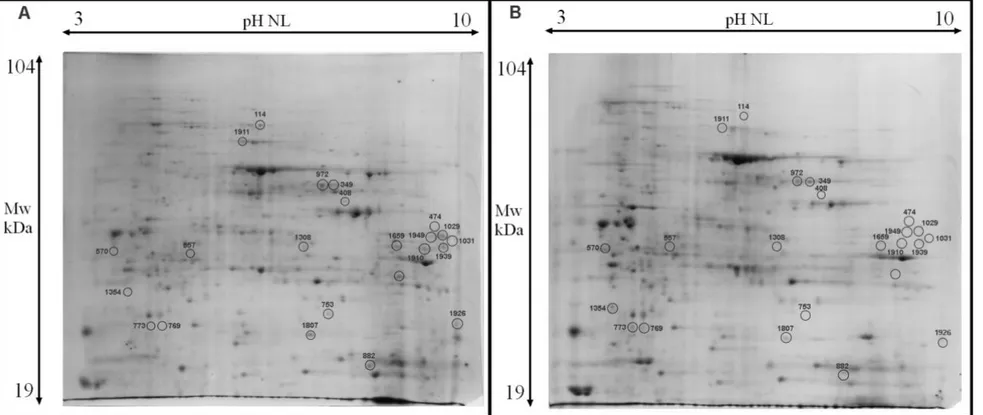
The primary mean using in proteomic studies is proteome maps generated by two-dimensional gel electrophoresis (2-DE).
Since the advent of the 2-DE methodology, numerous improvements have been introduced to develop the full potential of 2-DE and to make it an indispensable tool in biological research. Particularly fruitful was the establishment of annotated 2-D protein maps where proteins of model organisms separated on 2-D gels are identified.
Hence a great deal of effort has been spent in the development of a reference protein map of the yeast S.cerevisiae. Since this pioneering work, a large number of annotated yeast reference maps have been reported. They include proteins from total cell extracts (Maillet et al., 1996; Garrels et al., 1997; Gygi et al., 1999; Perrot et al., 2006; Kobi et al., 2004; Yin et al., 2004), strongly alkaline (Wildgruber et al., 2002), mitochondrial (Ohlmeier et al., 2004; Sickmann et al., 2003) or vacuolar luminal proteins (Sarry et al., 2007). Several hundred proteins have been identified on these maps. These annotated maps have proved to be helpful in solving numerous biological problems (Kolkman et al., 2005).
The first yeast reference map was reported by McLaughlin’s group in 1982 (Ludwig et al., 1982). After, Boucherie (Boucherie et al., 1996) have published onto Electrophoresis journal a manuscript in which show the identification of 250 protein spots. In later years, Perrot and coworkers have published 3 update of yeast proteome map. In 1999 (Perrot et al., 1999) their identification extended the number of protein spots identified on our yeast reference map to 401, in 2006 (Perrot et al., 2006) to 602 and, finally, in 2009 (Perrot et al., 2009) to 716 (Figure 9).
Chapter 1 Introduction
27
Figure 9. Standard yeast reference map with names of the identified spots
Hence proteomics represent a valid instrument to have an overview of what is happening when, for example, strain is knock out for a gene, such as in my PhD research for deletion of metacaspase gene YCA1.
Chapter 1 References R
28
REFERENCES
Adrian C. and Martin S.J. The mitochondrial apoptosome: a killer unleashed by the
cytochrome seas. Trends Biochem Sci, (2001), 26:390–397.
Ahn S. H., Henderson K. A., Keeney S., Allis C. D. H2B (Ser10) phosphorylation is
induced during apoptosis and meiosis in S. cerevisiae. Cell Cycle. (2005),4(6): 780-783.
Almeida B., OhlmeierS.,Almeida A.J., MadeoF., Leao C.,Rodrigues F., Ludovico P. Yeast protein expression profile during acetic acid-induced apoptosis indicates causal
involvement of the TOR pathway. Proteomics.(2009), 9:720–732.
Austriaco, N. Endoplasmic reticulum involvement in yeast cell death. Front Oncol.
(2012), 2: 87.
Bender C. E., Fitzgerald P., Tait S. W., Llambi F., McStay G. P., Tupper D. O., Pellettieri J., Sanchez Alvarado A., Salvesen G. S., Green D. R. Mitochondrial
pathway of apoptosis is ancestral in metazoans. Proc Natl Acad SciUSA (2012) 109(13): 4904-4909.
Bettiga M., Calzari L., Orlandi I., Alberghina L., Vai M. Involvement of the yeast
metacaspase Yca1 in ubp10Delta-programmed cell death. FEMS Yeast Res. (2004), 5(2):141-7.
Botstein D. Why yeast? Hosp Pract (Off Ed). (1991), 26(10):157-61, 164.
Boucherie H., Sagliocco F., Joubert R., Maillet I., Labarre J., Perrot M.
Two-dimensional gel protein database of Saccharomyces cerevisiae. Electrophoresis. (1996), 17(11):1683-99.
Bröker L.E., Kruyt F.A., Giaccone G. Cell death independent of caspases: a review.
Clin Cancer Res. (2005), 11(9):3155-62. Review.
Buttner S., Ruli D., Vogtle F. N., Galluzzi L., Moitzi B., Eisenberg T., Kepp O., Habernig L., Carmona-Gutierrez D., Rockenfeller P., Laun P., Breitenbach M., Khoury C., Frohlich K. U., Rechberger G., Meisinger C., Kroemer G., Madeo F. A
Chapter 1 References
29
yeast BH3-only protein mediates the mitochondrial pathway of apoptosis. Embo J.(2011), 30(14): 2779-2792.
Cai J. and Jones D.P. Superoxide in apoptosis. Mitochondrial generation triggered by
cytochrome c loss. J. Biol. Chem. (1998), 273:11401–11404.
Carmona-Gutierrez D., Eisenberg T., Buttner S., Meisinger C., Kroemer G., Madeo F. Apoptosis in yeast: triggers, pathways, subroutines. Cell Death Differ.
(2010), 17(5): 763-773.
Cássio F., Leão C., van Uden N. Transport of lactate and other short-chain
monocarboxylates in the yeast Saccharomyces cerevisiae. Appl Environ Microbiol (1987), 53:509–513.
Clifford J., Chiba H., Sobieszczuk D., Metzger D., Chambon P. RXRa-null F9
embryonal carcinoma cells are resistant to the differentiation,anti-proliferative and apoptotic effects of retinoids. EMBO (Eur. Mol. Biol.Organ.) J. (1996), 15:4142–4155.
Diaz-Ruiz R., Rigoulet M., Devin A. The Warburg and Crab-tree effects: on the origin
of cancer cell energy metabolism and of yeast glucose repression. Biochim. Biophys. Acta (2010), 1807, 568–576.
Eisenberg T., Büttner S., Kroemer G., Madeo F. The mitochondrial pathway in yeast
apoptosis. Apoptosis (2007) , 12(5):1011-23.
Eisenberg T., Carmona-Gutierrez D., Büttner S., Tavernarakis N., Madeo F.
Necrosis in yeast. Apoptosis (2010), 15(3): 257-68. Review.
Foury F. and Kucej M. Yeast mitochondrial biogenesis: a model system for humans?
Curr Opin Chem Biol (2001), 6, 106-111.
Garrels J.I. YPD-A database for the proteins of Saccharomyces cerevisiae. Nucleic
Acids Res. (1996), 24(1):46-9.
Garrels J.I., McLaughlin C.S., Warner J.R., Futcher B., Latter G.I., Kobayashi R., Schwender B., Volpe T., Anderson D.S., Mesquita-Fuentes R., Payne W.E.
Chapter 1 References
30
Proteome studies of Saccharomyces cerevisiae: identification and characterization of abundant proteins. Electrophoresis. (1997), 18(8):1347-60.
Ghaemmaghami S., Huh W.K., Bower K., Howson R.W., Belle A., Dephoure N., O'Shea E.K., Weissman J.S. Global analysis of protein expression in yeast. Nature.
(2003), 425(6959):737-41.
Ghibelli L., Coppola S., Rotilio G., Lafavia E., Maresca V., Ciriolo M.R.
Non-oxidative loss of glutathione in apoptosis via GSH extrusion. Biochem. Biophys. Res. Commun (1995), 216:313–320.
Giannattasio S., Atlante A., Antonacci L., Guaragnella N., Lattanzio P., Passarella S., Marra E. Cytochrome c is released from coupled mitochondria of yeast en route to
acetic acid-induced programmed cell death and can work as an electron donor and a ROS scavenger. FEBS Lett.(2008), 582(10): 1519-1525.
Giannattasio S., Guaragnella N., Arbini A.A., Moro L. Stress-related mitochondrial
components and mitochondrial genome as targets of anticancer therapy. Chem Biol Drug Des. (2013), 81(1):102-12.
Giannattasio S., Guaragnella N., Corte-Real M., Passarella S., Marra E. Acid stress
adaptation protects Saccharomyces cerevisiae from acetic acidinduced programmed cell death. Gene. (2005), 354: 93-98.
Goffeau A., Barrell B.G., Bussey H., Davis R.W., Dujon B., Feldmann H., Galibert F., Hoheisel J.D., Jacq C., Johnston M., Louis E.J., Mewes H.W., Murakami Y., Philippsen P., Tettelin H., Oliver S.G. Life with 6000 genes. Science (1996),
74(5287):546, 563-7.
Greenlund L.J.S., Deckwerth T.L., Johnson E.M.Jr. Superoxide dismutase delays
neuronal apoptosis: a role for reactive oxygen species in programmed neuronal death. Neuron. (1995), 14:303–315.
Griffin T.J., Gygi S.P., Ideker T., Rist B., Eng J., Hood L., Aebersold R.
Complementary profiling of gene expression at the transcriptome and proteome levels in Saccharomyces cerevisiae. Mol Cell Proteomics. (2002), 1(4):323-33.
Chapter 1 References
31
Guaragnella N., Antonacci L., Passarella S., Marra E., Giannattasio, S.
Achievements and perspectives in yeast acetic acid-induced programmed cell death pathways. Biochem Soc Trans.(2011), 39(5): 1538-1543.
Guaragnella N., Bobba A., Passarella S., Marra E., Giannattasio S. Yeast acetic
acid-induced programmed cell death can occur without cytochrome c release which requires metacaspase YCA1. FEBS Lett. (2010a), 584(1): 224-228.
Guaragnella N., Passarella S., Marra E., Giannattasio S. Knock-out of metacaspase
and/or cytochrome c results in the activation of a ROS-independent acetic acid-induced programmed cell death pathway in yeast. FEBS Lett. (2010b), 584(16): 3655-3660.
Guaragnella N., Pereira C., Sousa M. J., Antonacci L., Passarella S., Corte-Real M., Marra E., Giannattasio S. YCA1 participates in the acetic acid induced yeast
programmed cell death also in a manner unrelated to its caspase like activity. FEBS Lett. (2006), 580(30):6880-6884.
Guaragnella, N., Ždralević, M., Antonacci, L., Passarella, S., Marra, E. and Giannattasio, S. The role of mitochondria in yeast programmed cell death. Front
Oncol.(2012), 2: 70.
Gygi S.P., Rochon Y., Franza B.R., Aebersold R. Correlation between protein and
mRNA abundance in yeast. Mol Cell Biol. (1999), 19(3):1720-30.
Hauptmann P., Riel C., Kunz-Schughart L.A., Frohlich K.U, Madeo F., Lehle L.
Defects in N-glycosylation induce apoptosis in yeast. Mol Microbiol. (2006), 59:765-778.
Herker E., Jungwirth H., Lehmann K. A., Maldener C., Frohlich K. U., Wissing S., Buttner S., Fehr M., Sigrist S., Madeo F. Chronological aging leads to apoptosis in
yeast. J Cell Biol. (2004), 164(4): 501-507.
Ho P.K., Hawkins C.J. Mammalian initiator apoptotic caspases. FEBS J. (2005),
Chapter 1 References
32
Hockenbery D.M., Oltvai Z.N., Yin X.M., Milliman C.L., Korsmeyer S.J. Bcl-2
functions in an antioxidant pathway to prevent apoptosis. Cell. (1993), 75:241–251.
Hoeberichts F.A., ten Have A., Woltering E.J. A tomato metacaspase gene is
upregulated during programmed cell death in Botrytis cinerea-infected leaves. Planta (2003), 217: 517–522.
Huttemann M., Pecina P., Rainbolt M., Sanderson T. H., Kagan V. E., Samavati L., Doan J. W., Lee I. The multiple functions of cytochrome c and their regulation in
life and death decisions of the mammalian cell: From respiration to apoptosis. Mitochondrion (2011), 11(3): 369-381.
Ikeda S. and Kawasaki N. Isolation and characterization of the Schizosaccharomyces
pombe cDNA encoding the mitochondrial endonuclease(1). Biochim. Biophys. Acta (2001), 1519: 111–116.
Ivanovska I. and Hardwick J.M. Viruses activate a genetically conserved cell death
pathway in a unicellular organism. J. Cell Biol. (2005), 170:391–399.
Jeong H., Mason S.P., Barabási A.L., Oltvai Z.N. Lethality and centrality in protein
networks. Nature. (2001), 411(6833):41-2.
Kane D.J., Sarafian T.A., Anton R., Hahn H., Gralla E.B., Valentine J.S., Örd T., Bredesen D.E. Bcl-2 inhibition of neural death: decreased generation of reactive
oxygen species. Science (1993), 262:1274–1277.
Khan M.A., Chock P.B., Stadtman E.R. Knockout of caspase-like gene, YCA1,
abrogates apoptosis and elevates oxidized proteins in Saccharomyces cerevisiae. Proc NatlAcad Sci USA. (2005), 102(48):17326-31.
Kluck R. M., Ellerby L. M., Ellerby H. M., Naiem S., Yaffe M. P., Margoliash E., Bredesen D., Mauk A. G., Sherman F., Newmeyer D. D. Determinants of cytochrome
c pro-apoptotic activity. The role of lysine 72 trimethylation. J BiolChem.(2000), 275(21): 16127-16133.
Chapter 1 References
33
Kluck R., Bossy-Wetzel E., Green D.R., Newmeyer D.D. The release of cytochrome c
from mitochondria: a primary site for Bcl-2 regulation of apoptosis. Science (1997), 275:1132–1136.
Knorre D. A., Smirnova E. A., Severin, F. F. Natural conditions inducing
programmed cell death in the yeast Saccharomyces cerevisiae. Biochemistry (Mosc). (2005), 70(2): 264-266.
Kobi D., Zugmeyer S., Potier S., Jaquet-Gutfreund L. Two-dimensional protein map
of an "ale"-brewing yeast strain: proteome dynamics during fermentation. FEMS Yeast Res. (2004), 5(3):213-30.
Kolkman A., Slijper M., Heck A. J. Development and application of proteomics
technologies in Saccharomyces cerevisiae. Trends Biotechnol. (2005), 23:598–604.
Laun P., Pichova A., Madeo F., Fuchs J., Ellinger A., Kohlwein S., Dawes I., Frohlich K. U., Breitenbach M. Aged mother cells of Saccharomyces cerevisiae show
markers of oxidative stress and apoptosis. Mol Microbiol. (2001), 39(5): 1166-1173.
Leão C. and van Uden N. Transport of lactate and other monocarboxylates in the yeast
Candida utilis. Appl Microbiol Biotechnol (1986), 23:389-393.
Li L.Y., LuoX., Wang X. Endonuclease G is an apoptotic DNase when released from
mitochondria. Nature (2001), 412: 95–99.
Liang X.H., Jackson S., Seaman M., Brown K., Kempkes B. Hibshoosh H., Levine B. Induction of autophagy and inhibition of tumorigenesis by beclin 1. Nature (1999),
402:672–676.
Liu X., Kim C.N., Yang J., Jemmerson R., Wang X. Induction of apoptotic program
in cell-free extracts: requirement for dATP and cytochrome c. Cell (1996), 86:147–157.
Ludovico P., Rodrigues F., Almeida A.,Silva M.T., Barrientos A., Corte- Real M.
Cytochrome c release and mitochondria involvement in programmed cell death induced by acetic acid in Saccharomyces cerevisiae. Molecular Biology of the Cell. (2002), 13, 2598–2606.
Chapter 1 References
34
Ludovico P., Sousa M.J., Silva M.T., Leão C., Côrte-Real M. Saccharomyces
cerevisiae commits to a programmed cell death process in response to acetic acid. Microbiology (2001), 147: 2409-2415.
Ludwig J.R. 2nd, Foy J.J., Elliott S.G., McLaughlin C.S. Synthesis of specific
identified, phosphorylated, heat shock, and heat stroke proteins through the cell cycle of Saccharomyces cerevisiae. Mol Cell Biol. (1982), 2(2):117-26.
Madeo F, Fröhlich E, Ligr M, Grey M, Sigrist SJ, Wolf DH, Fröhlich KU. Oxygen
stress: a regulator of apoptosis in yeast. J Cell Biol. (1999), 145(4):757-67.
Madeo F., Carmona-Gutierrez D., Ring J., Büttner S., Eisenberg T., Kroemer G..
Caspase-dependent and caspase- independent cell death pathways in yeast. Biochem.Biophys.Res.Commun. (2009), 382:227–231.
Madeo F., Frohlich, E., Frohlich, K. U. A yeast mutant showing diagnostic markers of
early and late apoptosis. J Cell Biol.(1997), 139(3): 729-734.
Madeo F., Herker E., Maldener C., Wissing S., Lachelt S., Herlan M., Fehr M., Lauber K., Sigrist S.J., Wesselborg S., Frohlich K.U. A caspase-related protease
regulates apoptosis in yeast. Mol.Cell (2002), 9: 911–917.
Maillet I., Lagniel G., Perrot M., Boucherie H., Labarre J. Rapid identification of
yeast proteins on two-dimensional gels. J Biol Chem. (1996), 271(17):10263-70.
Martin S.J., Reutelingsperger C.P.M., McGahon A.J., Rader J.A., van Schie R.C.A.A.,. LaFace D.M, Green D.R.Early redistribution of plasma membrane
phosphatidylserine is a general feature of apoptosis regardless of the initiating stimulus: inhibition by overexpression of Bcl-2 and Abl. J. Exp. Med. (1995), 182:1545–1556.
Mazzoni C., Herker E., Palermo V., Jungwirth H., Eisenberg T., Madeo F., Falcone C. Yeast caspase 1 links messenger RNA stability to apoptosis in yeast.EMBO
Chapter 1 References
35
Morton C. O., Dos Santos S. C., Coote P. An amphibian-derived, cationic,
alpha-helical antimicrobial peptide kills yeast by caspase-independent but AIFdependent programmed cell death. Mol Microbiol. (2007), 65(2): 494-507.
Narasimhan M. L., Coca M. A., Jin J., Yamauchi T., Ito Y., Kadowaki T., Kim K. K., Pardo J. M., Damsz B., Hasegawa P. M., Yun D. J., Bressan R.A. Osmotin is a
homolog of mammalian adiponectin and controls apoptosis in yeast through a homolog of mammalian adiponectin receptor. Mol Cell. (2005),17(2): 171-180.
Ohlmeier S., Kastaniotis A. J., Hiltunen J. K., Bergmann U. The yeast mitochondrial
proteome, a study of fermentative and respiratory growth. J. Biol. Chem. (2004), 279: 3956–3979.
Pampulha M.A. and Loureiro-Dias M.C. Combined effect of acetic acid, pH and
ethanol on intracellular pH of fermenting yeast. Appl Microbiol Biotechnol (1989), 31: 547–550.
Parrish J., Li L., Klotz K., Ledwich D., Wang X., Xue D. Mitochondrial
endonuclease G is important for apoptosis in C. elegans. Nature (2001),412: 90–94.
Payne W.E., Garrels J.I. Yeast Protein database (YPD): a database for the complete
proteome of Saccharomyces cerevisiae. Nucleic Acids Res. (1997), 25(1):57-62.
Pereira C., Coutinho I., Soares J., Bessa C., Leão M., Saraiva L. New insights into
cancer-related proteins provided by the yeast model. FEBS J. (2012), 279(5):697-712.
Pereira C., Silva R.D., Saraiva L., Johansson B., Sousa M.J., Côrte-Real M.
Mitochondria-dependent apoptosis in yeast. Biochim Biophys Acta (2008), 1783(7):1286-302.
Perrot M., Guieysse-Peugeot A.L., Massoni A., Espagne C., Claverol S., Silva R.M., Jenö P., Santos M., Bonneu M., Boucherie H. Yeast proteome map (update
2006). Proteomics. (2007), 7(7):1117-20.
Perrot M., Moes S., Massoni A., Jenoe P., Boucherie H. Yeast proteome map (last
Chapter 1 References
36
Perrot M., Sagliocco F., Mini T., Monribot C., Schneider U., Shevchenko A., Mann M., Jenö P., Boucherie H. Two-dimensional gel protein database of Saccharomyces
cerevisiae (update 1999). Electrophoresis. (1999), 20(11):2280-98.
Pham N., Robison B., Hedley D. Simultaneous detection of mitochondrial respiration
chain activity and reactive oxygen in digitonin-permeabilized cells using flow cytometry. Cytometry (2000), 41:245–251.
Pinto I., Cardoso H., Leão C. High enthalpy and low enthalpy death in Saccharomyces
cerevisiae induced by acetic acid. Biotechnol Bioeng (1989), 33:1350-1352.
Reggiori F. and Klionsky D.J. Autophagic processes in yeast: mechanism, machinery
and regulation. Genetics. (2013), 194(2):341-61. Review.
Reiter J., Herker E., Madeo F., Schmitt M. J. Viral killer toxins induce
caspase-mediated apoptosis in yeast. J Cell Biol. (2005), 168(3): 353-358.
Ren Q., Yang H., Rosinski M., Conrad M. N., Dresser M. E., Guacci V., Zhang Z.
Mutation of the cohesin related gene PDS5 causes cell death with predominant apoptotic features in Saccharomyces cerevisiae during early meiosis. Mutat Res.(2005) 570(2): 163-173.
Resnick M.A. and Cox B.S. Yeast as an honorary mammal. Mutat Res. (2000),
451:1-11.
Ruckenstuhl C., Büttner S., Carmona-Gutierrez D., Eisenberg T., Kroemer G., Sigrist S.J., Fröhlich K.U., Madeo F. The Warburg effect suppresses oxidative stress
induced apoptosis in a yeast model for cancer. PLoS One (2009), 4(2):e4592. doi: 10.1371.
Saraiva L., Silva R. D., Pereira G., Goncalves J. Corte-Real, M. Specific modulation
of apoptosis and Bcl-xL phosphorylation in yeast by distinct mammalian protein kinase C isoforms. J Cell Sci.(2006 ), 119(Pt 15): 3171-3181.