Universit`
a degli Studi di Ferrara
DOTTORATO DI RICERCA IN
BIOCHIMICA, BIOLOGIA MOLECOLARE E BIOTECNOLOGIE
CICLO XXVII
COORDINATORE Prof. Francesco Bernardi
Development of cellular and animal models of coagulation factors deficiencies for the assessment of innovative therapeutic approaches
acting on transcriptional and post-transcriptional regulation
Settore Scientifico Disciplinare BIO/11
Dottorando Dott. Barbon Elena
Tutore Prof. Pinotti Mirko
Co-tutore Prof. Mingozzi Federico
Contents
Introduction v
1 TALE TFs 1
1.1 Brief overview of coagulation process . . . 1
1.1.1 Initiation-FVII/FVIIa . . . 2
1.1.2 Propagation–The tenase complex . . . 3
1.2 Focus on FVII: gene and protein features . . . 3
1.2.1 Factor FVII protein . . . 3
1.3 Factor FVII gene . . . 5
1.4 FVII deficiency . . . 6
1.4.1 Background and rationale . . . 7
1.5 Aim of the study . . . 9
1.6 Materials and Methods . . . 10
1.6.1 Design and assembly of TALE-TFs . . . 10
1.6.2 Construction of the reporter plasmids . . . 10
1.6.3 Cell culture and reporter activation assay . . . 11
1.6.4 FACS analysis . . . 12
1.6.5 RT-PCR and qPCR for FVII mRNA . . . 12
1.7 Results . . . 12
1.7.1 Assembly of engineered TALE-TFs and reporter plasmids . 12 1.7.2 In vitro models: effect of promoter mutations and valida-tion of the efficacy of eTFs . . . 14
1.7.3 Assessment of the specificity of TF4 . . . 14
1.7.4 Effect of TF4 in the endogenous context of HepG2 cells . 17 1.7.5 Effect of TF4 in the endogenous context of Hek293 cells . 17 1.7.6 Effect of the TF1, TF2 and TF3 in the endogenous context of HepG2 and Hek293 cells . . . 18
1.7.7 Discussion and future perspectives . . . 19
2 SB Transposon 23 2.1 Focus on FIX: gene and protein features . . . 23
2.1.1 Factor FIX protein . . . 23
2.1.2 Factor FIX gene . . . 23
2.2 FIX deficiency-Haemophilia B . . . 24
2.3 Background and rationale . . . 25 i
2.4 Exon Specific U1s (ExSpeU1s) as therapeutic tool . . . 26
2.5 Creation of a stable model: the Sleeping Beauty transposon system (SBTS) . . . 28
2.6 Aim of the study . . . 30
2.7 Materials and Methods . . . 31
2.7.1 Vectors and cloning of the transposon plasmids . . . 31
2.7.2 Cell culture and generation of Hek293 stable clones . . . . 32
2.7.3 Evaluation of the hFIX stable expression . . . 32
2.7.4 Evaluation of the gene copy number in Hek293 stable clones by Southern blot . . . 32
2.7.5 Transfection with U1snRNA sh9 . . . 33
2.7.6 AAV-expressing U1sh9 . . . 33
2.7.7 Transduction with AAV-U1sh9 in Hek293 stable clones . . 33
2.7.8 Generation of vectors . . . 34
2.7.9 Animal procedures . . . 35
2.7.10 Measurement of hFIX antigen and mRNA . . . 35
2.7.11 Assessment of the transgene copy number . . . 35
2.8 Results . . . 36
2.8.1 Generation of stable Hek293 clones expressing human FIX 36 2.8.2 SB100X activity in–vitro: assessment of the gene copy num-ber . . . 39
2.9 U1sh9 mediated correction: hFIX mRNA and protein rescue . . . 40
2.9.1 Cell transduction with an AAV8-vector expressing the mod-ified U1sh9 . . . 42
2.9.2 Generation and validation of the transposon plasmids for the creation of mouse models . . . 44
2.9.3 Generation of the mouse models . . . 45
2.9.4 Assessment of U1sh9 efficacy in–vivo: RNA and protein analysis . . . 46
2.9.5 Evaluation of the integration profile: gene copy number . 49 2.9.6 Discussion and future plans . . . 50
Ringraziamenti
Questo lavoro, e soprattutto l’esperienza e l’arricchimento scientifico e personale che ne sono scaturiti, non sarebbe stato possibile senza l’aiuto delle persone con cui ho lavorato in questi tre anni. Ringrazio Mirko e il Prof.Bernardi per avermi introdotto e guidato nel fantastico mondo della biologia molecolare, sostenendomi in tutte le fasi del mio percorso di dottorato e trasmettendomi i loro insegnamenti. Grazie a Matteo, un buon capo ma soprattutto un buon amico, per la sua grande
disponibilit`a e per aver condiviso con me la sua esperienza, le idee e gli spunti per
lavorare assieme sinergicamente. Grazie anche a Silvia e Mattia, perch´e parte di
questo lavoro `e merito del loro impegno, che ho apprezzato soprattutto quando
non ero fisicamente presente a seguirli. Un grazie enorme va a Federico, che `e
diventato per me un ottimo mentore oltre che mente scientifica da ammirare, per
la sua grande disponibilit`a verso di me e il mio progetto fin dai primi momenti, e
per avermi insegnato che la condivisione delle conoscenze e lo scambio scientifico
sono l’unica, onesta e produttiva strada da seguire. Merci beaucoup `a Giuseppe,
Christian, Fanny, S´everine, Amine et Francesco, parce-que chez eux j’ai trouv´e
une ´equipe fantastique du travail, unie et compacte. Grazie al mitico gruppo
Cartel, per aver contribuito a rendere magnifica la mia esperienza a Parigi. Grazie
a Silvia, perch´e avere la presenza di una cara amica come compagna di laboratorio
ha reso questi anni di dottorato pi`u pazzi e allegri. Grazie come sempre ai miei
genitori ed al loro incondizionato sostegno in tutte le scelte.
Grazie ad Alessandro, che ha reso perfetto il layout di questa tesi. . . ed anche tutto il resto.
Introduction
In the last decades, enormous efforts have been pushed toward the development of therapeutic approaches for human genetic diseases, and the research all over the world has obtained remarkable achievements. Advances in biotechnology have brought gene therapy to the forefront of medical research. Gene replacement, in which a normal copy of the defective gene is efficiently delivered and expressed into target cells, represents one of the most advanced strategies that produced encouraging results in patients with diseases caused by single gene recessive dis-orders (like haemophilia, muscular dystrophy, sickle cell anemia, cystic fibrosis etc.), some viral infections and inherited genetic diseases such as cancer and ADA deficiency [13]. Notwithstanding, the intense research also led to potential ther-apeutic strategies based on the correction of the specific disease-causing defects, which might circumvent some limitations (i.e. gene size, gene regulation) of gene replacement therapy. These approaches are of great interest for patients with coagulation deficiencies, since they would benefit from even small increase in functional protein levels. The low therapeutic threshold makes the rare bleeding disorders (RBDs) an ideal target for investigating therapeutic strategies based on counteracting the pathogenic molecular mechanisms. This work propose the development of in–vitro and in–vivo models of RBDs in order to explore correc-tive molecular approaches acting on the specific disease-causing defects, both at transcriptional and post-transcriptional level.
The chapter 1 describes the usage of engineered transcription factors (eTFs) as potential therapeutic strategy for FVII deficiency caused by promoter muta-tions. Transcription impairment by mutations in the promoter regions represents indeed a small, but considerable cause of severe coagulation factor defects and of all inherited human diseases. These mutations often affect only the untrans-lated control regions impairing to different extent the expression of completely unaffected genes. Different from gene therapy approaches inserting exogenous sequences that drive the expression of the missing factor, the induced restart of the transcription would maintain the natural chromosomal structure and context restoring gene expression. Engineered TFs have been shown to have wide-range potential in modulating desired gene expression through targeting their promot-ers [109]. A recent breakthrough with transcription activator-like effector proteins (TALEs) makes it possible to establish universal types of engineered TFs that can potentially target any selected gene. Hence, fusion of a TALE-derived binding domain to a transcriptional activator, such as VP64 [21], could generate TALE-transcription factors (TALE-TFs) that can target selected promoter regions and
modulate expression of corresponding genes [55]. Intensive studies on stem-cell maintenance and differentiation have been recently carried out with TALE-TFs [55, 27, 46, 56, 72], but to our knowledge only one example of these eTFs has been used for the recovery of transcription impaired by human disease-causing mutations [86]. As a model to exploit TALE-TF transcription restoration in coagulation factor deficiencies, we chose two severe promoter mutations impair-ing FVII transcription, the -94 C>G and -61 T>G transversions, fallimpair-ing in the binding site for the hepato-specific HNF-4 and the ubiquitous Sp1 transcription factors, respectively [18, 29]. The patients homozygous for these mutations expe-rience life-threatening hemorrhagic symptoms and require replacement therapy. Through the expression of gene reporter plasmids we first created a cellular model for the two F7 promoter variants. Then, we assembled 4 TALE-TFs (TF1-4) de-signed to target different regions on the F7 proximal promoter in order to test their efficacy in stimulating transcriptional activity on the target gene. The treat-ment with the different TALE-TFs demonstrated that TF4, targeting a sequence between the mutations, induced a robust increase of gene transcription in the presence of the defective promoter. Interestingly, TF4 appreciably increased the endogenous F7 transcription and mRNA levels in HepG2 cells and induced F7 expression in Hek293 cells that do not virtually express FVII.
The chapter 2 describes the exploitation of the Sleeping Beauty Transpo-son System (SBTS) to develop cellular and mouse model of haemophilia B (HB) caused by splicing mutations, in order to assess the efficacy of an RNA-based ther-apeutic approach. HB is a prototypical example of disease with a heterogeneous mutational pattern (www.factorix.org) and a relatively high frequency of splicing mutations (>15%) in severe disease forms. Intervention at the pre-mRNA splic-ing level is emergsplic-ing as a promissplic-ing therapeutic strategy for genetic disorders. Increasing attention has been given to the U1 small nuclear RNA (U1snRNA) that, in the earliest splicing step, mediates the recognition of the donor splice site (5’ss) by the ribonucleoprotein U1snRNP. Studies in various cellular models of human disease indicated the potential therapeutic effect of engineered U1snRNAs to rescue aberrant splicing caused by mutations at 5’ss, a relatively frequent cause of severe forms. In the last years, modified U1snRNAs have been exploited to correct splicing mutations causing severe coagulation factor VII deficiency and HB [37, 74, 75]. The rescue has been proven in cellular models by using F7/F9 minigenes. However, the F9 mutational pattern is extremely heterogeneous, with several different splicing mutations. Therefore, the evaluation of the U1snRNA-mediated correction strategy in–vivo implies the creation of mouse models, which are not yet available. Here we used the SBTS to develop cellular/mouse models of HB caused by the FIXex5-2C splicing variant, and subsequently assess the U1-mediated correction in chromatin context. Indeed, in contrast to other ap-proaches the transposon enables the transgene to be integrated into the genome, which represents the preferred physiologic situation for subsequent splicing stud-ies. Initially we have performed experiments to test the hyperactive transposase SB100X-mediated integration of the splicing-competent human FIX transgene into the genome using human embryonic kidney cells. We have generated Hek293
vii
stable clones expressing the normal or mutated human splicing-competent FIX cassettes integrated into the genome as a result of the transposase activity. These preliminary studies provided us with optimized experimental protocol to create in a relatively short time cellular models of human disease caused by splicing muta-tions. Moreover, it permitted the assessment of the modified U1 snRNA-mediated rescue in–vitro in a genomic expression context instead of a transient episomal system. This also provided with the rationale for the creation of mouse mod-els through hydrodynamic injection of the transposon plasmids and of SB100X transposase in C57BL/6 wt mice [62] in order to assess the efficacy of the U1 snRNAs-mediated rescue in–vivo.
Chapter 1
TALE TFs
1.1
Brief overview of coagulation process
Haemostasis is a physiological process that depicts a delicate balance between pro-coagulant and anti-coagulant activities. From many years researchers have been interested in deepening the mechanisms underlying the complex series of biochemical reactions occurring to maintain these equilibrium in-vivo. According to the classical coagulation waterfall/cascade model, described in 1964 by two independent groups [96, 112], several proteins with serine-protease activity are involved in the formation of blood clot whenever an injury in a vessel occurs. In physiological conditions these coagulation factors, circulating normally as zymo-gens, are converted by cleavage into their active form and interact both together and with the pro-coagulant surfaces in order to produce the fibrin formation [95]. In this traditional view a contact pathway (intrinsic) as well as a tissue-factor (TF) depending-pathway (extrinsic) have been described. The intrinsic pathway, so named because triggered by components already present in blood, is composed of FXII and pre-kallikrein (PK), with FXII being activated either by PK or plas-min to the active enzyme (FXIIa) when in contact with a negatively charged or artificial surfaces. In the intrinsic pathway the activation of a certain amount of FXII leads to the subsequent generation of FXIa from FXI, which then activates FIX. FIXa, together with phospholipids, calcium and FVIIIa, activates FX, which in turn converts prothrombin (FII) to thrombin (FIIa). A peculiar feature of FXII and PK zymogens is that they are calcium-independent, thereby able to “auto-activate” in a continuous manner even in the presence of anticoagulant molecules. In spite of this, it has been reported that individuals with deficiencies of FXII or PK do not experience any bleeding tendency in vivo, thereby suggesting that the contact pathway may not have such a fundamental role in blood coagulation, but rather in the thrombus formation [10, 97]. The first waterfall/cascade model has been implemented from the 1977, with the work of Osterud and Rapaport showing that FVIIa-TF can activate FIX and FX, so focusing attention on the role of the extrinsic pathway in interacting with the intrinsic one during clotting [3]. And in 1992 a group proposed the actual model of coagulation, called “cell-based” [105], which is composed of an initiation phase and a propagation phase (fig.1.1). The
clotting is triggered in the initiation phase when tissue factor (TF), also called thromboplastin or FIII, comes in contact with blood. TF is an integral membrane protein expressed on the surface of several cell types, like adventitial cell, smooth muscle cells and keratinocytes, that normally are not in contact with blood flow. When a lesion in the endothelial barrier occurs these cells become exposed to blood and consequently TF is able to bind the plasma serine protease FVIIa, forming a complex that is able to generate a modest amount of thrombin. In the following propagation phase, acting on the activated platelet surfaces, the tenase complex can be properly assembled together with the prothrombinase complex and calcium for massive production of thrombin. This “thrombin burst” arises at the end in the conversion of insoluble fibrinogen into soluble fibrin monomers forming the stable clot.
Figure 1.1: Cell-based model of coagulation
1.1.1
Initiation-FVII/FVIIa
Factor VII is a vitamin K-dependent serine protease produced in liver and circu-lating at a concentration of 500 ng/ml [33, 90]. It displays the shortest half-life as zymogen (approximately 5 hours) and is the only coagulation factor that is present in both the active and inactive forms. In humans, the active form (FVIIa) represents about 1% of total circulating FVII. FVII itself has a negligible activity and participates in the initiation phase principally after its activation to FVIIa. A number of coagulation enzymes including FIXa, FXa, FXIIa, thrombin, plasmin or FVII-activating proteases can activate FVII. However, FXa seems to be the most potent activator of FVII. Also FIX plays an important role in this activation because humans with haemophilia B display very low levels of FVIIa. Moreover, the FVII-TF complex can be auto-activated by FVIIa-TF complex. FVIIa has a long half-life in plasma because unless it is bound to TF it can not be inacti-vated by any protease inhibitor. The FVIIa-TF complex then activates FIX to
1.2 Focus on FVII: gene and protein features 3
FIXa and FX to FXa. It is worth noting that this initiation phase only results in a trace amount of thrombin after FXa activation, because FXa and thrombin are rapidly neutralized by antithrombin (AT) and the complex FVIIa-TF-FXa by the tissue factor pathway inhibitor (TFPI). The TFPI/AT provides a good regulatory system in order to prevent a massive thrombin generation for a false alarm. The pro-coagulant signal is provided only when TF is exposed at high enough levels to overcome inhibition by TFPI an AT. If this is the case, thrombin is able to enhance its own generation through the activation of FXI to FXIa and FV to FVa on the platelets surface, and also through the activation of FVIII to FVIIIa by cleaving von Willebrand factor (vWF). In this context recruited acti-vated platelets provide the pro-coagulant phospholipids membrane needed for the subsequent enzymatic reactions, and the further generation of FXa is dependent on the formation of the tenase complex.
1.1.2
Propagation–The tenase complex
Once FX, FIX and the cofactors FV and FVIII are activated after the generation of thrombin in the initiation phase the assembly of tenase complex can occurs. FIXa, in association with its cofactor FVIIIa, binds FX and calcium on negatively charged surfaces, mostly on platelets membrane, in order to form the tenase complex, leading to the generation of a large amount of FXa. Most of the FXa is physiologically produced in vivo by the tenase complex, which is considered to be around 50-fold more efficient in this process compared to the FVIIa-TF complex. Therefore, in the initiation phase the amount of FXa is insufficient to sustain hemostasis whereas during propagation a massive production of FXa is achieved through the formation of the tenase complex. FXa participates in the assembly of the prothrombinase complex together with FVa and calcium, which results in an explosive generation of thrombin and the subsequent stable clot formation. In physiological conditions the process is tightly controlled, in order to maintain the coagulative events just at the site of injury. As a matter of fact even if thrombin and FXa can diffuse from the site of damage to the adjacent healthy endothelium they are inhibited by the lack of pro-coagulant membranes and the presence of anticoagulants molecules like AT, TFPI and thrombomodulin, able to reduce their activity. It is worth noting that in the propagation phase both FVIII and FIX are required. Indeed, in the absence of FVIII and FIX the initiation occurs normally, but the propagation steps are severely diminished, resulting in a deficient fibrin clot formation and the incapacity of the hemostatic system to respond in repairing the damaged tissues.
1.2
Focus on FVII: gene and protein features
1.2.1
Factor FVII protein
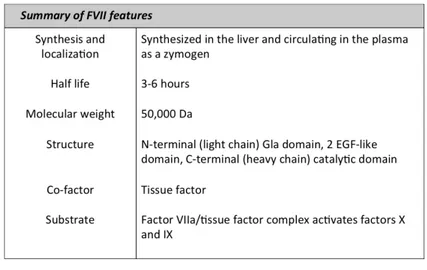
Human factor FVII is a zymogen for a vitamin K-dependent serine protease syn-thesized in liver and circulating in plasma at a concentration of approximately
0.5µg/ml (10nmol/L). It is secreted into blood as a single chain glycoprotein of 48KDa. All vitamin K-dependent coagulation zymogens share a similar protein domain structure consisting an N-terminal gamma-carboxyglutamic acid-rich do-main (Gla dodo-main, residues 1-38), a short hydrophobic segment (residues 39-45) that often is considered as part of the Gla domain, two epidermal growth fac-tor (EGF)-like domains (residues 47-84 and 85-131), and the C-terminal serine protease domain (residues 153-406). The Gla domain is responsible for the in-teraction of the protein with lipid membranes, while the EGF-like domain has a calcium ion binding site that to some degree mediates interaction with the TF exposed at the site of vessel injury (Tab 1.1). The major proportion of FVII
Table 1.1: Summary of factor VII features
circulates in plasma in the inactive form. Activation of FVII occurs by the cleav-age of a single peptide bond between Arg152 and Ile153, located in the region connecting the second EGF-like domain and the protease domain [111]. This results in the formation of a two-chain FVIIa molecule consisting of a light chain of 152 amino acids (containing the membrane-binding Gla domain) and a heavy chain of 254 amino acids (containing the catalytic domain) held together by a single disulfide bond (between Cys-135 and Cys-262). Conversion of factor VII to factor VIIa is catalyzed by a number of proteases, including thrombin, factor IXa, factor Xa, factor XIa, and factor XIIa [111]. Comparison of these proteins has shown that factor Xa, in association with phospholipids, has the highest po-tential to activate factor VII. Rapid activation also occurs when factor VII is combined with its cofactor, which is the tissue factor in the presence of calcium (autocatalysis). This reaction may be initiated by a small amount of preexisting factor VIIa. FVIIa is a trypsin-like enzyme and catalyses the hydrolysis of pep-tide bonds within a polypeppep-tide chain to produce two new smaller peppep-tides. In 1964, FVIIa proteolytic activity was observed first against FX, but the methods used did not reveal whether TF first reacted with FVII to form an intermedi-ate that then activintermedi-ated FX or whether FVII directly activintermedi-ated FX. A few years later, it was demonstrated that TF interacted with FVII and the formed
inter-1.3 Factor FVII gene 5
mediate was catalytically active [122]. The reaction product of TF and FVII is a potent activator of FX and also of FIX [3]. The interaction of FVIIa with TF
is Ca2+–dependent. Ca2+ saturation of the Gla domain is likely responsible for
this increase in affinity, since deletion of Gla in FVIIa results in a loss of affinity
for TF. Ca2+ may stabilize energetically important hydrophobic contacts of Gla
with TF [7]. In addition, Ca2+ increases FVIIa affinity for FX by conformational
changes in FVIIa and FX that are essential for the interaction of these proteins with phospholipids [120].
1.3
Factor FVII gene
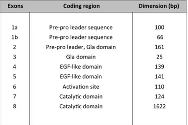
The human factor VII gene (F7) is a single copy gene located on chromosome 13 (13q34) which consists of eight exons spread over 12 kb of genomic DNA and produces a 2.4-kb mRNA encoding a secreted protein of 406 amino acids. The entire nucleotide sequence was reported for the first time in 1987 [70]. The introns ranged in size from 68 nucleotides (intron C) to nearly 2.6 kb (intron A). The exons also varied considerably in size, ranging from 25 nucleotides (exon 3) to 1.6 kb (exon 8). In 1986 different cDNAs coding for factor VII were isolated from a cDNA library prepared from a HepG2 cell line [45], by which it was possible to establish that 1b is an optional exon not always included in the mature mRNA. Its inclusion determines the length of the pre-pro leader sequence (from 38
Table 1.2: Factor VII gene exons organization
aminoacids when the mRNA lack 1b to 60 aminoacids when 1b is incorporated). In both cases the product is an in frame-mRNA that results in a functional transcript that codes for a biologically active FVII. In normal liver the most represented form is mRNA lacking exon 1b. As for other coagulation factor genes, F7 exons encode for discrete domains of the protein (Tab. 1.2): the pre-pro leader sequence (exon 1a, 1b and part of exon 2), the γ–carboxilase region (exons 2 and 3), the two EGF-like domains (exons 4 and 5), the activation domain (exon 6)and finally the catalytic domain (exons 7 and 8). The conservation of domains, exons
and introns positions among the members of the vitamin K-dependent protein family supports the theory of their evolution as modular protein by exon shuffling [100].
1.4
FVII deficiency
FVII, or proconvertin, deficiency was first recognized in 1951 [16]. Considered the most common of rare bleeding disorders its incidence is estimated at 1 per 300,000–500,000 [59]. It is inherited in an autosomal recessive fashion and affects males and females equally. Factor VII deficiency is very rare, but like all auto-somal recessive disorders, it is found more frequently in areas of the world where marriage between close relatives is common. Studies conducted on F7 knock-out mice suggest that a complete lack of FVII is incompatible with life [77], so it is thought that FVII deficiency can not be associated with complete absence of functional FVII. Triplett et al. [87] have classified FVII deficiency in CRM-(activity and antigen proportionally reduced), CRM+ (reduced activity, antigen normal) and CRMred (antigen is reduced but not as much as activity). This haemorragic disorder displays a variable clinical heterogeneity [65], which ranges from lethal to mild or even asymptomatic forms (in general when FVII:C >10%). The most frequent symptoms, like epistaxis and early bruising, indicate that the disease is mild in the majority of cases. However, severe to very severe cases are not infrequent, and are characterized by hemartrosis, muscle hematomas, or even central nervous system (CNS) and gastrointestinal (GI) bleeding. Nose bleeding is by far the most frequent symptom and is not gender-related. Other very com-mon symptoms are post–operative, skin and gum bleeds. As for gender, women are more prevalent among bleeders: this is mainly attributable to menorrhagia, which occurs in about two-thirds of women of fertile age but gum bleeding and easy bruising are also more frequent in females. Severe and life-threatening haem-orrhages are rare in general (about 5% of the bleeds) and occur most frequently during the first 6 months of life. In newborns (< 1 month) presenting bleed-ing manifestations were, as ranked for frequency, central nervous system (CNS), gastro-intestinal(GI), cephalohaematoma and umbilical bleeding. Several thera-peutic options are available for patients experience FVII deficiency, which may result in a very effective correction of the disease [99]. Fresh-frozen plasma is still used in developing countries, the most important drawback being the blood volume overload and a relatively high risk of transmitting blood-borne viruses. Plasma-derived FVII concentrates are essentially prothrombin complex concen-trates with a higher content of FVII. They are used for prophylactic treatment, as well as for controlling serious bleeding episodes, and bleeding during surgery. However, plasma-derived concentrates carry the risk of potential transmission of blood-borne pathogens. Recombinant FVIIa is to be considered nowadays the optimal protein replacement therapy. It is free of human plasma and albumin, so there is no risk of human viral transmission, but it is very expensive and not available for all patients, particularly in developing countries.
1.4 FVII deficiency 7
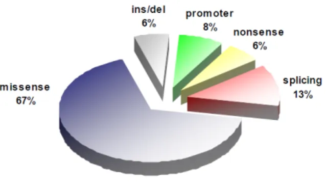
A considerable number of mutations have been reported to date in FVII gene (F7 mutation database: http://www.hgmd.cf.ac.uk/ac/index.php). Missense mutations represent the most frequent portion (fig.1.2) and occur in the 68% of cases, followed by splicing-site (13%), promoter (8%) and nonsense (6%)
mu-tations, small insertions and deletions (6%) [99, 60, 61]. A number of these
mutations have been identified as the cause of FVII deficiency but only a limited number of them have been well characterized.
Figure 1.2: Pie chart reporting the percentages of each specific mutation type found in F7 gene.
1.4.1
Background and rationale
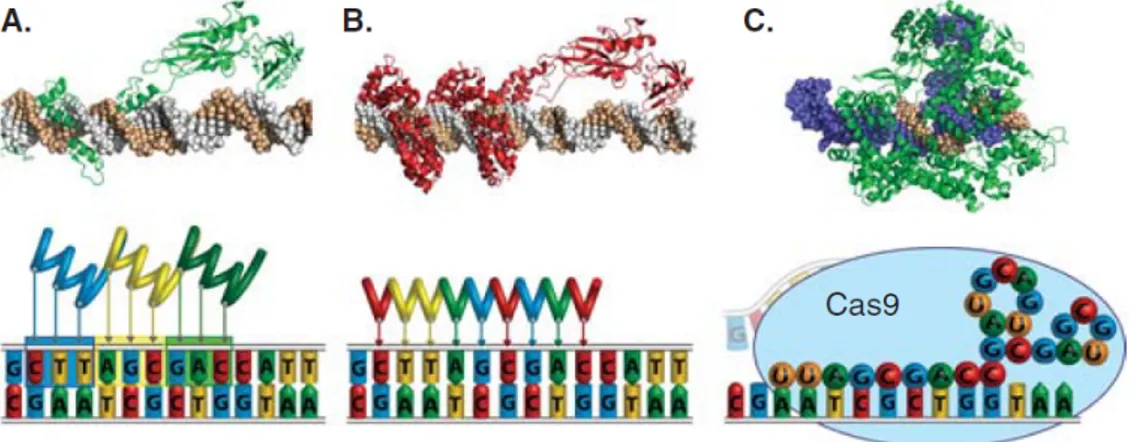
In the last years new technologies have been developed in order to control the expression of human genes by engineering synthetic transcription factors that can theoretically target any DNA sequence in a specific manner. The capability of regulating gene expression in the native chromosomal context may represent a way to address several genetic diseases. This approach also circumvent some of the traditional challenges of gene replacement, like gene delivery, specific ac-tivation and immune response related-aspects. Over the past 20 years several platforms have been developed in order to target a specific DNA sequence and the most widely used are represented by zinc finger-based transcription factor/nu-cleases, the transcription activator-like effectors and the CRISPR/Cas9 systems (figure 1.3). All these systems are based on the exploitation of particular DNA binding domains (DBDs) that can be associated with several effectors to target a DNA sequence in order to stimulate a specific biological effect [6]. The first syn-thetic transcription factors designed to modulate the expression of human genes were engineered from zinc-finger proteins [114], able to recognize a specific target through their modular DBD. Despite many achievements in using these modified zinc finger-based DBDs linked to several actuators in different biological applica-tion [22, 8, 121, 88], there are many issues related to their engineering that have limited the widespread adoption of this platform. More recently, another system protein-based has been developed, starting from a more detailed knowledge of the structure of the bacterial transcription activator-like effectors (TALEs). Similar to zinc finger proteins, they show a modular DBD that can be modified in order
Figure 1.3: Technologies for engineering programmable DNA-binding proteins, including (A) zinc finger proteins, (B) TALEs and (C) CRISPR/Cas9. Representative crystal structures of a (A) zinc finger protein or (B) TALE fused to the p65 transcriptional activation domain or (C) Cas9 (green) bound to a gRNA (blue) and the corresponding DNA target site (brown).
to determine the specificity for a particular sequence in the genome. The possi-bility in engineering these TALE proteins in a relatively simpler way compared to zinc fingers has determined their broad usage in the last years [94]. Moreover, a third technology has emerged based on the RNA- guided DNA endonuclease Cas9 from the type II bacterial adaptive immune system CRISPR [58]. It differs from the previous ones because the targeting of a specific DNA loci is due to the complementary pairing with a guide RNA (gRNA) that form a complex with Cas9 endonuclease, meaning that the system requires only the exchange of the nucleotides into the gRNA expression-cassette, and not the design and assembly of new proteins. Overall, despite the differences in the way of recognizing the DNA target site, the possibility to deliver a specific actuator to it makes the spectrum of action of these platform wide, spanning from the gene editing to the gene activation/suppression [110].
Transcription impairment by mutations in the promoter regions represents a small, but considerable cause of severe coagulation factor defects and of all inher-ited human diseases. These mutations often affect only the transcriptional regula-tory sequences affecting at different extent the expression of completely unaffected genes. The enhancement of the transcriptional activity could potentially restore gene expression at therapeutic levels, above all in the case of monogenic diseases. A recent breakthrough with transcription activator-like effector proteins (TALEs) makes it possible to establish universal types of engineered TFs (eTFs) that can potentially target any selected promoter. TALEs are natural effectors found in the gram-negative Xanthomonas plant pathogen bacteria, secreted into the host plant cell cytoplasm and then transported into the nucleus where they function as eukaryotic-like transcription factors. Recognition of specific promoters and the interaction with the transcriptional machinery lead to the selective expres-sion of specific host plant genes [102]. TALEs structure consists in a N-terminal
1.5 Aim of the study 9
translocation domain (which permits the protein to be delivered into the cell cy-toplasm), a C-terminal region composed of a nuclear localization signal (NLS) and a transcriptional activation domain (AD), and a unique central domain that determines the DNA binding specificity. Typically, the central region of the pro-tein contains tandem repeats of 34 amino acids (aa) in length termed monomers (ranging normally from 7 to 34). Although the sequence of each monomer is highly conserved, they differ essentially in two positions termed RVDs (repeat variable di-residues) in position 12 and 13. These two amino acids are respon-sible for preferential binding of the repeat module to a single specific nucleotide and a simple cipher specifies the target base of each RVD, where HD, NG/HG and NI recognize almost exclusively cytosine, thymine and adenine, respectively [24, 107]. On the contrary, NN has more degenerated specificity (adenine or gua-nine). Therefore, the linear sequence of the monomers in the protein determines the DNA target sequence in 5’ to 3’ orientation, such that each repeat contacts one specific DNA base pair via the RVD. The natural DNA binding site within plant genomes always start with a thymine, which is contained in a cryptic repeat of the N-terminal region (NTR) determining the overall basic charge of the TALE protein [40]. The tandem repeat DBD always ends with an half-repeat, consisting of 20aa. Hence, the length of the DNA binding site correspond to the number of full repeat plus two. This simple coding principle has enabled assembly of TALE repeat arrays for targeting almost any given DNA sequence. The fusion of TALEs to transcriptional activator, such as the monomer or oligomer of the VP16 acidic transactivator [21, 12], could generate TALE-TFs that may be used to specifically induce the expression of a gene as a potential treatment for haplo-insufficiency. Recently, synthetic TALE- based transcription factors (TALE-TFs) targeted to the frataxin gene have been used to enhance its expression in Friedreich ataxia (FRDA) patients fibroblast, down-regulated in presence of a trinucleotide repeat expansion in the first intron [31]. Moreover, TALE-TFs has been applied in the development of genetic reprogramming of induced pluripotent stem cells (iPSCs), thus providing a means for enhancing tissue regeneration and directing cell lineage specification [27]. However, to our knowledge there are no examples of TALE-TFs being used to enhance gene expression impaired by promoter mutations leading to human disease. Interestingly, coagulation factor deficiencies represent a good target for this type of therapeutic approach, as they would significantly benefit even from a modest increase in the production of the functional protein.
1.5
Aim of the study
As a model to exploit TALE-TF transcription restoration in coagulation factor deficiencies, we considered natural promoter mutations impairing F7 gene tran-scription. It is worth noting that in the F7 mutational pattern a relatively abun-dant percentage of the cases is represented by the regulatory variants (∼8%), all reported in the Human Gene Mutation Database (http://www.hgmd.cf.ac.uk/ ac/index.php). In this study we chose two natural promoter mutations found in patients experience severe FVII deficiency (tab 1.3), the -94C>G and -61T>G
transversions relative to the initiation codon [18, 29]. The patients homozygous for these mutations show life-threatening hemorrhagic symptoms and require re-placement therapy.
Sequence F7 gene Molecular Clinical Number change region defect severity of cases -94C>G 5’-flanking region Sp1 binding site Severe 1 -61T>G 5’-flanking region HNF-4 binding site Severe 1
Table 1.3: Synopsis of F7 promoter mutations considered in the study: the relative position respect the transcription starting site, the transcription factors affected, clinical severity and number of cases are listed. All the regulatory variants are reported in HGMD-Human Gene Mutation Database.
The main scope of this project was to explore the ability of engineered TALE-Transcription Factors (eTFs) to enhance the expression of F7 gene by acting at the transcriptional level, and to rescue promoter mutations that are associated to severe FVII deficiency. More specific objectives were the identification of eTFs able to enhance transcriptional activity on F7 promoter in presence of the muta-tions impairing gene expression, the assessment of the efficacy and the specificity of the promoter targeting and the evaluation of their capability in modulating F7 expression in the endogenous genomic context of human liver and non-liver cell lines. All these studies were conducted in-vitro to establish the feasibility of this molecular therapeutic approach, in order to subsequently assess the efficacy of eTF mediated-rescue of gene expression in specific mouse models.
1.6
Materials and Methods
1.6.1
Design and assembly of TALE-TFs
The 4 customized TALE-TFs targeting the FVII promoter were assembled using the protocol described by Zhang and colleagues [79] and reagents distributed by Addgene (Cambridge, MA). TALE-repeat domains were synthesized by hierar-chical ligation of individual repeat monomers, and cloned into TALE-TF cloning backbones (available by Addgene). The final TALE-TF expressing-plasmids con-tain the TALE-derived DNA binding domain, the nuclear localization signal (NLS), a transcriptional activation domain (VP64), and a 2A sequence followed by an enhanced EGFP-coding gene.
1.6.2
Construction of the reporter plasmids
– pGL3 FVII wt vector was obtained by amplification of a 520bp human FVII promoter region from genomic DNA, by using Pfu DNA polymerase (Thermo Scientific) and with primers containing Hind III recognition site
1.6 Materials and Methods 11
at 5’ends: FVII prom F,
5’-AAA AAGCTT CTGTGGCTCACCTAAGAAACCAG-3’ and FVII prom R,
5’-AAA AAGCTTGATG AAATCTCTGCAGTGCTGC-3’
. This region was subsequently cloned into a commercial pGL3 Basic Vector (Promega) through HindIII sites.
– pGL3 FVII -94G and pGL3 FVII -61G were obtained by site-directed mu-tagenesis with the primers FVII -94G F
5’-TCCTCCCCTCCGCCATCCCTCTG-3’ and R 5’-CAGAG GGATGGCGGAGGGGAGGA-3’ FVII -61G F 5’-GCAGAGAACGTTGCC CGTCAGTC-3’ and R 5’-GACTGACGGGCAACGTTCTCTGC-3’
respectively. The mutagenesis reaction was performed using QuickChange II Site-Directed Mutagenesis Kit (Agilent Technologies). All the restriction reactions were performed with enzymes from NEB (New England Biolabs).
1.6.3
Cell culture and reporter activation assay
The human hepatocyte carcinoma HepG2 and the human embryonic kidney
Hek293 cell lines were maintained under 37◦C, 5% CO2 using Dulbecco’s
mod-ified Eagle’s Medium supplemented with 10% FBS, 2mM GlutaMAX (Invitro-gen), 100U/ml penicillin and 100 µg/ml streptomycin. Firefly luciferase reporter activation was assessed by co-transfecting HepG2 cells with plasmids carrying Luciferase reporters and TALE-TFs. HepG2 cells were seeded into 12-well plates
the day before transfection at densities of 0.2×10⁶ cells/well. Approximately 24h
after initial seeding, cells were transfected using Lipofectamine 2000 (Invitro-gen). For 12-well plates we used 1 µg of each plasmid TALE-TF and Reporters, and 100ng of Renilla Luciferase. After about 48h from transfection the reporter activity was established by luminometer measurement in Dual-Luciferase assay (Promega). All fold-induction values were normalized to the activity level of Re-nilla Luciferase for each transfection. All experiments were performed according to manufacturer’s recommended protocol.
1.6.4
FACS analysis
HepG2 cells were seeded into a T75 flask 2 days before transfection at density
of 2.1 ×10⁶ cells/flask. Approximately 72h after initial seeding, cells were
trans-fected with 20 µg of the plasmid encoding TF4 using Lipofectamine 2000 (Invit-rogen). After 24h the transfection efficiency was established by evaluating the eGFP expression by a fluorescence microscope. After about 48h post-transfection HepG2 cells were trypsinized, resuspended in a PBS-5 EDTA solution and filtered through a 50 µm cell strainer to eliminate clumps and debris. eGFP-positive cells were recovered in DMEM F12 medium after FACS analysis performed with a BD Facs Aria II sorter (BD Biosciences). The total RNA was extracted with Trizol Reagent (Invitrogen). The cDNA was generated by iScript cDNA Synthesis Kit (Bio-Rad) according to the manufacturer’s recommended protocol. F7 transcript was detected by PCR with primer F7 ex5 F
5’-GAGAACG GCGGCTGTGAG-3’ and F7 ex7 R
5’-GTTCCTCCAGTTCTTGATTTTGTCG-3’
1.6.5
RT-PCR and qPCR for FVII mRNA
HepG2 and Hek293 cells were seeded in 12-well plates the day before transfection
at density of 0.2 ×10⁶ cells/well. After 24h from the seeding cells were
trans-fected with 2 µg of each TALE-TF. Approximately 48h after transfection the total RNA was extracted with Trizol Reagent (Invitrogen). The cDNA was gen-erated by iScript cDNA Synthesis Kit (Bio-Rad) according to the manufacturer’s recommended protocol. F7 transcript was detected by PCR with primer F7 ex5 F and F7 ex7 R. GAPDH, 18S and FVII mRNAs were detected by qPCR using SsoFast EvaGreen Supermix (Bio-Rad).
1.7
Results
1.7.1
Assembly of engineered TALE-TFs and reporter
plas-mids
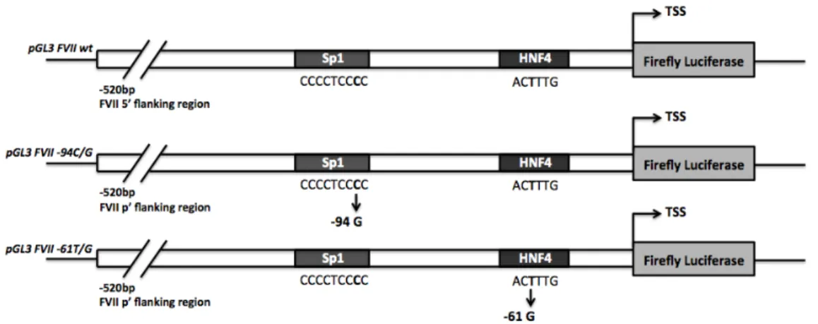
The two natural variants we considered, the -94 C>G and the -61 T>G transver-sions relative to the initiation codon, are reported to falling in the binding site for the hepato-specific HNF-4 and the ubiquitous Sp1 transcription factors, respec-tively (Fig. 1.4). The proximal promoter region of FVII gene has been shown to maintain the maximal promoter activity in HepG2 cells [76], with the HNF-4 and Sp1 binding sites included in the first 185 bp upstream the translation start site (TSS). We have therefore generated the reporters vectors for the F7 promoter, wt and mutants, by cloning 520 bp human F7 promoter region upstream the coding sequence for firefly luciferase (Fig.1.4), so obtaining the pGL3 FVII wt, pGL3
1.7 Results 13
Figure 1.4: Scheme representing the reporter plasmids pGL3 FVII wt, -94G and -61G. In each construct the firefly luciferase expression cassette is cloned downstream the F7 proximal promoter (520 bp). The reported Sp1 and HNF-4 binding sites with the point mutations are included in the first 185bp before the TSS.
FVII -94G and pGL3 FVII -61G plasmids. Based on computational analyses of the F7 promoter and of the transcription factor binding sites in relation with the position of the nucleotide changes, we performed a tailing design for a panel of TALE-TFs targeting the functional sequences and resulting in 4 TALE-TF platforms (TF 1, 2, 3 and 4) covering a region of 70 bp ranging from -122 to -52 of the F7 5’-flanking region (Fig.1.5). In order to reduce off-target activation we targeted sequences from 18 to 26 bp in length spanning the region between Sp1 and HNF-4 binding site on FVII promoter predicted to be unique sites in the human genome by performing an in–silico analysis. All the constructs are
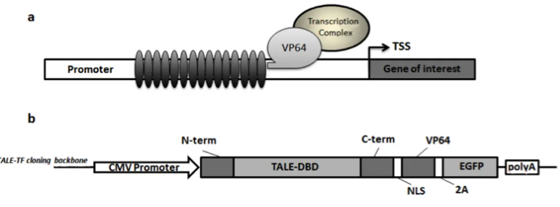
Figure 1.5: Scheme representing the reporter system. Firefly luciferase gene is regulated by the proximal promoter region of FVII gene (520bp), wt or mutated, targeted by four different TALE-TFs (TF1, TF2, TF3 and TF4).
based on a specific TALE-TF cloning backbone which permits the expression of the customized TALE protein fused with a transcription activator that is, four VP16 peptides (i.e.VP64), able to recruit the transcriptional machinery on the target promoter (Fig1.6a). The final TALE-TF expressing-plasmids contain also a 2A sequence followed by an enhanced EGFP-coding gene (Fig.1.6b).
Figure 1.6: a) Scheme representing the TALE-TF platform. Each synthetic TALE-TFs is expressed as a fusion protein composed of a TALE-derived DNA modular binding-domain and a VP64 transactivation domain, able to recruit the transcriptional machinery on a spe-cific target promoter to stimulate the transcription of the gene of interest. b) The TALE-TF expression plasmid is composed of the TALE-derived DNA binding domain, the nuclear local-ization signal (NLS), a transcriptional activation domain (VP64), and a 2A auto-cleavage peptide followed by an enhanced EGFP-coding gene. The expression cassette is driven by the CMV strong promoter.
1.7.2
In vitro models: effect of promoter mutations and
validation of the efficacy of eTFs
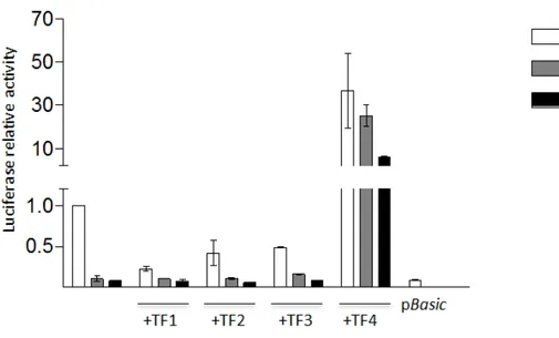
The expression of the reporter plasmids pGL3 -94G and pGL3 -61G in HepG2 cell line showed the causative effect of the mutations, with an abolishment of the firefly luciferase transcriptional levels compared to the pGL3 FVII wt (Fig.1.7). Most importantly, we observed a robust increase (25±4.9 fold increase for pGL3 -61G and 5.9±0.6 for pGL3 -94G) in the transcriptional levels of the reporter genes guided by promoter mutants by co-transfecting the cells with TALE-TF 4 (fig.1.7), and thus was confirmed also when it was co-expressed with the pGL3 FVII wt (36.6±17.3 fold increase). Conversely, TALE-TF 1, 2 and 3 did not show a significant transcriptional activity, either with the mutants and the F7 promoter wt. A negative control was represented by a pBasic vector carrying the firefly luciferase gene without an upstream promoter sequence (fig.1.7).
1.7.3
Assessment of the specificity of TF4
To investigate more in detail the specificity of TF4, our best candidate, we per-formed a series of experiments in HepG2 cells by modulating some in-cis and
in-trans elements of the reporter genes transcriptional system. First we
co-transfected TF-4 expressing-vector with the pBasic vector lacking the 5’ promoter region and, in parallel, with a pSlug vector containing the firefly luciferase guided by the ubiquitous Slug promoter derived from a Snail gene family of zinc-finger transcription factors [32]. In both conditions transcriptional levels of the reporter gene remained unaltered (Figure 1.8). A positive control was represented by the
1.7 Results 15
Figure 1.7: Co-expression of the reporter plasmids wt, pGL3 -94G or -61G with each synthetic TALE-TFs in HepG2 cell line. Histograms report luciferase relative activity as change fold relative to pGL3 FVII wt alone. The negative control is represented by a plasmid pBasic carrying firefly luciferase gene without an upstream promoter. All the results are expressed as mean ±SD derived from three independent experiments.
co-transfection of TF-4 with pGL3 FVII wt.
Moreover, to test the DNA binding specificity we introduced 5 mismatches in
Figure 1.8: Co-expression of the TALE-TF4 with pGL3 FVII wt, pBasic or pSlug reporter plasmids in HepG2 cell line. Histograms of the dual assay report firefly luciferase relative activity as change fold relative to pGL3 FVII wt alone. All the results are expressed as mean ±SD derived from three independent experiments.
the TF4 target sequence by site-specific mutagenesis of the plasmids pGL3 FVII wt and pGL3 FVII -61G, thus creating the pFVII wt ∆T4 and the pFVII -61G ∆T4 vectors. In presence of 5bp changes TF4 DNA binding and transactivation
capability is virtually abolished, as shown in Figure 1.9. In addition, we tested
Figure 1.9: Effect of TF4 co-transfected in HepG2 with pGL3 FVII wt ∆T4 or pGL3-61 ∆T4 reporter plasmids, expressing firefly luciferase guided by FVII proximal promoter carrying 5 mismatches in TALE-TF4 binding site. The negative control is represented by pBasic vector. All the results are expressed as mean ±SD derived from three independent experiments
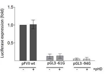
the TALE-DNA binding domain-play role by co-transfecting pGL3 FVII wt, -61G and -94G variants with a pHD plasmid expressing the VP64 transactivator with-out the TALE-derived DBD, and even in this case any substantial enhancement in firefly relative activity was appreciable (Figure 1.10).
Figure 1.10: Co-transfection of HepG2 cells with the pGL3 FVII wt, -61G or -94G with a pHD backbone vector expressing the VP64 transactivation domain alone. Histograms of the dual assay report firefly luciferase relative activity as change fold relative to pGL3 FVII wt alone. All the results are expressed as mean ±SD derived from three independent experiments.
1.7 Results 17
1.7.4
Effect of TF4 in the endogenous context of HepG2
cells
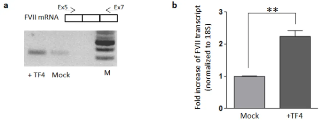
To assess the capability of TF4 in enhancing gene expression in the genomic context we targeted the endogenous F7 promoter in HepG2 cells. Because of the low transfection efficiency assessed in this cell line by using lipofectamine method (<10%), we took advantage of the eGFP expression from the TF4 coding-vector (fig.1.6b) thus selecting eGFP positive-HepG2 post-transfection by FACS analysis. Total RNA was then extracted from both eGFP-positive and negative cells, followed by a specific RT-PCR in order to detect F7 mRNA. In the eGFP-positive HepG2 (+TF4) the F7 transcript amount was increased compared to eGFP-negative cells (fig.1.11a). The quantification of F7 mRNA levels through qPCR showed an increase by 2.3±0.2 fold (P <0.01) after treatment with TF4 (fig.1.11b). Data analyses were performed with T-Student test comparing TF4-treated cells with untrasfected mock cells; * p < 0.05; ** p < 0.01.
Figure 1.11: Effect of TF4 on the FVII transcriptional levels in HepG2 endogenous context. a) RT-PCR performed on cDNA from HepG2 eGFP positive and negative (mock) cells. F7 transcript was amplified using a primer F on exon 5 (Ex5) and a primer R on exon 7 (Ex7). PCR products (312bp) were detected by gel electrophoresis on agarose gel 2%. b) Levels of FVII mRNA in transfected GFP positive-HepG2 cell compared to the mock, as determined by relative qPCR. All the results are expressed as mean ±SD derived from three independent experiments. The change folds are reported to 18S housekeeping gene expression.
1.7.5
Effect of TF4 in the endogenous context of Hek293
cells
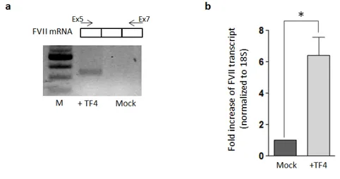
We tested TF4 transcriptional activity also in the endogenous context of Hek293 cells that normally do not express F7 gene (http://webserver.mbi.ufl.edu/ ~shaw/293.html), as confirmed from our experimental data. Hek293 cells were treated with TF4, and the presence of F7 mRNA was confirmed by specific RT-PCR. Conversely, in the mock non-treated Hek293 cells no F7 mRNA was de-tectable, as expected (fig.1.12a). The increase in F7 mRNA levels was quantified
by qPCR and established at 6.3±1.3 fold (P <0.05) compared to the mock cells (fig.1.12b). Data analyses were performed with T-Student test comparing TF4-treated cells with untrasfected mock cells; * P < 0.05.
Figure 1.12: Effect of TF4 on the FVII transcriptional levels in HeK293 endogenous context. a) RT-PCR performed on cDNA from Hek293 TF4-treated and non-treated cells. F7 tran-script was amplified using a primer F on exon 5 (Ex5) and a primer R on exon 7 (Ex7). PCR products (312bp) were detected by gel electrophoresis on agarose gel 2%. b) Levels of FVII mRNA in TF4-treated Hek293 cells compared to the mock (transfected with the lipo-fectamine vehicle), as determined by relative qPCR. All the results are expressed as mean ±SD derived from three independent experiments. The change folds are reported to 18S housekeeping gene expression.
1.7.6
Effect of the TF1, TF2 and TF3 in the endogenous
context of HepG2 and Hek293 cells
As experimental control HepG2 and Hek293 cells were transfected with TF1, 2 or 3, even if they did not show appreciable transcriptional activity in the gene reporter assays (fig1.7). F7 mRNA levels were quantified by qPCR and nor-malized to the untreated cells. The F7 mRNA was increased by 1-1.5 fold (P value=ns) in HepG2 cells (fig.1.13a) and by 2-3 fold in Hek293 cells (fig.1.13b), a non-significant effect compared to TF4 treatment (fig.1.12a). Data analyses were performed with one-way ANOVA and Bonferroni correction for multiple comparisons (TFs compared to the mock).
1.7 Results 19
Figure 1.13: Effect of TF1, TF2 and TF3 on the FVII transcriptional levels in HepG2 and HeK293 endogenous context. a) Levels of FVII mRNA in HepG2 cells treated with TF1, 2 and 3 compared to the mock untreated cells, as determined by relative qPCR. All change folds are reported to 18S housekeeping gene expression. b) Levels of FVII mRNA in Hek293 cells treated with TF1, 2 and 3 compared to the mock untreated cells, as determined by relative qPCR. All the results are expressed as mean ±SD derived from three independent experiments. All change folds are reported to 18S housekeeping gene expression.
1.7.7
Discussion and future perspectives
The in-vitro study is part of a wider project focused on the genetically-determined deficiencies of blood coagulation factors that, in the most severe cases, are asso-ciated to life-threatening bleeding symptoms, sometimes also fatal. It is worth noting that in these pathologies even modest increase of functional protein lev-els in plasma significantly ameliorates the bleeding phenotype. FVII deficiency due to promoter mutations represents a good model to investigate the ability of engineered TFs of enhancing gene transcription for therapeutic purposes. As a matter of fact, increased expression of F7 gene may reduce or completely reverse the symptoms of this deficiency, given that patients have been shown to receive benefit from restored level of proteins up to 6% [98]. In this in-vitro study the model of FVII deficiency has been validated for the promoter variants -94C>G and -61T>G by exploiting gene reporter assays in HepG2 cell line, due to the im-possibility of conducting the experiments in primary hepatocytes from patients, which are not available. These mutations, by altering the DNA binding sites for the natural transcription factors Sp1 and HNF-4 [18, 29], lead to a consequent decrease in the functionality of the promoter and reduce the transcriptional levels, as confirmed by the firefly luciferase reporter assays (fig.1.7). The screening with four engineered TFs designed to recognize different target sequences spanning from Sp1 to HNF-4 binding sites on F7 promoter allowed the identification of TF4, that showed a robust enhancement of the transcriptional activity (from ∼6 to 50 fold increase, depending on the variant considered, fig.1.7). The different changes in the expression levels for the mutants -94C>G and -61T>G (fig.1.7) could be explained by a different DNA-protein interaction framework governing
transcription in the different mutant context. Besides, it is important to note that the increased expression is observable also in the presence of the F7 pro-moter wt, while the treatment with the TF1, 2 and 3 is no effective in any case (fig.1.7), thus giving important information on the promoter accessibility. The reason why only TF4 is effective could be at least in part attributable to the position of its target sequence between the Sp1 and HNF-4 binding sites. This probably represents the best positioning from the TSS in close proximity to the endogenous TFs binding sites in order to recruit the transcriptional machinery, and to avoid the competition of the TF4 with the other TFs on their own DNA binding sites. An important issue that has to be considered in the correction approach with engineered TFs is the specificity, since there is a potential risk arising from non-specific DNA-TF interactions that would lead to the alteration of off-target gene expression. One way to improve the specificity is a careful design of the protein DBD. For this purpose the four TFs were assembled to recognize DNA sequences from 18 to 26bp in length that, by in-silico analyses, are predicted to be unique into the genome. We focused in particular on the potential off-target effects that could be induced by the treatment with TF4, the only effective, testing its binding specificity by modulating some elements in the fusion protein itself and in the target sequence. The co-transfection of TF4 expressing-plasmid with the pSlug vector carrying firefly luciferase under the control of the Slug promoter showed no increase in the reporter activity (fig.1.8), thus supporting the specificity for F7 promoter. Most importantly, we found that 5 mismatches in the TF4 target sequence (fig.1.9) are able to abolish the TF4 transactivation capability, thus strengthening a mechanism strictly dependent on the highly specific target sequence, as previously reported [41]. We confirmed that the recruitment of the transcriptional machinery by eTF4 on the promoter is specifically due to its binding with the DNA by testing the activity of VP64 lacking the TALE-derived DBD. The co-expression of VP64 alone in presence of the F7 promoter wt, as well as the two mutants, did not showed any change in the reporter relative activity (fig.1.10). Data obtained from the gene reporter assays showed the efficacy of TF4 that is able to efficiently recruit the transcrip-tional machinery and rescue the expression of the reporter otherwise abolished in presence of the disease-causing mutations. However, it is worth noting that the reporter genes episomal system may not be representative of the physiological context, because it does not permit to take into account the chromatin context. To address this issue we assessed the activity of the four eTFs on the endogenous F7 gene expression in HepG2 and Hek293, which express or not express FVII, respectively. In HepG2, a human hepatoma cell line, the treatment with TF1, 2 and 3 had a minor impact on F7 mRNA levels, with the maximum change fold measured around 1.5 (fig.1.13a), reflecting the previous data obtained with the gene reporter system. On the other hand, the treatment with eTF4 showed a more robust increase in F7 mRNA levels (fig.1.11a) established to be 2.3±0.2 fold. Surprisingly, this fold increase was markedly less pronounced as compared to that observed in the episomal plasmid context with the F7 promoter wt vec-tor (fig.1.7). It is therefore possible to argue that chromatin environment has a
1.7 Results 21
strong impact on the capability of the eTF to bind efficiently F7 promoter and/or to enhance transcription via VP64. This finding is supported by other literature data in which the magnitude of gene activation by eTFs at native chromosomal loci was relatively modest [66, 93].
Nevertheless, it has also to be considered that the enhancement achieved is referred to its action on the F7 promoter wt, thus supporting the idea that a major improvement could be reachable in presence of promoter mutations. Ex-periments conducted in human embryonic kidney Hek293 background showed F7 mRNA levels increased from 2.2±0.3 (+TF3, fig.1.13b) to 6.3±1.3 times (+TF4, fig.1.12b). It is worth noting that Hek293 cells normally do not express FVII (as revealed by RT-PCR and qPCR experiments in the transfected cells), though af-ter treatment with TF4 F7 mRNA was clearly detectable by RT-PCR (fig.1.12a). This data confirms the capability of TF4 in actively stimulating F7 gene ex-pression in Hek293 cells. The different behavior in HepG2 and Hek293, with a surprising activity in Hek293 cells in which the transcription on F7 promoter is normally down-regulated, may underlie a competition of TF4 with the liver specific HNF-4 in HepG2 (http://www.cisreg.ca/cgi-bin/tfe/articles.pl? tfid=140&tab=expression).
Overall, these results suggest that TF4 should be able to increase expression of the F7 gene in patient cells. This may potentially reduce or completely reverse the symptoms of FVII deficiency, as patients with FVII deficiency have been shown to receive benefit from restored levels of protein up to 6% [98]. We are conscious that the study in vitro, by exploiting reporter gene assay, does not permit the evaluation of eTFs effect on the proper chromosomal context and thereby the ef-ficacy of TF4 has to be validated in a proper model of FVII deficiency caused by promoter mutations. Future experiments will be addressed to test the detrimental impact of human F7 promoter mutations in the murine environment by trans-fecting expression vectors, either wt or mutated, in Hepa1-6 murine cells. Normal or impaired expression of the human FVII gene will be evaluated by qRT-PCR. Mutations that will affect transcription to similar extent in the human and mouse liver cell lines will provide the rationale for the subsequent experiments in-vivo. These will be conducted in specific mouse models for promoter mutations created by zygote injection of plasmids encoding CRISPR-Cas9 system to target the F7 murine promoter or a safe locus and insert the human F7 (hF7) gene under the control of the human promoter, either wild-type (positive control) or bearing the natural mutations. In our hypothesis, the mutations would prevent the apprecia-ble expression of the hFVII in mouse liver, and thus the presence of circulating protein. Injection of liver-specific AAV vectors expressing TF4 in these new an-imal models and evaluation of hFVII expression at the mRNA and protein level will permit the assessment of the correction efficacy in vivo. Moreover, a proper animal model will permit the determination of the optimal delivery method, the characterization of the specificity in the gene regulation and the evaluation of the immunogenicity of the synthetic proteins, very important issues when proposing an innovative therapy.
Chapter 2
SB Transposon
2.1
Focus on FIX: gene and protein features
2.1.1
Factor FIX protein
Human coagulation factor IX, also called Christmas factor or antiemophilic B factor, is a single chain glycoprotein synthesized in liver and circulating in blood like a zymogen at a physiologic concentration of 5 µg/ml [1]. As FVII it features a N–terminal γ–carboxyglutamic acid–rich domain followed by two EGF–like do-mains and the C–terminal serine protease domain. The mature protein consists of 415 aminoacids of 57 KDa molecular weight. The first 40 aminoacids constitute the γ–carboxyglutamic (Gla) domain, which contains post-translational modifica-tions of many glutamate residues by vitamin K–dependent carboxylation to form γ–carboxyglutamate (Gla). The Gla residues are responsible for the high-affinity binding of calcium ions [101, 117, 54], so conferring to FIX its biological activ-ity. Then the protein consists of a short hydrophobic segment (residues 41-46), two EGF–like domains (residues 47-84 (EGF1) and 85-127 (EGF2)), an activa-tion peptide region (residues 146-180), and a serine protease module (residues 181-415). Activation of FIX involves proteolytic cleavages at Arg145-Ala146 and Arg180 and Val181 bonds with a concomitant release of a 35-residue activation peptide (3, 40). The FIXa thus formed contains a light chain (residues 1-145) and a heavy chain (residues 181-415) held together by a single disulfide bond. The light chain consists of the Gla, EGF1, and EGF2 domains whereas the heavy chain contains the serine protease domain that features the catalytic triad of residues His-c57221, Asp-c102 269 and Ser-c195 365 [118, 103, 2].
2.1.2
Factor FIX gene
The gene of human factor IX (F9) is localized in q27.1-q27.2 region of chromosome X. Isolated and sequenced in 1985 [91] it is approximately 34kb in lenght and contains eight exons (spanning from 25bp to 548bp) and seven introns. The transcript is 2803 bases in length and comprises a short 5‘ UTR (29 bp), an open reading frame (1383 bp) and a 3‘ UTR (1390 bp). Exon 1 and part of exon
Exons Coding region Dimension (bp) 1 2 3 4 5 6 7 8
Pre-‐pro leader sequence Pre-‐pro leader, Gla domain
Gla domain EGF-‐like domain EGF-‐like domain Ac@va@on site Cataly@c domain Cataly@c domain 88 164 25 114 129 203 115 571
Table 2.1: Factor IX gene exons organization
2 encode for the pre-pro leader sequence, which is removed during the protein biosynthesis, while the last part of exon 2 together with exon 3 encode for the Gla domain containing 12 γ–carboxiglutamic acid residues. The γ–carboxilation is a modification needed for the proper folding of the FIX and for its calcium binding capacity. Exons 4 and 5 encode respectively for the first and the second epidermal growth factor-like domains (EGF-like 1 and 2). Exon 6 codes for the activation domain, in which factor IXa and the complex FVIIa-TF cut in order to activate FIX. The last two exons encode for the catalytic domain of the protein (tab.2.1) [17].
2.2
FIX deficiency-Haemophilia B
Haemophilia B, or Christmas disease, is an inherited, X-linked, recessive disor-der that results in deficiency of functional plasma coagulation factor IX [11]. It has a prevalence of around 1 in 30,000 live births (about five times rarer than haemophilia A). There are usually carrier females and affected males. The clas-sification of the severity of haemophilia B is based on either clinical bleeding symptoms or plasma pro-coagulant levels; the latter are the most widely used criteria.
• Severe disease occurs with a factor IX level below 1% of the reference and accounts for about 50% of cases.
• Moderate severity occurs with a level of 1-5% and accounts for around 30% of cases.
• Mild disease is with levels of 6-30% and accounts for around 20% of cases. People with mild haemophilia B typically experience bleeding only after serious injury, trauma or surgery and in many cases the disease is not diagnosed until an injury, surgery or tooth extraction results in prolonged bleeding. Women with
2.3 Background and rationale 25
mild haemophilia often experience menorrhagia, heavy menstrual periods and can hemorrhage after childbirth. People with moderate haemophilia tend to bleed af-ter injuries, and finally people with a severe phenotype may have also frequent spontaneous bleeding episodes, often into their joints and muscles. The current treatment is based on the intravenous administration of FIX (replacement ther-apy), either plasma derived or recombinant [106, 119]. Since even small increase in FIX levels (>2%) would result in significant amelioration of the clinical pheno-type, HB represents a model to investigate innovative therapeutic approaches in a quantitative manner, by virtue of functional and protein assays in plasma. Enor-mous efforts have been pushed on substitutive gene therapy, and very recently it has been demonstrated that intravenous infusion of a AAV vector encoding FIX in HB patients resulted in FIX expression ranging from 1% to 6% for periods of 2 years, with amelioration of bleeding phenotypes [104, 68]. DNA editing by using zinc finger nucleases has been also exploited in HB mouse models [53]. However, there are still limitations regarding the delivery method to the target site and safety of gene therapy that encourage research towards alternative therapeutic approaches.
Figure 2.1: Pie chart showing the F9 mutational pattern reported in the Haemophilia B Mutation Database.
2.3
Background and rationale
In the last years enormous efforts have been made to develop therapeutic ap-proaches for human genetic diseases alternative to protein replacement therapy. The most remarkable results arise from the gene therapy field that has shown its huge potential in the cure of diseases such as primary immunodeficiencies [15, 14, 44], Leber’s congenital amaurosis [57], cancer [9], Fanconi’s anemia [85]
and, recently, Haemophilia B [68, 69]. The concept behind gene replacement is very simple, concerning the possibility of efficiently transferring and expressing a normal copy of a defective gene into target cells [115]. Fascinatingly, the ad-vances in biotechnology have allowed the therapeutic technique to move from the conceptual stage to its application in patients for clinical trials for a variety of disorders. To date, from 1989 to 2014 over one thousand gene therapy clin-ical trials have been completed, are ongoing or have been approved worldwide (http://www.abedia.com/wiley). However, there are still issues that limit the wide applicability of these approaches. Limitations are related to the transgene size, the long-term expression and the regulation of the therapeutic gene, to the immune response, to the nature of the DNA vehicle and to harmful effects such as insertional mutagenesis when the transgene is integrated into the genome [5]. Therefore, research is still pushed towards alternative and/or complementary therapeutic strategies based on the correction of the specific disease-causing de-fects, in order to try to circumvent some of these limitations. These approaches are of great interest especially for patients that would benefit even by small increase in functional protein levels, as it happens in the case of coagulation dis-orders (http://www.haemophilia.org). HB is a prototypical example of disease with a heterogeneous mutational pattern (www.factorix.org) and a relatively high frequency of splicing mutations (>15%) in severe disease forms. Mutations inducing aberrant splicing, at splice junctions or within exons, can be addressed by intervention at the pre-mRNA level and this therapeutic approach have been intensively studied by us to correct splicing mutations causing severe HB and FVII deficiency [37, 74, 75, 19]. In particular, we proposed the development of modified U1 small nuclear RNA (U1 snRNA), fundamental component of the U1small nuclear ribonuclearparticle (U1snRNP) with an essential role in exon definition [108], to restore exon inclusion during pre-mRNA maturation impaired by exon-skipping causing mutations.
2.4
Exon Specific U1s (ExSpeU1s) as
therapeu-tic tool
An early event in exon definition is the recognition of the 5’ss by the U1 snRNP, which is composed by a 164bp-long U1 snRNA associated to U1-specific proteins (U1-70K, U1A and U1C) and Sm proteins that are also present in the other snRNPs [108]. The gene coding for the U1 RNA is present in multiple copies in the human genome, is highly evolutionary conserved and possesses its own specific Pol II promoter and polyA site in a short gene long ∼ 600bp . The as-sembly of the spliceosome in the early phase of the splicing process begins with the recognition of the 5’ splice site (5’ss) sequence by the U1snRNP through its 5’ RNA tail [78, 89], which U1 snRNP-associated proteins U1-70K and U1 stabilize this transient interaction. Another important step following the U1 snRNP-5’ss binding is the recognition of the 3’ss mediated by the U2 auxiliary factor [81]. The subsequent establishment of multiple interactions between the 3’ss and the