UNIVERSITY OF CATANIA
DEPARTMENT OF AGROFOOD AND ENVIRONMENTAL MANAGEMENT SYSTEMS
INTERNATIONAL PhD
PLANT HEALTH TECHNOLOGIES AND PROTECTION OF AGROECOSYSTEMS
CYCLE XXV 2010-2012
Detection of new Calonectria spp. and Calonectria Diseases and
Changes in Fungicide Sensitivity in Calonectria scoparia Complex
This thesis is presented for the degree of Doctor of Philosophy by
VLADIMIRO GUARNACCIA
COORDINATOR SUPERVISOR PROF. C. RAPISARDA PROF. G.POLIZZI
CHAPTER 1 - The genus Calonectria and the fungicide resistance... 1 1.1 Introduction... 2 1.1.1 Calonectria... 2 1.1.2 Importance of Calonectria ... 3 1.1.3 Morphology... 6 1.1.4 Pathogenicity... 9 1.1.5 Microsclerotia ... 9 1.1.6 Mating compatibility... 10 1.1.7 Phylogeny... 12
1.1.7.1 Calonectria scoparia species complex ... 13
1.1.7.2 Calonectria pauciramosa... 16
1.1.7.3 Calonectria polizzii ... 18
1.1.8 Calonectria diseases in Mediterranean environment... 19
1.1.9 Calonectria diseases control ... 27
1.2 Fungicide resistance... 33
1.2.1 Introduction... 33
1.2.2 Fungicide control of crop disease ... 33
1.2.3 Defining fungicide resistance... 34
1.2.4 Occurrence of resistance ... 36
1.2.5 Resistance mechanism ... 39
1.2.6 Monitoring ... 40
1.2.7 Assessing the risk... 42
1.2.8 Management strategies... 44
1.2.9 Specific management strategies ... 49
1.2.9.1 Benzimidazoles ... 50
1.2.9.2 SBIs (sterol biosynthesis inhibitors) ... 51
1.3 Fungicide resistance in Calonectria spp. ... 53
1.4 Thesis aims... 54
CHAPTER 2 - Detection of new disease on Laurus nobilis and identification of pathogen species responsible ... 61
2.1 Introduction... 62
2.2 Materials and methods ... 63
CHAPTER 3 - Calonectria spp. causing leaf spot, crown and root rot of ornamental
plants in Tunisia ... 70
3.1 Introduction... 71
3.2 Materials and methods ... 74
3.2.1 Disease survey and fungal isolates... 74
3.2.2 DNA sequence comparisons ... 74
3.2.3 Taxonomy ... 77
3.2.4 Pathogenicity... 77
3.3 Results... 78
3.3.2 Disease survey and fungal isolates... 78
3.3.3 DNA sequence comparisons ... 80
3.3.4 Taxonomy ... 82
3.3.4.1 Calonectria pseudomexicana... 82
3.3.4.2 Calonectria tunisiana... 84
3.3.5 Pathogenicity... 86
3.4 Discussion ... 91
CHAPTER 4 - Changes in sensitivity to prochloraz in Calonectria scoparia complex in southern Italy ... 93
4.1 Introduction... 94
4.2 Materials and methods ... 95
4.2.1 Sampling sites and fungal population ... 95
4.2.2 Molecular identification by using DNA sequence comparisons... 97
4.2.3 Morphological identification... 99
4.2.4 Assessment of fungicide sensitivity in vitro ... 100
4.2.5 Assessment of fungicide sensitivity on model plant-host... 102
4.2.6 Assessment of fungicide sensitivity on Feijoa sellowiana... 103
4.2.7 Statistical data analysis ... 104
4.3 Results... 106
4.3.1 Molecular identification by using DNA sequence comparisons... 106
4.3.2 Morphological identification... 109
4.3.3 Assessment of fungicide sensitivity in vitro ... 109
4.3.4 Assessment of fungicide sensitivity on model plant-host... 116
4.4 Discussion ... 122 Acknowledgements... 126 References... 128
1.1 Introduction
1.1.1 Calonectria
The genus Calonectria (Ca.) was erected in 1867 by De Notaris, based on Ca. daldiniana collected on leaves of Magnolia grandiflora (Magnoliaceae), in Italy (Rossman 1979a). Rossman (1979a) later reduced Ca. daldiniana to synonymy under Ca. pyrochroa, and defined this nectrioid fungus as having an ascocarp wall structure that is brightly coloured, changing to blood-red in 3 % KOH solution, warty to scaly and with a Cylindrocladium (Cy.) anamorph (Rossman 1993, Rossman et al. 1999). However, due to the restricted morphological characteristics of the teleomorph (Rossman 1979b, 1983), specimens can in many cases only be identified to species level if the anamorph is present (Schoch et al. 2000b, Crous 2002).
The anamorph genus Cylindrocladium, which is based on Cy. scoparium, was first described by Morgan (1892) in the U.S.A., where it was found as saprobe on a pod of Gleditsia triacanthos. Although Morgan (1892) failed to mention the stipe extension terminating in a vesicle of characteristic shape, he defined the genus as having branched conidiophores producing cylindrical conidia. This fungus has a wide distribution in sub-tropical and tropical regions of the world, and species are pathogenic to numerous plants (Crous 2002).
Calonectria resides in the Nectriaceae, one of three families in Hypocreales, an order that has been reviewed extensively (Rogerson 1970, Rossman 1983, Rossman et al. 1996, 1999). The Nectriaceae family is circumscribed as having uniloculate ascomata that are orange to purple and not immersed in well-developed stromata (Rossman et al. 1999). The family includes approximately 20 genera of socio-economic importance and of these, Calonectria are more clearly distinguished from the others by their Cylindrocladium anamorphs and relevance as plant pathogens.
The first monograph of Cylindrocladium, by Boedjin & Reitsma (1950), introduced seven Cylindrocladium species with a Calonectria connection to one of these species. Later, in her treatment of Calonectria, Rossman (1983) recognized five species including the novel Ca. ophiospora. However, this
species description did not include the anamorph state. The circumscribed type, Ca. pyrochoa, was also incorrectly reduced to synonymy with several other species based only on the teleomorph morphology. Peerally (1991a) highlighted this in a monograph of Cylindrocladium, where he regarded the anamorph morphology as important in distinguishing species of Calonectria. He subsequently recognized 10 Calonectria species with their Cylindrocladium anamorphs, including an additional 16 Cylindrocladium species not associated with a teleomorph. However, he mistakenly reduced Cylindrocladiella, a genus that accommodates Cylindrocladium-like species with small conidia (Boesewinkel 1982), to synonymy with Cylindrocladium.
The monograph of Cylindrocladium by Crous & Wingfield (1994) entrenched the importance of anamorph characteristics in the taxonomy of Calonectria spp. In this monograph, 22 Cylindrocladium species and one variety were recognised, associated with 16 Calonectria species. Five species were assigned to the genus Cylindrocladiella based on morphological characters of the holomorph. The focus on anamorph characteristics is perpetuated in the most recent monograph (Crous 2002), which recognized 28 Calonectria species, all associated with Cylindrocladium anamorphs and an additional 18 Cylindrocladium species for which teleomorph states were not known. Of the latter group, seven taxa were of doubtful authenticity. Actually, 109 Calonectria and 96 Cylindrocladium species are recognised (Crous 2002, Crous et al. 2004b, 2006a, Gadgil & Dick 2004, Lombard et al. 2009, 2010d).
A general search on MycoBank (www.mycobank.org; Crous et al. 2004a, Robert et al. 2005) and Index Fungorum (www. indexfungorum.org) provide a total of 291 and 306 name records respectively for Calonectria. A similar search for Cylindrocladium species on both electronic databases indicated a total of 98 and 92 names records respectively.
1.1.2 Importance of Calonectria
The genus Calonectria was initially regarded as a saprobe as no disease symptoms could be induced by inoculating a suspected host (Graves 1915). The first proof of pathogenicity of these fungi was provided by Massey (1917), and
subsequently by Anderson (1919), who showed pathogenicity of Ca. morganii (as Cy. scoparium). Subsequently, Calonectria species have been associated with a wide range of disease symptoms on a large number of hosts worldwide (Crous 2002). In the past, several authors showed that Calonectria species cause disease on plants residing in approximately 30 plant families (Booth & Gibson 1973, French & Menge 1978, Peerally 1991a, Wiapara et al. 1996, Schoch et al. 1999). Upon closer inspection, the number of plant families is actually closer to 100 and approximately 335 plant host species (Crous 2002). The plant hosts include important forestry, agricultural and horticultural crops and the impact of these plant pathogens has likely been underestimated.
The majority of disease reports associated with Calonectria species in forestry include hosts in five plant families, of which the most important are associated with Fabaceae (Acacia spp.), Myrtaceae (Eucalyptus spp.) and Pinaceae (Pinus spp.). Disease symptoms include cutting rot (Crous et al. 1991, Crous 2002, Lombard et al. 2009, 2010d), damping-off (Batista 1951, Cox 1953, Terashita & Itô 1956, Sharma & Mohanan 1982, Sharma et al. 1984, Crous et al. 1991, Brown & Ferreira 2000, Crous 2002, Taniguchi et al. 2008) leaf diseases (Cox 1953, Hodges & May 1972, Barnard 1984, Sharma et al. 1984, El-Gholl et al. 1986, Peerally 1991b, Crous et al. 1993b, Crous & Wingfield 1994, Crous et al. 1998b, Schoch & Crous 1999, Schoch et al. 1999, Booth et al. 2000, Park et al. 2000, Crous & Kang 2001, Gadgil & Dick 2004), shoot blight (Sharma et al. 1984, Crous et al. 1991, 1998b, Crous & Kang 2001), stem cankers (Cox 1953, Sharma et al. 1984, 1985, Crous et al. 1991, Lombard et al. 2009) and root rot (Cox 1953, Hodges & May 1972, Cordell & Skilling 1975, Mohanan & Sharma 1985, Crous et al. 1991, Lombard et al. 2009). The majority of these diseases are associated with seedling and cutting production in forestry nurseries, but in a few cases Cylindrocladium species have also been reported from older, established commercial plantations. In these cases the pathogens have been reported to cause leaf diseases and shoot blight resulting in defoliation of trees leading to loss of vigour (Hodges & May 1972, Sharma et al. 1985, Booth et al. 2000, Park et al. 2000, Crous & Kang 2001, Crous 2002, Old et al. 2003, Rodas et al. 2005).
In agriculture, Calonectria species have been reported to cause diseases on several economically important crops. Several plant families of agricultural
importance are susceptible to Calonectria infections, including Fabaceae and Solanaceae. Important diseases in these families are Cylindrocladium black rot of Arachis hypogea (peanut) and red crown rot of Glycine max (soybean) caused by Ca. ilicicola and Ca. pyrochroa in the USA (Bell & Sobers 1966, Beute & Rowe 1973, Rowe et al. 1973, Sobers & Littrell 1974, Rowe & Beute 1975, Phipps et al. 1976, Johnson 1985, Dianese et al. 1986, Berner et al. 1988, 1991, Culbreath et al. 1991, Porter et al. 1991, Varon 1991, Hollowell et al. 1998, Kim et al 1998) and Cylindrocladium tuber rot of Solanum tuberosum (potato) (Boedijn & Reitsma 1950, Bolkan et al. 1980, 1981) by Ca. brassicae (as Cy. gracile) in Brazil. Other diseases associated with Calonectria species on agricultural crops include root rot and leaf diseases of fruit bearing and spice plants (Jauch 1943, Wormald 1944, Sobers & Seymour 1967, Nishijima & Aragaki 1973, Milholland 1974, Krausz & Caldwell 1987, Hutton & Sanewski 1989, Anandaraj & Sarma 1992, Risède 1994, Jayasinghe & Wijesundera 1996, Risède & Simoneau 2001, Vitale & Polizzi 2008), post-harvest diseases of fruits (Fawcett & Klotz 1937, Boedijn & Reitsma 1950, Sepiah 1990, Fitzell & Peak 1992, Vaidya & Roa 1992, Sivapalan et al. 1998), root and crown rot of Medicago sativa (alfalfa) (Ooka & Uchida 1982, Hwang & Flores 1987), and sheath net blotch of Oryza sativa (rice) (Crous 2002).
On horticultural crops, Calonectria species have been reported mostly from the Northern Hemisphere, especially in gardens and ornamental commercial nurseries in Europe and Asia (Polizzi & Crous 1999, Polizzi 2000, Crous 2002, Henricot & Culham 2002, Pérez-Sierra et al. 2007, Polizzi et al. 2007a, b, Hirooka et al. 2008, Polizzi et al. 2009a, Vitale et al. 2009). Hosts in this sector include ornamental trees, shrubs and cut flowers in several plant families, most commonly in Arecaceae, Asteraceae, Ericaceae and Rosaceae. A wide range of disease symptoms are recorded including crown-, collar- and root rot, leaf spots, and cutting rot (Massey 1917, Anderson 1919, Aragaki et al. 1972, 1988, Peerally 1991b, Uchida & Kadooka 1997, Polizzi & Crous 1999, Polizzi 2000, Crous 2002, Henricot & Culham 2002, Henricot & Beales 2003, Poltronieri et al. 2004, Lane et al. 2006, Pérez-Sierra et al. 2006, 2007, Polizzi et al. 2006a, b, 2007a, b, Vitale & Polizzi 2007, Aghajani et al. 2008, Hirooka et al. 2008, Vitale et al. 2008, Polizzi et al. 2009a, Vitale et al. 2009).
1.1.3 Morphology
Morphological or phenotypic characters have played a major role in the description of fungal species (Brasier 1997, Taylor et al. 2000) and form the basis of new fungal descriptions as required by the ICBN (McNeill et al. 2005). In recent years, the use of morphological characters alone to delimit new species has been set aside because considered not enough, with more focus being placed on biological and phylogenetic characters (Rossman 1996, Brasier 1997, Taylor et al. 2000). This trend is also evident in recent studies on Calonectria species (Crous et al. 2004b, 2006a).
The morphology of Calonectria and to a greater extent its anamorph, Cylindrocladium, has been important in the taxonomic history of these fungi. Prior to the 1990s, identification of species was based on morphological characteristics and to a lesser extent on sexual compatibility using standardised media (Boedijn & Reitsma 1950, Peerally 1991a, Crous et al. 1992, Crous & Wingfield 1994, Crous 2002). This resulted in the establishment of several species complexes, as many Cylindrocladium species are morphologically very similar. These include the Ca. scoparia complex (Schoch et al. 1999), Ca. brassicae (as Cy. gracile) complex (Crous et al. 2004b) and Ca. kyotensis complex (Crous et al. 2006a). Characteristics of the anamorphs that are extensively employed in identifications include vesicle shape, stipe extension length and macroconidial septation and dimensions (Boesewinkel 1982, Peerally 1991a, Crous & Wingfield 1994, Crous 2002). The morphological characteristics of the teleomorph those are important for identifications are ascospore septation and dimensions, ascospore number within the asci and perithecial colour. Perithecia of Calonectria species are morphologically very similar and these are not typically useful in identifications (Crous & Wingfield 1994, Crous 2002).
Recently, for morphological identification of the anamorphs and teleomorph, single conidial cultures were prepared on synthetic nutrient-poor agar (SNA; Nirenburg 1981, Lombard et al. 2009, 2010). Inoculated plates were incubated at room temperature and examined after 7d. Gross morphological characteristics were determined by microscopic observations. The measurements of conidia, optimal growth temperatures were determined generally on MEA at 5–35 °C in
5 °C intervals in the dark. Colony colours were determined after 7 d on MEA at 25 °C in the dark. Descriptions, nomenclature, and illustrations were deposited in MycoBank (Crous et al. 2004a). This kind of morphological characterization was applied in last years (Lombard et al. 2010b,c,d Lombard et al.2011, Chen et al. 2011), but any difficulties experienced in morphological identification due to the high level of similarity among the species observed, have led to several molecular approaches being employed to identify Calonectria spp.
The use of biochemical techniques can also be used in phenotypic characterisation. These include substrate utilisation and cell wall polysaccharide analysis. The use of aminopeptidase specificity (Stevens et al. 1990) and utilisation of nitrogen and carbon (Hunter & Barnett 1978, Sharma et al. 1992) have been used successfully to separate several Cylindrocladium species. The use of polysaccharides obtained from cell walls of Cylindrocladium positively identified linkages between asexual species and their respective Calonectria teleomorphs (Ahrazem et al. 1997). However, this method has been found to limit value as some species in complexes could not be distinguished (Crous 2002).
Figure 1. a-b. Conidiophores with terminal vesicles of Calonectria spp. c. Cluster of chlamydospores. d. Perithecium with asci and ascospores. e. Ascospores.
a
b
c
d
1.1.4 Pathogenicity
Representatives strains of Cylindrocladium were initially considered to be saprophytic (Graves, 1915). This is not totally surprising, as most species are readily retrieved from soil samples, and are also found to sporulate a lot on debris collected from damp areas. The first disease reports of Cy. scoparium by Massey (1917) and Anderson (1919) were on plants of rose. Subsequent to these reports, numerous others have been made, documenting a wide range of symptoms such as damping-off, root rot, crown canker, fruit rot, stem lesions, tuber rot, etc. (Crous et al. 1991). The germination of conidia, microsclerotia or ascospores, is the first step of infections, which requires free water, infact the role of rain, dew and irrigation practices is very important. High humidity and free water increase diseases caused by species of Cylindrocladium and related genera. Conidia are easily splash-disperded, which underlines the importance or seedling spacing and general nursery hygiene. The disease also appears to be more severe if seedlings are exposed to nutrient stress (Arentz 1991).
Conidia of Cy. reteaudii (as Cy. quinqueseptatum) were observed to germinate faster in vivo than in vitro (Sharma & Mohanan 1990). Germination occurred after 3 h on leaves of 2 month old Eucalyptus grandis seedlings. After germination, germ tubes originating from the same or different conidia were observed to anastomose. Earlier reports (Bolland et al. 1985) stated that Cy. reteaudii could only infect eucalypts through stomata. This was refuted by Sharma & Mohanan (1990), who observed that isolates of Cy. reteaudii favoured direct penetration, and rarely formed appressoria over stomata for stomatal penetration. Direct penetration on eucalypt leaves has on several occasions also been observed for Cy. pauciramosum and is probably the more common mode of penetration for most species.
1.1.5 Microsclerotia
In proximity of infection, chlamydospores and microsclerotia (chain of chlamydospores) have been observed to develop in substomatal chambers in pine needles or in the inner cortex cells (Bugbee & Anderson 1963). When the
infected plant material disintegrates or the plants are harvested, the leaves and the other plant materials fall to the ground, releasing microsclerotia into the soil. Microsclerotia can survive in the absence of the host for periods of 15 years or more (Thies & Patton 1970, Sobers & Littrell 1974), and are the primary survival structure in soil (Phipps et al. 1976). A large number of fungicides have been reported as effective in inhibiting conidial germination or mycelial growth, but not so for microsclerotia (Sharma & Mohanan 1991a). Microsclerotia din not survive in soil with a low water content (Sung et al. 1980), but they survived for longer periods when buried, as the cooler conditions favoured a higher moisture content (Pataky & Beute 1983). A temperature of 25 °C was optimal for microsclerotial survival, irrespective of soil moisture (Almeida & Bolkan 1981). Microsclerotia have been reported to be present at depths of up to 66 cm below the soil surface (Anderson 1919). Dumas et al. (1998) found that an Egedal® bed steamer produced sufficient heat to kill microsclerotia of Cy. floridanum at 5-10 cm soil depths in a bare root forest seedlings nursery, but did not affect the microsclerotia at or below 15 cm.
Very low soil temperatures and severe drought were also found to affect the number of viable microsclerotia recovered (Phipps & Beute 1979, Roth et al. 1979, Taylor et al. 1981). Moreover, preliminary study, in which solarization effectively suppressed C. pauciramosa microsclerotia (Polizzi et al. 2003), were confirmed by recent research showing that different tested solarizing materials, had potential in eradicating Calonectria inocula from soil acting on microsclerotia (Vitale et al. 2012b).
1.1.6 Mating compatibility
Mating strategies have been employed in the taxonomy of Calonectria and have played an important role in identifying new species of the genus (Schoch et al. 1999, Crous 2002). Based on these studies, there were approximately 18 species of Calonectria considered homothallic and 34 heterothallic (Crous 2002, Crous et al. 2004b, Gadgil & Dick 2004, Crous et al. 2006a). Studies in the female fertility of Cylindrocladium by Schoch et al. (1999, 2000a, 2001a) have also shown that several species are self-sterile hermaphrodites requiring fertilisation
from an opposite mating type. This is typical of heterothallic ascomycetes (Leslie & Klein 1996).
Several difficulties associated with applying the BSC have been highlighted (Brasier 1997, Taylor et al. 1999, 2000, Kohn 2005). The most relevant problem occurs where genetically isolated fungal strains retain the ancestral ability to recombine to produce viable progeny (Brasier 1997). This phenomenon has also been found with several phylogenetic species that are closely related in Calonectria. Cy. hawksworthii, Ca. insularis and Ca. morganii were capable of recombining, but that the progeny had low levels of fertility (Crous 2002). Other mating studies done by Overmeyer et al. (1996) and Neubauer & Zinkernagel (1995) have found that induction of fertile perithecia requires the presence of an additional isolate that, however, does not contribute to the genetic make-up of the progeny. Isolates of Ca. polizzii (species very closed to Ca. pauciramosa) were not capable of mating with the
Ca. pauciramosa mating-tester strains or other Ca. pauciramosa isolates from
different geographic regions, while Ca. colombiana and Ca. zuluensis have a homothallic mating system, showing that the presence of homothallic and heterothallic mating strategies in closely related fungi is interesting and could well provide another opportunity to analyse the genetics of mating systems in ascomycetes (Lombard et al. 2010b).
All this data clearly highlights the need for further studies regarding the mechanism of perithecial formation and recombination in Calonectria.
Figure 2. Production of perithecia on CLA medium.
1.1.7 Phylogeny
During last years phylogenetic studies on Calonectria and its Cylindrocladium anamorphs have substantially influenced the taxonomy of these genera. Application of new molecular techniques and particularly DNA sequence comparisons to distinguish between species, showed news in the recognition of numerous cryptic species. Several molecular approaches have been employed that include total protein electrophoresis (Crous et al. 1993a, El-Gholl et al. 1993a), isozyme electrophoresis (El-Gholl et al. 1992, 1997, Crous et al. 1998a), random amplification of polymorphic DNA (RAPD) (Overmeyer et al. 1996, Victor et al. 1997, Schoch et al. 2000a, Risède & Simoneau 2004) restriction fragment length polymorphisms (RFLP) (Crous et al. 1993b, 1995, 1997b, Jeng et al. 1997, Victor et al. 1997; Risède & Simoneau 2001) and DNA hybridisation (Crous et al. 1993b, 1995, 1997a, Victor et al. 1997). All these mentioned techniques have been useful, but DNA sequence comparisons and associated phylogenetic inference had the most important influence on the taxonomy of Calonectria and are most widely applied today.
In the first study using 5.8S ribosomal RNA gene and flanking internally transcribed spacers (ITS) sequences Jeng et al. (1997) were able to distinguish between Cy. scoparium and Cy. floridanum isolates. Subsequently, it was found that this gene region contains few informative characters (Crous et al. 1999, Schoch et al. 1999, Risède & Simoneau 2001, Schoch et al. 2001b). Therefore, the β-tubulin (Schoch et al. 2001b) and histone H3 (Kang et al. 2001a) gene regions have been applied in order to allow for improved resolution in separating species.
The first complete DNA sequence-based phylogenetic study using partial β-tubulin gene sequences (Schoch et al. 2001b) compared phenotypic, biological and phylogenetic concepts used in the taxonomy of Cylindrocladium. This also highlighted the fact that Calonectria represents a monophyletic lineage (Schoch et al. 2000b, 2001b). Subsequently, combined DNA sequence data for the ITS, β-tubulin and histone H3 gene regions have been widely used in studies relating to taxonomic issues surrounding Cylindrocladium and Calonectria (Crous et al. 1999, Schoch et al. 2000a, 2000b, Crous & Kang 2001, Kang et al. 2001a, 2001b, Henricot & Culham 2002, Crous et al. 2004b, 2006a, Lombard et al. 2009, 2010d). Other partial gene sequences recently used include translation elongation 1-alpha (TEF-1α) and calmodulin (Crous et al. 2004b, Lombard et al. 2010d). For Cylindrocladium and Calonectria, there are only six studies (Kang et al. 2001a 2001b, Crous et al. 2004b, 2006a, Lombard et al. 2009, 2010d) that provide files on TreeBase (www.treebase.org).
1.1.7.1 Calonectria scoparia species complex
Several past studies have focused on the taxonomy of Calonectria spp. with small, 1-septate macroconidia and ellipsoidal to obpyriform vesicles (Crous et al. 1993, Overmeyer et al. 1996, Schoch et al. 1999, 2000a). These Calonectria spp. were initially regarded as either Ca. morganii (= Cylindrocladium scoparium) or Ca. scoparia (= Cy. candelabrum) based on their morphological similarities. However, the anamorph state of Ca. morganii was circumscribed as having ellipsoidal to pyriform vesicles and Ca. scoparia having ellipsoidal to obpyriform vesicles by Crous et al. (1993a). Later studies, incorporating DNA
sequence data, have shown that Ca. morganii is restricted to the Northern Hemisphere and Brazil (Crous et al. 1993a, Overmeyer et al. 1996, Schoch et al. 2000a). In contrast, Ca. scoparia is found worldwide and forms part of a species complex consisting of four mating groups, each representing a different Calonectria species that includes Ca. pauciramosa (anamorph: Cy. pauciramosum), Ca. scoparia, Ca. mexicana (anamorph: Cy. mexicanum) and Ca. insularis (anamorph: Cy. insulare) (Schoch et al. 1999).
Calonectria pauciramosa has been reported worldwide on numerous plant hosts (Schoch et al. 1999, Koike et al. 1999, Koike & Crous 2001, Polizzi & Crous 1999, Polizzi 2000, Polizzi & Catara 2001, Polizzi & Vitale 2001, Crous 2002, Polizzi et al. 2006a, 2007b, 2009a, Vitale et al. 2009), where it causes different symptoms such as cutting rot, damping-off, root rot and leaf blight. In South Africa and Australia, Ca. pauciramosa is regarded as the most important pathogen in commercial forest nurseries (Crous 2002) and it is also found on various horticultural crops in commercial nurseries in Italy and the U.S.A. (Schoch et al. 2001, Crous 2002, Polizzi et al. 2006a, 2007b, 2009a,c, Vitale et al. 2009).
Schoch et al. (2001a) considered female fertility in populations of Ca. pauciramosa from various geographical regions to determine the ratio of mating types present, and based on these data suggested that Ca. pauciramosa was endemic to South America given that the ratio of both mating types approached 1:1. Furthermore, the study also indicated that Ca. pauciramosa isolates from California were represented by only one mating type, supporting the view that this represented an introduced pathogen. Isolates from Italy showed higher ratios of hermaphrodites and some variation was observed in the β-tubulin sequences. In contrast, South African isolates had close to a 1:1 mating type ratio and showed variation in β-tubulin sequence data (Schoch et al. 1999, 2001a), indicating that this was either a native pathogen or that there had been multiple introductions into the country.
Initial investigations using DNA sequence comparisons and mating studies on Ca. pauciramosa isolates from South Africa and Colombia showed some variation amongst isolates. These findings and those of Schoch et al. (2001a) suggested that Ca. pauciramosa might accommodate a number of cryptic species. A recent study, considering a variation observed amongst isolates of
Ca. pauciramosa from different geographical localities, had the aim to consider the phylogenetic relationships, morphological characters and mating compatibility of available isolates of Ca. pauciramosa and to determine whether this species represented an assemblage of cryptic taxa. This study revealed the presence of three cryptic species accommodated in cultures that have collectively been treated as Ca. pauciramosa (Lombard et al. 2010d). This result is homogeneous with the results of previous studies (Schoch et al. 1999, 2001a), which noted variation within Ca. pauciramosa, although at that time the sample size was inordinately small to consider the matter further. The descriptions of Ca. colombiana, Ca. zuluensis and Ca. polizzii add three new species to the Ca. scoparia species complex. This complex is characterised by species having ellipsoidal to obpyriform vesicles and producing 1-septate macroconidia (Schoch et al. 1999, Crous 2002). The complex was previously regarded as having a biallelic, heterothallic mating system (Schoch et al. 1999, 2001a). However, both the newly described Ca. colombiana and Ca. zuluensis are homothallic. Schoch et al. (2001a) considered female fertility of Ca. pauciramosa, and found variation in BT sequence data for isolates from Italy. This new species has thus been shown as unique based on morphological, phylogenetic inference and biological characteristics, separating it from Ca. pauciramosa. Morphologically, Ca. polizzii can be distinguished from Ca. pauciramosa by its smaller 1-septate macroconidia. Isolates of Ca. polizzii were also not capable of mating with the Ca. pauciramosa mating-tester strains or other Ca. pauciramosa isolates from different geographic regions (Lombard et al. 2010b).
Figure 3. Morphological variability among different strains belonging to Calonectria
scoparia species complex.
1.1.7.2 Calonectria pauciramosa
Ca. pauciramosa C.L. Schoch & Crous, anamorph Cy. pauciramosum C.L. Schoch & Crous, has been described as a member of the species complex Cy. candelabrum (Schoch et al. 1999). It is a polyphagous fungal species widely reported in Australia, New Zealand, Brazil, Colombia, Mexico, South Africa, USA, and Europe, probably introduced from Australia or South Africa by the trade of plant-propagation material, new plant-species and cultivars.
The first reports of this species concerned Medicago truncatula (Lamprecht 1986), Acacia longifolia (Hagemann & Rose 1988), Rododendron spp., Azalea spp., Eucalyptus spp. and Protea spp. (Botha & Crous 1992).
Subsequently, this pathogen has been reported for the first time in North America on Erica capensis (Koike et al. 1999), while Polizzi & Crous (1999) have confirmed the discovery in Europe on plants of Polygala myrtifolia.
In Italy Ca. pauciramosa is a species widely present in the nurseries of eastern Sicily and other parts of southern Italy, responsible for significant losses on
several ornamental plants, especially on young seedlings, where it finds the optimal conditions for its development. This pathogen was found in several plants such as Fejioa sellowiana, Arbutus unedo, Acacia retinodes and Dodonaea viscosa (Polizzi & Catara 2001). Ca. pauciramosa also causes leaf spots, defoliation, stem blight on different species belonging to the family of Mirtaceae which bottlebrushes, blue eucalyptus, red eucalyptus, melaleuca, myrtle, and Metrosideros spp. (Polizzi 1996).
Until some years ago, for the identification of this species the application of the keys of Crous & Wingfield (1994) and Schoch et al. were considered necessary and enough (1999).
Difficulties experienced in morphological identification, have led to several molecular approaches being employed to identify Calonectria spp. These include total protein electrophoresis (Crous et al. 1993a, El-Gholl et al. 1993), isozyme electrophoresis (El- Gholl et al. 1992, El-Gholl et al. 1997, Crous et al. 1998a), random amplification of polymorphic DNA (RAPD) (Overmeyer et al. 1996, Victor et al. 1997, Schoch et al. 2000a, Risède & Simoneau 2004), restriction fragment length polymorphisms (RFLP) (Crous et al. 1993b, Crous
et al. 1995, Crous et al. 1997, Jeng et al. 1997, Victor et al. 1997, Risède &
Simoneau 2001) and DNA hybridisation (Crous et al. 1993a, 1995, 1997, Victor
et al. 1997). However, DNA sequence comparisons and associated phylogenetic
inference has the most significant impact on the taxonomy of the group. It is also most widely applied in contemporary species descriptions. The 5.8S ribosomal RNA gene and flanking internally transcribed spacer (ITS) sequences made it possible for Jeng et al. (1997) to distinguish between Cy. scoparium and
Cy. floridanum isolates. Subsequently, it was found that this gene region
contains few informative characters for members of the genus (Crous et al. 1999, Schoch et al. 1999, Risède & Simoneau 2001, Schoch et al. 2001b). As a consequence, this resulted in the β-tubulin (BT) (Schoch et al. 2001b) and histone H3 (HIS3) (Kang et al. 2001b) gene regions being widely employed to improve the resolution of phylogenetic trees for species of Calonectria.
In addition, DNA sequence comparisons and mating studies on Ca.
pauciramosa isolates from South Africa and Colombia showed some variation
amongst isolates. These findings and those of Schoch et al. (2001) suggested that Ca. pauciramosa might accommodate a number of cryptic species.
The morphological measures of perithecia, conidia, terminal vesicle, conidiophores and other morphological characters, still are fundamental for the distinction among different species (Peerally 1991, Crous et al. 1992). In Ca. pauciramosa, macroconidiophores form a stipe. This represents a sterile extension from which the bundle of branches fertile started. The stipe is septate, hyaline, 120-230 µm in length, terminating in a vesicle of variable shape from ellipsoidal to pyriform and with diameter of (5-) 7-9 (-11) µm. The primary branches are aseptate or with only one septum (12-45 x 5-6 µm), the secondary branches are aseptate (15-20 x 5 µm), and also the tertiary (12-15 x 5 µm). The terminal branches produce 2-6 phialides, with shape variable from doliiform to reniform, hyaline, without septum, (10-13 x 2.5-4 µm), with a small enlargement at the apex.
The conidia are cylindrical, hyaline, 1-septate, lacking a visible abscission scar, held in parallel cylindrical clusters by colourless slime. The microconidiophores have never been observed. Chlamydospores extensive throughout the medium, represent the preservation organs; dark brown in colour, aggregated to form microsclerotia, while the teleomorph produces perithecia subglobose to ovoid with height of 250-400 µm and width of 170-300 µm, with a variable colour from orange to red to brown. Asci 8-spored, clavate, 70-140 µm, with filiform stipe, contain 8 ascospores, hyaline, fusoid, septate, guttulate and slightly curved with rounded tip.
Colonies fast growing (35–40 mm diameter after 7 d) with optimal growth temperature at 25 oC (growth at 10– 30 oC) on MEA, reverse amber to
sepia-brown after 7 d; abundant white aerial mycelium with sparse sporulation. 1.1.7.3 Calonectria polizzii
Calonectria polizzii L. Lombard, Crous & M.J. Wingf. is a cryptic species belonging to Calonectria scoparia complex. The teleomorph is unknown. Conidiophores with a stipe bearing a penicillate suite of fertile branches, stipe extensions, and terminal vesicles. Stipe are septate, hyaline, smooth, 58–108 × 5–7 µm; stipe extensions septate, straight to flexuous, 111–167 µm long, 5–6 µm wide at the apical septum, terminating in an obpyriform to ellipsoid vesicle, 6–9 µm diam. The conidiogenous apparatus 27–57 µm long, and 28–51 µm
wide; primary branches aseptate or 1-septate, 15–35 × 4–6 µm; secondary branches aseptate, 12–26 × 3–5 µm; tertiary branches aseptate, 10–15 × 4–5 µm, each terminal branch producing 2–6 phialides; phialides doliiform to reniform, hyaline, aseptate, 8–13 × 3–4 µm; apex with minute periclinal thickening and inconspicuous collarette. Macroconidia are cylindrical, rounded at both ends, straight, (31–)32–42(–49) × 3–5 µm (av. = 37 × 4 µm), 1-septate, lacking a visible abscission scar, held in parallel cylindrical clusters by colourless slime. Megaconidia and microconidia absent.
Colonies are fast growing (35-40 mm diameter after 7 d) with optimal growth temperature at 25oC (growth at 10-30 oC) on MEA, reverse amber to sepia-brown after 7 d; abundant white aerial mycelium with sparse sporulation; chlamydospores extensive throughout the medium, forming microsclerotia. Ca. polizzii is morphologically similar to Ca. pauciramosa and Ca. zuluensis. The macroconidia of Ca. polizzii (av. 37 × 4 µm) are smaller to those of Ca. pauciramosa (av. 50 × 4.5 µm).
1.1.8 Calonectria diseases in Mediterranean environment
Several studies of last twenty years reported a broad plant-host group susceptible to Calonectria diseases. Different symptoms such as cutting rot, damping-off, crown rot, root rot, leaf blight and petiole rot are caused by Calonectria spp. in different countries of Mediterranean basin. In Italy, the most widespread species is Ca. pauciramosa reported as agent causal of leaf spots on Arbutus unedo, Acacia retinodes, Feijoa sellowiana, Dodonaea viscosa, Pistacia lentiscus, Brahea armata, Agonis flexuosa (Polizzi & Catara 2001, Vitale & Polizzi 2007, Polizzi et al. 2007a, Polizzi et al. 2010), crown rot and stem blight on plants of Pistacia lentiscus (Vitale & Polizzi 2007), defoliation and stem blight on plants of Eugenia myrtifolia (Polizzi et al. 2009a), crown and root rot on Ceanothus thyrsiflorus, Arbutus unedo, Eugenia myrtifolia and Feijoa sellowiana (Polizzi et al. 2006b, Vitale et al. 2008, Vitale et al. 2009, Polizzi et al. 2009a).
Ca. morganii was also widely reported as responsible of crown rot, leaf spot, stem blight and defoliation on Pistacia lentiscus (Polizzi et al. 2006a),
damping-off, crown, root rot and leaf spot on Callistemon spp. (Polizzi et al. 2007b), leaf spot and shoot blight on Melaleuca acuminata (Polizzi et al. 2009b).
Moreover, Carrai & Garibaldi (1990) observed during 1987 in several Italian nurseries initial symptoms such as leaf spot, petiole rot, stem blight and subsequently the death of plants of Spathiphyllum. The causal agent of these symptoms was identified as Cylindrocladium spathiphylli (Schoultles & El-Gholl 1980) reported for the first time as the agent of crown and root rot of Spathiphyllum plants cv. Clevelandii in 1977 in Florida (Schoulties & El-Gholl, 1980).
In Italy, a further species was reported on plants of Buxus sempervirens in which defoliation and stem blight were observed; the fungal species involved in this disease was identified as Cylindrocladium buxicola (Saracchi et al.
2008), due to the recent outbreaks in northern Europe, in 2007 Cy. buxicola was placed in a list established by the European Parliament
(www.eppo.org/quarantine/quarantine.htm), as regards quarantine pathogens. In Spain Ca. pauciramosa was reported as responsible of leaf spot on Callistemon citrinus (Perez-Sierra et al. 2007), while in Portugal was observed on plants of Myrtus communis causing crown and root rot (Henricot & Beales 2003).
Recently three isolates collected in Italy from symptomatic plants of Arbutus unedo and Callistemon citrinus were identified as Ca. polizzii, one of the three cryptic species recognized within Ca. pauciramosa; the pathogenicity of these isolates was not still established.
Table 1. Ornamental-plants host of Calonectria spp. in Mediterranean basin.
HOST SYMPTOMS FUNGAL
SPECIES REFERENCE
Acacia retinodes Leaf spot, stem
blight Cy. pauciramosum Polizzi & Catara 2001 Agonis flexuosa Leaf spot Cy. pauciramosum Polizzi et al. 2010 Arbutus unedo Leaf spot, stem blight Cy. pauciramosum Polizzi & Catara 2001 Brahea armata Leaf spot Cy. pauciramosum Polizzi et al. 2007 Buxus
sempervirens
Leaf spot, stem
blight, defoliation Cy. buxicola Saracchi 2008 Callistemon spp. Leaf spot, defoliation Cy. scoparium Polizzi et al. 2007 Callistemon spp. Leaf spot, stem blight Cy. pauciramosum Polizzi 1996 Ceanothus
thyrsiflorus
Crown and root
rot Cy. pauciramosum Polizzi et al. 2006 Chamaerops
humilis Leaf spot Cy. pauciramosum Polizzi et al. 2007 Dodonaea viscosa Leaf spot Cy. pauciramosum Polizzi & Catara 2001 Eugenia myrtifolia Crown and root rot Cy. pauciramosum Polizzi et al. 2009a Eucalyptus
globulus Leaf spot Cy. pauciramosum Polizzi 1996 Eucalyptus
rostrata Leaf spot Cy. pauciramosum Polizzi 1996 Eucalyptus spp. Leaf spot Cy. pauciramosum Polizzi 1996
Feijoa sellowiana Leaf spot Cy. pauciramosum Polizzi & Catara 2001 Melaleuca
acuminata
Leaf spot, shoot
blight Cy. scoparium Polizzi 2009b
Melaleuca fulgens Crown and root rot Cy. pauciramosum Polizzi 2009c Melaleuca
hypericifolia Leaf spot Cy. pauciramosum Polizzi 1996 Metrosideros
robustus
Leaf spot, stem
blight Cy. pauciramosum Polizzi 1996 Myrtus communis
Leaf spot, stem blight, crown and root rot
Cy. pauciramosum Polizzi 1996
Pistacia lentiscus
Leaf spot, stem blight, crown and root rot,
defoliation
Cy. scoparium Polizzi et al. 2006
Pistacia lentiscus Leaf spot, stem blight, Cy. pauciramosum Vitale & Polizzi 2008 Polygala myrtifolia Crown and root rot Cy. pauciramosum Polizzi & Crous 1999 Spathiphyllum spp. Leaf spot, petiole
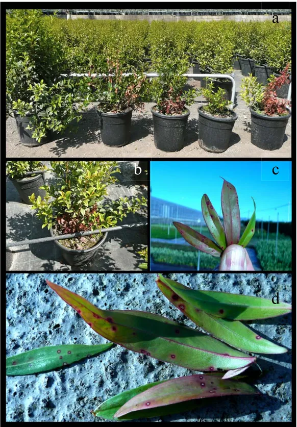
Figure 4. Symptomatic plants of Myrtus communis. a. wilting . b. crown-rot with presence of mycelia. c-d. leaf spot.
a
b
d
c
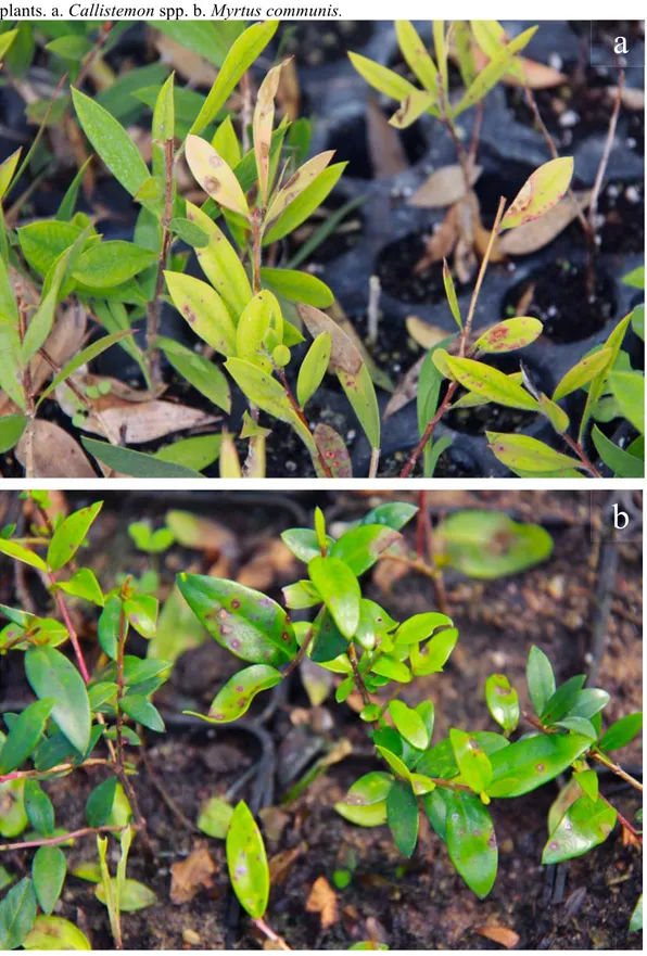
Figure 5. a. Leaf spot and b. severe defoliation on Metrosideros spp. plants. c,e. Leaf spots and d,f. damping-off on plants of Callistemon spp.
c
b
a
d
e
f
Figure 6. a-b. Different severity of disease on plants of Eugenia myrtifolia c-d. Leaf spots on Agonis flexuosa.
a
c
d
b
Figure 7. a-c. Wilting and crown and root rot on Polygala myrtifolia cuttings.
c
a
Figure 8. a. Defoliation and b. leaf spots on Melaleuca fulgens. c. Leaf spot on Melaleuca hypericifolia. Leaf spots on Dodonaea viscosa. e. Death of Arbutus unedo plants as a consequence to crown and root rot.
a
b
c
d
1.1.9 Calonectria diseases control
Various fungicides and different methods of application have been proposed to control diseases caused by Calonectria spp.
Soil fumigation or sterilization has also been reported as effective in reducing soil populations of Calonectria (Jauch 1943). Soil temperature, potting medium, compaction and pH were found to influence Cylindrocladium root and petiole root of Spathiphyllum, with the fungus favored by warm, moist conditions with lower pH and higher soil compaction. However higher pH only appeared to delay the onset of disease development (Chase & Poole 1987). At least, soil pH could play a positive role in disease control when temperatures were sub-optimal. Diseases increased from 25-30 °C, but decreased at 32 °C (Chase & Poole 1987). Only preventative measures were found effective, while no curative effects could be obtained for this disease. In Florida (U.S.A.), Calonectria diseases were controlled by use of fungicide in association with good nursery practices, including immediate removal of diseased plants, use of new, uninfected potting medium, and minimal watering.
To control Calonectria diseases in cuttings during rooting, effort must be made to reduce primary inoculum by using healthy shoots and adopting management practices such as selective and continuous shoot harvesting and the use of inoculum-free trays (Silveira 1996). Good control of Calonectria rot was obtained on azaleas when mother plants were treated with benomyl 9-12 days before cutting were taken (Roos 1980, 1981). Control was further improved if cuttings were dipped in a benomyl solution. Care should be taken, however, as Alfenas et al. (1988) observed that constant use of benomyl to control Calonectria in nursery cuttings led to the selection of benomyl-resistant strains. This led to recommendation that fungicides with different modes of action be used in rotation.
Since chemical control of Cylindrocladium Leaf Blight (CLB) is expensive (Sharma & Mohanan 1992), the long-term solution should be to control CLB by selecting resistant provenances or species. Sharma & Mohanan (1991b, 1992) found, however, that the susceptibility rating of a provenance to different species of Cylindrocladium was related to the eucalypt species or subgenus. These results also provided the first evidence for the existence of physiological
strains in Cy. reteaudii (Sharma & Mohanan 1991b). In an experiment screening 22 fungicides against several species of Cylindrocladium and Cylindrocladiella Sharma & Mohanan (1991a) also demonstrated a differential effect of many fungicides depending on the species of Cylindrocladium involved, and that the ED100 also varied with species.
In Florida the disease was contained by benomyl, triflumizole and prochloraz treatments associated with good agronomic practices such as using uninfected potting medium (Chase 1987).
For some fungicide, the activity to prevent and eradicate the disease and the activity to inhibit the sporulation of Cy. candelabrum were valuated. Tebuconazole, eposiconazole and eposiconazole + pyraclostrobin showed an high ability to inhibit conidial germination and mycelial growth of Cy. candelabrum. In in vivo experiments these active ingredients reduced the foliar infection severity to 96, 98 and 95% respectively (Ferreira et al. 2006).
Grigoletti & Auer (2003) tested several fungicides to manage foliar infections on Ilex paraguariensis caused by Cy. spathulatum. Benomyl and captan resulted the most effective.
Experiments with fungicides were carried out on Polygala myrtifolia showing a good efficacy on different strains of Cy. pauciramosum (Polizzi 2000). In the first trial the preventive activity of prochloraz and tebuconazole was valuated to control root rot and both reduced the symptoms; moreover, the fresh and the dry weight of roots not treated were lower than those of the treated plants. Tebuconazole provide symptoms of stunting with marked reduction of leaf edges on treated plants. The second trial, again on potted plants of P. myrtifolia, showed good effectiveness of repeated applications of prochloraz every 10-12 days.
One more assay with fungicide to manage natural infections caused by Cy. pauciramosum was carried-out on young plants of Callistemon citrinus var. ‘Captain Cook’. The results showed stunting symptoms, reduced average length of plants and a reduced total number of shoot on plants treated with tebuconazole, the Copper hydroxide caused necrotic, dark red dot on young leaves and delay of blooming. Foliar treatments were applied also on two cultivars (‘baetica’ e ‘lusitanica’) of Myrtus communis for infection caused by Cy. pauciramosum on leaves. A lower susceptibility of cultivar ‘lusitanica’ and
a good effectiveness of copper hydroxide were observed; no effectiveness of prochloraz on plants of M. communis was observed, underlining the variability of activity of this fungicide (Vitale et al. 2003).
Brand (2006) tested the activity of 9 active ingredient for the management of Cy. buxicola. The conidial germination on amended medium was inhibited for 16 hours at low rates of tolylfluanide, mancozeb, chlorothalonil and fludioxonil + cyprodinil. The mycelial growth was totally inhibited by prochloraz, propiconazole, thiophanate-methyl and carbendazim + flusilazole for 2 weeks. Chemical control experiments carried out on plants of myrtle showed the effectiveness of regular applications with copper oxychloride for management of foliar infections caused by Cy. pauciramosum while, benomyl, chlorothalonil, prochloraz and dithianon were ineffective (Polizzi & Azzaro 1996).
Experimental trials were carried out with the aim to evaluate the efficacy of the antagonist Trichoderma harzianum T22 and prochloraz in management of crown and root rot of P. myrtifolia caused by Cy. pauciramosum. The biological antagonist provided an excellent control compared to the untreated control and to the chemicals treatments. In addition, plants treated with the strain of T. harzianum, were more vigorous and larger than plants of others treatments, showing a higher commercial value (Polizzi & Vitale 2002).
Recently, Aiello et al. (2013) showed the effectiveness of preventive applications of tebuconazole, fosetyl-Al, Cu hydroxide, thiophanate-methyl, prochloraz + cyproconazole, trifloxystrobin, azoxystrobin and prochloraz in controlling leaf spots caused by both Ca.morganii and Ca. pauciramosa on bottlebrush plants, while cyproconazole, propamocarb + fosetyl-Al and K phosphite were discouraged. Moreover, K phosphite, fosetyl-Al, prochloraz + cyproconazole, cyproconazole, Cu hydroxide, thiophanate-methyl, trifloxystrobin and azoxystrobin provided a significant reduction of crown and root rot caused by Ca. pauciramosa in feijoa plants, detecting variable results for prochloraz applications and suggesting further investigations about the efficacy of this compound.
Soil fumigation (metham-sodium and 1, 3-dichloropropene + methyl isothiocyanate) combined with resistant cultivars significantly reduced the build-up of Cylindrocladium black rot (CBR) in groundnuts (Phipps 1990).
Good control of CBR was obtained using a combination of metham sodium fumigation and resistant genotypes (Cline & Beute 1986). Genetic breeding proved promising in obtaining durable resistance to Sclerotinia minor and Cy. parasiticum (Coffelt et al. 1994).
Practices for controlling CBR and red collar rot (RCR) include the use of resistant varieties (Berner et al. 1988) and cultural practices such as reducing the inoculum and delayed planting (Sidebottom & Beute 1989). A direct correlation was observed between weed-free and disease-free areas in soybean fields (Berner et al. 1991). This led to the investigation of the possible fungicidal activity of herbicides that contain glyphosate. Other recommendations for CBR control include crop rotation with non-legumes, control of nematodes, use of resistant cultivars, and soil fumigation with products containing methyl isothiocyanate (Phipps 1990).
CBR of groundnut was slowed when soil temperatures exceeded 25 °C and stopped when temperatures exceeded 35 °C. Sidebottom & Beute (1989) proposed cultural practices to modify soil temperature, and hence control the disease. In experiments inoculating resistant and susceptible groundnut cultivars, Hadley et al. (1979) concluded that races of Cy. parasiticum could occur, even though host resistance appeared to be quantitatively inherited. The use of antagonistic microorganism is a viable alternative for suppression of plant pathogens in cutting nurseries, since the rooting substrate and the humidity and temperature conditions required for the development of roots favor biocontrol agents (Kunieda-de-Alonso 1997). The incorporation and pre-incubation of Trichoderma isolates in Eucalyptus bark compost resulted in 100% suppression of Rhizoctonia and 90% of Cylindrocladium (A.C. Alfenas). Most species of Cylindrocladium are soil-borne, and many have been associated with root disease problems. In Canada, isolates of Cy. floridanum species complex have also been isolated from asymptomatic seedlings (Juzwik et al. 1988). To date these pathogens have primarily been controlled by means of soil fumigation with either methyl bromide or chloropicrin. The present mandate, however, is that these synthetic pest control agents should be reduced, and that environmentally safe alternatives be explored for disease control. Species of Trichoderma are well-known agents of biocontrol. Dumas et al. (1996) therefore evaluated isolates of T. harzianum, T. viride, T. koningii, T.
polysporum, T. hamatum and T. virense for their ability to inhibit Cy. floridanum in vitro. Isolates that inhibited mycelial growth and microsclerotia production were found to produce 6-n-pentyl-2H-pyran-2-one in culture. However, Cy. floridanum isolates were also found to produce a substance that inhibited the mycelial growth of Trichoderma species. Isolates of Trichoderma or fluorescent Pseudomonas reduced root disease caused by Cy. scoparium and Cy. gracile (Blum & Lin 1991).
Some Bacillus isolates obtained from Eucalyptus grandis leaves were antagonistic to Cy. scoparium (Bettiol et al. 1988). By applying a bacterial suspension from a 10-d-old culture to detached leaves 1, 24 and 48 h before inoculation with the pathogen, disease control similar to that obtained with benomyl (0,5g/l) was obtained. A Bacillus sp. was also observed to provide good control of Cy. scoparium on eucalypt seedlings at the pre-emergence stage (Santos et al. 1993) Under greenhouse conditions Bacillus and Streptomyces spp. were the most effective in controlling the disease.
The effect of various ectomycorrhizal fungi to control common root pathogens were screened by Natarajan & Govindasamy (1990), who reported Suillus brevipes to control most pathogens, including Cy. scoparium. Ectomycorrhizal fungi such as Paxillus involutus and Hebeloma cylindrosporum also inhibited Cy. canadense in vitro (Morin et al. 1999).
Soils treated with Phaeotheca dimorphospora led to an increase in the number of propagules of T. harzianum, whereas the population of Cy. scoparium decreased rapidly and was not detectable 30 days after treatment (Yang et al. 1995). An extract of the bark of Barringtonia ceylanica was found to inhibit growth of Cy. reteaudii by up to 50% (Palanakumbura et al. 1996). Field applications of compost and dazomet controlled Cy. scoparium (Lyons et al. 1997): while field application of sewage and spent mushroom compost also suppressed Cy. scoparium (Hunter et al. 1995).
Cell-wall homogenates of Cy. spathiphylli markedly stimulated the accumulation of tolytoxin, an antifungal secondary metabolite, in cultures of the cyanobacterium Scytonema ocellatum (Patterson & Bolis 1997). These results suggest that tolytoxin is an inducible chemical defense agent (phytoalexin) capable of protecting S. ocellatum against fungal invasion.
Activities and effective dose of different types of Ginkgo biloba have been tested in vitro against Cy. colhouni. The greatest effect was obtained from extracts diluted in ethanol with an efficiency of 37.4% followed by extracts diluted in ether-petroleum (23.7%), and from fresh extracts (18.4%). When the concentration of the extracts has reached high values it is obtained an efficacy of 100% (Feng et al. 2007). The C184 strain of T. harzianum isolated from banana was tested in vitro against Cy. pteridis, the causal agent of necrosis of the roots on Musa acuminata. Culture filtrates of the antagonist reduced the mycelial growth of the pathogen from 52 to 87% (Ngueko & Tong 2002). T. harzianum, T. viride and Pseudomonas fluorescens were tested to evaluate their effectiveness against damping-off of seedlings of Eucalyptus tereticornis and E. grandis caused by Rhizoctonia solani and Cy. quinqueseptatum. Potential antagonists were inoculated alone or in combination with each other, in the soil and on the seeds by dipping. The treatment to the soil with T. harzianum was the most effective in limiting disease (Mohanan 2007).
Other studies showed a good activity of T. harzianum T-22 (Rootshield) for the management of roots infections caused by Cy. pauciramosum on P. myrtifolia. The treatment with the antagonist showed a higher activity than prochloraz and than the association between the same active ingredient and antagonist. In addition, the inoculation of the antagonist determined an increase of vegetative growth and commercial value of plants (Polizzi & Vitale 2002). T. harzianum T-22 was also tested on plants of M. communis subspecies 'tarentina' for the control of natural infections caused by Cy. pauciramosum and Phytophthora palmivora. Contrary to the results obtained previously, the treatments to the collar and soil, showed a reduced activity of the antagonist in the control of mixed infections (Vitale et al. 2003).
T. harzianum strain T22 showed the potential ability to reduce microsclerotia production on carnation leaf tissue used as cultural-like debris and in controlling collar and root rot on selected hosts. However, antagonist activity was not always complete and variable effects were detected among the tested isolates. As regards its effect on microsclerotia, reduction of the primary resting structures of Ca. pauciramosa was dependent on the application timings of the antagonist and on the tested isolate. Even T22 effectiveness in controlling Ca. pauciramosa infections on red clover was related to treatment timing and
pathogen isolate. Based on our results, the T22 efficacy in controlling Ca. pauciramosa varied among the isolates tested. However, greatest T22 effectiveness in reducing the collar and root rot of selected hosts was related to the lowest virulence of the pathogen (Vitale et al. 2012a).
1.2 Fungicide resistance
1.2.1 Introduction
Since the first cases of widespread fungicide resistance arose, agrochemical manufacturers, academic and government scientists, and crop advisers, have put a great work into analysing the phenomenon and establishing countermeasures.
1.2.2 Fungicide control of crop disease
Fungicides have been used for over 200 years to protect plants against disease attack by fungi. From the beginnings of using, mainly to protect cereal seeds and grape-vines, the number of crops and crop diseases treated, the range of chemicals available, the area and frequency of their use, and the effectiveness of treatments, have increased enormously, especially since the second world war. Two very old-established remedies, copper-based formulations and sulphur, are still used widely and effectively. A broad number of effective fungicides, of novel structure and mostly with systemic activity not found in the earlier products, were introduced in the late 1960s and 1970s. These included 2-amino-pyrimidines, benzimidazoles, carboxanilides, phosphorothiolates, morpholines, dicarboximides, phenylamides, and sterol demethylation inhibitors (DMIs). Introductions in the 1980s mainly were analogues of existing fungicides, particularly DMIs, with generally similar though sometimes improved properties. Over the past decade, however, a number of novel compounds have been introduced commercially or have reached an advanced stage of development – these include phenylpyrroles, anilinopyrimidines, quinone outside inhibitors (QoIs, including strobilurin analogues), benzamides, carboxylic acid amides, azanapthalene (AZN, quinoxyfen and proquinazid) and
succinate-dehydrogenase (SDHI). Spraying has always been the principal method of fungicide application, and the conventional hydraulic sprayer still predominates. Reduction in spray volume, and more stable and safer formulation, are probably the most significant advances that have been made in application technology. Systems of integrated crop management involving minimum necessary chemical and energy inputs, and use of complementary non-chemical protection measures wherever possible, have been widely adopted and to some extent have led to a reduction in spray number and dose in some situations.
At present some 150 different fungicidal compounds, formulated and sold, are used in world agriculture. The total value of fungicide sales to end-users is approximately 7.4 billion US dollars. Nearly half of the usage is in Europe, where fungal diseases cause the most economic damage to crops. Most of the recommended treatments generally provide 90% or greater control. Although many fungicides are marketed, any one major crop disease typically is well controlled by only three or four different types of fungicide, so that any fall in effectiveness caused by resistance development can be a very serious matter for the grower.
1.2.3 Defining fungicide resistance
A potential new fungicide is identified in laboratory and glasshouse tests on different types of fungal pathogen, and is then tested in field trials against an appropriate range of crop diseases in different regions and countries. Only if it works uniformly well against pathogens involved in important crop and in numerous trials over several seasons is it considered effective and good for marketing. The pathogens it works against are considered to be ‘sensitive’, and those that it does not affect or hardly affects are regarded as ‘naturally’ or ‘inherently resistant’. Reasons for natural resistance are seldom investigated, although sometimes they can be comprised from studies of the mode of action. The fungicide resistance is a different phenomenon, sometimes called ‘acquired resistance’. Sooner or later during the years of commercial use of a fungicide, populations of the target pathogen can arise that are no longer sufficiently
sensitive to be controlled adequately. They generally appear as a response to repeated use of the fungicide, or to repeated use of another fungicide which is related to it chemically and/or biochemically through a common mechanism of antifungal action. This emergence of resistant populations of target organisms, which were formerly well controlled, has been widely known for antibacterial drugs (e.g. sulphonamides, penicillin, streptomycin) and for agricultural and public health insecticides (e.g. DDT) for almost sixty years.
Some people prefer to call this phenomenon ‘insensitivity’ or ‘tolerance’. The former term is preferred by some plant pathologists, because they believe that fungicide resistance is easily confused with host-plant resistance to certain species or pathotypes of fungi. Some agrochemical companies have also tended to use ‘insensitivity’, ‘loss of sensitivity’ or ‘tolerance’, because these sound less alarming than ‘resistance’. On the other hand, two studies on terminology recommended that ‘resistance’ should be the preferred term (Anon 1979; Delp & Dekker 1985). Also ‘resistance’ has been in use for many years to describe precisely the same phenomenon in bacteriology and entomology, and it is now very widely used with reference to fungicides also.
Workers within the agrochemical industry have objected from time to time to the use of ‘resistance’ to describe shifts in fungicide sensitivity occurring either in non-crop situations such as the laboratory or experimental glasshouse, or in the field but to a degree which is too small to affect disease control. They recommend that ‘resistance’ should denote only situations where failure or diminution of crop disease control is known to have resulted from a change in sensitivity. ‘Field resistance’ (in contrast to ‘laboratory resistance’) has been used sometimes to denote specifically a crop disease control problem caused by resistance. However, detection of some signs of resistance in the field can still be a far cry from having a control failure. It seems preferable to use ‘field resistance’ to indicate the presence of resistant variants in field populations (at whatever frequency or severity), and ‘practical resistance’ to indicate consequent, observable loss of disease control, whenever such precise terminology is necessary. ‘Laboratory resistance’ or ‘artificially induced resistance’ also are useful and precise terms.