SCUOLA DOTTORALE
IN BIOLOGIA
XXVIII Ciclo
Sezione “Scienze Biomolecolari e Cellulari”
Molecular and physiological aspects regarding the
Arabidopsis polyamine oxidase gene family
Dr. Osama "Mohammad Ali" Ahmad Alabdallah
2016
Docente Guida/ Supervisor : Prof.ssa Paraskevi Tavladoraki Coordinatore/Coordinator: Prof. Paolo Mariottini
I
TABLE OF CONTENTS
ABBREVIATIONS ... III
ABSTRACT ... V
INTRODUCTION ... 1
1. Polyamines ... 1
1.1. General characteristics and physiological roles ... 1
1.2. Polyamine metabolism ... 3
1.2.1. Polyamine biosynthesis ... 3
1.2.2. Polyamine catabolism ... 4
1.2.2.1. Arabidopsis thaliana PAOs ... 5
2. Auxin, cyokinin, Therm-Spm and their interplay in vascular
tissue differentiation
... 8
3. Roles of polyamines under abiotic and biotic stress conditions ... 11
AIM OF THE WORK ... 14
RESULTS ... 15
1. AtPAO5 is involved in the control of polyamine homeostasis ... 15
2. AtPAO5 is up-regulated by cytokinins and auxin in an organ
specific way ... 20
3. AtPAO5 is involved in the cytokinin-mediated control of root
xylem differentiation ... 20
4. AtPAO5 is involved in the control of stem and root development..23
5. AtPAO5 is involved in the control of lateral roots ... 27
6. AtPAO5 and atpao5 plants exhibit altered vasculature thickness
in hypocotyls, cotyledons and leaves ... 31
II
7. Auxin- and xylem differentiation-related genes are differently
expressed in AtPAO5-1 and atpao5-1 plants ... 31
8. AtPAO1 contributes, together with AtPAO5, in stem and
root development ... 35
9. AtPAO5, together with AtPAO1, participates in the control of
plant responses to salt and drought stress. ... 36
DISCUSSION ... 41
MATERIAL AND METHODS ... 49
REFERENCES ... 56
SUPPLEMENTARY DATA ... 75
PUBLICATIONS ... 80
III
ABBREVIATIONS
ABA Abscisic acid
ACL5 ACAULIS5; Thermospermine synthase
ADC Arginine decarboxylase
Agm Agmatine
AHP6 HISTIDINE PHOSPHOTRANSFER PROTEIN 6
Arg Arginine
ATHB8 ARABIDOPSIS THALIANA HOMEOBOX8
AtPAO Arabidopsis thaliana polyamine oxidase
BAP 6-benzylaminopurine
b-HLH Basic helix-loop-helix
Cad Cadaverine
CNA CORONA
CuAO Copper-containing amine oxidases
Dap 1,3diaminopropane
Dc-SAM Decarboxylated S-adenosylmethionine H2O2 Hydrogen peroxide
HD-ZIP III III homeo-domain leucine zipper
IAA Indole-3-acetic acid
MmAPAO Murine APAO
MmSMO Murine SMO
MP MONOPTEROS
Nor-Spd Norspermidine
Nor-Spm Norspermine
ODC Ornithine decarboxylase
PA Polyamine
PAO Polyamine oxidase
PAT Polar auxin transport
PCD Programmed cell death
PHB PHABULOSA PHV PHAVOLUTA PIN1 PIN-FORMED1 Put Putrescine QC Quiescent centre REV REVOLUTA
ROS Reactive oxygen species
IV
SAM S-adenosyl methionine
SAMDC S-adenosylmethionine decarboxylase
SCR SCARECROW
SHR SHORT ROOT
SMO Spermine oxidase
Spd Spermidine SPDS Spermidine synthase Spm Spermine SPMS Spermine synthase SSAT Spd/Spm N1-acetyltransferase Therm-Spm Thermospermine TZ Transition zone VND6 VASCULAR-RELATED NAC-DOMAIN6
V
ABSTRACT
In plants, the polyamines (PAs) putrescine (Put), spermidine (Spd), spermine (Spm) and thermospermine (Therm-Spm) are involved in several physiological processes. In particular, Spd is important for survival, while Put and Spm have been implicated in plant responses to drought, high salt stress, wounding and pathogens. Furthermore, Therm-Spm is involved in the control of xylem differentiation having an auxin antagonizing effect. PA oxidases (PAOs) are FAD-dependent enzymes involved in PA catabolism. In
Arabidopsis thaliana, five PAO genes (AtPAO1–AtPAO5) have been
identified. AtPAO1 and AtPAO5 are cytosolic enzymes catalyzing the back-conversion of Spm and Therm-Spm to Spd. AtPAO5 is also able to oxidize
N1-acetyl-Spm. Conversely, the other three members of the Arabidopsis gene family (AtPAO2, AtPAO3 and AtPAO4) have a peroxisomal localization and are able to oxidize both Spd and Spm, but not Therm-Spm.
To investigate the physiological role(s) of AtPAO5 during plant growth and development, two 35S::AtPAO5-6His Arabidopsis transgenic lines that ectopically express AtPAO5, one with 70-fold (AtPAO5-1) and the other 4-fold (AtPAO5-2) higher expression levels than the endogenous gene, were characterized. Parallel studies were also performed with two loss-of-function mutants lacking AtPAO5 expression (atpao5-1 and atpao5-2 mutants). Analysis of PA levels showed decreased levels of Spm, Therm-Spm, and N1-acetyl-Spm in AtPAO5-1 seedlings and increased levels in
atpao5-1 and atpao5-2 whole seedlings, as well as in specific organs (stem
and leaves), as compared to the wild-type plants. Instead, the AtPAO5-2 transgenic line does not present differences in PA levels from the wild-type plants. These data are in agreement with the AtPAO5 substrate specificity in
vitro and indicate that Spm, Therm-Spm and N1-acetyl-Spm are the substrates of AtPAO5 also in vivo, and that AtPAO5 contributes in a dose-dependent way to PA homeostasis along the entire plant. Additionally, analysis of the expression levels of Therm-Spm biosynthetic genes thermospermine synthase (ACAULIS5; ACL5) and S-adenosylmethionine decarboxylase 4 (SAMDC4) showed that they are up-regulated in AtPAO5-1 plants and down-regulated in
atpao5 mutants, but not affected at all in AtPAO5-2 plants. Instead, no
change in spermine synthase (SPMS) expression levels was observed in any of the AtPAO5 and atpao5 plants as compared to the wild-type plants. These data suggest that AtPAO5 participates in a feedback mechanism controlling Therm-Spm homeostasis.
VI
Phenotypical analyses of AtPAO5 and atpao5 plants evidenced some developmental differences in different plant organs (stems, roots, leaves and hypocotyls). In particular, the two atpao5 mutants produce longer and thicker flowering stems, while conversely the AtPAO5-1 transgenic plants produce thinner and shorter ones compared to the wild-type plants. Similarly,
AtPAO5-1 transgenic plants present shorter roots with a higher number of
lateral roots than the wild-type plants, while atpao5 mutant plants longer ones. Transverse sections showed that the AtPAO5-1 transgenic plants undergo excessive xylem differentiation, while the atpao5 mutants reduced. Furthermore, AtPAO5-1 and atpao5 plants present altered vasculature thickness in hypocotyls and leaves as compared with the wild-type plants. Taken together, these phenotypical differences indicate that AtPAO5 contributes to plant development controlling xylem differentiation, which is consistent with the high expression levels of AtPAO5 in the vascular system, as shown by histochemical analysis of AtPAO5::GFP-GUS transgenic plants. In the present study it has been also shown that AtPAO5 expression is up-regulated by cytokinins and Therm-spm, specifically in the roots. Furthermore, cytokinin and Therm-Spm treatment differently affected xylem differentiation in the roots of the AtPAO5-1, atpao5 and wild-type plants, suggesting that AtPAO5 is involved in the cytokinin and Therm-Spm-mediated control of root xylem differentiation. To understand the mechanism(s) through which AtPAO5 is involved in vascular system differentiation and considering that both auxin and cytokinin have an important role in these processes, the expression of some cytokinin-, auxin- and xylem-related genes were analyzed in AtPAO5-1, atpao5 and wild-type plants by qRT-PCR. The results showed that several auxin- and xylem-related genes are up-regulated in AtPAO5-1 plants and down-regulated in
atpao5 mutants compared to the wild type plants. Moreover, some
cytokinin-related genes were differently regulated in AtPAO5-1, atpao5 and wild-type plants following cytokinin treatment. These data altogether suggest altered auxin and cytokinin signaling, together with altered xylem differentiation, in the atpao5 mutants and the AtPAO5-1 transgenic plants comparing with the wild-type plants. To further investigate on the auxin and cytokinin signaling in the AtPAO5-1 and atpao5 plants, sexual crossings of these plants with
DR5::GUS transgenic plants, DR5 being an artificial auxin–regulated
promoter, are in progress.
Since AtPAO1 presents some similarities to AtPAO5, the atpao1 single mutant and the atpao1/atpao5 double mutant (DM15) were additionally analyzed. Data evidenced that AtPAO1 participates, together
VII
with AtPAO5, to Spm and Therm-Spm homeostasis, as well as to the control of stem and root development, with AtPAO5 playing however a major role in these processes.
Several studies evidenced an important role of PAs in plant defense responses to biotic and abiotic stresses. To determine whether AtPAO1 and
AtPAO5 contribute to these processes, the AtPAO5-1 transgenic plants, as
well as the atpao5 and DM15 mutants were observed under drought and salt stress. Preliminary results indicate that the atpao5 and DM15 mutants are more tolerant to both salt and drought stress as compared to the wild-type plants, the AtPAO5-1 plants appearing more sensitive to these stresses. Experiments are still in progress to further understand the role of the AtPAO1 and AtPAO5, as well as the other members of the AtPAO gene family, under conditions of environmental stress.
In conclusion, our studies further support a tightly controlled interplay between Therm-Spm, auxin and cytokinins necessary for proper xylem differentiation and plant growth. AtPAO5 and AtPAO1 redundantly contribute to this regulatory network participating in the feedback mechanisms which control Therm-Spm levels. On the other hand, AtPAO5, together with AtPAO1, participate in the control of plant responses to salt and drought stress.
1
INTRODUCTION
1. Polyamines
1.1. General characteristics and physiological roles
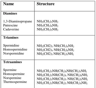
Polyamines (PAs) are small organic molecules found in all living organisms (Cohen, 1998) and they can be considered as one of the oldest group of substances known in biochemistry (Galston, 1991). The diamine putrescine (Put), the triamine spermidine (Spd) and the tetramine spermine (Spm) constitute the most common PAs in eukaryotes (Galston and Sawhney, 1990). In addition, other PAs, such as 1,3-diaminopropane (Dap), cadaverine (Cad), thermospermine (Therm-Spm), norspermidine (Nor-Spd) and norspermine (Nor-Spm) (Table 1) are found in many organisms as minor components of the cellular PA pool and are referred to as uncommon PAs (Tavladoraki et al., 2011). PAs occur in the free form or as conjugates bound to phenolic acids and other low molecular weight compounds or to macromolecules, such as proteins and nucleic acids (Kusano et al., 2008).
PAs are involved in the regulation of a diverse range of vital cellular processes in both eukaryotic and prokaryotic cells, including cell proliferation, signal transduction, membrane stabilization, maintenance of DNA structure, RNA processing, regulation of translation, modulation of enzyme activities (Cohen, 1998; Wang et al., 2003; Kusano et al., 2008). In animals, they are additionally involved in the control of programmed cell death (PCD), particularly apoptosis (Seiler & Raul, 2005; Groppa and Benavides, 2008; Alcázar et al., 2010), while in bacteria an essential role for PAs in biofilm formation and adaptation to various stresses has been demonstrated (Lee et al., 2009; Morimoto et al., 2010).
In plants, PAs have been suggested to play important roles including the control of the N:C balance (Mattoo et al., 2006; Moschou et al., 2012), xylem differentiation (Muñiz et al., 2008; Tisi et al., 2011), membrane fluidity, protein regulation (Baron and Stasolla, 2008), morphogenesis, growth, embryogenesis, organ development, leaf senescence, abiotic and biotic stress response (Alcázar et al., 2006; Groppa et al., 2007; Kusano et
al., 2008). Indeed, Spd is important for survival in that the Arabidopsis spds1/spds2 double mutant for the two genes encoding for spermidine
synthase (SPDS) shows embryonic lethality (Imai et al., 2004). Furthermore, Put and Spm have been implicated in plant responses to drought, high salt stress, wounding and pathogens (Tavladoraki et al., 2012; Takano et al.,
2
Table 1. Common and uncommon PAs
2012; Jiménez-Bremont et al., 2014; Minocha et al., 2014). Put is also required for the synthesis of tropane and nicotine alkaloids in plants (Tavladoraki et al., 2012). Therm-Spm, which is a structural isomer of Spm and was first detected in the thermophilic bacterium Thermus thermophilus, was recently identified in plants (Knott et al., 2007; Naka et al., 2010; Rambla et al., 2010). Genome analyses in many organisms suggest that it may be present in most plants, but not in animals and fungi (Minguet et al., 2008). Several recent studies support an important role of Therm-Spm in controlling xylem differentiation. Indeed, disruption of the Therm-Spm biosynthetic gene, thermospermine synthase (ACAULIS5; ACL5) gene, in
Arabidopsis, which is expressed specifically in procambial cells and xylem
precursor cells during vascular differentiation (Clay and Nelson, 2005; Muñiz et al., 2008), caused impaired stem elongation, over-proliferation of xylem vessels and thicker veins in hypocotyls, leaves and inflorescence stems (Hanzawa et al., 1997; Hanzawa et al., 2000; Clay and Nelson, 2005; Kakehi
et al., 2008; Muñiz et al., 2008). Conversely, increased ACL5 expression
levels or exogenously supplied Therm-Spm suppressed xylem differentiation (Kakehi et al., 2010; Milhinhos et al., 2013; Baima et al., 2014). Also, mutation of the Arabidopsis BUD2/SAMDC4 gene, one of the four
Name Structure Diamines 1,3-Diaminopropane Putrescine Cadaverine NH2(CH2)3NH2 NH2(CH2)4NH2 NH2(CH2)5NH2 Triamines Spermidine Homospermidine Norspermidine NH2(CH2)3 NH(CH2)4NH2 NH2(CH2)4 NH(CH2)4NH2 NH2(CH2)3 NH(CH2)3NH2 Tetraamines Spermine Homospermine Norspermine Thermospermine NH2(CH2)3NH(CH2)4NH(CH2)3NH2 NH2(CH2)3NH(CH2)4 NH(CH2)4NH2 NH2(CH2)3NH(CH2)3 NH(CH2)3NH2 NH2(CH2)3NH(CH2)3 NH(CH2)4NH2
3
Arabidopsis S-adenosylmethionine decarboxylase (SAMDC) genes involved
in PA biosynthesis, which has been shown to be down-regulated by Therm-Spm and has been proposed to predominantly mediate Therm-Therm-Spm synthesis (Kakehi et al., 2010; Kim et al., 2014), produced plants with vascular defects similar to those of acl5 (Ge et al., 2006). Therm-Spm metabolism has been also shown to be involved in the plant defence responses to pathogens (Sagor
et al., 2012; Marina et al., 2013).
To adjust the overall intracellular concentration of PAs to the levels required by the physiological state of the cells, various organisms have evolved complex homeostatic mechanisms involving PA biosynthesis, catabolism, conjugation, transport, and uptake (Martin-Tanguy, 1997, Tiburcio et al., 1997; Angelini et al., 2010; Moschou et al., 2012).
1.2. Polyamine metabolism
1.2.1. Polyamine biosynthesis
In animals and most plants, Put is synthesized directly by decarboxylation of ornithine via the enzyme ornithine decarboxylase (ODC) or indirectly from arginine (Arg) by arginine decarboxylase (ADC) via agmatine (Agm) (Tabor and Tabor, 1985). In Arabidopsis thaliana, Put is produced by the ADC pathway, since no ODC gene has been identified in the sequenced genome of this plant and the corresponding enzyme activity has not been detected (Hanfrey et al., 2001; Alcázar et al., 2010). In bacteria, in addition to ADC and ODC, another enzyme is present involved in Put biosynthesis, agmatinase, which directly produces Put from agmatine (Wortham et al., 2007). After Put synthesis, next biosynthetic steps require the activity of SAMDC to produce from decarboxylation of S-adenosyl methionine (SAM), decarboxylated S-adenosylmethionine (dc-SAM) which acts as a donor of aminopropyl groups in the successive reactions catalyzed by SPDS, spermine synthase (SPMS) and ACL5 for the synthesis of Spd, Spm and Therm-Spm, respectively (Fig. 1). The Arabidopsis genome carries together with the four SAMDCs (Urano et al. 2003), two ADC genes (Soyka and Heyer, 1999), two SPDS genes (SPDS1and SPDS2), a single SPMS gene and a single ACL5 gene (Hanzawa et al., 2002; Knott et al., 2007).
4
Fig. 1. PA biosynthetic pathways. Figure taken from Takahashi and Kakehi, 2010.
1.2.2. Polyamine catabolism
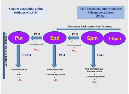
PA catabolism contributes greatly to PA homeostasis and is involved in several physiological processes (Angelini et al., 2010). Copper-containing amine oxidases (CuAO) oxidize Put to 4-aminobutanal with concomitant production of NH3 and H2O2 in a terminal catabolic pathway (Tavladoraki et
al., 2012; Planas-Portell et al., 2013). The flavin-containing amine oxidases
(polyamine oxidases; PAOs) catalyze the oxidation of the free form and/or the acetylated derivatives of Spm, Spd and Therm-Spm either through a terminal catabolic pathway or through a PA back-conversion pathway (Fig. 2). The PAOs involved in the terminal catabolic pathway, among which the extracellular Zea mays PAO1 (ZmPAO1) is the best characterized (Tavladoraki et al., 2006; Fincato et al., 2011), oxidize the endo-side of the
N4-nitrogen of the free forms of Spd and Spm with the production of 4-aminobutanal and N-(3-aminopropyl)-4-4-aminobutanal, respectively, together with 1,3-diaminopropane and H2O2. Conversely the PAOs participating in the PA back-conversion pathway oxidize the exo-side of N4-nitrogen of Spm or Spd to produce Spd or Put respectively, in addition to 3-aminopropanal and
5
H2O2 (Angelini et al., 2010; Tavladoraki et al., 2012). Among the PAOs involved in PA back-conversion, the animal spermine oxidases (SMOs) which have a cytosolic/nuclear localization and oxidize only the free form of Spm, are the best characterized (Wang et al., 2001; Vujcic et al., 2003; Cervelli et al., 2003; Landry and Sternglanz, 2003). The animal peroxisomal PAOs (Vujcic et al., 2003; Wu et al., 2003; Cona et al., 2006) which preferentially oxidize N1-acetyl-Spm, N1-acetyl-Spd, and N1,N12 -bis-acetyl-Spm, whereas the yeast Fms1 oxidizes Spm and N1-acetyl-Spm (Adachi et
al., 2012; Landry and Sternglanz, 2003) are also involved in PA
conversion. The best so far characterized plant PAOs involved in PA back-conversion are those of Arabidopsis.
Copper containing amine oxidases (CuAOs)
FAD-dependent amine oxidases Polyamine oxidases (PAOs) PAO
Spm
Spd
Put
4-aminobutanal + 1,3-diaminopropane + H2O2 N-(3-aminopropyl)-4-aminobutanal + 1,3-diaminopropane + H2O2 4-aminobutanal + NH3 + H2O2Polyamine back-conversion Pathway
T er m in al c atab ol ic p ath w ay 3-aminopropanal + H2O2 3-aminopropanal + H2O2
CuAO PAO PAO
T-Spm PAO
Fig. 2. Polyamine catabolic pathways.
1.2.2.1. Arabidopsis thaliana PAOs
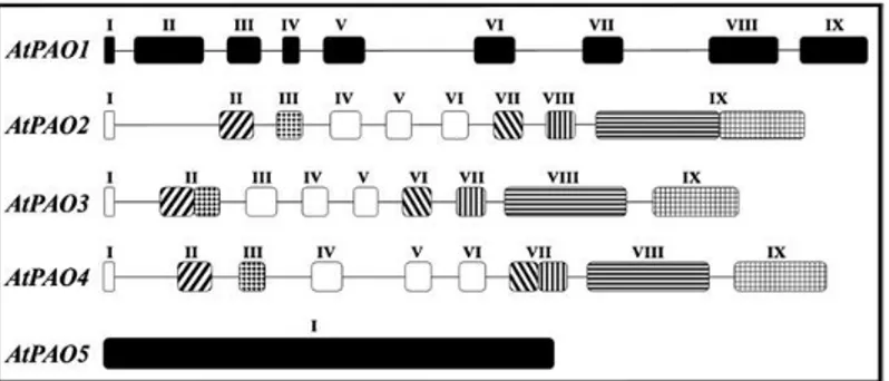
In Arabidopsis thaliana, there are five PAO genes (AtPAO1-AtPAO5), which present some common characteristics, but also important differences in gene structure (Fig. 3), substrate specificity, subcellular localization, and expression pattern, differences which may reflect differences in physiological
6
roles (Fincato et al., 2011). AtPAO1, which has a predicted cytosolic localization, shares with the extracellular ZmPAO1 a 45% homology at the amino acid level and a similar intron/exon organization (Tavladoraki et al., 2006). AtPAO1, differently from ZmPAO1, catalyzes the back-conversion of Spm and Therm-Spm to Spd (Tavladoraki et al., 2006). Promoter activity studies using AtPAO1::GFP-GUS transgenic plants showed that AtPAO1 is highly expressed in the transition region between the meristematic and the elongation zone of the root. In young seedlings, the expression was shown in cotyledons, shoot apex and newly emerging leaves. Later in development, the expression was also observed at the tips and hydathodes of the cotyledons and expanded leaves respectively (Fincato et al., 2012).
AtPAO2, AtPAO3 and AtPAO4, display low sequence homology (23% - 24% homology) with ZmPAO and the other two AtPAOs, but a high sequence homology to each other. In particular, the amino acid sequence identity between AtPAO2 and AtPAO3 is 85%, while it is 58% between AtPAO2 and AtPAO4 and 50% between AtPAO3 and AtPAO4 (Fincato et
al., 2011). Furthermore, AtPAO2, AtPAO3 and AtPAO4 have a very similar
intron/exon organization to each other bearing eight introns at highly conserved positions (Fig. 3). This, together with the elevated sequence homology to each other, suggest that these three Arabidopsis genes are recent derivatives from a common ancestor, thus forming a distinct PAO subfamily (AtPAO2–AtPAO4 subfamily). AtPAO2, AtPAO3 and AtPAO4 have a peroxisomal localization and are able to oxidize both Spd and Spm, but not Therm-Spm (Moschou et al., 2008c; Kamada-Nobusada et al., 2008; Fincato
et al., 2011; Ono et al., 2012). AtPAO2, AtPAO3 and AtPAO4 are expressed
in the root cap. Interestingly, at the root cap differences exist between
AtPAO2 and AtPAO3, although they belong to the same PAO subfamily.
Indeed, while AtPAO2 is expressed only near the quiescent center and columella initials, AtPAO3 is expressed in lateral root cap and in the whole columella. The expression of the three genes were also observed in the different plant organs and cells sharing some common expression pattern. In particular, AtPAO2, AtPAO3 and AtPAO4 are expressed in the guard cells, which may reflect a distinct physiological role (Fincato et al., 2012).
AtPAO5 has low amino acid sequence homology with the other AtPAOs and ZmPAO1 (19–23%) and a higher sequence homology with murine SMO (MmSMO) and murine APAO (MmAPAO) (31% ; Ahou et al., 2014). Furthermore, differently from the other AtPAO genes and ZmPAO1,
AtPAO5 gene bears no intron (Fig. 3; Fincato et al., 2011). AtPAO5 is a
7
proteasome (Ahou et al., 2014). Furthermore, AtPAO5 catalyzes the back-conversion of Spm and Therm-Spm to Spd. It is also able to catalyze the back-conversion of N1- acetyl-Spm to Spd with a kcat higher than with Spm, and Therm-Spm (Ahou et al., 2014). In this way, AtPAO5 represents the first plant enzyme characterized so far involved in PA catabolism with a good activity with acetylated PAs. (Tavladoraki et al., 2006; Moschou et al., 2008b; Fincato et al., 2011). Furthermore, differently from the other animal and plant PAOs so far characterized, AtPAO5 has been classified as a dehydrogenase rather than as an oxidase, since it has been shown that O2 is a poor electron acceptor in the reaction catalyzed by this enzyme (Ahou et al., 2014). Only in bacteria has the existence of spermidine dehydrogenases with an important role in the utilization of PAs as carbon and nitrogen source been reported so far (Tabor and Kellog, 1970; Hisano et al., 1990, 1992; Dasu et
al., 2006). The function of AtPAO5 as a dehydrogenase and its involvement
in PA back-conversion suggest that AtPAO5 has a role in PA homeostasis rather than in H2O2 production, in contrast to the other PAOs so far characterized, as for example ZmPAO1, for which involvement in important physiological processes through H2O2 production has been shown (Cona et
al., 2006; Angelini et al., 2010; Tisi et al., 2011). AtPAO5 is expressed
mainly in the vascular tissue of the roots, hypocotyls and cotyledons. It is also expressed in leaves showing a diffused pattern and in flower buds, in particular in the anthers (Fincato et al., 2012).
Fig. 3. Schematic representation of the exon/intron organization of AtPAO genes. Introns are
represented by lines and exons by boxes. Exons are numbered in Roman numerals. Open and filled boxes indicate shared and unshared exons among the various AtPAO genes, respectively. Stripes and stipples show shared exon domains which are found either joined to each other or separated by the presence of an intron, according to the specific gene considered. Exons and introns are drown in scale. Figure taken from Fincato et al., 2011.
8
2. Auxin, cyokinin, Therm-Spm and their interplay in vascular
tissue differentiation
Vascular plants possess a regulatory network to coordinate the different phases of xylem maturation, including secondary cell-wall formation, cell death, and finally autolysis of the cell contents. Auxins and cytokinins are important members of this network interacting antagonistically on many levels.
Recent genetic studies revealed the molecular basis of the xylem differentiation and specification by auxin and cytokinin. One of the earliest expressing transcriptional regulators in the preprocambial cells is the ARABIDOPSIS THALIANA HOMEOBOX8 (ATHB8), a class III homeo-domain leucine zipper (HD-ZIP III) transcription factor family gene. The transcription of ATHB8 is activated directly by auxin responsive transcription factor, MONOPTEROS (MP). ATHB8 subsequently directs the formation of preprocambial cells and induces the expression of PIN-FORMED1 (PIN1) auxin enflux proteins (Scarpella et al., 2006). These processes establish a positive feedback loop of auxin-MP-ATHB8-PIN1-auxin flow which contribute in the initiation and specification of vascular stem cells and in the restriction of procambium precursor cells to continuous and narrow regions (Ohashi-Ito and Fukuda, 2010; Nieminen et al., 2014). Besides ATHB8, the Arabidopsis genome contains four other HD-ZIP III genes PHABULOSA (PHB), PHAVOLUTA (PHV), REVOLUTA (REV),
CORONA (CNA)/ATHB15 which were shown to be regulated by SHORT
ROOT (SHR), SCARECROW (SCR) and miR165/166 (Kondo et al., 2014).
All of them are expressed in vascular tissues in somewhat overlapping fashion, suggesting a likely redundancy in their function. In Arabidopsis root, a loss of all the five HD-ZIP IIIs results in the failure of xylem vessel development (Carlsbecker et al., 2010). On the contrary, over-expression of
ATHB8 causes enhanced xylem differentiation (Baima et al., 2001),
indicating that HD-ZIP IIIs are de novo regulators of the xylem vessel formation. LONESOME HIGHWAY (LHW), a bHLH transcriptional activator, regulates the bilateral symmetry of vascular pattern and proliferation of procambium cells in roots. The TARGET OF
MONOPTEROS (TMO5) has been found as one of the interactors with LHW
and the auxin-dependent TMO5/LHW transcriptional complex triggers
several rounds of periclinal cell divisions response to signaling by auxin
9
VASCULAR-RELATED NAC-DOMAIN6 (VND6) and VND7 also play an essential role in xylem vessel differentiation in both Arabidopsis and Populus. Indeed, it has been demonstrated that VND6 and VND7 act as central regulators of metaxylem and protoxylem vessel differentiation, respectively (Kubo et al., 2005; Ohashi-Ito and Fukuda, 2010).
The first step of cytokinin perception requires the binding of cytokinin to a transmembrane histidine kinase receptor. This binding induces, within the receptor, the autophosphorylation of a conserved His residue in its kinase domain. In A. thaliana, there are three histidine kinase receptors (AHKs). Following autophosphorylation, the phosphate group is then transferred to a conserved Asp residue within the receiver domain of the AHK protein, and subsequently transferred to a member of the ARABIDOPSIS HIS PHOSPHOTRANSFER PROTEIN (AHP) family. Following transfer to the nucleus, phosphorylated AHPs transfer the phosphate group to the nuclear-localized ARABIDOPSIS RESPONSE REGULATOR (ARR) proteins. AHP6 is an AHP that lacks the conserved histidine residue required for phosphotransfer and therefore acts as a negative regulator of cytokinin
signalling by competing with other AHPs (Mähönen et al., 2006). The ARRs
are encoded by a gene family and are classified into type A, type B and type C, depending on their protein domains and cytokinin responsiveness. Type-A ARRs (ARR3–ARR9 and ARR15–ARR17) possess short C-termini, while type-B ARRs (ARR1, ARR2, ARR10–ARR14 and ARR18–ARR21) have longer C-termini, which contain a conserved GARP (GOLDEN2/ARR/Psr1) DNA-binding domain. Type-A ARRs act as negative regulators of cytokinin responses, via an as yet unknown mechanism. Type-B ARRs, which are transcription factors, play a positive role in mediating cytokinin-regulated gene expression. The type-C ARRs are more distantly related to type-A and type-B ARRs. AHPs also mediate the accumulation in the nucleus of other plant-specific cytokinin-responsive genes, the CYTOKININ RESPONSE
FACTORS (CRFs), members of the A. thaliana APETALA 2 family of
transcription factors. In response to cytokinin, activated type-B ARRs and CRFs act together to regulate gene expression of common and unique targets, including type-A ARRs. The transcriptional activation of the type-A ARRs represents a negative feedback loop required to dampen the response upon high or prolonged signal input. Cytokinins have a dual role during vascular development acting both as a promoter of periclinal cell divisions and an inhibitor of protoxylem differentiation (Mähönen et al., 2000; Mähönen et
10
promotes protoxylem development by negatively regulating cytokinin signaling (Mähönen et al., 2006).
AHP6 expression is auxin-dependent, and loss of AHP6 function
results in expansion of the expression domains of cytokinin-response genes
ARR5 and ARR15 from the procambium into the protoxylem position
(Mähönen et al., 2006; Bishopp et al., 2011). Cytokinin signaling, in turn, regulates auxin availability. High cytokinin signalling in the procambium promotes the efflux of auxin from the procambium into the xylem axis by stimulating lateralization of PIN1 protein and by increasing the expression of the laterally localized PIN7, and perhaps also PIN3 (Bishopp et al., 2011). This mutually inhibitory interaction between cytokinin and auxin in adjacent locations maintains the bisymmetric vascular pattern in the primary root (Bishopp et al., 2011). A connection between the auxin-MP-TMO5/LHW and the cytokinin-AHP6 pathways also exists. Indeed, the TMO5/LHW dimer promotes the expression of the xylem precursor-specific genes LONELY GUY 3 (LOG3) and LOG4, which encode enzymes catalyzing the final reaction step of cytokinin biosynthesis (De Rybel et al., 2014; Ohashi-Ito et al., 2014). Since TMO5/LHW also promotes the expression of AHP6, the protoxylem pre-cursor cells have low cytokinin signaling levels despite being the site of cytokinin synthesis, and therefore display a reduced rate of periclinal cell division. However, cytokinin is able to move from the xylem precursor cells to the neighboring procambial cells where it activates the cytokinin signaling pathway and thus promotes periclinal cell division (Mähönen et al., 2006; De Rybel et al., 2014; Ohashi-Ito et al., 2014).
PAs are implicated in several different processes during xylem differentiation, including cell wall formation, lignin biosynthesis, and auxin– cytokinin signaling (Ge et al., 2006; Cui et al., 2010; Vera-Sirera et al., 2010). In particular, Therm-Spm has important role on xylem differentiation (Muñiz et al., 2008). Indeed, acl5 loss-of-function mutants exhibit incorrect or incomplete secondary cell-wall formation, as well as early expression of xylem cell death markers, and consequently early vessel cell death compared with the wild type, suggesting that Therm-Spm has a protective role against premature xylem maturation and cell death (Muñiz et al., 2008). Recently, many studies have evidenced the regulation of ACL5 and BUD2/SAMDC4 by auxin and Therm-spm. Indeed ACL5, together with BUD2/SAMDC4, is positively regulated by auxin (Hanzawa et al., 2000; Cui et al., 2010), and negatively regulated by exogenous Therm-Spm (Kakehi et al., 2010) through a feedback mechanism involving the regulation of the basic helix-loop-helix (b-HLH) transcription factor SUPPRESSOR OF ACAULIS 51 (SAC51) at the
11
translational level (Imai et al., 2006). In turn, SAC51, as well as SAC51-like proteins heterodimerizes with LHW, thus competing TMO5/LHW interactions and preventing activation of TMO5/LHW target genes to suppress the over-proliferation caused by excess TMO5/LHW activity (Katayama et al., 2015; Vera-Sirera et al., 2015). It has been also shown that Therm-Spm and auxin have opposite action in the control of xylem differentiation (Yoshimoto et al., 2012). Indeed, increased Therm-Spm levels delay xylem differentiation by negatively affecting the expression of auxin-regulated transcription factors belonging to HD–ZIP III gene family and key auxin signaling genes resulting in alteration of auxin-mediated processes (Yoshimoto et al., 2012; Milhinhos et al., 2013; Baima et al., 2014). In this regulatory mechanism, which involves a well-controlled feedback circuit operating to fine-tune formation and differentiation of xylem, the ATHB8 transcription factor of the HD–ZIP III gene family has an important role directly regulating ACL5 expression (Baima et al., 2014).
3. Roles of polyamines under abiotic and biotic stress conditions
Plants are exposed continuously to a variety of adversely changing environmental (abiotic) stresses including salinity, drought, freezing, heat, hypoxia, ozone , heavy metal toxicity and mechanical wounding. Plants are also exposed to pathogen infections (biotic) stress, such as viruses, fungi and bacteria. Several studies from different plant species has shown that PA accumulation increases in response to abiotic and biotic stress (Marina et al., 2013; Kusano et al., 2008; Takahashi et al., 2010; Alcázar et al., 2006; Groppa and Benavides, 2008). In particular, transcript profiling by using qRT-PCR has revealed that water stress induces the expression of ADC2,SPDS1 and SPMS (Alcázar et al., 2006; Phuc., et al 2014). It has also shown
that cold enhances the expression of ADC1, ADC2 and SAMDC2 genes while heat shock induces SPMS expression (Urano et al., 2003; Cuevas et al., 2008; Cuevas et al., 2009; Sagor et al., 2012). Furthermore, recent studies using either transgenic over-expression or loss-of-function mutants support a protective role of PAs under abiotic stresses (Alcázar et al., 2006; Kusano et
al., 2008; Gill and Tuteja, 2010). High Put levels induced by over-expression
of homologous ADC1 enhanced freezing tolerance in Arabidopsis (Altabella
et al., 2009). Similarly, elevated levels of Put by over-expression of ADC2
produces drought tolerance in Arabidopsis, which may be related to reduction of water loss by the induction of stomata closure (Alcázar et al., 2010). Moreover, acl5/spms Arabidopsis double mutants that do not produce Spm
12
are hypersensitive to salt and drought stresses, and the phenotype is mitigated by application of exogenous Spm (Kusano et al., 2007; Yamaguchi et al., 2006). All these observations support the protective role of PAs in plant response to abiotic stress (Alcazar et al., 2010).
Abscisic acid (ABA) is a plant hormone that reduces water loss through stomatal pores on the leaf surface. Enhanced biosynthesis of ABA occurs in response to water deficit, resulting in the redistribution and accumulation of ABA in guard cells (Bray, 1997). The expression of some of the PA biosynthetic genes, such as ADC2, SPDS1 and SPMS are altered in ABA-deficient (aba2-3) and ABA-insensitive (abi1-1) mutants subjected to water stress (Alcázar et al., 2006). In particular, these three genes display reduced transcriptional induction in the stressed aba2-3 and abi1-1 mutants compared to the wild-type plants, indicating that ABA modulates PA metabolism at the transcription level by up-regulating the expression of
ADC2, SPDS1 and SPMS genes under water stress conditions (Alcázar et al.,
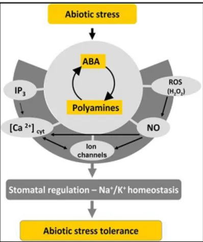
2006). In addition, Put accumulation in response to drought is also impaired in the aba2-3 and abi1-1 mutants compared to wild-type plants. This result is further supported by metabolomic studies showing that PA responses to dehydration are also impaired in loss-of-function mutants for NCED3, a key biosynthetic gene of ABA (Urano et al., 2009). The ABA signaling pathway in stomata regulation involves many different components such as ABA receptors, G-proteins, protein kinases and phosphatases, transcription factors and secondary messengers, including Ca2+, reactive oxygen species (ROS) and NO (Kuppusamy et al., 2009). PAs are also linked with reactive oxygen species (ROS) and NO signaling, since PAs catabolism generates H2O2, and also there is evidence that PAs enhance NO production (Tun et al., 2006 ). It has been also reported that Put, Spd and Spm regulate stomatal responses (Liu et al., 2000; An et al., 2008). which indicates that PAs participates in the hormonal pathways, including ABA regulation of abiotic stress responses (Fig. 4) (Alcazar et al., 2010; Yamasaki and Cohen, 2006).
Furthermore, it has been shown that biotic and abiotic stresses induce PA transport in the apoplast (Yoda et al., 2003; Yoda et al., 2006; Marina et al., 2013; Moschou et al., 2009; Toumi et al., 2010). Stress-related factors that have been shown to induce PA transport in the apoplast are: incompatible and compatible plant pathogen interactions, salt stress and treatment with the stress-related hormone ABA (Yoda et al., 2003; Yoda et
al., 2006; Marina et al., 2008; Moschou et al., 2009; Toumi et al., 2010).
This suggests that PA catabolism in the apoplast is a general defence response against several stresses. It has been also shown that H2O2 produced
13
Fig. 4. Scheme represents the integration of PAs with ABA, ROS (H2O2), NO, Ca2+ homeostasis
and ion channel signaling during abiotic stress responses. Figure taken from Alcazar et al., 2010.
by PA catabolism in the apoplast contributes to the second phase of ROS production during Tobacco mosaic virus (TMV)-induced HR, a plant response which is developed during an incompatible plant-pathogen interaction and consists of rapid ROS production, PCD and induction of defence responses aiming to restrict pathogen expansion (Yoda et al., 2003). Similar approaches showed that H2O2 produced by PA catabolism in the apoplast contributes to the synthesis of the ROS that accumulate under abiotic stress conditions (Moschou et al., 2008b). PCD was also shown to be induced by oxidation of extracellular Spd also under salt stress.
14
AIM OF THE WORK
PAOs contribute to various physiological processes through regulation of PA levels and reaction products. PAOs are characterized by a broad variability in substrate specificity, catalytic mechanism and subcellular localization. Extracellular PAOs have been shown to have crucial roles during plant growth under physiological and stress conditions, giving rise to increased apoplastic H2O2 which in turn signals cell wall development, xylem differentiation and defense responses. Conversely, little is known about the physiological roles of intracellular PAOs, such as those of Arabidopsis
thaliana.
The aim of the present work is to study the contribution of the five members of the Arabidopsis thaliana PAO gene family in developmental and defense processes, with particular focus on the two cytosolic PAOs (AtPAO1 and AtPAO5), which present some similarities to each other, but also important differences, as concerned substrate specificity, catalytic mechanism and tissue-specific expression pattern. Among them, AtPAO5 results of particular interest since it has been shown to be regulated at the post-transcriptional level by proteasome and to function mainly as a dehydrogenase, differently from all other PAOs so far characterized which are classified as oxidases. This study will permit to elucidate the functional complexity characterizing PA metabolism.
15
RESULTS
1. AtPAO5 is involved in the control of polyamine homeostasis
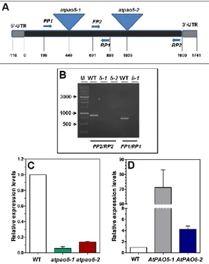
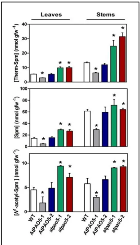
To investigate the physiological role(s) of AtPAO5 during plant growth and development, two 35S::AtPAO5-6His Arabidopsis transgenic lines that ectopically express AtPAO5, one with 70-fold (AtPAO5-1; Fig. 5; Ahou et al., 2014) and the other 4-fold (AtPAO5-2; Fig. 5) higher expression levels than the endogenous gene, were characterized during plant growth and development. Parallel studies were also performed with two loss-of-function mutants, atpao5-1 (Ahou et al., 2014) and atpao5-2 which lack AtPAO5 expression (Fig. 5).The AtPAO5 transgenic plants and the atpao5 mutants were analyzed for PA levels. This analysis has been performed both in whole seedlings and in specific organs in taking into consideration that AtPAO5 is expressed in all organs (Fig. 6; Fincato et al., 2012). Results evidenced decreased levels of Spm, Therm-Spm, and N1-acetyl-Spm in mature leaves and inflorescence stems of AtPAO5-1 transgenic plants and increased levels in those of
atpao5-1 and atpao5-2 plants, as compared to the wild-type ones (Fig. 7). Similar
results were obtained in whole seedlings (data not shown). In contrast to the
AtPAO5-1 transgenic plants, the AtPAO5-2 transgenic line does not present
differences in the levels of Spm, Therm-Spm and N1-acetyl-Spm from those of the wild-type plants (Fig. 7), which suggests a dose-dependent contribution of AtPAO5 in PA homeostasis. The analysis of the PA levels further evidenced higher Spm and Therm-Spm levels in stems than in leaves, in agreement with previous studies (Fig. 2; Naka et al., 2010). This is probably determined by the relative amounts of the anabolic and catabolic enzymes specific for these PAs (such as ACL5, SAMDC4, AtPAO1 and
AtPAO5), considering their similar organ-specific gene expression pattern
(Fig. 6A). Contrary to Spm and Therm-Spm, comparable N1-acetyl-Spm levels are present in stems and leaves (Fig. 7).
16
Fig. 5. AtPAO5 expression levels in atpao5 loss-of-function mutants and 35S::AtPAO5-6His
Arabidopsis transgenic plants. (A) Schematic genome structure of AtPAO5. Triangles indicate
the positions of the two T-DNA insertions and arrows the position of the primers used for mutant characterization. (B) Analysis of selected homozygous lines of atpao5-1 and atpao5-2 mutants for AtPAO5 expression levels by RT-PCR. (C and D) Relative AtPAO5 expression levels in young seedlings of the two atpao5 mutants (atpao5-1 and atpao5-2), the two independent
35S::AtPAO5-6His Arabidopsis transgenic lines (AtPAO5-1 and AtPAO5-2) and the wild-type
(WT) plants. The expression levels were determined by qRT-PCR. Very similar results for
AtPAO5 expression levels were obtained when specific organs (leaves, roots and stems) of AtPAO5-1 transgenic plants were tested.
17
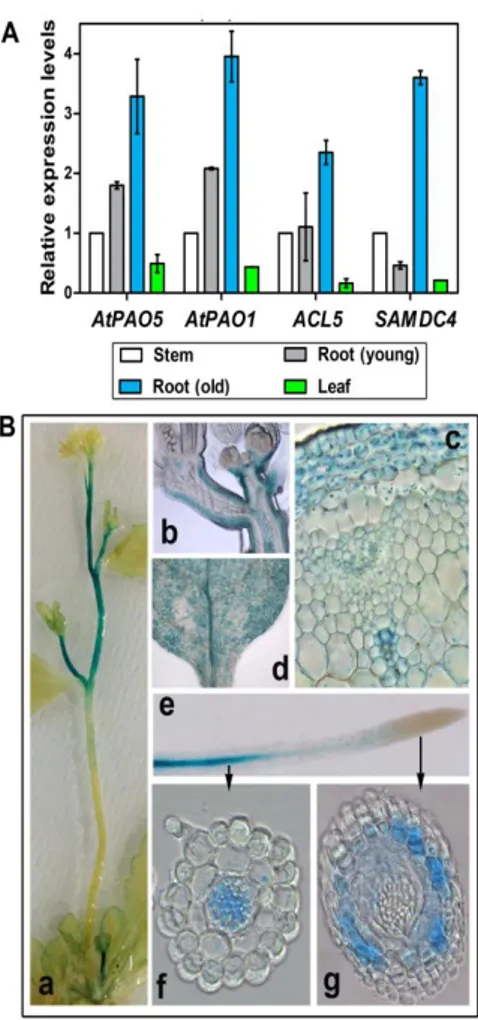
Fig. 6. Expression of AtPAO1, AtPAO5, ACL5 and SAMDC4 in various plant organs. (A)
Relative expression levels of AtPAO1, AtPAO5, ACL5 and SAMDC4 in inflorescence stems, leaves and roots of 10-day-old (young) and 3-week-old (old) plants as determined by qRT-PCR. Mean values ± SE of two independent experiments are shown. (B) AtPAO5 promoter activity in stems (a, b, c), leaves (d) and roots (e, f, g) determined by histochemical GUS staining of
AtPAO5::GFP-GUS transgenic plants. Longitudinal (b) and transverse (c) sections of stems and
18
Fig. 7. Spm, Therm-Spm and N1
-acetyl-Spm levels in AtPAO5 and atpao5 plants. PA levels were determined in rosette leaves and inflorescence stems of 5-week-old seedlings by GC-MS. The analysis was repeated three times and a representative experiment is shown. Numbers are mean values ± SE of three replicates obtained from the same pooled fresh material. Statistical analysis was performed by one-way ANOVA test (P<0.05). Asterisks indicate statistically significant differences from the wild-type plants.
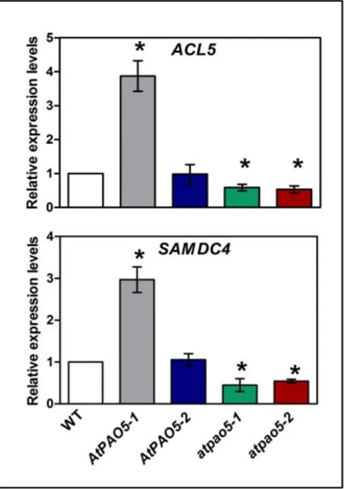
It is well established that the expression levels of ACL5 and SAMDC4 are regulated by Therm-Spm levels through a negative feedback loop (Hanzawa et al., 2000; Clay and Nelson, 2005; Kakehi et al., 2008; Kakehi et
al., 2010; Baima et al., 2014). To address whether such a regulatory
mechanism is activated upon modulation of AtPAO5 expression levels, the
AtPAO5 over-expressing transgenic plants, as well as the atpao5-1 and atpao5- 2 mutants were analyzed for ACL5 and SAMDC4 expression levels
by qRT-PCR. As shown in Fig. 8, ACL5 and SAMDC4 genes are up-regulated in AtPAO5-1 seedlings and down-up-regulated in the atpao5-1 and
atpao5-2 ones, but not affected at all in AtPAO5-2 seedlings with low
expression levels of the transgene and unaltered PA content. Instead, no change in SPMS expression levels was observed in any of the AtPAO5 and
19
(Supplemental Fig. S1), as well as leaves and flowers (data not shown), were analyzed.
These data, together with recently published data showing AtPAO5 up-regulation by Therm-Spm (Ahou et al., 2014), suggest that AtPAO5 participates in the feedback mechanism controlling ACL5 and SAMDC4 expression levels, as well as Therm-Spm homeostasis along the entire plant. The activation of this feedback mechanism in AtPAO5-1 plants may explain the reduction of Spm, Therm-Spm, and N1-acetyl-Spm levels to only 50% of those in wild-type plants despite the very high expression levels of ectopic
AtPAO5 in the AtPAO5-1 plants.
Fig. 8. ACL5 and SAMDC4 expression levels in AtPAO5 and atpao5 plants. Relative expression
levels were determined by qRT-PCR analysis of 10-day-old seedlings. Numbers are mean values ± SE of three independent repetitions of the experiment. Statistical analysis was performed by one-way ANOVA test. Asterisks indicate statistically significant differences from the wild-type (WT) plants.
20
2. AtPAO5 is up-regulated by cytokinins and auxin in an
organ-specific way
AtPAO5 promoter activity studies using AtPAO5prom::GFP-GUS
transgenic plants showed that in roots AtPAO5 expression is extended from the hypocotyl–root junction region throughout the maturation and elongation zones until when the spiral secondary cell wall thickenings of the protoxylem elements first become evident. In particular, the AtPAO5 expression in the roots is tissue-specific involving the whole vascular system (xylem, phloem and procambial/cambial cells) and pericycle (Fig. 6B, e and f; Fincato et al., 2012; Ahou et al., 2014), similarly to the tissue-specific expression of
AtPAO5 in hypocotyls (Fincato et al., 2012). Occasionally, AtPAO5 appeared
to be expressed also in the root meristematic region, mainly in cortical tissues (Fig. 6B, g). In cotyledons and leaves, AtPAO5 promoter activity is extended to the entire area (Fincato et al., 2012), including veins (Fig. 6B, d). In stems,
AtPAO5-related staining was specifically present in cortex and xylem vessels
(Fig. 6B, b and c), mainly in the upper internodes (Fig. 6B, a). Treatment of
AtPAO5::GFP-GUS transgenic plants with 6-benzylaminopurine (BAP)
increased the intensity of GUS staining in the root vasculature and the meristematic region (Fig. 9A). Instead, no increase in AtPAO5-related GUS staining was evident in the above-ground part of the plants. These data indicate that AtPAO5 expression is under the control of cytokinins specifically in the roots. A root-specific up-regulation of AtPAO5 was additionally observed following treatment with indole-3-acetic acid (IAA) (Fig. 9A). The up-regulation of AtPAO5 by cytokinins and auxin was further confirmed by qRT-PCR analysis (Fig. 9B).
3. AtPAO5 is involved in the cytokinin-mediated control of root
xylem differentiation
Cytokinins have a critical role in root elongation, root apical meristematic activity and protoxylem differentiation (Perilli et al., 2010; Del Bianco et al., 2013; Kondo et al., 2014). Indeed, increased cytokinin levels shift the position of the meristematic/elongation transition zone (TZ) more distally, shortening the meristematic region and impairing root elongation, whereas decreased cytokinin levels shift the TZ proximally, producing longer
21
meristems and roots. Furthermore, cytokinins negatively regulate protoxylem specification (Mähönen et al., 2006; Bishopp et al., 2011; Perilli et al., 2010).
Fig. 9. AtPAO5 up-regulation by cytokinins and auxin. (A) Histochemical GUS staining of
7-day-old AtPAO5::GFP-GUS Arabidopsis transgenic plants treated with 1 μM BAP or 10 μM IAA for 24 h. Bar indicates 100 μm. (B) Quantitative RT-PCR analysis of AtPAO5 expression levels in whole seedlings (BAP) or roots (IAA) of 10-day-old wild-type plants treated with 5 μM BAP or 10 μM IAA for 3 h. Mean values ± SE of relative quantitation from two independent experiments are shown.
To determine the physiological significance of the root–specific effect of cytokinins on AtPAO5 expression levels, root elongation rate of the
AtPAO5 over-expressor and mutant plants in the absence and presence of
BAP, was compared with that of wild-type plants. However, no difference among the different genotypes was apparent even though BAP impaired root elongation (Fig. 10A). Despite the lack of differences in root elongation rate among the transgenic and the wild-type plants in the presence and the absence of exogenous cytokinins, a detailed analysis of the timing of protoxylem and metaxylem differentiation in roots was performed determining the distance between the first protoxylem and metaxylem elements from the quiescent centre (QC). Root apical meristematic activity was additionally analyzed measuring the distance between TZ and QC. In the
22
absence of exogenous BAP, no difference was observed in root protoxylem and metaxylem differentiation (Fig. 11), nor in root apical meristematic activity among the AtPAO5 over-expressor plants, the atpao5 mutants, or the wild-type plants (Fig. 10 B).
Fig. 10. Effect of cytokinins on root elongation and
meristematic activity of AtPAO5-1 and atpao5 plants. ) Effect of BAP on root elongation. (B) Effect of BAP on root tip length. AtPAO5-1, atpao5 and wild-type (WT) seedlings of 5 days were transferred onto plates containing 1 μM BAP. Root length was measured at various time intervals after transferring. Root tip length was determined 6 days after transferring to the BAP-containing medium measuring the distance from the quiescent center to the first cells displaying elongation under confocal microscope. NT: not treated control.
Addition of BAP in the growth medium, delayed protoxylem differentiation in all plant genotypes, thus shifting the position of the first protoxylem vessels more proximally than in the mock treated plants. However, this delay was less pronounced in AtPAO5-1 transgenic plants and more pronounced in atpao5-1 and atpao5-2 mutants than in wild-type plants (Fig. 11). Further, BAP treatment highly reduced apical meristematic activity, but at the same level in all genotypes (Fig. 10B). This is despite the fact that BAP increases AtPAO5 expression levels not only in the root vasculature but also in the meristematic region. BAP treatment additionally caused shortening of the distance from the quiescent center (QC) of the first metaxylem vessels probably due to the reduced apical meristematic activity. The effect, however, appeared genotype-independent. These data suggest that
AtPAO5 is specifically involved in the cytokinin-mediated control of root
A
23
protoxylem differentiation, this specific signaling pathway being impaired in the presence of increased AtPAO5 expression levels and enhanced in the absence of AtPAO5 expression.
Since vascular system differentiation is controlled also by auxins and considering the organ-specific effect of IAA in AtPAO5 expression levels,
atpao5-1 and AtPAO5-1 plants together with wild-type plants were analyzed for root growth (data not shown) and xylem differentiation (Supplemental Fig. S2) in the presence and absence of IAA. This analysis showed that IAA inhibited root growth and reduced the distance of the first protoxylem and
metaxylem elements from the QC at the same level in all plant genotypes.
Fig. 11. Cytokinin-mediated xylem differentiation in
the AtPAO5-1 and atpao5 plants. (A) Distance of the first protoxylem cells from QC (Protoxylem distance). (B) Distance of the first metaxylem elements from QC (Metaxylem distance). Protoxylem and Metaxylem distances were measured after 5 days of treatment or not with 1 μM BAP of 7-day-old plants initially grown under physiological conditions. For protoxylem and metaxylem visualization seedlings were stained with fuchsin and observed under confocal microscope. The analysis was repeated at least three times and a representative experiment is shown. Numbers are mean values ± SE (n≥4) and asterisks denote statistically significant differences from the corresponding wild-type (WT) plants (P<0.05; One-way ANOVA). NT: untreated control.
4. AtPAO5 is involved in the control of stem and root development
As regards the aerial part of the plants, at early development stages theatpao5 mutants and the AtPAO5 over-expressing transgenic plants display
the same growth pattern and phenotype as the wild-type plants and only after the transition from vegetative to reproductive growth some differences become evident. In particular, while the atpao5-1 and atpao5-2 mutants as well as the AtPAO5-1 and AtPAO5-2 transgenic plants initiated vegetative
24
growth and transition to the inflorescence meristem in the same manner as the wild-type plants, the two atpao5 mutants produced longer and slightly thicker flowering stems than the wild-type plants, while, conversely, the
AtPAO5-1 transgenic plants produced shorter and thinner flowering stems
leading to a semi-dwarf phenotype with reduced apical dominance (Fig. 12). Instead, the stems of the AtPAO5-2 transgenic plants which have lower transgene expression levels than the AtPAO5-1 transgenic plants did not show differences in length and thickness compared to the wild-type plants (Fig. 12).
Fig. 12. Phenotype of AtPAO5 and atpao5 transgenic plants. (A) Stem phenotype of 5-week-old
soil-grown AtPAO5, atpao5 and wild-type (WT) plants. (B) Stem length of 5-week-old
AtPAO5-1, atpao5 and WT plants. The analysis has been repeated for more than 6 times (n=4 to 10) with
highly reproducible results. (C) Stem thickness of AtPAO5 and atpao5 transgenic plants. Stem thickness was determined by measuring the area of transverse sections from the stem basal ends using ImageJ analysis. Numbers are mean values ± SE (n≥4) from a representative experiment. Statistical analysis was performed by one-way ANOVA test and asterisks indicate statistically significant differences compared with WT plants.
25
To understand these differences in inflorescence development among the atpao5 mutants, the AtPAO5 over-expressing transgenic plants and the wild-type plants at the cellular level, the inflorescence anatomy of these plants were examined analyzing transverse sections of the first and second internodes at the basal end of the inflorescence stem (Fig. 13). These analyses showed that the AtPAO5-1 transgenic plants on the one hand undergo excessive primary xylem differentiation presenting a much higher number of large-diameter, thick-walled metaxylem vessels than the wild-type plants (Fig. 14), while on the other hand display a highly reduced secondary growth (Fig. 13). On the opposite, the atpao5-1 and atpao5-2 mutant plants present a slightly lower number of metaxylem vessels and a more extensive secondary growth than the wild-type plants (Fig. 13; Fig. 14). These differences in the xylem development were observed both in young inflorescences before appearance of differences in stem length and in later
Fig. 13. Histological analysis of inflorescence stems of AtPAO5-1 and atpao5 plants. (A)
Transverse sections of the second internodes at the basal end of inflorescence stems stained with toluidine blue. (B) Vascular bundles of inflorescence stems as observed from transverse sections stained with toluidine blue. (C) Vascular bundles of inflorescence stems observed under UV light. Black and white bars indicate 100 μm. Similar results were obtained from transverse sections of the first internodes at the basal end of inflorescence stems.
26
Fig. 14. Number of metaxylem vessels and size of
secondary xylem in bundles of AtPAO5-1, atpao5-1,
atpao5-2 and wild-type (WT) stems. (A) An
example of quantification of the number of metaxylem cells and the size of secondary xylem area in a stem transverse section stained with toluidine blue. Measurement of secondary xylem area was performed following image acquisition through ImageJ software. (B) Number of metaxylem vessels in bundles of AtPAO5-1, atpao5-1, atpao5-2 and WT stems. (C) Size of secondary xylem in bundles of AtPAO5-1, atpao5-1, atpao5-2 and WT stems. Numbers are mean values ±SE. Asterisks indicate statistically significant differences from the wild-type (WT) plants (P<0.05; one-way ANOVA test). A representative experiment is shown.
developmental stages. The different degree of xylem differentiation in the stems of the AtPAO5-1, atpao5-1 and atpao5-2 mutants as compared to the wild-type plants most probably interfered with the normal organ growth.
Additionally to the differences in stem elongation and thickness, the
AtPAO5 and atpao5 plants present some differences in root length from the
wild-type plants in advanced developmental stages. Indeed, despite the lack of differences in root elongation at early developmental stages among the different genotypes, mature AtPAO5-1 transgenic plants present slightly shorter roots than the wild-type plants, while atpao5-1 and atpao5-2 longer ones (Fig. 15 A and B). Furthermore, observation of root transverse sections
27
at the maturation zone of 15-day-old plants growing in vitro evidenced increased number of metaxylem vessel elements in the AtPAO5-1 transgenic plants as compared with the wild-type plants and decreased in the atpao5-1 and atpao5-2 mutants (Fig. 15 C). These data indicate that AtPAO5 contributes to xylem development both in stems and roots, the effect appearing more pronounced in stems than in roots most likely due to the longer growth period of the stems comparing with the roots. However, a major role of AtPAO5 in stem than in root xylogenesis cannot be excluded.
Fig. 15. Root phenotype of AtPAO5 over-expressing transgenic plants and atpao5 mutants. (A)
Roots from 16-day-old plants grown in hydroponic cultures. To help visualization, roots were stained with fuchsin. (B) Root length of 16-day-old plants grown in hydroponic cultures. Numbers are mean values ± SE (n≥4) from a representative experiment. Statistical analysis was performed by one-way ANOVA test and asterisks indicate statistically significant differences compared to WT plants. (C) Transverse sections of roots stained with toluidine blue. Sections at 2 cm below root-hypocotyl junction were made.
5. AtPAO5 is involved in the control of lateral roots
A further analysis of root development showed that the AtPAO5-1 transgenic plants present a higher number of lateral roots than the wild-type plants (Fig. 16B). Instead, no statistically significant difference was evidenced in the number of lateral roots for the atpao5-1 and atpao5-2 plants in respect to the wild-type plants (Fig. 16). The addition of Therm-Spm in the growth medium did not alter significantly the number of lateral roots in the different genotypes as compared with the untreated controls, the number of the lateral roots in the AtPAO5-1 transgenic plants being higher than that of
28
Fig. 16. Modulation of lateral roots by AtPAO5 ectopic expression. (A) Root growth of
AtPAO5-1, atpao5-AtPAO5-1, atpao5-2 and wild-type (WT) plants in the presence and the absence of 100μM
Therm-Spm. (B) Number of lateral roots of AtPAO5-1, atpao5-1, atpao5-2 and wild-type (WT) plants grown in the absence or in the presence of 100 μM Therm-Spm. A representative experiment is shown. Bars denote standard errors and asterisks indicate statistically significant differences in respect to the corresponding wild-type (WT) plants (P<0.05; One-way ANOVA).
29
the wild-type plants also under these conditions (Fig. 16). However, in the presenceof Therm-Spm, lateral roots of atpao5 mutant plants present reduced axial extension comparing with those of AtPAO5-1 transgenic plants and wild-type plants, the latter plants presenting an intermediate phenotype (Fig. 16A). On the contrary, no difference was observed among the different genotypes, as considered root elongation and growth of the aerial parts of the plants, both in the presence and the absence of Therm-Spm, during the 5- to 8-day period of observation.
The reduced axial extension of the lateral roots may be due to reduced strength of the roots, being thus unable to withstand the forces of stretching that occur during organ expansion inside the agar plates. Indeed, in the presence of Therm-Spm, protoxylem and metaxylem differentiation appeared highly delayed in atpao5-1 and atpao5-2 mutants but not in AtPAO5-1 transgenic plants and the wild-type plants (Fig. 17A). Additionally, in the presence of Therm-Spm, several discontinuities were observed in the protoxylem elements of the main roots in atpao5-1 and atpao5-2 mutants, but not in those of AtPAO5-1 transgenic plants (Fig. 17B). The discontinuities in the protoxylem elements of atpao5 roots were more prominent in the presence of 100 µM Therm-Spm than in the presence of 50 µM (Fig. 17B). Wild-type roots only occasionally presented discontinuities in protoxylem elements. Contrary to Therm-Spm, Spm affected neither the axial extension of the lateral roots nor disconnected protoxylem elements (data not shown).
These data are consistent on the one hand with the high catalytic activity of AtPAO5 towards Therm-Spm, thus back-converting the available amount of Therm-Spm in the medium to Spd and on the other hand to the opposing effects of Therm-Spm and auxin on xylem differentiation (Yoshimoto et al., 2012). It is probably because of this negative effect of Therm-Spm on xylem integrity that prolonged growth of atpao5 mutants in the presence of Therm-Spm inhibited growth of the aerial part of the plants as recently reported (Kim et al., 2014).
30
Fig. 17. Modulation of xylem development by Therm-Spm. (A) The distances from QC of the
first protoxylem (PQ) and metaxylem (MQ) vessels were measured in 10-day-old seedlings growing for the 5 last days in the presence or the absence of 100 μM Therm-Spm. Mean values ± SE (n≥6) from a representative experiment are shown. Asterisks indicate statistically significant differences in respect to the corresponding wild-type (WT) plants (P<0.05; One-way ANOVA). (B) Protoxylem discontinuities in the maturation zone of roots of atpao5-1 plants. grown in the presence of 50 μM or 100 μM Therm-Spm. Yellow arrows show regions in atpao5-1 roots in which metaxylem vessels are missing and protoxylem discontinuity is observed. White bars indicate 50 μm distance.