UNIVERSITÀ DEGLI STUDI DI SASSARI
SCUOLA DI DOTTORATO DI RICERCA Scienze e Biotecnologie dei Sistemi Agrari e Forestali e delle Produzioni Alimentari
Indirizzo Biotecnologie Microbiche Agroalimentari
Ciclo XXVI
Phenomic analysis and cell wall remodulation of Saccharomyces
cerevisiae flor strains in the presence of different nitrogen sources
dr. Marc Bou Zeidan
Direttore della Scuola prof. Alba Pusino
Referente di Indirizzo prof. Severino Zara
Abstract
Flor yeasts of Saccharomyces cerevisiae are the main actors in the biological ageing of Sherry wines by developing an air-liquid biofilm, called velum at the surface of wine at the end of fermentation. The air-liquid biofilm occurs due to a complex mechanism which is similar to many microbial biofilms. The velum phenotype is mainly regulated by the expression of a highly O-glycosylated cell wall protein Flo11 which by turn contributes to the hydrophobicity and electrostatic charge of the cell wall which are crucial for unspecific interactions and adhesion capacity of yeasts. In this study we characterized the interactions of flor yeasts with a series of nitrogen compounds known for their hydrophobicity and/or charge. We find that, different flor yeast strains characterized by different degree of functional FLO11 are diversely affected in terms of biofilm formation and adhesion capacity when interact with different nitrogen sources. The positive role of the synthetic hexapeptide PAF26 in enhancing the biofilm formation was first discovered. On the contrary, cationic amino acids were able to inhibit biofilm formation and adhesion ability. Results together highlight on the involvement of the Flo11p in hydrophobic and electrostatic interactions and open to new investigations, addressed to the control of microbial adhesion and biofilm formation.
Table of content:
INTRODUCTION ... 1
1. Peculiarities of flor yeasts ... 3
2. Cell wall role and molecular structure ... 5
2.1. Chitin. ... 6
3. Cell wall mannoproteins. ... 10
3.1. GPI anchored cell wall mannoproteins. ... 11
3.2. Cell wall Adhesins. ... 12
3.3. Sugar-sensitive flocculins and flocculation. ... 13
3.4. Sugar-insensitive flocculins. ... 15
4. Genetic characteristics and regulation of FLO11 ... 16
4.1. FLO11 signaling pathways and transcription factors. ... 17
4.2. Elongation and tandem repeats. ... 19
5. Adhesion, biofilm formation and phenotypic variation ... 20
AIM OF THIS STUDY ... 33
CHAPTER I: FLO11 gene is involved in the interactions of flor strains of Saccharomyces cerevisiae with biofilm promoting synthetic hexapeptide ... 36
Introduction ... 50
Materials and methods ... 52
Results ... 56
Discussion ... 67
CHAPTER II: Inhibitory effect of L-histidine on biofilm formation by Saccharomyces cerevisiae flor yeasts with functional FLO11 gene ... 48
Introduction ... 50
Materials and methods ... 52
Results ... 56
Discussion ... 67
CHAPTER ΙΙΙ: The biofilm formation and adhesion ability of Saccharomyces
cerevisiae flor yeasts are affected by the presence of amino acids ... 77
Introduction ... 78
Materials and methods ... 80
Results ... 83
Discussion ... 90
GENERAL CONCLUSIONS ... 97
Acknowledgments………...…….-100-
Yeast have been used spontaneously by human from approximately 10.000 B.C in wine and bread production (Piskur et al., 2006), and recently also as producers of bio-ethanol, vitamins and pharmaceutical products like hormones and protein drugs. Despite these “good” purposes, several yeast species are pathogenic to animals and plants. The most studied yeast is Saccharomyces cerevisiae, a unicellular eukaryotic, very well known for its ability to ferment, under fully aerobic conditions, glucose to ethanol and carbon dioxide, thus been classified as a Crabtree positive yeast (Gelinas, 2009). Besides its common usage in wine production, brewing and baking products,
S. cerevisiae is intensively used as a model system to study numerous cellular
processes such as eukaryotic gene regulation and evolution, cell cycle, metabolic pathways, apoptosis and ageing (Giaever et al., 2002; Petranovic and Nielsen, 2008). Yeasts also organize into multicellular communities which is critical for their surviving in harmful environments. Natural Saccharomyces cerevisiae strains form complex structured colonies, which share many typical properties with biofilms infections of Candida albicans and Candida glabrata strains in the human body (Lionakis and Netea, 2013). Microbial biofilms are widespread in nature and can develop on biotic or abiotic surfaces and are enclosed in an extracellular matrix, the whole forming a complex three-dimensional architecture. The formation of biofilms, whether bacterial or fungal, consists of cell-cell aggregation, adhesion of cells to a surface, initial formation of colonies and secretion of extracellular polymeric matrices (Stoodley et al., 2002; Mowat et al., 2009). Adhesion and biofilm formation mechanisms are considered as an adaptive response to adverse environmental conditions (Hall-Stoodley and Stoodley, 2009; Ning et al., 2013).
At the biotechnological level, the multicellular behavior of S. cerevisiae is applied in the sedimentation and the removal of the biomass at the end of fermentations processes (Verstrepen and Klis, 2006; Bauer et al., 2010). Flor yeasts are natural S.
Sherry-like wines. In Sardinia, sherry-like wine is produced traditionally, and flor yeasts found are endogenous (ex: Vernaccia di Oristano) (Budroni et al., 2000; Budroni et al., 2005; Zara et al., 2008).
1. Peculiarities of flor yeasts
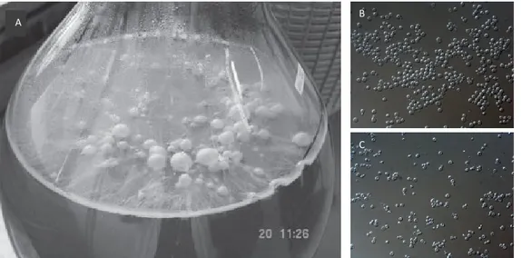
Flor yeasts belong to S.cerevisiae species and have the ability of cell and cell-surfaces aggregation, invasive and pseudohyphal growth and in particular, they can form biofilm at air-liquid interfaces (Fig.1). The dimorphic unicellular to multicellular growth swift occurs when flor yeasts are exposed to critical environmental conditions, such as depletion of favorable carbon and/or nitrogen sources. Indeed, in wine making, at the end of alcoholic fermentation, when nutritional resources are depleted, the further growth becomes dependent on the access to oxygen (Freiberg and Cruess, 1955; Zara et al., 2005; Fidalgo et al., 2006). The formation of the air–liquid biofilm allows the yeast cells to grow aerobically thru the uptake of preferably glycerol, acetic acid and ethanol respectively, as non fermentable carbon sources (Zara et al., 2010).
FIG 1 A) Air-liquid biofilm formation by S. cerevisiae flor strain on Vernaccia sherry-like wine (Zara et al., 2005). B) Microscopic view on the multicellular tendency of flor yeasts. C) Microscopic view on the unicellular tendency of laboratory yeast S288c.
A B
The formation of the air–liquid biofilm allows the yeast cells to grow aerobically thru the uptake of preferably glycerol, acetic acid and ethanol respectively, as non fermentable carbon sources (Zara et al., 2010). This aerobic growth induces the substantial production of acetaldehyde, and activates complex metabolisms of amino acids, such as Proline, which are used directly as nitrogen sources or electron acceptors (Mauricio et al., 2001). Moreover, studies evidenced a high accumulation of unsaturated long-chain fatty acids in flor yeast cells in fermentation phase, which subsequently increases in biofilm formation phase. The oleic acid was shown to be the most copious in flor yeasts (Mannazzu et al., 2008; Marques et al., 2008). It was suggested that the excessive presence of these unsaturated fatty acids is related to the enhanced ethanol tolerance and hydrophobicity of flor yeasts, and to the biofilm formation process in general, by increasing the flor yeast cell density, which enable them to grow on air-liquid surfaces (Zara et al., 2012).
The emergence of molecular techniques has greatly improved the genetic characterization of flor yeasts. Restriction analysis of the intergenic region of 5.8S rDNA has identified a 24 base pair deletion in all analyzed flor strains (Esteve-Zarzoso et al., 2001). In the last decade, a series of studies uncovered the key factor in biofilm formation. These studies revealed that the FLO11 gene, which encodes for a cell wall mannoprotein, Flo11p is the main factor in biofilm formation and multicellular growth. Besides to FLO11 gene, it was shown that the whole cell wall is involved in the multicellular response to threatening environments (Cid et al., 1995; Galitski et al., 1999; Reynolds, 2001; Zara et al., 2005; Dranginis et al., 2007).
2. Cell wall role and molecular structure
The cell wall is a principal compartment in yeast and is largely involved in the dimorphic shift of the cell (Verstrepen et al., 2004). It has main functions toward mechanical and osmotic stresses. i) It provides protection from osmotic shock by limiting the influx of water to avoid bursting and to maintain cell the intracellular water activity (Hohmann, 2002); ii) it is essential for the strength and elasticity required to maintain the shape of the cell, as well as it provide an effective barrier against sheer and compression forces (Klis et al., 2006); iii)it regulates the permeability of solutes (Lipke and Ovalle, 1998).
Apart of the protective role of the cell wall, it also serves as a tool for cell-cell and cell-environment interactions. In fact, one of the most important functions of the cell wall is the ability to adhere to other cells, biotic and abiotic surfaces (Zara et al., 2005). This includes adhesion of sexual partner cells as well as vegetative adhesion. Sexual adhesion of budding yeast is well understood and is mediated by cell-type-specific adhesins called agglutinins, which are produced by mating partners after exchange of pheromones and confer cell–cell adherence by high-affinity heterotypic protein– protein interactions (Lipke and Kurjan, 1992; Chen and Thorner, 2007; Dranginis et
al., 2007). On the other hand, the vegetative adhesion includes cell and
cell-surfaces bindings, flocculation, biofilm formation and multicellular growth, which leads to an increased resistance to unfavorable chemical and physical conditions (Guo et al., 2000; Kojic and Darouiche, 2004; Stovicek et al., 2012).
All these crucial functions attributed to the cell wall reflect its complexity and high specificity. Therefore, yeast cells use considerable energy in the construction of the cell wall, which comprises some 10–25% of the cell mass depending on growth conditions (Smits et al., 1999; Aguilar-Uscanga and Francois, 2003; Levin, 2011). It is mainly composed of chitin, β-glucans and mannoproteins arranged into two layers.
The inner layer is a load-bearing polysaccharides, acting as a scaffold for a protective outer layer of mannoproteins that extend into the medium. The mechanical strength of the wall is mainly due to the inner layer, which consists of β-1,3-glucan and chitin, and represents about 40-50% of the wall dry weight. The outer layer, which consists of heavily glycosylated mannoproteins emanating from the cell surface, is involved among others in cell-cell recognition events (Cappellaro et al., 1994; Teunissen and Steensma, 1995; Reynolds, 2001). It also limits the accessibility of the inner part of the wall and the plasma membrane to foreign enzymes such as cell wall-degrading enzymes in plants tissues (Fig.2) (de Nobel et al., 1990; Lipke and Ovalle, 1998; Klis et
al., 2002; Yin et al., 2005). This macromolecular confirmation confers an
electron-transparent internal layer and an electron-dense outer layer (Osumi, 1998).
2.1. Chitin. Chitin is a linear, insoluble homopolymer composed of β-1,4-linked
subunits of the acetylated amino sugar N-acetylglucosamine. After cellulose, chitin is the second most abundant polymer found in the biosphere. It is the main compound of invertebrate exoskeletons and an essential structural component of the cell walls of yeast and filamentous fungi (Rabea et al., 2003; Tharanathan and Kittur, 2003).
Even though it is considered as minor component of the yeast cell wall, it is structurally important for cell surviving. Chitin forms in normal growth conditions 1–2% of the yeast cell wall by dry weight (Klis, 1994; Klis et al., 2002) whereas the cell walls of filamentous fungi, such as Neurospora and Aspergillus, are reported to contain 10–20% chitin (de Nobel et al., 2000).
In Saccharomyces cerevisiae, the synthesis of chitin is mediated by expression of chitin synthases CHS1, 2 and 3 genes, which encode for an integral membrane enzymes that catalyze the transfer of N-acetylglucosamine from uridine diphosphate (UDP)-N-acetylglucosamine to a growing chitin chain (Roncero, 2002).
Chs1p functions in regenerating chitin polymers lost during cytokinesis and Chs2p is required for the formation of the primary septum within the dividing yeast cell (Shaw et al., 1991; Latge, 2007). Chs3p generates approximately 80–90% of the total cellular chitin and its activity includes the i) synthesis of the bulk chitin of the cell wall, ii) increase of chitin synthesis as a response to cell wall stress, iii) chitin ring formation during bud emergence as well as iiii) the chitin linked covalently to the β-1,3-glucan fraction of the cell wall, and particularly, to β-1,6- glucan, as a response to certain cell wall stress (Bulawa, 1992; Kollar et al., 1995; Latge, 2007).
Mutants affected in the Chs3p chitin synthase have vastly reduced chitin levels and rates of growth, accompanied by defects in cell wall integrity. The deletion of all three genes results a lethal phenotype, due to a high disorder in cell wall, cell malformation and osmotic instability, demonstrating that chitin is an indispensable component of the cell wall of S. cerevisiae (Bulawa, 1993). This is appropriated to the inter-chain hydrogen bonding between chitin microfibrils which forms polymers with high tensile strength and contribute to the overall integrity of the cell wall. Such hydrogen bondings occur mainly between the newly formed polymers of chitin, leading to the formation of microfibrils and subsequent crystallization of chitin in the extracellular space immediately adjacent to the plasma membrane. In yeast and filamentous fungi, this occurs mostly in sites of active growth and cell wall remodeling such as the bud tip during polarized growth and the bud neck during cytokinesis, cell wall synthesis and hyphal apex areas (Bowman and Free, 2006; Latge, 2007).
2.2. β-glucans. β-glucans are naturally occurring polysaccharides and are prevalent
among the Saccharomyces cerevisiae cell wall by β-1,3 or β-1,6-links. β-1,3-glucans consist of chains with a degree of polymerization of almost 1,500 glucose units/chain, found integrally in a variety of bacteria, plants and fungi. β-1,3-glucans share a coiled
spring-like structure that confers elasticity and tensile strength to the cell wall. They constitute the 30-45% of the dry weight of the cell wall and 80-90% of the inner part of the cell wall. β-1,3-glucans network are characterized by their non-reducing ends, which are cross-linked to the reducing ends of chitin and β-1,6-glucans respectively at the lower and outer sides of the network (Kollar et al., 1997; Lesage and Bussey, 2006). β-1,6-glucans represent the 10–15% of the total yeast cell wall polysaccharides, with an average size of 350 glucose units/chain. β-1,6-glucan polymers are amorphous in structure, and acts as a flexible join by forming covalent cross-links to β-1,3-glucan, to chitin and most importantly to cell wall mannoproteins (Kollar et al., 1997; Shahinian and Bussey, 2000; Lesage and Bussey, 2006).
The synthesis of β-1,3-glucans occurs in the plasma membrane through the 1,3-β-D-glucan synthase (GS) enzymatic complex. The GS complex consists of a catalytic subunit and a regulatory subunit, both of which are essential for the complex activity. The regulatory subunit is a GTP-binding protein encoded by RHO1, which also regulates protein kinase C and acts as an activator responsive to cell morphogenesis signals (Mazur and Baginsky, 1996; Qadota et al.,
1996). FKS1 and GSC2 encode the catalytic subunit with the activity of UDP-glucose:1,3-β-D-glucan 3-β-glucose transferase, which catalyzes the transfer of a glucose moiety from UDP-glucose to the glucan chain. FKS1 is mainly expressed during vegetative growth, whereas GSC2 is induced under starvation, during sporulation, and in response to mating pheromones. Single mutation of each of these genes is not lethal, but the double null mutant fks1 gsc2 is not viable, indicating that the GS function is essential.(Douglas et al., 1994; Mazur et al., 1995; Qadota et al., 1996; Lesage et al., 2004; Levin, 2011). Regarding the β-1,6-glucans, the mechanism and the genes involved in their synthesis seem to be more complex respect to other polysaccharides and still not well understood (Cabib and Arroyo, 2013).
Many genes throughout the secretory pathway to the cell wall were found to be involved indirectly in β-1,6-glucan synthesis, which has prevented identification of the gene(s) encoding the 1,6-glucan synthase (Page et al., 2003). Interestingly, the β-1,6-glucan formed in Knh1 and/or kre9 mutants is significantly smaller than its wild type equivalent and have an abnormal structure (Brown and Bussey, 1993; Klis et al., 2006).
FIG 2 General structure and cell wall assembly in Saccharomyces cerevisiae. After synthesis in the ER and Golgi, mannoproteins are packed in vesicles and transported to the plasma membrane, whereas polysaccharides, such as chitin, β-1,3-glucan, and hypothetically β-1,6-glucan, emerge from the plasma membrane into the periplasmic space. Chitin is attached to β-1,6-glucan through a β-1,3-linked glucose branch and to the non-reducing terminal glucose of β-1,3-glucan (in the box, lighter-coloured circles represents non-non-reducing ends and reducing ends are shown as triangles). Mannoproteins are linked to β-1,6-glucan by the glycosylphosphatidylinositol remnant. The Pir proteins which are attached in the inner part of the wall to β-1,3-glucan through an alkali-labile linkage are not shown. Et, ethanolamine; Glc, Glucose; GlcNAc,
3. Cell wall mannoproteins.
To the cell wall β-glucans are linked a varied set of highly glycosylated mannoproteins which represent the electron-dense and fibrillar outer layer of the wall (Lesage and Bussey, 2006). Cell wall mannoproteins form the 30-50% of the cell wall dry weight. In fact, the actual protein content is about 4–5%; the remaining mass is from protein-linked, mannose-containing side-chains. These carbohydrate side chains are added to cell wall proteins by N-glycosylation or O-mannosylation which are the two types of oligosaccharidic post-transcriptional protein modifications that have been described in S.cerevisiae so far. Both N- and O-linked carbohydrate side chains of yeast mannoproteins contain phosphodiester groups (Jigami and Odani, 1999). The outer surface of yeast contains abundant negatively charged groups at a pH ≥3.0 (Klis et al., 2007).
N-glycosylated cell wall proteins receive an oligosaccharide through an N-glycosidic bond between a GlcNAc and an asparagine residue in the ER, than extensively mannosylated in the Golgi, with a final structure of α-1,6-linked mannose chain of up to 50 mannose residues extending from the N-glycan core and to which are attached shorter chains of α-1,2 residues terminating in α-1,3-linked mannose residues, forming a highly branched structure containing as many as 200 mannose residues (Dean, 1999; Dempski and Imperiali, 2002).
O-mannoslyation consists of attaching by glycosidic bonds, short oligomannose chains of up to five mannose units, to the hydroxyl group of serine and Threonine residues of cell wall proteins. The first two mannose residues being α-1,2 linked and subsequent ones α-1,3 linked. Mannosyltransfer takes place in the ER and is catalyzed by a conserved family of protein O-mannosyltransferases, encoded by
PMT genes family (Loibl and Strahl, 2013). Despite the small size of the O-linked
chains, the abundance of cell wall proteins with rich tandem repeats of serine and Threonine residues leads to heavy O-mannosylation, and so the number of O-linked
chains per protein can be high and the amount of O-linked mannose in the cell wall significant (Strahl-Bolsinger et al., 1999).
The wall of S. cerevisiae contains nearly 20 different covalently linked mannoproteins (Yin et al., 2005). The majority of cell wall proteins responsible for vegetative adhesions are GPI-anchored and are thus indirectly linked to the β-1,3-glucans network thru a glycosidic linkage with branched β-1,6 glucans (Kollar et al., 1997; Cabib and Arroyo, 2013). In addition, a minor group of cell mannoproteins is directly linked to the β-1,3-glucan fibril chain through an unidentified linkage which is sensitive to mild alkali. These proteins are called ASL (alkali-sensitive linkage) and include the PIR genes family of cell wall mannoproteins (Pir, proteins with internal repeats) (De Groot et al., 2005). ASL and GPI mannoproteins are distributed throughout the inner and the outer cell wall skeletal layer respectively, which is consistent with their being directly connected to β-1,3-glucan and β-1,6-glucan macromolecules (Kapteyn et al., 1999b). Last, some cell wall proteins, such as Aga2p, are not directly linked to cell wall polysaccharides but are linked to other cell wall proteins through disulphide bridges (Cappellaro et al., 1994; Moukadiri et al., 1999).
3.1. GPI anchored cell wall mannoproteins. GPI-anchored proteins are conserved
among yeast, protozans, plants and animals (Ferguson, 1999). They are proteins post transcriptionally modified with glycosyl-phosphatidyl-inositol. In S.cerevisiae, around 50 genes encode for GPI-anchored proteins, found attached to the plasma membrane or as an intrinsic part of the cell wall. Also, they are functionally diverse and important for signal transduction, cell-cell interaction, cell adhesion and host defense (Kapteyn et al., 1999a; Ikezawa, 2002). FLO genes family involved in cell and cell-surface adhesions are GPI-anchored proteins (Cid et al., 1995; Teunissen and Steensma, 1995). The structure of a GPI anchor consists of an ethanolaminephosphate (EthN-P) linked to the Manα1-2Manα1-6Manα1-4GlcN tetrasaccharide, which in
turn is linked to myo14 inositol in α1-6 linkage. On the ER membrane, the protein C-terminal is covalently linked to EthN-P, by the putative GPI-protein transamidase complex (Caro et al., 1997). Than GPI mannoproteins are directed to cell surface by their N-terminal through the secretory pathway. This modification enables GPI contained proteins either the anchor the membrane by incorporation of the GPI moiety into the lipid bilayer, or by conferring covalent attachment to the β-1,6-glucan of the cell wall, which is in turn, linked glycosidically to β-1,3-glucan or to chitin (Frieman and Cormack, 2004; Verstrepen and Klis, 2006; Brückner and Mösch, 2012).
3.2. Cell wall Adhesins. Multicellular growth of S. cerevisiae has been observed since
the 19th century, but the molecular basis for adhesion remained unclear until the isolation of the involved genes and proteins. To date, molecular techniques helped to isolate and identify at least eight different adhesins that confer adhesions, from different industrial and laboratory strains. These adhesins are all GPI-anchored cell wall mannoproteins, sharing similar molecular core structure with some differences between adhesins. Mainly, the core structure consists of a C-terminal GPI-anchor, a large and highly O-mannosylated central domain rich in serine and threonine, and an N-terminal secretion signal domain (Teunissen and Steensma, 1995). These adhesins are composed mainly of i) sexual adhesins families AGA1, AGA2 and FIG2 responsible of adhesions during mating (Lipke and Kurjan, 1992; Cappellaro et al., 1994; Zhang et al., 2002; Chen and Thorner, 2007; Dranginis et al., 2007) ii) and vegetative adhesions family, divided into sugar sensitive adhesins FLO1, FLO5, FLO9 and FLO10 responsible of flocculation and cell-cell interactions (Goossens et al., 2010) and sugar-insensitive adhesin FLO11 also involved in adhesion to biotic and abiotic surfaces, biofilm formation and interaction with hosts (Dranginis et al., 2007; Van Mulders et al., 2009). While mating adhesions arise as a pheromone sensing response, vegetative adhesions are reactions to affront environmental stresses.
Moreover, the genetic diversity and background between distinct S. cerevisiae strains and the different environmental conditions, highly influence the expression rate of
FLO genes and on the potential adhesion of flocculins, which create very different
phenotypes (Zara et al., 2009a). Microbial cell development and survive along with their metabolic activity are strongly affected by cell adhesion, which represents the initial step in biofilm formation (Baror, 1990; Dague et al., 2012).
3.3. Sugar-sensitive flocculins and flocculation. Genes which encode these
flocculins are FLO1 FLO5, FLO9 and FLO10 and are carried in subtelomeric loci at chromosomes I, VIII, I and XI respectively (Teunissen and Steensma, 1995). Generally, in a sugar-depleted medium, S. cerevisiae encodes for adhesins Flo1p, Flo5p, Flo9p and Flo10p, which contain a lectin-like domain on their N-terminal, and can bind to mannose-containing carbohydrate structures present at the cell surface of neighboring cells, causing flocculation (Guo et al., 2000; Govender et al., 2008; Van Mulders et al., 2009). This phenomenon was extensively studied in industrial S.
cerevisiae strains because its importance as an environmental friendly way to
sediment and remove yeast cells at the end of fermentation processes in the production of beer, wine, ethanol and biodiesel (Verstrepen and Klis, 2006; Bauer et
al., 2010). The classic definition of flocculation is the asexual, reversible and Ca2+
-dependent aggregation of thousands of vegetative cells into flocs (Lindquist, 1952; Bony et al., 1997). Recent molecular and bioinformatic studies suggest that the N-terminus of sugar-sensitive flocculins contain approximately 250 amino acids, with highly similar sequence identities and conserved domains, such as Ca2+ and
carbohydrates binding motifs, and a conserved PA14-like domain (Fig 3) (Veelders et
PA14 domain is shared by a wide variety of bacterial and eukaryotic proteins, which include many glycosidases, proteases, amidases and bacterial toxins such as anthrax protective antigen (PA), and are involved in carbohydrate binding (Rigden et al., 2004). Moreover, the study suggested that the particular conformation of the N-termimus implicates a lectin mode of carbohydrate binding via Ca2+ mediated
recognition of the 2′- and 3′-hydroxyl groups, where the calcium ion is bound between sugar and protein in a distorted pentagonal bi-pyramidal coordination.
FIG 3 The novel flocculation molecular basis. These designs represent the novel molecular mode of action of flocculation, based on studying the N-terminus of Flo5p. A) Protein topology diagram showing the PA14-like domain (green) and the five-stranded Flo5 subdomain (light green). B) The Flo5-mannose complex and the conserved domains involved in the interaction (Veelders et al., 2010).
In details, two calcium binding sites were noted in the PA14-like domain, and identified as Carbohydrate Binding Loops, CBL1 and CBL2 which consist of a conserved Asp160-Asp161 motif and the two carbonyl groups the side chain of Asp24 respectively. In parallel, the conserved motif Val226-Ser227-Try228-Gly229-Thr230, and precisely the residue Try228, recognizes the 2-hydroxyl group of
mannose and not of glucose or other sugars (Fig.3) (Kobayashi et al., 1998; Goossens
et al., 2010; Veelders et al., 2010).
Similar conserved motifs in the human pathogenic Candida glabrata, on the N-terminus domain of the epithelial adhesin Epa1, were found to be functionally homologous to these motifs found in S.cerevisiae flocculins (Zupancic et al., 2008; Ielasi et al., 2012).
FIG 4 Domain organization of Saccharomyces cerevisiae adhesins. Proteins were analyzed using the Pfam protein families’ database at http://pfam.sanger.ac.uk/ (Finn et al., 2010). Known domains are shown in different colors. The broad partition into N-terminal (A), middle (B) and C-terminal (C) domains is indicated (Brückner and Mösch, 2012).
3.4. Sugar-insensitive flocculins. Unlike other flocculins, Flo11p is a mannose
insensitive and its N-terminal domain was shown to handle homotypic interactions, with affinity to peptides instead of sugar, and exerts hydrophobic interactions between cells and abiotic surfaces, leading to biofilm formation (Reynolds, 2001; Goossens and Willaert, 2012b). At the molecular level, FLO11 gene is located far away from telomere region on the chromosome IX and the N-terminus of Flo11p is less similar to the rest of flocculins. It doesn’t possess a PA14 ligand-binding domain,
but contains a unique domain called Flo11 domain, covering residues 42-195 (160 amino acids) and responsible of homotypic interactions.
Finally, Flo11p N-terminal showed a high self-binding capacity, high O-glyosylation and its secondary structure contains almost 40% of β-sheets and 3% α-helices (Goossens and Willaert, 2012a). Despite its essential role in the multicellular growth phenotypes of S. cerevisiae, the real mode of action of this flocculin remains unclear and less studied, respect to other flocculins. In parallel, FLO11 gene is being extensively studied at the genetic level, because it possesses the longest promoter in the S. cerevisiae spp. genome.
Therefore, many scientists study FLO11 at different levels, from cell sensing of external environment to the signaling pathways, transcription regulation, epigenetic and post transcription, to arrive to the final product Flo11p and all the phenotypes related.
4. Genetic characteristics and regulation of FLO11
FLO11 gene has the largest promoter described in S.cerevisiae (≈ 3kb) (Steffen Rupp et al., 1999). In general, this large promoter is shown to be a target for many regulatory
pathways and transcription factors, which influence directly or indirectly the final phenotype. For now, it was uncovered the involvement of the cAMP-protein kinase A (PKA), Snf1-Nrg1/Nrg2, the mitogen-activated protein (MAP) kinase cascade as well as the Target of Rapamycin (TOR) signaling pathways as a response to glucose and/or nitrogen sources depletion (Kuchin et al., 2002; Vinod et al., 2008). FLO11 gene expression was shown to be influenced by various transcription factors. At the level of flor yeasts of S. cerevisiae, the FLO11 promoter carries several mutation points and a deletion of 111 bp, respect to laboratory strains, which was related to their high
level of expression of FLO11, leading to the formation of air-liquid biofilm (Fidalgo et
al., 2006; Zara et al., 2009b).
4.1. FLO11 signaling pathways and transcription factors. MAPK signaling pathway
is conserved among all eukaryotes. In S. cerevisae, it is crucial for the response to environmental stresses. It regulates mating, filamentous and invasive growth and cell wall integrity, in response to pheromone, nutrient limitation and osmotic stress respectively (Elion, 2000; Granek and Magwene, 2010). When it comes to nutrients depletion, the MAPK pathway is known to activate the transcription factor Ste12-Tec1 complex that binds on the FLO11 promoter. The GTP-binding protein Ras2 activates the Rho-type GTPase Cdc42, which along with Ste20 activate MAPK
cascade comprising of Ste11, Ste7 and Kss1. The MAPK Kss1 activates Ste12-Tec1 to bind to the promoter of FLO11 (Mosch et al., 1996) (Fig.5). In parallel, the MAPK Fus3, which is involved in mating, phosphorylates Tec1 and triggers rapid ubiquitin-mediated degradation of the transcription factor and inhibits the transcription of
FLO11 (Bao et al., 2004; Chou et al., 2004).
In addition to the MAPK cascade, the activation of FLO11 gene as a response to nutrient depletion also requires a functional Ras-cAMP-PKA pathway. cAMP-PKA activates the transcription factor Flo8 which binds to a specific region on the FLO11 promoter. In details, Ras2 and Gpa2 (nucleotide binding alpha subunit of Ras2 and other GTP-binding proteins) activate adenylate cyclase Cyr1 to synthesize cAMP, which in turn relieves the catalytic subunits Tpk1, Tpk2 or Tpk3 from the regulatory subunit Bcy1 (Tamaki, 2007). Tpk2 was shown to stimulate FLO11 expression by activating Flo8 and inhibiting Sfl1, whereas Tpk1 and Tpk3 were shown to inhibit the synthesis of cAMP, by activating the repressing transcription factor Sfl1and as a result, the inhibition of FLO11 expression (Gancedo, 2001; Sengupta et al., 2007)
(Fig.5). Tpk1 phosphorylates Yak1 at high glucose levels (Zhu et al., 2000; Malcher et al., 2011), which targets Sok2 for binding and repression of the FLO11 promoter (Borneman et al., 2006). At low glucose levels, this Tpk1 repression is relieved and
FLO11 activated.
Moreover, some studies demonstrated that both MAPK and cAMP-PKC pathways could be also triggered by Mep2, a high affinity ammonium permease, when low concentration of ammonium sulphate is available as nitrogen source (Lorenz and Heitman, 1998a). Genetic evidences also indicated the involvement of the Snf1 in connecting nitrogen starvation to FLO11 expression by targeting the transcription factors Flo8, Sfl1, Nrg1 and Nrg2 (Lorenz and Heitman, 1998b, a; Van Nuland et al., 2006). Studies also confirmed the contribution of the target of rapamycin complex (TORC1), which generally controls the nitrogen discrimination pathway (NDP) and senses theintracellular nutrients to regulate growth, in the signaling of FLO11 (Schmelzle and Hall, 2000; Cutler et al., 2001; Vinod et al., 2008).
However, the connection between TOR and the FLO11 promoter still not clear, but recent studies evidenced the involvement of the transcription factor Gln3 and its regulator Ure2, controlled by TOR pathway, in regulating positively the expression of FLO11 and the filamentation, under nitrogen starvation (Lorenz and Heitman, 1998a, b; Cooper, 2002).Promoter analysis identified one upstream activation sequence and one repression site that confer regulation by amino acid starvation, which are mainly regulated by the Ssy1-Ptr3-Ssy5 (SPS) and General Amino Acid Control (GAAC) pathways (Braus, 2003; Brückner and Mösch, 2012). Further transcription factors with unknown mode of action as Mss11, Mga1, Phd1 and Haa1 were noted to control the expression of FLO11. Studies showed that Mss11 is required for the activation of FLO11 by many other regulators including Tec1, Flo8, Phd1, Nrg1, Nrg2, Sok2 and Sfl1 (van Dyk et al., 2005). In parallel, Haa1 transcription factor is required for adaptation of yeast to acidic stress (Aranda and del Olmo, 2004; Fernandes et al., 2005). Even more, Mga1 and Phd1 were find to bind in vivo to
FLO11 promoter and to have a key role in the complex regulatory network for
adhesion and filamentation (Borneman et al., 2006).
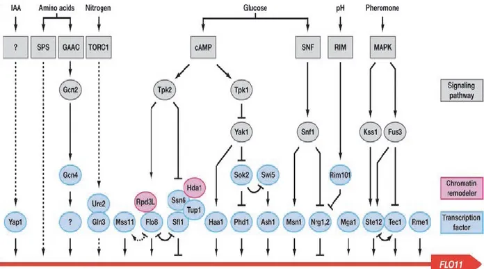
FIG 5 Regulation of FLO11 expression. Wiring diagram showing the complex regulation of the FLO11 promoter by conventional and epigenetic mechanisms. Arrows indicate positive regulation and inhibition is shown by bars. Different environmental stimuli and corresponding signaling pathways targeting FLO11 are indicated at the top (Iaa= indole acetic acid; ?= unknown pathway). Downstream acting protein kinases are shown in gray. Chromatin remodelers found in the FLO11 region are shown in magenta. Transcription factors targeting the FLO11 promoter (red line) are shown in light blue (? = unknown transcription factor). The input of the different transcription factors is only shown schematically and does not correspond to the positions of known binding sites. (Bruckner and Mosch, 2012).
4.2. Elongation and tandem repeats. Besides the effects of expression signaling
and/or silencing of FLO11 gene on the biofilm formation process, it was also shown that the tandem repeats and the core length of its central domain, are also involved in affecting this phenotype. Indeed, the intragenic repeats are residues of Serine and Threonine, which in turn, are sites for O-mannosylation and N-glycosylation.
The central domain of FLO11 gene varies between strains and results in a variation of the final product, in terms of gene length, glycosylation sites and high phenotype
variability (Verstrepen et al., 2005) (Douglas et al., 2007). Therefore, length variations in this single gene provide a combinatorial diversity, which may contribute to a very rapid adaptation to fluctuating environments (Fidalgo et al., 2008).
Indeed, the cloning of two FLO11 alleles from two different flor strains, of 3.1 and 5.0 kb length, into their appropriate locus in a lab strain BY4742 resulted in a significant correlation between biofilm-forming ability and FLO11 length both in different and in the same genetic backgrounds (Zara et al., 2009).
5. Adhesion, biofilm formation and phenotypic variation
- All these described peculiarities related to FLO11 gene, along with the whole cell wall complex, generate a high variability in the multicellular process of S. cerevisiae yeasts. In S. cerevisiae flor yeasts, the innate diversity of FLO11 and cell wall related genes background, generate variable adhesion ability between strains. These diversities comprise the FLO11 gene length, expression level and the frequency of tandem repeats of serine and threonine residues resulting as O-mannosyled sites on the cell wall (De Groot et al., 2005; Klis et al., 2006). All these parameters influence crucially the physiochemical characteristics and the adhesion forces of yeasts cell wall, which affect directly the cell-cell and cell-surface adhesion and biofilm formation. These adhesion forces involve specific and non-specific interactions; flocculation phenotype is considered as a specific interaction due to lectin-carbohydrate binding motifs, meanwhile biofilm formation and adhesion to substrates are known to be non-specific interactions. Non-specific interactions include mainly electrostatic and hydrophobic interactions (van Oss, 1990; Bos et al., 1999).
In aqueous solutions, the cell wall chemical groups are ionized and confer an electric charge to the cell surface which enables electrostatic interactions between the cell and the charged ions or molecules present in the external medium (Vu et al., 2011). Electrostatic interactions are short-ranged repulsive or attractive interactions that occur between ions and charged surfaces and are subdivided into van der Waals interactions, permanent diapoles, and hydrogen bonding.
These individual subdivisions are relatively weak and transitory forces, but when aligned with each other and depending on the size of the molecule; the larger the molecule the stronger the force; electrostatic interactions could modulate significantly the total adhesion ability (Leckband and Israelachvili, 2001).
Hydrophobic interactions are considered as the predominant interactions mediating microbial binding to biotic and abiotic surfaces, of molecules dissolved in polar liquids. They are strong and long-ranged interactions, with a factor of 5-10 times efficiency respect to the rest of adhesion interaction forces (Vu et al., 2011). Mechanisms behind hydrophobic interactions are still not well understood, even though, the classical description regarding the hydrophobicity is the rapid aggregation of non-polar liquid i.e. oil, when dissolved in polar liquid i.e. water, as the exclusion of hydrophobic molecules from water results in a squeezing effect of theses surfaces (Van Oss, 1995).
The mechanism leading to microbial aggregation is triggered by these adhesion forces when two cells start attracting and interacting thru the hydrophobic long-range forces, than reinforced by the electrostatic short-long-range forces, polar interactions, hydrogen bonds, and specific interactions (Van Oss, 1995; Holder et al., 2007). In S. cerevisiae, accordingly with these interacting forces, flo11 mutant strains present a fall in cell wall hydrophobicity and electrostatic charge, which lead to a loss of their adhesion capacity and their ability to form biofilm (Reynolds, 2001; Zara et
al., 2005; Fidalgo et al., 2006; Barrales et al., 2008). This includes also flocculation
mechanism in brewing, where strains with low cell wall hydrophobicity are less flocculants (Azeredo et al., 1997; Holle et al., 2012). Even more, at the biomedical level, the hydrophobicity of invasive Candida species is much higher than non invasive spp (Borghi et al., 2011).
These physiochemical characteristics generate a high variability in adhesion and biofilm formation and structure. This is the reason why many recent studies are focusing on finding molecules which affect directly or indirectly the cell hydrophobicity, thru the interaction with cell wall components, mainly cell wall mannoproteins. The aim of such interactions studies is to intercept the hydrophobic ligands, which can lead to the collapse of adhesion and invasion capacity of yeast strains. Antimicrobial peptides (AMPs) are leader components in this approach. Many AMPs were found to modulate the adhesion and biofilm formation of some yeasts and fungi, thru hydrophobic and electrostatic interactions. For example, Histidine rich glycoproteins greatly inhibits Candida albicans biofilm formation thru binding and rupturing cell wall components (Rydengard et al., 2008). In contrast, histatin-5 and LL-37 AMPs were shown to be sheded by the cell wall mucin Msb2 of
Candida albicans, enhancing the cell resistance toward such AMPs
References:
Aguilar-Uscanga, B., Francois, J.M., 2003. A study of the yeast cell wall composition and structure in response to growth conditions and mode of cultivation. Lett Appl Microbiol 37, 268-274.
Alexandre, H., 2013. Flor yeasts of Saccharomyces cerevisiae—Their ecology, genetics and metabolism. International Journal of Food Microbiology 167, 269-275.
Aranda, A., del Olmo, M.L., 2004. Exposure of Saccharomyces cerevisiae to acetaldehyde induces sulfur amino acid metabolism and polyamine transporter genes, which depend on Met4p and Haa1p transcription factors, respectively. Appl Environ Microbiol 70, 1913-1922.
Azeredo, J., Ramos, I., Rodriguks, L., Oliveira, R., Teixeira, J., 1997. Yeast flocculation: A new method for characterizing cell surface interactions. Journal of the Institute of Brewing 103, 3.
Bao, M.Z., Schwartz, M.A., Cantin, G.T., Yates, J.R., 3rd, Madhani, H.D., 2004. Pheromone-dependent destruction of the Tec1 transcription factor is required for MAP kinase signaling specificity in yeast. Cell 119, 991-1000.
Baranyi, J., Roberts, T.A., 1994. A dynamic approach to predicting bacterial growth in food. Int J Food Microbiol 23, 277-294.
Barnett, J.A., 2003. A history of research on yeasts 6: the main respiratory pathway. Yeast 20, 1015-1044.
Baror, Y., 1990. The effect of adhesion on survival and growth of microorganisms. Experientia
46, 4.
Barrales, R.R., Jimenez, J., Ibeas, J.I., 2008. Identification of novel activation mechanisms for FLO11 regulation in Saccharomyces cerevisiae. Genetics 178, 145-156.
Bauer, F.F., Govender, P., Bester, M.C., 2010. Yeast flocculation and its biotechnological relevance. Appl Microbiol Biotechnol 88, 31-39.
Bochner, B.R., 2001. Phenotype MicroArrays for High-Throughput Phenotypic Testing and Assay of Gene Function. Genome Research 11, 1246-1255.
Bochner, B.R., 2009. Global phenotypic characterization of bacteria. FEMS Microbiology Reviews 33, 191-205.
Bony, M., Thines-Sempoux, D., Barre, P., Blondin, B., 1997. Localization and cell surface anchoring of the Saccharomyces cerevisiae flocculation protein Flo1p. J Bacteriol 179, 4929-4936.
Borghi, E., Sciota, R., Biassoni, C., Cirasola, D., Cappelletti, L., Vizzini, L., Boracchi, P., Morace, G., 2011. Cell surface hydrophobicity: a predictor of biofilm production in Candida isolates? J Med Microbiol 60, 689-690.
Borglin, S., Joyner, D., DeAngelis, K.M., Khudyakov, J., D’haeseleer, P., Joachimiak, M.P., Hazen, T., 2012. Application of phenotypic microarrays to environmental microbiology. Current Opinion in Biotechnology 23, 41-48.
Borneman, A.R., Leigh-Bell, J.A., Yu, H., Bertone, P., Gerstein, M., Snyder, M., 2006. Target hub proteins serve as master regulators of development in yeast. Genes Dev 20, 435-448.
Bos, R., van der Mei, H.C., Busscher, H.J., 1999. Physico-chemistry of initial microbial adhesive interactions--its mechanisms and methods for study. FEMS Microbiol Rev 23, 179-230.
Bou Zeidan, M., Carmona, L., Zara, S., Marcos, J.F., 2013. FLO11 Gene Is Involved in the Interaction of Flor Strains of Saccharomyces cerevisiae with a Biofilm-Promoting Synthetic Hexapeptide. Appl Environ Microbiol 79, 6023-6032.
Bowman, S.M., Free, S.J., 2006. The structure and synthesis of the fungal cell wall. Bioessays 28, 799-808.
Braus, G.H., 2003. Amino Acid Starvation and Gcn4p Regulate Adhesive Growth and FLO11 Gene Expression in Saccharomyces cerevisiae. Molecular Biology of the Cell 14, 4272-4284.
Brown, J.L., Bussey, H., 1993. The yeast KRE9 gene encodes an O glycoprotein involved in cell surface beta-glucan assembly. Mol Cell Biol 13, 6346-6356.
Brückner, S., Mösch, H.-U., 2012. Choosing the right lifestyle: adhesion and development in Saccharomyces cerevisiae. FEMS Microbiology Reviews 36, 25-58.
Bruckner, S., Mosch, H.U., 2012. Choosing the right lifestyle: adhesion and development in Saccharomyces cerevisiae. FEMS Microbiol Rev 36, 25-58.
Budroni, M., Giordano, G., Pinna, G., Farris, G.A., 2000. A genetic study of natural flor strains of Saccharomyces cerevisiae isolated during biological ageing from Sardinian wines. J Appl Microbiol 89, 657-662.
Budroni, M., Zara, S., Zara, G., Pirino, G., Mannazzu, I., 2005. Peculiarities of flor strains adapted to Sardinian sherry-like wine ageing conditions. FEMS Yeast Res 5, 951-958.
Bulawa, C.E., 1992. CSD2, CSD3, and CSD4, genes required for chitin synthesis in Saccharomyces cerevisiae: the CSD2 gene product is related to chitin synthases and to developmentally regulated proteins in Rhizobium species and Xenopus laevis. Mol Cell Biol 12, 1764-1776.
Bulawa, C.E., 1993. Genetics and molecular biology of chitin synthesis in fungi. Annu Rev Microbiol 47, 505-534.
Cabib, E., Arroyo, J., 2013. How carbohydrates sculpt cells: chemical control of morphogenesis in the yeast cell wall. Nature Reviews Microbiology 11, 648-655.
Cappellaro, C., Baldermann, C., Rachel, R., Tanner, W., 1994. Mating type-specific cell-cell recognition of Saccharomyces cerevisiae: cell wall attachment and active sites of a- and alpha-agglutinin. EMBO J 13, 4737-4744.
Caridi, A., 2006. Enological functions of parietal yeast mannoproteins. Antonie van Leeuwenhoek 89, 417-422.
Caro, L.H., Tettelin, H., Vossen, J.H., Ram, A.F., van den Ende, H., Klis, F.M., 1997. In silicio identification of glycosyl-phosphatidylinositol-anchored plasma-membrane and cell wall proteins of Saccharomyces cerevisiae. Yeast 13, 1477-1489.
Cartwright, S.P., Bill, R.M., Hipkiss, A.R., 2012. L-carnosine affects the growth of Saccharomyces cerevisiae in a metabolism-dependent manner. PLoS One 7, e45006.
Chen, H., 2006. Feedback control of morphogenesis in fungi by aromatic alcohols. Genes & Development 20, 1150-1161.
Chen, R.E., Thorner, J., 2007. Function and regulation in MAPK signaling pathways: lessons learned from the yeast Saccharomyces cerevisiae. Biochim Biophys Acta 1773, 1311-1340.
Chou, S., Huang, L., Liu, H., 2004. Fus3-regulated Tec1 degradation through SCFCdc4 determines MAPK signaling specificity during mating in yeast. Cell 119, 981-990.
Cid, V.J., Duran, A., del Rey, F., Snyder, M.P., Nombela, C., Sanchez, M., 1995. Molecular basis of cell integrity and morphogenesis in Saccharomyces cerevisiae. Microbiol Rev 59, 345-386.
Cooper, T.G., 2002. Transmitting the signal of excess nitrogen in Saccharomyces cerevisiae from the Tor proteins to the GATA factors: connecting the dots. FEMS Microbiol Rev 26, 223-238.
Cutler, N.S., Pan, X., Heitman, J., Cardenas, M.E., 2001. The TOR signal transduction cascade controls cellular differentiation in response to nutrients. Mol Biol Cell 12, 4103-4113.
Dague, E., Potthoff, E., Guillaume-Gentil, O., Ossola, D., Polesel-Maris, J., LeibundGut-Landmann, S., Zambelli, T., Vorholt, J.A., 2012. Rapid and Serial Quantification of Adhesion Forces of Yeast and Mammalian Cells. PLoS One 7, e52712.
Damberg, B.E., Blumberg Ia, E., 1983. [Toxic effect of cysteine on cells of Saccharomyces cerevisiae growing on media of various compositions]. Mikrobiologiia 52, 68-72.
De Groot, P.W., Ram, A.F., Klis, F.M., 2005. Features and functions of covalently linked proteins in fungal cell walls. Fungal Genet Biol 42, 657-675.
de Nobel, H., van Den Ende, H., Klis, F.M., 2000. Cell wall maintenance in fungi. Trends Microbiol 8, 344-345.
de Nobel, J.G., Klis, F.M., Priem, J., Munnik, T., van den Ende, H., 1990. The glucanase-soluble mannoproteins limit cell wall porosity in Saccharomyces cerevisiae. Yeast 6, 491-499.
Dean, N., 1999. Asparagine-linked glycosylation in the yeast Golgi. Biochim Biophys Acta 1426, 309-322.
Dempski, R.E., Jr., Imperiali, B., 2002. Oligosaccharyl transferase: gatekeeper to the secretory pathway. Curr Opin Chem Biol 6, 844-850.
DeRisi, J.L., Iyer, V.R., Brown, P.O., 1997. Exploring the metabolic and genetic control of gene expression on a genomic scale. Science 278, 680-686.
Douglas, C.M., Foor, F., Marrinan, J.A., Morin, N., Nielsen, J.B., Dahl, A.M., Mazur, P., Baginsky, W., Li, W., el-Sherbeini, M., et al., 1994. The Saccharomyces cerevisiae FKS1 (ETG1) gene encodes an integral membrane protein which is a subunit of 1,3-beta-D-glucan synthase. Proc Natl Acad Sci U S A 91, 12907-12911.
Douglas, L.M., Li, L., Yang, Y., Dranginis, A.M., 2007. Expression and characterization of the flocculin Flo11/Muc1, a Saccharomyces cerevisiae mannoprotein with homotypic properties of adhesion. Eukaryot Cell 6, 2214-2221.
Dranginis, A.M., Rauceo, J.M., Coronado, J.E., Lipke, P.N., 2007. A biochemical guide to yeast adhesins: glycoproteins for social and antisocial occasions. Microbiol Mol Biol Rev 71, 282-294.
Elion, E.A., 2000. Pheromone response, mating and cell biology. Curr Opin Microbiol 3, 573-581.
Esteve-Zarzoso, B., Peris-Toran, M.J., Garcia-Maiquez, E., Uruburu, F., Querol, A., 2001. Yeast Population Dynamics during the Fermentation and Biological Aging of Sherry Wines. Applied and Environmental Microbiology 67, 2056-2061.
Fazly, A., Jain, C., Dehner, A.C., Issi, L., Lilly, E.A., Ali, A., Cao, H., Fidel, P.L., Jr., R, P.R., Kaufman, P.D., 2013. Chemical screening identifies filastatin, a small molecule inhibitor of Candida albicans adhesion, morphogenesis, and pathogenesis. Proc Natl Acad Sci U S A 110, 13594-13599.
Ferguson, M.A., 1999. The structure, biosynthesis and functions of glycosylphosphatidylinositol anchors, and the contributions of trypanosome research. J Cell Sci 112 ( Pt 17), 2799-2809.
Fernandes, A.R., Mira, N.P., Vargas, R.C., Canelhas, I., Sa-Correia, I., 2005. Saccharomyces cerevisiae adaptation to weak acids involves the transcription factor Haa1p and Haa1p-regulated genes. Biochem Biophys Res Commun 337, 95-103.
Fidalgo, M., Barrales, R.R., Ibeas, J.I., Jimenez, J., 2006. Adaptive evolution by mutations in the FLO11 gene. Proc Natl Acad Sci U S A 103, 11228-11233.
Fidalgo, M., Barrales, R.R., Jimenez, J., 2008. Coding repeat instability in the FLO11 gene of Saccharomyces yeasts. Yeast 25, 879-889.
Finn, R.D., Mistry, J., Tate, J., Coggill, P., Heger, A., Pollington, J.E., Gavin, O.L., Gunasekaran, P., Ceric, G., Forslund, K., Holm, L., Sonnhammer, E.L., Eddy, S.R., Bateman, A., 2010. The Pfam protein families database. Nucleic Acids Res 38, D211-222.
Forsberg, H., Ljungdahl, P.O., 2001. Sensors of extracellular nutrients in Saccharomyces cerevisiae. Curr Genet 40, 91-109.
Freiberg, K.J., Cruess, W.V., 1955. A study of certain factors affecting the growth of flor yeast. Appl Microbiol 3, 208-212.
Frieman, M.B., Cormack, B.P., 2004. Multiple sequence signals determine the distribution of glycosylphosphatidylinositol proteins between the plasma membrane and cell wall in Saccharomyces cerevisiae. Microbiology 150, 3105-3114.
Galitski, T., Saldanha, A.J., Styles, C.A., Lander, E.S., Fink, G.R., 1999. Ploidy regulation of gene expression. Science 285, 251-254.
Gancedo, J.M., 2001. Control of pseudohyphae formation in Saccharomyces cerevisiae. FEMS Microbiol Rev 25, 107-123.
García-Rodriguez, L.J., Trilla, J.A., Castro, C., Valdivieso, M.H., Durán, A., Roncero, C., 2000. Characterization of the chitin biosynthesis process as a compensatory mechanism in the fks1 mutant of Saccharomyces cerevisiae. FEBS Lett. 478, 84-88.
Gelinas, P., 2009. Inventions on baker's yeast strains and specialty ingredients. Recent Pat Food Nutr Agric 1, 104-132.
Giaever, G., 2002. Functional profiling of the Saccharomyces cerevisiae genome. Nature 418, 387-391.
Godard, P., Urrestarazu, A., Vissers, S., Kontos, K., Bontempi, G., van Helden, J., Andre, B., 2007. Effect of 21 Different Nitrogen Sources on Global Gene Expression in the Yeast Saccharomyces cerevisiae. Molecular and Cellular Biology 27, 3065-3086.
Goossens, K.V., Willaert, R.G., 2012a. The N-terminal domain of the Flo11 protein from Saccharomyces cerevisiae is an adhesin without mannose-binding activity. FEMS Yeast Res 12, 78-87.
Goossens, K.V.Y., Stassen, C., Stals, I., Donohue, D.S., Devreese, B., De Greve, H., Willaert, R.G., 2010. The N-Terminal Domain of the Flo1 Flocculation Protein from Saccharomyces cerevisiae Binds Specifically to Mannose Carbohydrates. Eukaryotic Cell 10, 110-117.
Goossens, K.V.Y., Willaert, R.G., 2012b. The N-terminal domain of the Flo11 protein from Saccharomyces cerevisiae is an adhesin without mannose-binding activity. FEMS Yeast Research 12, 78-87.
Govender, P., Domingo, J.L., Bester, M.C., Pretorius, I.S., Bauer, F.F., 2008. Controlled expression of the dominant flocculation genes FLO1, FLO5, and FLO11 in Saccharomyces cerevisiae. Appl Environ Microbiol 74, 6041-6052.
Granek, J.A., Magwene, P.M., 2010. Environmental and genetic determinants of colony morphology in yeast. PLoS Genet 6, e1000823.
Guo, B., Styles, C.A., Feng, Q., Fink, G.R., 2000. A Saccharomyces gene family involved in invasive growth, cell-cell adhesion, and mating. Proc Natl Acad Sci U S A 97, 12158-12163.
Hall-Stoodley, L., Stoodley, P., 2009. Evolving concepts in biofilm infections. Cell Microbiol 11, 1034-1043.
Hans, M.A., Heinzle, E., Wittmann, C., 2003. Free intracellular amino acid pools during autonomous oscillations in Saccharomyces cerevisiae. Biotechnol Bioeng 82, 143-151.
Hazelwood, L.A., Daran, J.M., van Maris, A.J.A., Pronk, J.T., Dickinson, J.R., 2008. The Ehrlich Pathway for Fusel Alcohol Production: a Century of Research on Saccharomyces cerevisiae Metabolism. Applied and Environmental Microbiology 74, 2259-2266.
Hinnebusch, A.G., 2005. Translational regulation of GCN4 and the general amino acid control of yeast. Annu Rev Microbiol 59, 407-450.
Hohmann, S., 2002. Osmotic stress signaling and osmoadaptation in yeasts. Microbiol Mol Biol Rev 66, 300-372.
Holder, D.J., Kirkland, B.H., Lewis, M.W., Keyhani, N.O., 2007. Surface characteristics of the entomopathogenic fungus Beauveria (Cordyceps) bassiana. Microbiology 153, 3448-3457.
Holle, A., Machado, M.D., Soares, E.V., 2011. Flocculation in ale brewing strains of Saccharomyces cerevisiae: re-evaluation of the role of cell surface charge and hydrophobicity. Applied Microbiology and Biotechnology 93, 1221-1229.
Holle, A.V., Machado, M.D., Soares, E.V., 2012. Flocculation in ale brewing strains of Saccharomyces cerevisiae: re-evaluation of the role of cell surface charge and hydrophobicity. Appl Microbiol Biotechnol 93, 1221-1229.
Homann, O.R., Cai, H., Becker, J.M., Lindquist, S.L., 2005. Harnessing natural diversity to probe metabolic pathways. PLoS Genet 1, e80.
Horisberger, M., Clerc, M.F., 1988. Ultrastructural localization of anionic sites on the surface of yeast, hyphal and germ-tube forming cells of Candida albicans. Eur J Cell Biol 46, 444-452.
Ielasi, F.S., Decanniere, K., Willaert, R.G., 2012. The epithelial adhesin 1 (Epa1p) from the human-pathogenic yeast Candida glabrata: structural and functional study of the carbohydrate-binding domain. Acta Crystallogr D Biol Crystallogr 68, 210-217.
Ikezawa, H., 2002. Glycosylphosphatidylinositol (GPI)-anchored proteins. Biol Pharm Bull 25, 409-417.
Ishigami, M., Nakagawa, Y., Hayakawa, M., Iimura, Y., 2006. FLO11 is the primary factor in flor formation caused by cell surface hydrophobicity in wild-type flor yeast. Biosci Biotechnol Biochem 70, 660-666.
Jigami, Y., Odani, T., 1999. Mannosylphosphate transfer to yeast mannan. Biochim Biophys Acta 1426, 335-345.
Kapteyn, J.C., Van Den Ende, H., Klis, F.M., 1999a. The contribution of cell wall proteins to the organization of the yeast cell wall. Biochim Biophys Acta 1426, 373-383.
Kapteyn, J.C., Van Egmond, P., Sievi, E., Van Den Ende, H., Makarow, M., Klis, F.M., 1999b. The contribution of the O-glycosylated protein Pir2p/Hsp150 to the construction of the yeast cell wall in wild-type cells and beta 1,6-glucan-deficient mutants. Mol Microbiol 31, 1835-1844.
Karunanithi, S., Vadaie, N., Chavel, C.A., Birkaya, B., Joshi, J., Grell, L., Cullen, P.J., 2010. Shedding of the Mucin-Like Flocculin Flo11p Reveals a New Aspect of Fungal Adhesion Regulation. Current Biology 20, 1389-1395.
Klis, F.M., 1994. Review: cell wall assembly in yeast. Yeast 10, 851-869.
Klis, F.M., Boorsma, A., De Groot, P.W., 2006. Cell wall construction in Saccharomyces cerevisiae. Yeast 23, 185-202.
Klis, F.M., de Jong, M., Brul, S., de Groot, P.W., 2007. Extraction of cell surface-associated proteins from living yeast cells. Yeast 24, 253-258.
Klis, F.M., Mol, P., Hellingwerf, K., Brul, S., 2002. Dynamics of cell wall structure in Saccharomyces cerevisiae. FEMS Microbiol Rev 26, 239-256.
Kobayashi, O., Hayashi, N., Kuroki, R., Sone, H., 1998. Region of FLO1 proteins responsible for sugar recognition. J Bacteriol 180, 6503-6510.
Kojic, E.M., Darouiche, R.O., 2004. Candida infections of medical devices. Clin Microbiol Rev 17, 255-267.
Kollar, R., Petrakova, E., Ashwell, G., Robbins, P.W., Cabib, E., 1995. Architecture of the yeast cell wall. The linkage between chitin and beta(1-->3)-glucan. J Biol Chem 270, 1170-1178.
Kollar, R., Reinhold, B.B., Petrakova, E., Yeh, H.J., Ashwell, G., Drgonova, J., Kapteyn, J.C., Klis, F.M., Cabib, E., 1997. Architecture of the yeast cell wall. Beta(1-->6)-glucan interconnects mannoprotein, beta(1-->)3-glucan, and chitin. J Biol Chem 272, 17762-17775.
Kolodkin-Gal, I., Romero, D., Cao, S., Clardy, J., Kolter, R., Losick, R., 2010. D-Amino Acids Trigger Biofilm Disassembly. Science 328, 627-629.
Kregiel, D., Berlowska, J., Szubzda, B., 2012. Novel permittivity test for determination of yeast surface charge and flocculation abilities. Journal of Industrial Microbiology & Biotechnology 39, 1881-1886.
Krogerus, K., Gibson, B.R., 2013. Influence of valine and other amino acids on total diacetyl and 2,3-pentanedione levels during fermentation of brewer’s wort. Applied Microbiology and Biotechnology 97, 6919-6930.
Kuchin, S., Vyas, V.K., Carlson, M., 2002. Snf1 protein kinase and the repressors Nrg1 and Nrg2 regulate FLO11, haploid invasive growth, and diploid pseudohyphal differentiation. Mol Cell Biol 22, 3994-4000.
LaRue, T.A., Spencer, J.F., 1967. The utilization of D-amino acids by yeasts. Can J Microbiol 13, 777-788.
Leckband, D., Israelachvili, J., 2001. Intermolecular forces in biology. Q Rev Biophys 34, 105-267.
Lee, J.C.Y., Tsoi, A., Kornfeld, G.D., Dawes, I.W., 2013. Cellular responses toL-serine inSaccharomyces cerevisiae: roles of general amino acid control, compartmentalization, and aspartate synthesis. FEMS Yeast Research 13, 618-634.
Lei, H., Zheng, L., Wang, C., Zhao, H., Zhao, M., 2013. Effects of worts treated with proteases on the assimilation of free amino acids and fermentation performance of lager yeast. Int J Food Microbiol 161, 76-83.
Lesage, G., Bussey, H., 2006. Cell wall assembly in Saccharomyces cerevisiae. Microbiol Mol Biol Rev 70, 317-343.
Lesage, G., Sdicu, A.M., Menard, P., Shapiro, J., Hussein, S., Bussey, H., 2004. Analysis of beta-1,3-glucan assembly in Saccharomyces cerevisiae using a synthetic interaction network and altered sensitivity to caspofungin. Genetics 167, 35-49.
Levin, D.E., 2005. Cell Wall Integrity Signaling in Saccharomyces cerevisiae. Microbiology and Molecular Biology Reviews 69, 262-291.
Levin, D.E., 2011. Regulation of cell wall biogenesis in Saccharomyces cerevisiae: the cell wall integrity signaling pathway. Genetics 189, 1145-1175.
Liao, S.-M., Du, Q.-S., Meng, J.-Z., Pang, Z.-W., Huang, R.-B., 2013. The multiple roles of histidine in protein interactions. Chemistry Central Journal 7, 44.
Lindquist, W., 1952. Cell surface constituents and yeast flocculation. Nature 170, 544-545.
Lionakis, M.S., Netea, M.G., 2013. Candida and host determinants of susceptibility to invasive candidiasis. PLoS Pathog 9, e1003079.
Lipke, P.N., Kurjan, J., 1992. Sexual agglutination in budding yeasts: structure, function, and regulation of adhesion glycoproteins. Microbiol Rev 56, 180-194.
Lipke, P.N., Ovalle, R., 1998. Cell wall architecture in yeast: new structure and new challenges. J Bacteriol 180, 3735-3740.
Liu, H., Styles, C.A., Fink, G.R., 1996. Saccharomyces cerevisiae S288C has a mutation in FLO8, a gene required for filamentous growth. Genetics 144, 967-978.
Ljungdahl, P.O., 2009. Amino-acid-induced signalling via the SPS-sensing pathway in yeast. Biochem Soc Trans 37, 242-247.
Ljungdahl, P.O., Daignan-Fornier, B., 2012. Regulation of Amino Acid, Nucleotide, and Phosphate Metabolism in Saccharomyces cerevisiae. Genetics 190, 885-929.
Loibl, M., Strahl, S., 2013. Protein O-mannosylation: what we have learned from baker's yeast. Biochim Biophys Acta 1833, 2438-2446.
Lorenz, M.C., Heitman, J., 1998a. The MEP2 ammonium permease regulates pseudohyphal differentiation in Saccharomyces cerevisiae. EMBO J 17, 1236-1247.
Lorenz, M.C., Heitman, J., 1998b. Regulators of pseudohyphal differentiation in Saccharomyces cerevisiae identified through multicopy suppressor analysis in ammonium permease mutant strains. Genetics 150, 1443-1457.
Magasanik, B., Kaiser, C.A., 2002. Nitrogen regulation in Saccharomyces cerevisiae. Gene 290, 1-18.
Mannazzu, I., Angelozzi, D., Belviso, S., Budroni, M., Farris, G.A., Goffrini, P., Lodi, T., Marzona, M., Bardi, L., 2008. Behaviour of Saccharomyces cerevisiae wine strains during adaptation to unfavourable conditions of fermentation on synthetic medium: cell lipid composition, membrane integrity, viability and fermentative activity. Int J Food Microbiol 121, 84-91.
Marques, F., Lasanta, C., Caro, I., Perez, L., 2008. Study of the lipidic and proteic composition of an industrial filmogenic yeast with applications as a nutritional supplement. J Agric Food Chem 56, 12025-12030.
Mauricio, J.C., Valero, E., Millan, C., Ortega, J.M., 2001. Changes in nitrogen compounds in must and wine during fermentation and biological aging by flor yeasts. J Agric Food Chem 49, 3310-3315.
Mazur, P., Baginsky, W., 1996. In vitro activity of 1,3-beta-D-glucan synthase requires the GTP-binding protein Rho1. J Biol Chem 271, 14604-14609.
Mazur, P., Morin, N., Baginsky, W., el-Sherbeini, M., Clemas, J.A., Nielsen, J.B., Foor, F., 1995. Differential expression and function of two homologous subunits of yeast 1,3-beta-D-glucan synthase. Mol Cell Biol 15, 5671-5681.
Mortimer, R.K., Johnston, J.R., 1986. Genealogy of principal strains of the yeast genetic stock center. Genetics 113, 35-43.
Mosch, H.U., Roberts, R.L., Fink, G.R., 1996. Ras2 signals via the Cdc42/Ste20/mitogen-activated protein kinase module to induce filamentous growth in Saccharomyces cerevisiae. Proc Natl Acad Sci U S A 93, 5352-5356.
Moukadiri, I., Jaafar, L., Zueco, J., 1999. Identification of two mannoproteins released from cell walls of a Saccharomyces cerevisiae mnn1 mnn9 double mutant by reducing agents. J Bacteriol 181, 4741-4745.
Mowat, E., Williams, C., Jones, B., McChlery, S., Ramage, G., 2009. The characteristics of Aspergillus fumigatus mycetoma development: Is this a biofilm? Medical Mycology 47, S120-S126.
Muller, M., Mentel, M., van Hellemond, J.J., Henze, K., Woehle, C., Gould, S.B., Yu, R.Y., van der Giezen, M., Tielens, A.G., Martin, W.F., 2012. Biochemistry and evolution of anaerobic energy metabolism in eukaryotes. Microbiol Mol Biol Rev 76, 444-495.
Murray, D.B., Haynes, K., Tomita, M., 2011. Redox regulation in respiring Saccharomyces cerevisiae. Biochim Biophys Acta 1810, 945-958.
Nakagawa, Y., Toda, Y., Yamamura, H., Hayakawa, M., Iimura, Y., 2011. FLO11 is essential for pellicle formation by wild pellicle-forming yeasts isolated from contaminated wines. J. Biosci. Bioeng. 111, 7-9.
Naz, S., Gueguen-Minerbe, M., Cretenet, M., Vernoux, J.P., 2013. Aromatic amino acids as precursors of antimicrobial metabolites in Geotrichum candidum. FEMS Microbiol Lett 344, 39-47.
Ning, Y., Hu, X., Ling, J., Du, Y., Liu, J., Liu, H., Peng, Z., 2013. Candida albicans survival and biofilm formation under starvation conditions. Int Endod J 46, 62-70.
Osumi, M., 1998. The ultrastructure of yeast: cell wall structure and formation. Micron 29, 207-233.
Page, N., Gerard-Vincent, M., Menard, P., Beaulieu, M., Azuma, M., Dijkgraaf, G.J., Li, H., Marcoux, J., Nguyen, T., Dowse, T., Sdicu, A.M., Bussey, H., 2003. A Saccharomyces cerevisiae genome-wide mutant screen for altered sensitivity to K1 killer toxin. Genetics 163, 875-894.
Pallotta, M.L., 2013. L-Proline uptake in Saccharomyces cerevisiae mitochondria can contribute to bioenergetics during nutrient stress as alternative mitochondrial fuel. World J Microbiol Biotechnol.
Petranovic, D., Nielsen, J., 2008. Can yeast systems biology contribute to the understanding of human disease? Trends Biotechnol 26, 584-590.
Piskur, J., Rozpedowska, E., Polakova, S., Merico, A., Compagno, C., 2006. How did Saccharomyces evolve to become a good brewer? Trends Genet 22, 183-186.
Popolo, L., Gilardelli, D., Bonfante, P., Vai, M., 1997. Increase in chitin as an essential response to defects in assembly of cell wall polymers in the ggp1Δ mutant of Saccharomyces cerevisiae. J. Bacteriol. 179, 463-469.
Qadota, H., Python, C.P., Inoue, S.B., Arisawa, M., Anraku, Y., Zheng, Y., Watanabe, T., Levin, D.E., Ohya, Y., 1996. Identification of yeast Rho1p GTPase as a regulatory subunit of 1,3-beta-glucan synthase. Science 272, 279-281.