1
UNIVERSITÀ POLITECNICA DELLE MARCHE
S
CUOLA DID
OTTORATO DIR
ICERCA DELLAF
ACOLTÀ DIM
EDICINA EC
HIRURGIACurriculum Medicina Clinica e Molecolare – XIV ciclo
MECCANISMI DEL DANNO CELLULARE DA
CHEMIOTERAPICI SU CELLULE NORMALI E
NEOPLASTICHE
D
OCENTET
UTOR: Chiar.mo
D
OTTORANDOProf. Pietro Leoni
Dott. Jacopo Olivieri
C
OORDINATORE:
Chiar.mo
Prof. Roberto Di Primio
2
MECHANISMS OF CHEMOTHERAPY-INDUCED CELLULAR
DAMAGE IN NORMAL AND NEOPLASTIC CELLS
SUMMARY
INTRODUCTION ... 3
PATIENTS AND METHODS ... 5
CARDIAC ASSESSMENT ... 5
PLANNED TREATMENTS ... 6
STATISTICAL ANALYSES ... 7
RESULTS ... 10
BASELINE CHARACTERISTICS AND PLANNED TREATMENTS ... 10
TREATMENT DELIVERY AND OUTCOME... 11
CARDIAC TOXICITY: ECOCARDIOGRAPHIC EVALUATION ... 14
CARDIAC TOXICITY: BIOMARKER EVALUATION ... 16
FEASIBILITY ... 18
DISCUSSION ... 19
3
INTRODUCTION
Anthracyclines (AC) are the mainstay of first line treatment in many lymphoma patients. A specific concern of AC-containing regimens is the occurrence of cardiac toxicity [1]. Although cardiac adverse effects of AC were recognized shortly after their discovery, more than 40 years ago, still we face many uncertainties about basic aspects of AC-induced cardiotoxicity (AIC) [2]. Recent published guidelines acknowledge the lack of elementary knowledge and strong evidences to guide management of AC-treated patients [3]. First, the prevalence of AIC is highly variable in different settings and several risk factors have been reported to increase its occurrence but only age and cumulative AC dose are consistently recognized as relevant determinants [4]. Further uncertainty is added when dealing with haematological patients, as most recommendations are inferred by studies conducted in solid tumors, primarily breast and lung cancer [5,6] The exact prevalence occurring after widely used regimens as R-CHOP for non-Hodgkin lymphomas (NHL) or ABVD for Hodgkin’s disease (HD) is thus unknown in the real life: in fact, the large clinical studies which tested these interventions were mainly focused on efficacy. Second, several monitoring methods have been proposed but there is no clear indication about the best technique, although echocardiogram is generally preferred due to large accessibility and easy of use [7]; notably, the sensitivity of the detection method will also impact the reported prevalence of AIC. Thus, the only population-based study addressing AIC in lymphoma inherently lacks sensitivity to detect subclinical AIC [8].
Several strategies have been suggested to reduce the occurrence of AIC, without affecting negatively their antitumor efficacy: the most applied is limitation of the cumulative dose, according to current prescribing recommendations [9]; also, weekly or continuous infusion protocols have been shown to be equally effective but less toxic than classic scheduling every 2 or 3 weeks; however such protocols have limited applicability in outpatient settings. Dexrazoxane is a cardioprotectant with iron-chelating activity which has shown to decrease AIC when administered concomitantly with AC. However, concerns of reduced antitumor activity and a reported increase in second tumors in pediatric populations [10] led to warnings from regulatory agencies [11] and discouraged dexrazoxane use in clinical practice. Liposomal formulations of doxorubicin offer pharmacokynetic advances over the free drug resulting in longer half-life, reduced peak AC dose and increased tumor penetration, thereby enhancing the therapeutic index [12]. Non-pegylated liposomal doxorubicin (NPLD) showed reduced AIC and preserved antitumor in two randomized multicenter trials of
4
metastatic breast cancer [13]; although direct comparison of NPLD to the pegylated form has not been reported, NPLD may be preferable because it is devoid of a specific cutaneous toxicity of liposomal doxorubicin, i.e. palmo-plantar erythrodysesthesia. Positive data on metastatic breast cancer prompted use in lymphoma patients: several single-arm trials suggested an encouraging efficacy and reduced incidence of AIC, although we lack a randomized comparison with conventional AC [14,15].
A major advance in the management of heart failure was the recognition and aggressive treatment of subclinical structural heart damage to prevent further deterioration of cardiac function leading to symptomatic heart failure [16]. Translation of this approach to AIC has shown that, similarly to CHF from other causes, signs of cardiac dysfunction may be detected a long time before clinical manifestations occur [17]. Specifically, with regard to AIC, a monitoring strategy including serial measurements of cardiac troponins seems to identify the patients at very high risk for AIC in their earliest phase [18]. This observation supports the existence of a temporal window which may be exploited by therapeutic interventions aiming to stop and reverse the cardiac damage.
The aim of our study was to quantify the occurrence of cardiotoxicity in a real life population of lymphoma patients, combining novel and traditional monitoring methods including clinical, echocardiographic and laboratory parameters. To improve the feasibility of such a combined monitoring approach in an outpatient setting, we tested the performance of a telemedicine system to integrate data and to allow constant interdisciplinary management with an expert cardiologist. Regarding to biomarkers, we aimed to evaluate the occurrence and kinetics of troponin I rises and their correlation to clinical and subclinical cardiotoxicity.
5
PATIENTS AND METHODS
This is a prospective observational trial in lymphoma patients undergoing 1st line treatment with
conventional or liposomal AC at our center (Hematology Clinic, Ancona). Inclusion criteria were: age greater than 18 years, diagnosis of lymphoma according to WHO classification (2008), planned treatment with a chemotherapy protocol containing conventional or liposomal AC. Exclusion criteria were: lack of informed consent, previous treatment with chemotherapy or radiotherapy (with the exception of high-dose corticosteroids administered for less than 30 days); fertile women unwilling to undergo contraception.
The study was approved by our institutional review board and registered in the AIFA (Agenzia Italiana del Farmaco) registry of observational studies (Study ID: RSO-581). All patients provided informed consent. Patients were enrolled from February 2012 to March 2014. The primary objective of the study was to determine the incidence rate of AC-induced cardiotoxicity (early and late, subclinical and clinical) in a real life population of lymphoma patients; the secondary objectives were: to determine (1) the incidence of cardiac adverse effects possibly related to AC cardiotoxicity (sudden death, acute heart failure, arrhythmia requiring treatment, acute myocardial infarction or unstable angina, worsening of NYHA class, symptomatic pericarditis); (2) the occurrence of significant echocardiographic abnormalities other than LVEF reduction (increase in size of cardiac chambers and walls, valvulopathies, pericardial diseases); (3) the occurrence of troponin I rises (above the pre-specified cut-offs of 0.03 ng/ml and 0.08 ng/ml) not related to ischaemic events; (4) the feasibility of a combined monitoring approach using a telemedicine system; (5) the response to the chemotherapy treatment (according to 2007 IWG criteria [21]); (6) the overall survival.
CARDIAC ASSESSMENT
Echocardiographic cardiotoxicity was defined as a significant drop in LVEF as in Swain et al. [6] (> 10% to a final value less than 50%; > 20%; drop to a final value > 45%, irrespective of the initial value). Baseline and final (1 year after the end of treatment) echocardiographic evaluations were done using the Vivid 9 system (GE Healthcare, Little Chalfont, UK)[19]; performed analysis included: measurement of LVEF (Simpson biplane method), measurement of global strain rate
6
with speckle-tracking 2D-strain imaging and of systolic myocardial velocity (Sa) at the lateral mitral annulus with tissue Doppler imaging (TDI) [20].
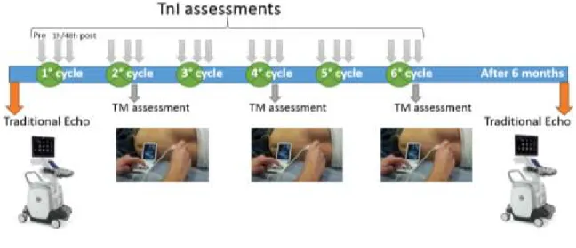
Other data were collected within a telemedicine (TM) system, which was developed as an electronic platform for cardiology consultations. TM assessments were performed at least every 100 mg/m2 of doxorubicin cumulative dose and consisted of brief history and physical examination, ECG and echocardiographic evaluation using a portable device (VScan, GE Healthcare, Little Chalfont, UK). The TM software was developed by Telemedware (Jesi, Italy). The front end TM assessment was performed by a hematologist with expertise in cardiac ultrasonography; the back end TM report was created by one expert cardiologist participating to the study. TnI measurements were performed before, 1h and 24-72h after each chemotherapy cycle (Advia Centaur, Siemens Healthcare). The monitoring schedule in a typical 6-cycle chemotherapy treatment is depicted in Fig. 1.
Figure 1: Monitoring schedule in a 6-cycle chemotherapy treatment
PLANNED TREATMENTS
Chemotherapy treatments for lymphomas were administered according to current standard protocols; the only types of AC used were doxorubicin hydrochloride (DOX) and non-pegylated liposomal doxorubicin hydrochloride (Myocet®, Teva) (NPLD). According to our internal policy (filed in October 2011 with the local health administration), standard doses of conventional AC are applied to all adults with less than 65 years, without a history of coronary heart disease, absence of structural heart disease at the baseline echocardiographic evaluation and serum BNP <100
7
pg/ml. On the contrary, patients with severely compromised left ventricular function (LVEF ≤ 35%) or having clinical evidence of refractory heart failure, untreated severe coronary artery disease or serum BNP > 400 pg/ml, are generally excluded from AC-containing protocols. Patients older than 65 years or those with moderately decreased left ventricular function (LVEF >35 and ≤50%) or having evidence of structural heart disease without significant coronary artery disease, are treated with liposomal AC.
When advised by our internal policy, DOX was substituted by NPLD at the same dose of DOX. Dose reductions for other drugs were applied at baseline according to age and comorbidities and subsequently according to toxicity. Prophylaxis for Pneumocystis Jiroveci was administered concomitantly to all CHOP-like regimens, but not to ABVD. Granulocyte-colony-stimulating factors for febrile neutropenia were administered as primary prophylaxis in all patients older than 65 years undergoing CHOP-like regimens repeated every 21 days; or in all patients undergoing CHOP-like regimens repeated every 14 days; as secondary prophylaxis in younger patients treated with CHOP-like regimens repeated every 21 days. In ABVD-treated patients, G-CSF was not routinely administered.
STATISTICAL ANALYSES
Descriptive statistics are reported for all the variables categorized according to type of AC used (DOX or NPLD). Binary outcomes (e.g. occurrence of cardiotoxicity, occurrence of other echocardiographic alterations, rises of TnI above pre-specified cut-offs) and continuous outcomes (e.g. change in LVEF, change in other echocardiographic parameters, change in biomarkers values) are reported for the entire cohort and separately according to type of AC used (DOX or NPLD). The rates and proportions were compared using chi-square or Fisher's exact test. Continuous and discrete variables were compared using Student t-test or Mann-Whitney test. The distribution of time-dependent variables (e.g. overall survival) was estimated using the Kaplan-Meier product limit method. The univariate assessment for the significative time-dependent covariates was carried out with the log-rank test.
8
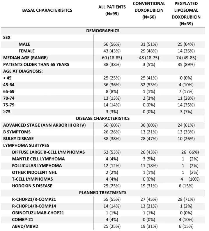
TABLE 1: Baseline characteristics of the 99 patients undergoing at least 1 cycle of CHT, related
to demographics, disease characteristics and planned treatments.
BASAL CHARACTERISTICS ALL PATIENTS (N=99) CONVENTIONAL DOXORUBICIN (N=60) NON-PEGYLATED LIPOSOMAL DOXORUBICIN (N=39) DEMOGRAPHICS SEX MALE 56 (56%) 31 (51%) 25 (64%) FEMALE 43 (43%) 29 (48%) 14 (35%)
MEDIAN AGE (RANGE) 60 (18-85) 48 (18-75) 74 (49-85) PATIENTS OLDER THAN 65 YEARS 38 (38%) 3 (5%) 35 (89%) AGE AT DIAGNOSIS: < 45 25 (25%) 25 (41%) 0 (0%) 45-64 36 (36%) 32 (53%) 4 (10%) 65-69 8 (8%) 1 (1%) 7 (17%) 70-74 13 (13%) 2 (3%) 11 (28%) 75-79 14 (14%) 0 (0%) 14 (35%) ≥75 3 (3%) 0 (0%) 3 (7%) DISEASE CHARACTERISTICS
ADVANCED STAGE (ANN ARBOR III OR IV) 60 (60%) 36 (60%) 24 (61%)
B SYMPTOMS 26 (26%) 13 (21%) 13 (33%)
BULKY DISEASE 38 (38%) 28 (47%) 10 (26%)
LYMPHOMA SUBTYPES
DIFFUSE LARGE B-CELL LYMPHOMAS 52 (53%) 26 (43%) 26 66%)
MANTLE CELL LYMPHOMA 4 (4%) 3 (5%) 1 (2%)
FOLLICULAR LYMPHOMA 12 (12%) 11 (18%) 1 (2%) OTHER INDOLENT NHL 2 (2%) 1 (1%) 1 (2%) T-CELL LYMPHOMAS 4 (4%) 0 (0%) 4 (10%) HODGKIN'S DISEASE 25 (25%) 19 (31%) 6 (15%) PLANNED TREATMENTS R-CHOP21/R-COMP21 55 (55%) 27 (45%) 28 (71%) R-CHOP14/R-COMP14 14 (14%) 13 (21%) 1 (2%) OBINOTUZUMAB-CHOP21 1 (1%) 1 (1%) 0 (0%) COMEP-21 4 (4%) 0 (0%) 4 (10%) ABVD/MBVD 25 (25%) 19 (31%) 6 (15%)
9
TABLE 2: Baseline characteristics of the 99 patients undergoing at least 1 cycle of CHT, related
to cardiac risk factors and comorbidities, laboratory values and cardiac evaluation before treatment.
BASAL CHARACTERISTICS ALL PATIENTS (N=99) CONVENTIONAL DOXORUBICIN (N=60) NON-PEGYLATED LIPOSOMAL DOXORUBICIN (N=39) TEST FOR DIFFERENCE BETWEEN GROUPS (P-VALUE) CARDIAC RISK FACTORS
OBESITY (BMI ≥ 30 KG/MQ) 5 (5%) 5 (8%) 0 (0%) NS
DIABETES MELLITUS 6 (6%) 1 (1%) 5 (12%) 0.034
HISTORY OF HYPERTENSION 29 (29%) 11 (18%) 18 (46%) 0.003
HISTORY OF HYPERLIPIDEMIA 18 (18%) 7 (11%) 11 (28%) 0.037
HISTORY OF SMOKING TOBACCO 28 (28%) 14 (23%) 14 (35%) NS
CURRENT SMOKER WITH >10 PACK-YEARS 12 (12%) 7 (11%) 5 (12%) NS
NUMBER OF CARDIAC RISK FACTORS - MEDIAN
1 (0 – 4) 0 (0 – 3) 1 (0 – 4) <0.001
NONE OF THESE 5 CARDIAC RISK FACTORS 46 (46%) 34 (57%) 12 (30%) 0.012
HISTORY OF CORONARY ARTERY DISEASE 8 (8%) 0 (0%) 8 (20%) 0.000
ATRIAL FIBRILLATION 5 (5%) 0 (0%) 5 (12%) 0.008
HISTORY OF HYPOTHYROIDISM 7 (7%) 1 (1%) 6 (15%) 0.014
AT LEAST 1 CARDIAC RISK FACTOR OR
COMORBIDITY 60 (60%) 29 (48%) 31 (79%) 0.002
BASELINE LABORATORY VALUES
BNP (PG/ML) – MEDIAN 24 17 51 <0.001
BNP >100 PG/ML 7 (7%) 1 (1%) 6 (15%) 0.014
TROPONIN I > 0.03 NG/ML 4 (4%) 1 (1%) 3 (7%) NS
CARDIAC EVALUATION BEFORE TREATMENT
LVEF (%) – MEAN (RANGE) 62.4 (47 – 75) 63.1 (50 – 75) 61.3 (47 – 75) NS
LVEF <50% (N) 6 (6%) 1 (1%) 5 (12%) 0.034
ABNORMAL TDI (< 8 CM/SEC) 10 (10%) 6 (10%) 4 (10%) NS
10
RESULTS
BASELINE CHARACTERISTICS AND PLANNED TREATMENTS
We enrolled a total of 104 patients, of whom 5 never started therapy at our center: 3 died before starting treatment (UPN 3,28,87) and 2 were treated in another Hematology center due to logistic preferences (UPN 53 and 66). Data are reported for the remaining 99 patients who underwent at least 1 cycle of chemotherapy (modified intention-to-treat population).
The baseline characteristics of the 99 patients are shown in Table 1. The median age was 60 years (range 18-85 years) and 38 patients were older than 65 years. Twenty-five were HD and 74 NHL. The most represented NHL subtype was Diffuse Large B Cell Lymphoma (DLBCL) in 53% of patients. All HD patients were treated with the ABVD protocol, with NPLD substituting for DOX in 6 patients. All 74 NHL patients were treated with CHOP-like regimens, with NPLD substituting for DOX in 33 patients: etoposide was added for T-cell lymphomas; in the 70 B-cell NHL the anti-CD20 antibodies rituximab (in 69 patients) or obinutuzumab were added (1 patient). Overall, NPLD was used in 39 patients: 35 had more than 65 years, 4 were younger than 65 years but had evidence of structural heart disease or significant past history for heart diseases. Of 38 patients older than 65 years, 3 were treated with DOX, contradicting our internal policy: substitution with NPLD was not possible as they were enrolled in interventional trials not allowing the use of NPLD; they all had excellent performance status and no comorbidities. The average cumulative AC dose was 262 mg/m2 (268mg/m2 for DOX-treated patients and 254 mg/m2 for NPLD-treated patients). Over half the patients (54%) had at least one of the following traditional cardiac risk factors for increased AIC (Table 2), excluding age > 60 years: obesity (body mass index > 30 kg/m2, n = 5), diabetes mellitus (n = 6), hypertension (n = 29), hyperlipidemia (n = 18), and/or a history of tobacco use (n = 28). Other important comorbidities that increase the risk of developing heart failure included a history of coronary artery disease (CAD, n = 8), atrial fibrillation (n = 5) and a history of hypothyroidism (n = 7) (Table 2).
The distribution of cardiac risk factors and comorbidities showed obvious imbalances across the two groups, as expected by the categorization recommended by our internal policy. Overall, the subgroup of patients treated with NPLD was significantly older than the subgroup treated with DOX (mean age 72.3 and 46.1 years in NPLD and DOX subgroups, respectively; p < 0.001); they had significantly more risk factors for heart disease (median number 1 and 0 in NPLD and DOX
11
subgroups, respectively; p < 0.001); in detail, in the NPLD subgroup there was increased prevalence of diabetes mellitus, hypertension, hyperlipidemia, but not of obesity or tobacco use. Eight patients reported previous history of CAD; of these, 3 had concurrent atrial fibrillation; other 2 patients had atrial fibrillation without previous history of CAD: according to our internal policy, all these patients were treated with NPLD.
Echocardiographic evaluation at study entry did not reveal significant differences between NPLD and DOX subgroups (mean LVEF at baseline in all patients = 62.4%; 61.3% and 63.1%, in the NPLD and DOX subgroups respectively); 3 patients had baseline LVEF lower than 50%: they were all treated with NPLD. Patients in the NPLD subgroup had higher frequency of abnormal global longitudinal strain (greater than -18%, n = 15 in NPLD subgroup; n = 9 in DOX subgroup, p = 0.015) but not of abnormal TDI.
As regards biomarker evaluation at baseline, the BNP was above the upper normal limit (100 pg/ml) in 7 patients: 6 and 1 in the NPLD and DOX subgroups, respectively (p < 0.001); only one patient in the NPLD subgroup had a baseline TnI value above the upper normal limit (0.08 ng/ml); 4 patients had a baseline TnI above 0.03 ng/ml, 3 and 1 in the NPLD and DOX subgroups respectively (p = NS).
TREATMENT DELIVERY AND OUTCOME
Of the 99 patients receiving ≥ 1 cycle of chemotherapy, 14 (10 treated with NPLD) did not complete the planned induction treatment: 7 (6 in the NPLD subgroup) died during treatment, 5 (4 in the NPLD subgroup) had unbearable toxicity and in 2 patients treated with DOX showed insufficient response at planned interim restaging, which prompted treatment change.
At the end of induction treatment, 92 patients (33 treated with NPLD) were evaluable for response: complete response (CR) was achieved in 79% (n = 73, of which n = 23 treated with NPLD); partial response (PR) in 14% (n = 13, of which n = 5 treated with NPLD); stable (SD) or progressive disease (PD) in 7% (n = 6, of which n = 1 treated with NPLD). The corresponding response rates for DLBCL, which represented the largest disease category (n = 48 evaluable for response, of which 23 treated with NPLD), are: CR in 83% of patients (87% in the NPLD subgroup), PR in 13% (13% in the NPLD subgroup), PD in 4% (no patients % in the NPLD subgroup).
Fifteen patients (5 treated with NPLD) underwent further lines of treatment after induction: 10 because they had resistant (n = 5) or refractory (n = 5) disease, 5 after relapse. Seven patients (3
12
treated with NPLD) underwent autologous transplantation and one allogeneic transplantation as salvage treatment. Radiotherapy was used as a complimentary therapy in 18 patients (4 treated with NPLD)
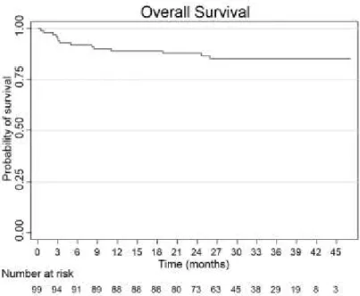
With a median follow-up of 31.7 months, the estimated probability of overall survival (OS) at 3 years is 85.3% in the entire cohort; in the DOX and NPLD subgroups the corresponding rates are 94.7% and 71.0%, respectively (p < 0.001). Restricting the analysis to patients with DLBCL, the 3 year-OS is 81.8% in the entire cohort; 92.3% and 71.8% in the DOX and NPLD subgroups, respectively (p = 0.08).
13
Figure 3: Overall survival according to type of anthracycline
Figure 4: Overall survival in the subgroup of patients with DLBCL, in the entire cohort (A) and according to type of anthracycline (B).
At last follow-up, 76 patients (24 treated with NPLD) are alive and in CR, of which 68 (23 treated with NPLD) are in continuous CR with no further treatments; 9 (4 treated with NPLD) patients are alive but not in CR: of these, 6 (3 treated with NPLD) are currently being treated with salvage treatments. Overall, 14 patients have died: 7 during induction treatment and 7 thereafter. Causes of death were: disease progression in 5 patients (2 treated with NPLD), cerebrovascular accidents
14
in 2 patients treated with NPLD, treatment-related in 4 patients treated with NPLD, cardiac disease in 3 patients treated with NPLD. Details of cardiac deaths are the following:
(1) A 67-year-old female patient with multiple cardiovascular comorbidities died of congestive heart failure after the 1st cycle with R-COMP for follicular lymphoma;
(2) A 75-year-old male patient with severe chronic obstructive pulmonary disease and chronic cor pulmonale died 15 months after completing chemotherapy for DLBCL (R-COMP) because of the worsening of congestive heart failure;
(3) A 49-year-old male with prosthetic aortic valve (he had underwent mediastinal radiotherapy 30 years before for malignant histiocytosis), achieved PR after 6 R-COMP cycles for high-risk DLBCL; while attaining mobilization for autologous transplantation, he had sudden death (likely due to myocardial infarction facilitated by leukocytosis).
CARDIAC TOXICITY: ECOCARDIOGRAPHIC EVALUATION
The primary endpoint of the study (reduced LVEF greater than 10% to a final value <50%), was evaluable in in 85 patients (28 treated with NPLD). Of the remaining 15 patients, 7 died during induction treatment, 3 died shortly after completing induction treatment, and 4 were unavailable to recall evaluations due to logistic difficulties: however, at last follow-up they did not report significant cardiac events and recent cardiac evaluations were normal. Two patients (2.4%) had LVEF reductions meeting the criteria for the primary endpoint.
(1) A 20-year-old female patient without cardiac risk factors had been treated for HD with ABVD followed by autologous transplantation because of resistant disease; at one follow-up visit she had a 14% absolute LVEF reduction (from 61% to 47%); she also had mild sinus tachycardia (105 bpm). Treatment was instituted with low doses of valsartan and bisoprolol, which were not tolerated due to hypotension; she was started on ivabradine and monitored monthly; 3 months after, the LVEF and the other cardiac abnormalities had completely recovered; 3 months after, ivabradine was stopped.
(2) A 51-year-old female patient without cardiac risk factors had been treated for DLBCL with 6 cycles of R-CHOP; 3 months after completing treatment, at one follow-up visit she had a 12% absolute LVEF reduction (from 58% to 46%); she reported mild dyspnea. She was
15
started on low doses of bisoprolol, and monitored monthly; 3 months after, the LVEF and the other cardiac abnormalities had completely recovered.
Five other patients developed other cardiac abnormalities without a significant LVEF reduction: in 3 patients (1 treated with NPLD) we found appearance or mild worsening of valvular diseases; one patient treated with DOX developed mild enlargement of the left ventricle; one patient treated with NPLD had marked and progressive worsening of a pre-existing right ventricular dysfunction, which ultimately led him to death.
The mean LVEF change from the baseline in the entire cohort was -1.25% (SD = 6.67, range -18 to +18; t-test for comparison between baseline and follow-up values: p = 0.09); it did not differ in the patients treated with DOX (-1.61%, SD = 6.61, range -16 to +14) or NPLD (-0.5%, SD 6.86, range -18 to +18; t-test for comparison between DOX and NPLD: p = 0.47). Overall, 8 patients had absolute LVEF reductions greater than 10%, 7 in the DOX subgroup (including the 2 patients meeting the primary endpoint) and 1 in the NPLD subgroup.
A complete echocardiographic evaluation, including measurements of global strain rate and tissue Doppler velocity performed both at baseline and at follow-up, was available in 73 patients (50 and 23 in the DOX and NPLD subgroups, respectively). There was a significant increase of the global strain rate (meaning reduced ventricular function) from the baseline in the entire cohort (mean change +1.2%, SD = 3.2, range -8.9 to +9.2, p = 0.003); also, there was a trend toward higher increases in the patients treated with DOX (+1.61%, SD = 2.8, range -8.9 to +7.3) with respect to patients treated with NPLD (-0.6%, SD 3.7, range -7.4 to +9.2; t-test for comparison between DOX and NPLD: p = 0.05).
As regards TDI evaluation, there was a significant decrease from baseline to follow-up values in the entire cohort (mean change -12.1 cm/sec, SD 20.8, range -70 to +30; t-test for comparison between baseline and follow-up values: p < 0.001); TDI reduction did not differ in the patients treated with DOX (-13.7%, SD = 21.6, range -70 to +30) or NPLD (-8.6%, SD 19.1, range -60 to +30; t-test for comparison between DOX and NPLD: p = 0.35).
16
CARDIAC TOXICITY: BIOMARKER EVALUATION
We performed a total of 1307 TnI measurements. Compliance to the protocol was excellent for the measurements planned before and 1h after chemotherapy (95%), but lower for those planned 48 hours after chemotherapy; to this end, logistic difficulties were the main reason of reduced compliance. Only 1.8% of TnI measurements resulted above 0.08 ng/ml; 9.6% above the 0.03 ng/ml.
There was a significant correlation between the cumulative dose of doxorubicin and the scale and frequency of TnI rises above the pre-specified cut-offs: mean TnI values at cumulative doxorubicin doses < and ≥ 200 mg/m2 were 0.013 and 0.023, respectively; TnI increases occurred more frequently at cumulative doxorubicin doses ≥ 200 mg/m2 considering both the cut-off of 0.03 ng/ml (3.6% and 20.7% at doses < and ≥ 200 mg/m2, respectively, p <0.001) and of 0.08 ng/ml (1.2% and 3.0% at doses < and ≥ 200 mg/m2, respectively, p <0.029); also, the frequency of TnI rises above 0.03 ng/ml had excellent linear correlation with the cumulative dose of doxorubicin (Fig. 5A; Pearson’s rho = 0.84; p = 0.019).
Thirteen patients (13%) developed a TnI increase above 0.08 ng/ml and 42 patients (42%) above 0.03 ng/ml. In two patients, the TnI rises reflected the occurrence of an asymptomatic acute coronary syndrome, detected by the telemedicine system:
(1) A 76-year-old male with hypertension, hyperlipidemia and history of CAD and previous endoarterectomy, underwent treatment with R-COMP for MCL. At the routine telemedicine evaluation before the 3rd chemotherapy cycle, he presented with diffuse
T-wave inversion and a mild TnI rise (0.09 ng/ml). He underwent coronary angiography which disclosed a severe stenosis of the left anterior descending artery which required percutaneous transluminal coronary angioplasty (PTCA) and positioning of a bare metal stent (BMS). After this intervention, the patient was able to complete the planned chemotherapy treatment.
(2) A 67-year-old male with atrial fibrillation, diabetes mellitus, hyperlipidemia, hypertension underwent treatment with MBVD for HD. TnI measurements after the 1st MBVD cycle showed a marked increase (up to 2 ng/ml). He underwent coronary angiography which disclosed a severe stenosis of the left circumflex artery which was treated with PTCA and positioning of a BMS. After this intervention, the patient was able to complete the planned chemotherapy treatment.
17
Overall, TnI increases above 0.03 ng/ml occurred more frequently in patients treated with NPLD (Fig. 5B and C; 11.9% and 8.3% in the NPLD and DOX subgroups, respectively; p = 0.034).
Figure 5: Biomarker evaluation: (A) relative frequency of the TnI rises above 0.03 ng/ml at different cumulative doxorubicin doses; (B) relative frequency of the TnI rises above 0.03 ng/ml according to the anthracycline type; (C) patients with TnI rises above 0.03 ng/ml according to the anthracycline type.
In DOX-treated patients we could observe the same excellent linear correlation between TnI rises above 0.03 ng/ml and the cumulative doxorubicin dose (pearson’s Rho = 0.86; p = 0.013). However, in the NPLD subgroup, the frequency of TnI rises above 0.03 ng/ml no longer retained this correlation (Fig. 6; Pearson’s rho = 0.69; p = 0.084). Thus, at cumulative doxorubicin doses ≤ 200 mg/m2, TnI rises above 0.03 ng/ml occur more frequently in the NPLD subgroup (9.3% and 0.6% in the NPLD and DOX subgroups, respectively; p <0.001); however, at doses > 200 mg/m2 the relationship is reversed, with higher frequency in the DOX subgroup (16.0% and 21.4% in the NPLD and DOX subgroups, respectively: p = 0.047).
18
Figure 6: Kinetics of the TnI rises at increasing cumulative doxorubicin doses: at cumulative doxorubicin doses < 200 mg/m2, TnI rises above 0.03 ng/ml occur more frequently in the NPLD subgroup (9.3% and 0.6% in the NPLD and DOX subgroups, respectively; p <0.001); however, at doses > 200 mg/m2 the relationship is reversed, with higher frequency in the DOX subgroup (16.0% and 21.4% in the NPLD and DOX subgroups, respectively: p = 0.047).
FEASIBILITY
Regarding feasibility, the average time to perform an echocardiogram was 7 minutes, while it took about 4 minutes on average by the cardiologist to produce a report. Echocardiograms were not evaluable only in 2.6% of cases.
19
DISCUSSION
Despite the recent advances of cancer treatment with the advent of targeted therapy, we are still a long way from abandoning chemotherapy in lymphoma. We are tempted to speculate that novel compounds will blow away the old cytotoxic drugs, but recent examples tell a different story. The introduction of the anti-CD20 monoclonal antibody Rituximab in the treatment of CD20+ NHL has clearly improved the survival of this disease [22]. However, at the current time, chemotherapy-free regimens are rarely employed in front-line settings and often restricted to relapsed and refractory patients who already failed several lines of chemotherapy; the vast majority of first-line regimens still provides the combination of a chemo-immunotherapy. This is particularly true in the setting of DLBCL, where the use of AC in the front-line seems undeniable and experimental protocols are trying to improve the outcomes by adding novel drugs to the old reliable CHOP backbone and not by substituting it [1,23].
The improvement in cancer therapeutics has also led to an apparent paradox in some situations: in HD, treatment-related deaths are exceeding those related to the underlying disease [24]. To cure more patients, modern oncology has undergone a paradigm shift: from the maximum tolerated dose we got to the minimum amount of chemotherapy needed to preserve the antitumor efficacy. While chemotherapy regimens have been constantly refined to reduce long-term toxicity, at the same time life expectancy has increased; furthermore new diagnostic techniques are now able to detect even minimal subclinical signs of organ damage that would have been ignored a few decades ago. Thus, it could happen that old-fashioned therapeutics which are indeed still very effective, look a lot more toxic than what they are nowadays. An example is radiotherapy in HD: newer radiation techniques are able both to retain efficacy and to minimize acute and late sequelae, but today we are still scared of the dramatic effect of old radiotherapy doses [25]. A similar situation may be encountered when dealing with AC. Although we are now using lower doses than in the past, we have improved diagnostic techniques, we have new cardioprotective strategies, the approach to AIC has remained quite the same in the last 40 years.
Recently, routine monitoring of TnI during chemotherapy has shown to detect cardiotoxicity in its earliest phase, long before any reduction in LVEF has occurred [26,27,28]. In those patients with a TnI rise, starting a cardioprotective treatment with ACE-inhibitors (enalapril) may prevent LVEF deterioration and other cardiac events [29]. The predictive value of TnI for AIC has been confirmed in various studies [30]. Moreover, monitoring with TnI during treatment with AC has
20
been recently recommended by the guidelines of the European Society of Medical Oncology to prevent from chemotherapy-induced cardiotoxicity [31].
Advanced echocardiographic techniques are currently explored to improve detection of subclinical signs of impaired ventricular function, which would be missed by conventional echocardiography [20]. Such novel approaches are under investigation also for chemotherapy-induced cardiotoxicity: a recent systematic review focused on the role of strain rate imaging for the early detection of myocardial changes and prediction of cardiotoxicity in patients receiving cancer therapy. The study found that, in late survivors of cancer, measures of global radial and circumferential strain are consistently abnormal, even in the context of normal LVEF; however, the authors concluded that the clinical value of global strain imaging in predicting subsequent ventricular dysfunction or heart failure has not been established [32].
Finally, the availability of new AC liposomal formulations opens up new ways to improve the therapeutic window of this class of drugs. To this end, the lymphoma setting has yet to be explored: the use of NPLD in Italy is approved for the treatment of aggressive NHL in elderly patients or in those with cardiac comorbidities (according to an extension of the approved indication [33]); however, yet there are no study comparing conventional and liposomal AC. Such data would certainly add valuable informations to guide the clinical decision between the two choices.
In our study we sought to take advantage of the recent technical, pharmacological and clinical advances in the field of cardiotoxicity to build a comprehensive strategy aimed to prevent, detect and treat AIC.
To our knowledge, our study is the first describing a direct comparison between AC and liposomal AC in lymphoma patients. Although this is not a randomized controlled trial but a prospective observational study, we can get a number of useful informations from the comparison between the subgroups of patients treated with DOX or NPLD. To this end, we should mention that the GRADE approach provides a methodological basis to upgrade the level of an evidence even if deriving from study designs assigned to a low quality of evidences (such as observational studies). The factors that can be considered to upgrade the quality of evidence are: a large effect size; dose response relations; residual confounding bias working to reduce the demonstrated effect. The latter reason may be used when all plausible biases from observational studies are working to underestimate an apparent treatment effect [34].
21
In our study, there were obvious imbalances in the baseline distribution of cardiac risk factors and comorbidities: the NPLD subgroup was unquestionably older and sicker than the DOX subgroup. Such inequality was intentional as it served to select patients at high risk for AIC, in order to allocate them to the less toxic treatment. However, evaluation of cardiotoxicity did not show an increased incidence of negative outcomes for the NPLD subgroup: the primary endpoint of the study (reduced LVEF greater than 10% to a final value <50%) occurred in only two patients treated with DOX; the mean LVEF and other echocardiographic endpoints (such as structural or valvular abnormalities, TDI measurements) did not differ significantly in the two subgroups. On the contrary, the global strain rate evaluated after the end of treatment was significantly more impaired in the DOX-treated patients with respect to the NPLD subgroup. The biomarkers data further suggest that NPLD may reduce subclinical damage in patients at high risk for AIC. Overall, TnI rises were significantly more frequent in patients treated with NPLD; this is not surprising, considering that the two populations had different baseline risk for AIC. This result is even clearer if we focus on low cumulative doxorubicin doses (p-value < 0.001 for TnI rises above 0.03 ng/ml with doses ≤ 200 mg/m2); however, at higher cumulative doxorubicin doses, the relationship was reversed, with more TnI rises in the DOX subgroup (p-value = 0.047 for TnI rises above 0.03 ng/ml with doses > 200 mg/m2).
As regards efficacy evaluation, the current study has the obvious limitation of including several lymphoma subtypes. However, in we focus on DLBCL, the largest homogenous NHL group (n = 52), we observed similar rates of complete response between DOX and NPLD subgroups. The NPLD subgroup had a trend toward lower overall survival (log-rank p = 0.08); however, it should be acknowledged that early treatment-related deaths (likely not attributable to reduced antitumor efficacy) had a significant impact on the survival curve of the NPLD subgroup. Baseline IPI was significantly higher in the NPLD subgroup (p = 0.049), likely resulting from the higher mean age in these patients.
In conclusion, we aimed to measure the prevalence of AIC in a setting of lymphoma patients where AC still constitute the mainstay of the 1st line treatment. We observed a very low incidence of AIC
(2.4%). Possible explanations for this finding include a spurious effect due to the relatively small sample size of our study; however, it is plausible that a comprehensive monitoring strategy compelling prompt initiation of a cardioprotective treatment may have reduced the occurrence of AIC. Beyond answering to an epidemiologic issue, our effort was also directed to improve detection of AIC. To this end, we used a comprehensive approach including echocardiography,
22
ECG and biomarkers. Such an intensive monitoring resulted feasible thanks to the integration in a telemedicine system.
23
REFERENCES
1. Luminari S, Montanini A, Federico M. Anthracyclines: a cornerstone in the management of non-Hodgkin's lymphoma. Hematol Rep. 2011 Oct 28;3(3s):e4. doi: 10.4081/hr.2011.s3.e4.
2. Di Marco A, Gaetani M, Scarpinato B. Adriamycin (NSC-123,127): a new antibiotic with antitumor activity. Cancer Chemother Rep 1969 Feb;53(1):33-7.
3. NCCN Clinical Practice Guidelines in Oncology (NCCN Guidelines®) Survivorship version 2.2015, available at http://www.nccn.org/professionals/physician_gls/pdf/survivorship.pdf, accessed Nov 7 2015
4. Smith LA, Cornelius VR, Plummer CJ, Levitt G, Verrill M, Canney P, Jones A. Cardiotoxicity of anthracycline agents for the treatment of cancer: systematic review and meta-analysis of randomised controlled trials. BMC Cancer. 2010 Jun 29;10:337. doi: 10.1186/1471-2407-10-337.
5. Von Hoff, D. D., M. W. Layard, et al. (1979). "Risk factors for doxorubicin-induced congestive heart failure." Ann Intern Med 91(5): 710-717.
6. Swain, S. M., F. S. Whaley, et al. (2003). "Congestive heart failure in patients treated with doxorubicin: a retrospective analysis of three trials." Cancer 97(11): 2869-2879.
7. Plana JC, Galderisi M, Barac A, Ewer MS, Ky B, Scherrer-Crosbie M, Ganame J,Sebag IA, Agler DA, Badano LP, Banchs J, Cardinale D, Carver J, Cerqueira M, DeCara JM, Edvardsen T, Flamm SD, Force T, Griffin BP, Jerusalem G, Liu JE, Magalhães A, Marwick T, Sanchez LY, Sicari R, Villarraga HR, Lancellotti P. Expert consensus for multimodality imaging evaluation of adult patients during and after cancer therapy: a report from the American Society of Echocardiography and the European Association of Cardiovascular Imaging. Eur Heart J Cardiovasc Imaging. 2014 Oct;15(10):1063-93. doi: 10.1093/ehjci/jeu192. PubMed PMID:25239940; PubMed Central PMCID: PMC4402366.
8. Hershman, D. L., R. B. McBride, et al. (2008). "Doxorubicin, cardiac risk factors, and cardiac toxicity in elderly patients with diffuse B-cell non-Hodgkin's lymphoma." J Clin Oncol 26(19): 3159-3165. 9. Prescribing informations for doxorubicin hydrochloride. Available at
http://www.bccancer.bc.ca/drug-database-site/Drug%20Index/ Doxorubicin_monograph
_1Apr2015.pdf, accessed 7 Nov 2015
10. Tebbi CK, London WB, Friedman D, Villaluna D, De Alarcon PA, Constine LS, et al. Dexrazoxane-associated risk for acute myeloid leukemia/myelodysplastic syndrome and other secondary malignancies in pediatric Hodgkin's disease. J Clin Oncol. 2007; 25:493-500
11. Available at http://www.fda.gov/Drugs/DrugSafety/ ucm263729.htm; accessed 7 Nov 2015. 12. Mross, K., B. Niemann, et al. (2004). "Pharmacokinetics of liposomal doxorubicin (TLC-D99;
Myocet) in patients with solid tumors: an open-label, single-dose study." Cancer Chemother Pharmacol 54(6): 514-524.
13. van Dalen, E. C., E. M. Michiels, et al. (2010). "Different anthracycline derivates for reducing cardiotoxicity in cancer patients." Cochrane Database Syst Rev(5): CD005006.
14. Luminari S, Montanini A, Caballero D, Bologna S, Notter M, Dyer MJ, Chiappella A, Briones J, Petrini M, Barbato A, Kayitalire L, Federico M Nonpegylated liposomal doxorubicin (MyocetTM) combination (R-COMP) chemotherapy in elderly patients with diffuse large B-cell lymphoma (DLBCL): results from the phase II EUR018 trial. Ann Oncol. 2010 Jul;21(7):1492-9. doi: 10.1093/annonc/mdp544. Epub 2009 Dec 11.
15. Visani G, Ferrara F, Alesiani F, et al. R-COMP 21 for frail elderly patients with aggressive B-cell non-Hodgkin lymphoma: a pilot study. Leuk Lymphoma 2008; 49:1081-6.
24
16. The SOLVD Investigators. Effect of enalapril on mortality and the development of heart failure in asymptomatic patients with reduced left ventricular ejection frations. N Engl J Med 1992; 327(10): 685-691.
17. Cardinale D, Colombo A, Bacchiani G, Tedeschi I, Meroni CA, Veglia F, Civelli M, Lamantia G, Colombo N, Curigliano G, Fiorentini C, Cipolla CM. Early detection of anthracycline cardiotoxicity and improvement with heart failure therapy. Circulation. 2015 Jun 2;131(22):1981-8. doi: 10.1161/CIRCULATIONAHA.114.013777. Epub 2015 May 6. PubMed PMID: 25948532;131(22):1981-8. 18. Cardinale, D., M. T. Sandri, et al. (2004). "Prognostic value of troponin I in cardiac risk stratification
of cancer patients undergoing high-dose chemotherapy." Circulation 109(22): 2749-2754
19. Gardin JM, Adams DB, Douglas PS, Feigenbaum H, Forst DH, Fraser AG, Grayburn PA, Katz AS, Keller AM, Kerber RE, Khandheria BK, Klein AL, Lang RM, Pierard LA, Quinones MA, Schnittger I; American Society of Echocardiography. Recommendations for a standardized report for adult transthoracic echocardiography: a report from the American Society of Echocardiography's Nomenclature and Standards Committee and Task Force for a Standardized Echocardiography Report. J Am Soc Echocardiogr. 2002 Mar;15(3):275-90. PubMed PMID: 11875394.
20. Dandel, M., Lehmkuhl, H., Knosalla, C., Suramelashvili, N., & Hetzer, R. (2009). Strain and strain rate imaging by echocardiography–basic concepts and clinical applicability. Current cardiology reviews, 5(2), 133.
21. Cheson BD, Pfistner B, Juweid ME, Gascoyne RD, Specht L, Horning SJ, Coiffier B, Fisher RI, Hagenbeek A, Zucca E, Rosen ST, Stroobants S, Lister TA, Hoppe RT, Dreyling M, Tobinai K, Vose JM, Connors JM, Federico M, Diehl V; International Harmonization Project on Lymphoma. Revised response criteria for malignant lymphoma. J Clin Oncol. 2007 Feb 10;25(5):579-86. 22. Molina A. A decade of rituximab: improving survival outcomes in non-Hodgkin's lymphoma.
Annu Rev Med. 2008;59:237-50.
23. Vitolo U, Chiappella A, Franceschetti S, Carella AM, Baldi I, Inghirami G, Spina M, Pavone V, Ladetto M, Liberati AM, Molinari AL, Zinzani P, Salvi F, Fattori PP, Zaccaria A, Dreyling M, Botto B, Castellino A, Congiu A, Gaudiano M, Zanni M, Ciccone G, Gaidano G, Rossi G; Fondazione Italiana Linfomi. Lenalidomide plus R-CHOP21 in elderly patients with untreated diffuse large B-cell lymphoma: results of the REAL07 open-label, multicentre, phase 2 trial. Lancet Oncol. 2014 Jun;15(7):730-7.
24. Aleman BM, van den Belt-Dusebout AW, De Bruin ML, van 't Veer MB, Baaijens MH, de Boer JP, Hart AA, Klokman WJ, Kuenen MA, Ouwens GM, Bartelink H, van Leeuwen FE. Late cardiotoxicity after treatment for Hodgkin lymphoma. Blood. 2007 Mar 1;109(5):1878-86. 25. Specht L, Yahalom J, Illidge T, Berthelsen AK, Constine LS, Eich HT, Girinsky T, Hoppe RT,
Mauch P, Mikhaeel NG, Ng A; ILROG. Modern radiation therapy for Hodgkin lymphoma: field and dose guidelines from the international lymphoma radiation oncology group (ILROG). Int J Radiat Oncol Biol Phys. 2014 Jul 15;89(4):854-62.
26. Cardinale, D., M. T. Sandri, et al. (2000). "Left ventricular dysfunction predicted by early troponin I release after high-dose chemotherapy." J Am Coll Cardiol 36(2): 517-522.
27. Sandri, M. T., D. Cardinale, et al. (2003). "Minor increases in plasma troponin I predict decreased left ventricular ejection fraction after high-dose chemotherapy." Clin Chem 49(2): 248-252. 28. Cardinale, D., M. T. Sandri, et al. (2004). "Prognostic value of troponin I in cardiac risk
stratification of cancer patients undergoing high-dose chemotherapy." Circulation 109(22): 2749-2754
29. Cardinale, D., A. Colombo, et al. (2006). "Prevention of high-dose chemotherapy-induced cardiotoxicity in high-risk patients by angiotensin-converting enzyme inhibition." Circulation 114(23): 2474-2481.
25
30. Cardinale, D., A. Colombo, et al. (2010). "Anthracycline-induced cardiomyopathy: clinical relevance and response to pharmacologic therapy." J Am Coll Cardiol 55(3): 213-220.
31. Curigliano G, Cardinale D, Suter T, Plataniotis G, de Azambuja E, Sandri MT, Criscitiello C, Goldhirsch A, Cipolla C, Roila F; ESMO Guidelines Working Group. Cardiovascular toxicity induced by chemotherapy, targeted agents and radiotherapy: ESMO Clinical Practice Guidelines. Ann Oncol. 2012 Oct;23 Suppl 7:vii155-66
32. Thavendiranathan P, Poulin F, Lim KD, Plana JC, Woo A, Marwick TH. Use of myocardial strain imaging by echocardiography for the early detection of cardiotoxicity in patients during and after cancer chemotherapy: a systematic review. J Am Coll Cardiol. 2014 Jul 1;63(25 Pt A):2751-68. doi:10.1016/j.jacc.2014.01.073.
33. Gazzetta Ufficiale della Repubblica Italiana del 23/05/2011; 152(118): 114
34. Guyatt GH, Oxman AD, Vist GE, Kunz R, Falck-Ytter Y, Alonso-Coello P, Schünemann HJ, for the GRADE Working Group. GRADE: an emerging consensus on rating quality of evidence and strength of recommendations. BMJ 2008 Apr 26;336(7650):924-926.