1
UNIVERSITA’ DEGLI STUDI DI NAPOLI “FEDERICO II”
Department of Chemical, Materials and Industrial Production Engineering
POLYTECHNIC AND BASIC SCIENCES SCHOOL
PhD in “Industrial Product and Process Engineering”
XXXI cycle
Photo-responsive oil core based polymer nanocapsules
Supervision:
PhD student:
Prof. Dr. Paolo Antonio Netti Chiara Di Cicco
Advisor:
Dr. Ing. Raffaele Vecchione
Coordinator:
Prof. Giuseppe Mensitieri
2
Table of Contents.
Chapter 1 ‘State of art’………..……….4
1.1 Nanomedicine: O/W drug delivery systems………....5
1.2 Endogenous and exogenous stimuli-responsive nanocarriers……….9
1.3 The o-nitrobenzyl ether as photocleavable linker………..14
1.4 Nanotechnology for nutraceuticals………...19
1.5 Ferric oxide nanocubes as contrast agents in MRI………...…..22
1.6 Brief mention on nano-‘theranostic’ delivery systems………..24
1.7 Aim of the PhD project………..26
1.8 References………28
Chapter 2 ‘Photo-responsive corr-linked drug delivery systems based on Layer by Layer oil-in-water nanoemulsions’………..31
Abstract………...32
2.1 Introduction………...32
2.2 Materials and Methods………..34
2.3 Results and Discussions………....45
2.4 Conclusions………...59
2.5 References……….…60
Chapter 3 ‘Cardioprotective effects of nanoemulsions loaded with anti-inflammatory nutraceuticals against doxorubicin-induced cardiotoxicity’……….…62
Abstract………...…63
3.1 Introduction………...63
3.2 Materials and Methods………..64
3.3 Results………...71
3.4 Discussions………...83
3.5 Conclusions………...84
3.6 References……….86
Chapter 4 ‘Oil core-PEG shell nanocarrier for in vivo MRI imaging’………..89
Abstract………...90
3
4.2 Results and Discussions………....91
4.3 Conclusions………...102
4.4 Materials and Methods………102
4.5 Supporting Information………...109
4.6 References………...113
Appendix ‘Future perspectives using a molecular formulation of Fe(III) in O/W nanoemulsion for MRI application’………...115
4
Chapter 1
5
1.1 Nanomedicine: O/W drug delivery systems
The application of nanotechnology to clinical treatments brings to the birth of nanomedicine. Nanomedicine’s main aims are early diagnostics and targeted therapies in several human diseases, such as cancer therapy.1 The fusion between nanotechnology and biomedicine developed novel
materials with highly specific and unique therapeutic properties. Compared to traditional therapies, nanotechnology devices offer new advantages, such as targeted delivery, controlled drug release, and improvement of cell barrier crossing.2,3
Over the past decades, nanotechnology world, engineering nano-scaled devices, has led to the development of a lot of particle systems, such as gold nanoparticles (NPs), silica NPs, liposomes, and polymeric NPs. Drug delivery systems’ aim is to obtain a particle design to improve drug specificity to the desired site of action, so higher efficacy and fewer adverse effects.4 In fact, literature strongly
underlines that the use of polymeric nanocarriers to transport active compounds like small-molecular drugs, peptides, or genes found an increased attention throughout the different fields of natural sciences. Not only these nanocarriers enhance the properties of already existing drugs in terms of solubility, bioavailability, and prolonged circulation times, furthermore they can be tailor-made in such a manner that they selectively release their cargo at the desired site of action.3,5 It is known that
thanks to drugs’ encapsulation, we can protect not only the cargo but also the living cells from the possible toxic effect of cargo. So we insist on the fact that nanomedicine could contribute with appropriated biocompatible multifunctional systems, to avoid these unwished effects. Structured nano-devices are able to navigate into living cells and can be activated for the local release of their cargo, increasing drug’s bioavailability too.2,6
In the early 1990s, Decher and co-workers were the first to develop Layer-by-Layer (LbL) thin films (Figure 1.1).1 Their protocol consists in the preparation of multilayer ultrathin organic films by
alternated deposition of oppositely charged polyelectrolytes on a charged solid subrate. So this technique is based on the electrostatic attractions polyanions and polycations. One of the advantages of LbL films is that the film thickness can be precisely tuned at the nanometer level by changing the number of deposited layers.2,4,7 In 1998, LbL technique was transferred from planar films to spherical
colloidal particles by Donath and co-workers, referring to the strategy on planar substrate.1 Since their
introduction in 1998, LbL capsules have obtained an increasing interest, largely because of the possibility to tailor their properties, such as size, composition, porosity, stability, surface functionality, colloidal stability (Figure 1.2). In addition, the LbL technique has a special protocol which enables to engineer new materials introducing unprecedented structure and function.3 The
variety of templates and materials has given rise to an ever-expanding catalogue of LbL-assembled multi-layered particles varying in sizes, shapes, and chemical compositions. A main feature of the
6
LbL technique is the precision of the final particles. Thanks to such versatility, multilayered particles have become a powerful platform for drug encapsulation and triggered drug release.4
Figure 1.1. (A) Schematic of the film deposition process. Steps 1 and 3 represent the adsorption of a polyanion and polycation, respectively, and steps 2 and 4 are washing steps. The four steps are the basic sequence for the simplest film architecture. (B) Simplified molecular picture “model” of the first two adsorption steps, starting with a positively charged substrate. Counter ions are omitted for clarity.8
The LbL NPs were first reported by Caruso and his group. In their protocol they prepared via LbL some polyelectrolyte nanocapsules around a solid template, when obtained monodisperse emulsions by filling them with oil.9,10 So more in general, LbL technology is about polymeric nanocapsules and
takes advantage of the charge–charge interaction between substrate and monolayers of polyelectrolytes to create multiple layered nano-architecture held together by electrostatic forces. The formation of LbL systems are attributed to electrostatic interactions, hydrogen bonding, hydrophobic interactions and van der Waals forces.5
7
In 2014, our team developed biodegradable oil-in-water (O/W) nano-emulsions coated with a molecular layer of biodegradable polyelectrolytes. In dependence of the polymer concentration, they controlled the level of coating that resulted in a tunable stability. Our O/W nano-emulsions consists of a biocompatible formulation: small lipidic droplets with soybean oil and Lipoid E80 lecithin dispersed in an aqueous medium. They have the ability to dissolve large quantities of hydrophobic drugs and protect their cargo from hydrolysis and enzymatic degradation. The stability over time and good behaviour of these systems, strictly connected to their monodisperisity, were obtained using a high-pressure homogenizer.10 In 2016, a protocol to obtain optimized liquid–liquid interfaces capable
to sustain multiple depositions of biodegradable polymers has also been developed in our lab. With this protocol, it was possible to perform around the oil core of the nanoemulsions, progressive depositions of the natural polymers shells via the classical LbL technique but with control almost at the molecular scale (Figure 1.3).12
Figure 1.3. Example of controlled deposition of polymeric multilayer film on oil core based nanocapsules.12 In drug delivery field, from a circulation point of view, another important point, respected by our formulations, is that the ideal size of drug carriers is in a range below 200 nm.6,10 In fact, bigger
capsules can obstruct the smallest blood capillaries and cannot reach the specific tissue.6 On the other
hand, the selective delivery of a drug loaded nanocapsule through systemic administration routes is often attributed to passive targeting via the “enhanced permeability and retention” (EPR) effect which is mainly a molecular size-based phenomenon (Figure 1.4). This EPR effect is the basis for development of macromolecular anticancer therapy which was first introduced by Matsumura and Maeda in 1986. They showed that most solid tumors have blood vessels with defective architecture. This effect is attributed to poor tumor vascularization and lymphatic drainage. So most solid tumors exhibit enhanced vascular permeability, which will ensure a sufficient supply of nutrients and oxygen to tumor tissues for rapid growth. The EPR effect is a unique anatomical–pathophysiological nature
8
of tumor blood vessels that facilitates transport of macromolecules into tumor tissues. In this way the nanocapsules manage to accumulate themselves within the tumor tissue. In contrast, this EPR effect-driven drug delivery does not occur in normal tissues.4,13
In addition to passive targeting, recent studies have demonstrated that cell selectivity in vitro can be improved conjugating targeting ligands to multilayer nanoparticles. This is called “active” targeting, because to promote specific interaction, nanocapsules can be decorated with functional ligands which recognize the tumor tissue, to delivery and release their drug selectively. This strategy can be an effective approach to complement the EPR effect.4 More in general, surface modifications are helpful
not only to deliver the nanocapsules in specific sites but also to prevent the body from recognizing the capsules as foreign material.3
Figure 1.4. Targeting of nanomedicines by the enhanced permeability and retention (EPR) effect. Notes: (A) normal and (B) tumor vessels.14
For all said above, LbL polyelectrolytes deposition on nanoemulsion has emerged as one of the best techniques to deposit ultrathin polymer layers. However, it is known that electrostatic interactions are weak and easily breakable in physiological environment.15 To improve emulsion stability a strategy
is the cross-linking between the polymer shells by click chemistry.10,16 In a previous work of our
group, it has been shown that the stability of biopolymer multilayers (glycol chitosan and heparin) obtained via layer by layer can be enhanced introducing a covalent bonds among layers by means of a metal catalyst-free, light initiated ‘click’ reaction, namely the thiol-ene reaction. In particular, the reaction involves a thiol moiety on glycol chitosan and an allylic moiety on heparin. Interestingly, the reaction resulted effective even without a photoinitiator, mainly due to the high degree of
9
modification of the biopolymers, proving a completely non-cytotoxic final product (Figure 1.5). Cross-linked multilayers resulted in more stable systems than those stabilized with just electrostatic forces. In addition, the application of this approach to biopolymers surrounding an O/W nanoemulsion demonstrated that thiol-ene cross-linkage can be nicely exploited in the field of completely biocompatible and biodegradable nanocarriers.15
Figure 1.5. Thiol-ene reaction between allylated heparin and thiolated glycol chitosan. Reaction was triggered throughout UV irradiation.15
1.2 Endogenous and exogenous stimuli-responsive nanocarriers.
Conventional chemotherapy is far from successful because of the lack of tumor selectivity, or toxicity of the drugs. So it is of extreme importance the development of highly selective systems for tumors, to reduce drug toxicity and enhance the therapeutic purpose.13,17 We have just discussed above about
the EPR effect-based drug delivery connected to pathophysiological heterogeneity of the tumors. However, EPR effect is absent in the central area of metastatic cancers, and so there the accumulation is lower than in other parts.13
Considering the difference between normal and pathological tissues, the design of stimuli-responsive nanocarriers is a promised approach for the future to improve the therapeutic effects in the tumor tissues and reduce the undesired ones in the healthy cells. The use of stimuli-responsive drug delivery nanocapsules could improve the cellular uptake and intracellular stability. According to the diagnostic or therapeutic purpose, these systems can include different organic and inorganic materials, such as polymers, liposomes, nanoemulsions, dendrimers and so on.18
In the 1970s stimuli-responsive drug delivery was introduced with the use of thermosensitive liposomes for a controlled release of drugs through hyperthermia. Since then, stimuli-responsive nanocarriers for drug delivery became of particular interest.19
About stimuli-responsive nanocarriers, Keun Sang Oh et.al. were the first to introduce LbL nanocarrier for cancer-targeting. First of all, the LbL approach improves the target ability by
10
prolonging the nanocarries systemic circulation.20 LbL nanocapsules surface can be modified with
the aim to prevent the recognition of these materials as foreign for the body and to deliver them to specific tissue by targeting. The application of micro-/nanocapsules as drug delivery vehicles shows great promise.3
The easy of the preparation process and the variety of applications in drug delivery have recognized the stimuli-sensitive LbL systems as a promising approach .11
Typically, stimuli-responsive mechanisms to release the drugs can be of two types (Figure 1.6): endogenous stimuli (e.g., pH, enzymes, redox potential, glucose concentration, ions) or exogenous stimuli (temperature, magnetic fields, electrical fields, ultrasound and light). The choice of release mechanism is connected to the desired application. Intracellular mechanisms are the basis of the endogenous stimuli mainly used to release the encapsulated drugs from biodegradable coated nanocarriers. Indeed, exogenous stimuli are mainly used for a remoted control of the opening of the nanocarries with external source.1,3,6 Among the variety of nanocarriers, polyelectrolytes LbL systems
is a powerful method because of their versatility in shape, size, modulated swelling capacity.21
Figure 1.6. On top: General scheme of a stimuli-responsive nanocarrier for the transport of active compounds. On bottom: General scheme of stimuli-responsive release of a drug from a nanocarrier.5
Before introducing the nucleus of this PhD project, which is based on a photo-responsive LbL O/W nanoemulsion, we carry on a brief summary of the different stimuli.
11
pH stimuli-responsive systems. In these systems the triggering of the drugs is based on the different
pH gradient between the tumor tissues (pH 5.7-6.8) and the extracellular environment (pH 7.4), and the acid pH of endosomal and lysosomal compartments.18,22 So pH stimuli-responsive systems are
based on pH sensitive polymeric materials. The change in pH environment lead to some changes in nanocapsules’ permeability with the followed release of the drugs.20 Typical pH-stimuli responsive
polymers reported in literature include poly(acrylamide) (PAM), poly(acrylic acid) (PAA),
poly(methacrylicacid) (PMAA), poly(diethylaminoethylmethacrylate) (PDEAEMA),
poly(dimethylaminoethyl methacrylate) (PDMAEMA), polymers with phosphoric acid derivates, and some natural polymers such as albumin and gelatin.17 An example for pH-responsive nanocarriers
was reported by Liang et al. In their studies the capsules were based on poly(2-diisopropylaminoethyl methacrylate) (PDPA). They combined PDPA with a minor component of lauryl methacrylate (PDPAC12). In this way they obtained the endocytic pH-induced degradation of their capsules. Their
multilayers containing C12 groups in the PDPA increased the overall hydrophobicity, and so a non-covalent stabilization of the multilayers at pH 7.4. On the other hand, the capsules degraded for the cellular pH shift.23
Redox potential stimuli-responsive systems. Between the extracellular and intracellular compartments
there is a different redox potential. In fact, the extracellular space is oxidative while the intracellular is reductive. This is due to the different concentration of glutathione (GSH) in these two compartments (~2–10 µM in the extracellular part and ~2–10 mM in the intracellular one) and in tumor tissues as compared to healthy ones.5,18,19 So the release of the drug in the cytosol can be
triggered by this different redox potential.19 Since disulphide bonds can be cleavaged by reducing
agent (GSH) and the thiol groups can return to disulphide by oxidation, it is possible to built redox-sensitive carriers by incorporating disulphide bonds which release the drug as a consequence of the redox potential triggering.24 An example of redox potential stimuli-responsive nanocarrier was
reported by Lee and co-workers. Their synthesized biocompatible polymer micelles with disulfide bonds in their shells responded to the typical reduced conditions of the cancer environment.25
Enzyme stimuli-responsive systems. In pathological states, such as cancer or inflammation, the
expression of vary enzymes (e.g. protease, phospholipases or glycosidases) is altered. So in those sites, it is possible to design drug delivery systems acting via specific enzyme-mediation.19,24 For
example, literature studies demonstrated that esterases or proteases can promote the cleavage of esters or short peptide sequences contained in the enzyme-responsive nanocarriers.5 Other examples of
enzymes used for trigger are proteases, glucuronidase, or carboxylesterases, which are expressed differentially in the normal and tumor cells. To promote recognition of the nanocarriers by these
12
enzymes, specific enzyme substrates are encapsulated into the nanocarriers or linked to external segments containing the drugs and bonded to the nanocarriers.5
Ion stimuli-responsive systems. In the case that there are coordinative bonds between the nanocarrier
and the guest, it is possible to trigger the release by competitive ligands or ions. For example, in literature, it was reported that micelles based on PEG-PAsp and PEG-PGlu and loaded with cis platin coordinated to the carboxyl groups in the core of the micelle, did not show release in absence of competitive ligands. Instead, in a phosphate buffered saline, 50% of cis platin was released after 24 h in the case of PEG-PAsp based micelles and after 72 h in the case of PEG-PGlu ones, this because chloride ions in the buffer were favorited ligands for cis platin than carboxyl groups.5
Glucose stimuli-responsive systems. The introduction of enzymes such as catalase (CAT) and glucose
oxidase (GOx) in the nanocapsules brings to the development of glucose stimuli-responsive systems. Generally, as reported by Qi et. al., these nanocarriers were loaded with insulin and were assembled by LbL depositions. Their shells contained imine bonds. The treatment with a glucose solution caused the conversion of glucose to gluconic acid. In fact, Qi et al.in 2009 noticed that CAT/GOD in the shells interaction with the glucose solution was responsible of a pH reduction in the microenvironment, connected to H+ production. In that acid conditions there was the cleavage of the
imine bonds, an increase of capsules’ permeability and so insulin release.5,20
Thermo stimuli-responsive systems. A thermosensitive polymer is characterized by a lower critical
solution temperature (LCST) in aqueous solution. Such polymer is water soluble below its LCST and becomes water insoluble above that temperature.18 The behaviour of thermosensitive polymers is
generated by the balance of hydrophilic and hydrophobic moieties constituted the amphiphilic polymer chain. It is known that there is a higher temperature of 1-2°C in the tumor microenvironment than that of normal tissue. So when the thermos-stimuli-responsive nanocarrier is in the tumor tissue, its thermal responsive polymers change their phase form into a hydrophobic collapsed one with the consequent release of the drug.24 For the past 20 years the study of hyperthermia to improve radiation
or chemotherapy therapies, has grown. In addition, tumor cells exhibit more sensibility to heat-induced damage than normal cells. So for example, in different clinical studies, liposomes and nanoparticles containing iron oxide particles were used to localize the tumor tissue by the magnetic properties of the iron oxide and to heat the specific tissue. Another advantage of the thermo responsive drug delivery systems is the creation of micro-emboli in the tumor vasculature, the lower intake of oxygen and nutrients into the tumor mass.18
Magneto stimuli-responsive systems. A magnetic field can have different uses and provide different
applications. A permanent magnetic field can be a magnetic guidance, an alternating magnetic field can increase temperature, or it is possible to have both the effects when both the strategies are
13
alternately used. So the same magnetic stimuli-responsive drug delivery system can be used for diagnostics and therapy within a single system (the so-called theranostic approach).19 For example,
in literature studies the magnetic control of permeability of the nanocapsules was obtained by the introduction of ferromagnetic gold-coated cobalt in nanocarriers’ shells. Applying an oscillating magnetic fields, the rotation of the embedded nanoparticles caused the distortion of the nanocapsules’ structure and so an increase of their permeability. In addition, for nanocapsules based on polyelectrolytes, lipid bilayers, and magnetic nanoparticles, a local heating could be induced by the magnetic field. This changes the membranes phase and brings to an easier release of the encapsulated drugs.26 Lu et al. study is an example of magneto responsive drug delivery systems based on LbL
assembly, where the magnetic field changed the permeability of the polyelectrolyte shells.17 Hu et al.
studied LbL nanocapsules containing magnetic nanoparticles in their shells under a high-frequency magnetic field to trigger the release of the drug. They demonstrated that the magnetic field proved nanocavities in the shells and consequently the nanocapsules rupture.11
Electrical stimuli-responsive systems. When an electrical stimulus is applied to a charged drug or to
polyelectrolyte multilayers, an influx of counterions and solvent molecule takes place. These influxes increase the osmotic pressure between the shells and so bring to a volumetric expansion. The osmotic pressure is balanced by polymers’ deformation and swelling.11,20 Electro-sensitive polymers are
generally polyelectrolytes. Only few natural polymers show this behaviour in non-conducting media. Typically in such systems it is necessary the functionalization with charged or polarizable component which respond to an electric field. For example, a lightly cross-linked poly(dimethyl siloxane) gel containing electrosensitive colloidal TiO2 particles was synthesized by Zrinyi and co-workers. TiO2
particles were trapped in the matrix. So the gel deformation was obtained by the electrical force acting on these particles which was transferred to the polymer.25
Photo stimuli-responsive systems. Photo-responsive polymers present specific functional groups
which are light sensitive. So upon light irradiation of appropriate wavelength, these polymers change their structural properties. An important aspect is that the use of light is a non-invasive mechanism. Some examples of photo stimuli-responsive groups incorporated into polymer structures are azobenzene, spiropyran, cinnamate, which present a light-induced isomerization behaviour. Other groups have a photo-labile behaviour, i.e. light cleavages them changing their polarity, such as some photo-labile esters.25 Also metal nanoparticles, such as silver, platinum, gold and gold sulfide or
magnetite are incorporated within polyelectrolyte multilayer capsule walls. These nanoparticles absorb light and dissipate adsorbed energy as heat. This behaviour causes irreversible changes in the shells integrity/permeability and capsules’ shell rupture.3,6,26 An example of photo stimuli-responsive
14
components introduced into the polyelectrolyte shells. Either Ag nanoparticles or IR dyes adsorb light. Under laser illumination with a low-power near-infrared continuous-wave diode, the capsules containing Ag nanoparticles or IR dye were deformed or cut. In this way, with a remote control, there was the release of the encapsulated materials.27 Being our systems based on a photo-stimuli linker, in
the next paragraph we are going to deepen more this typology of stimuli-responsive nanocarriers.
Ultrasound stimuli-responsive systems. Ultrasound application in drug delivery is highly
advantageous because it is non-invasive and has a good penetrating deep into the body. Moreover it can be focused and controlled by frequency, power, density, time of application and duty cycles.17
Ultrasound mechanism is connected to the cavitation phenomena, which is activated by ultrasound. Cavitation is the alternating growth and shrinkage of microbubbles that means alternation of high and low pressure waves. When these microbubbles implode, generate local waves that perturb and can disrupt the polymeric shells of the capsules. Typical ultrasound stimuli-responsive polymeric systems are generally based on gels, polymeric micelles, LbL coated microbubbles. Moreover, cavitation and microbubble implosion can mediate enhancement of vessel permeability to facilitate the nanocarriers’ encapsulation. This process is known as “sonoporation”.19,25 An example of ultrasound
stimuli-responsive systems by Nelson et al. was based on micelles which released doxorubicin (Dox) upon application of low-frequency ultrasound in an in vivo rat model of a colon carcinoma. They demonstrated that ultrasound induced a selective release of doxorubicin at the tumor site providing a reduction of the tumor volume as compared to the noninsonated tumors.28
1.3 The o-nitrobenzyl ether as photocleavable linker.
The world of photo-cleavable linker and the research in this field have found a great interest in these years in particular when applied to drug delivery systems. Now, we wish to introduce the photo-cleavable linker which has been adopted in this project, but before, by appropriate examples, we want to explain better the world of trigger molecules and systems describing the variety of photo-responsive linkers, their versatility, their fields of application and their advantages as shown in literatures.
First of all, as just said previously, the use of an external stimulus on drug delivery systems brings benefits such as protection of the drugs until they enter into the desired site and reduction of side effects. Among the external stimuli, light is of particular interest for its clinical relevance and the advantage to ensure a spatio-temporal control that enhance efficiency and minimize toxicity. The light-tissue interactions are mainly scattering and absorption, whose attenuate surface power density of the light beam. The attenuation coefficient and the penetration depth of light are inversely proportional where the attenuation coefficient depend from the absorption and scattering coefficients,
15
i.e. from the light wavelength and the type of tissue. Using NIR, penetration depth of light is in the order of centimetre scale. The penetration depth decreases going towards visible and UV light. Phototoxicity to tissues is connected to photochemical reaction or photothermal effects which are involved by the emission phenomena that rise when a tissue is irradiated. The maximum permissible exposure (MPE) depends on wavelengths and irradiation time. In general, even a photon of light can carry out a reaction through photo-responsive nanoparticles. Example of these photochemical reactions are: photo-isomerization, photo-cleavage, photo-crosslinking (Figure 1.7). These photochemical reactions can only be initiated by UV/vis radiation because of their higher energy but suffers from disadvantages like lower penetrating power. While lower wavelength radiations in NIR region have deeper penetration capabilities but are not energetic enough to initiate the reaction. The use of two-photon absorption is a strategy to combine the deeper penetration power of NIR and the high energy of UV/vis radiation to trigger chemical reaction. Photresponsive groups, such as o-nitrobenzyl and coumarin, absorb two NIR photons which are converted in single UV/vis photons.29,30
This could be a good opportunity since there are very few organic chromophores which absorb in NIR region, while the majority just responds to UV light.31
Figure 1.7. Examples of responsive groups used in photochemical reactions, such as cleavage, photo-isomerization, photo-induced rearrangement, and photo-crosslinking.29
Yan et al. demonstrated the multiphoton effect of core-shell lanthanide-doped upconverting nanoparticles (UCNPs) in a photosensitive hybrid hydrogel of biomedical interest. The UV light generated by the UCNPs induces the photo-cleavage of a photolinker present in the structure of the hydrogel, the o-nitrobenzyl moieties.32,33Another example of photodegradable hydrogels is formed
16
by the self-assembly of short peptides modified with a photo-responsive biaryl-substituted tetrazole moiety (Figure 1.8). This moiety, upon mild light irradiation, forms a fluorescent pyrazioline by a rapid intramolecular photoclick ligation. This photo intramolecular transformation, disturbing the system interaction, induces the disassembly of the hydrogel.34
Figure 1.8. (A) Components of the photo-responsive hydrogel. (B) Photo-response mechanism.34
Also inorganic materials are used for photo-based applications, such as titanium oxide nanotubes, calcium phosphosilicate, nanoscintillators, copper selenide nanoparticles and silica-cored gold nanoparticles.30 For example, a photo-responsive protein delivery system, studied by Luo and
co-workers, used TiO2 nanoparticles containing hydroxyl. They covalently attached hemoglobin onto
the surface of TiO2 nanoparticles by 3,4-dihydroxyl benzoic acid (DB). Visible light triggered the
controlled release of Hb by the cleavage of the coordination bonds between DB and TiO2 surfaces24
Another example of photo-responsive linker used in drug delivery system was the perylene-3-ylmethanol (Figure 1.9). In biological studies, this linker contained in chlorambucil, nanoparticles exhibited good biocompatibility, cellular uptake and it was efficient in photo-responsive anticancer therapy. Perylene-3-ylmethyl is an efficient fluorescent photo-trigger for carboxylic acids and alcohols in aqueous media under visible light.35
17
In literature, they are also shown photo-responsive nano drug delivery system based on fluorescent carbon dots and a photo-responsive 7-methoxy quinoline moiety. In a work on HeLa cells, carbon dots were used for their fluorescence in vitro cellular imaging application, while quinolone for controlled photo-induced anticancer drug release upon both one-photon and two-photon excitation.36
Some studies report examples of photo-responsive polymers introduced into nanocarriers. An examples is the bis(4-dimethylamino)phenylmethyl leucocyanide. Under UV irradiation it dissociates into ion pairs producing triphenyl methyl cations. The introduction of this photo-responsive linker in hydrogels brought to a swelling into the hydrogel via UV irradiation due to the formation of the cyanide ions after UV irradiation increasing the osmotic pressure within the gel.17
A widely used photo-responsive linker for its photo-isomerization property is azobene. Its trans-cis photo-triggered transition opens the possibility of a controlled release of the molecules encapsulated in the nanocarrier that contains this photo-linker. Trans-cis isomerization is activated by UV irradiation while the return to trans form is originated by visible light irradiation or heating.37
Azobene was used, for example, in UV-light responsive cross-linked polyamide nanocapsules obtained by O/W miniemulsion interfacial polycondensation. In this study, it was shown that the smaller the capsule size, the faster the achieved release. Moreover here, an azobenzene acyl chloride acted as co-surfactant and as crosslinking agent.1,38 In a study on hydrogel, Peng and coworkers
attached azobenzene groups to dextran decorated with a cyclodextrin. Trans-azobenzene fit into the cavities of the cyclodextrin but when the hydrogel was irradiated, cis-azobenzene did not fit into the cyclodextrin. This mechanism dissociated the cross-linking points and the encapsulated guest were released.5 Photo-isomerization property are typical of stilbene moieties too. In comparison with
azobene, trans-cis photo-isomerization requires a shorter wavelength and cis form has a higher thermal stability. On the other hand, azosulfonate, diphenyliodonium-2-carboxylate, and pyrenylmethyl ester give photolysis and are applied in photo-responsive hydrogel systems. Coumarin, cinnamylidene acetate, nitrocinnamate, anthracene, and poly(cinnamic acid) group have been used as a reversible crosslinking point. They have been used for hydrogel formation by photo-dimerization as photo-reversible crosslinking points.37
Among the myriad of photo-cleavable protecting groups which have been studied, the o-nitrobenzyl group is certainly the predominant one since it enables the caging of a wide range of functionalities, such as carboxy, amine, hydroxy, and thiol. Furthermore, the precise positioning of o-nitrobenzyl groups in polymer chains has allowed for the controlled alteration of polymer properties upon light stimulus.39 The o-nitrobenzyl moiety, variously functionalized, was studied in a lot of fields of
applications. An interesting feature of o-nitrobenzyl, over its biocompatibility,37 is that its
18
of near infrared.40 It has been employed in photo-responsive amphiphilic block copolymer (BCP)
micelles,40,41 in photo-responsive hydrogel systems for biomedical applications,37 dendrons,42
dendrimers,43 functionalized mesoporous silica nanospheres (MSNs),44 and so on.
So the o-nitrobenzyl group is one of the most extremely studied photo-responsive linker because of its versatility of application, its biocompatibility, the possibility to modify its structure with desired functionalities thanks to its functional groups, its capacity to respond to UV by also two-photons irradiation and so the possibility to improve the depth of penetration in tissue applications. For all these properties, it is clear that it is not a case if it is the most studied photo-responsive linker, as emerged by literature studies. In this PhD project, as far as we know, for the first time, the o-nitrobenzyl moiety has been employed in a LbL O/W nanoemulsion and not only as photo-trigger but also as component of a free initiator thiol-ene ‘click’ reaction. In our strategy, we started from a work on polymer-based hydrogels by DeForest et al., in which they studied a reversible strategy based on the combination of two orthogonal and biocompatible photoreactions. Visible light (λ=490–650 nm) and eosin Y as photoinitiator triggered the thiol-ene reaction involving the radical-mediated addition of a thiol to an alkene. Indeed UV light (λ=365 nm) was the trigger of the photoscission of the o-nitrobenzyl ether, which gives a nitrous compound and an acid by-product. They synthesized a biological molecule containing both the thiol group (adding that function on a starting azide by reaction with a N-acetyl cysteine) for the thiol-ene reaction and the photo-labile o-nitrobenzyl moiety (Figure 1.10). In this way the thiol-ene and photo-cleavage reactions was used to attach and subsequently remove covalently bound bioepitopes in hydrogel networks, respectively. Because of these reactions are photo-mediated with different wavelengths, by light both the introduction and the removal of relevant biomolecules could be controlled in space and time, indipendently.45
Furthermore, both the reactions could be initiated by multiphoton irradiation (λ=860 nm for addition, 740 nm for cleavage). In this paper, DeForest et al. showed a useful chemistry performable in the presence of cells because the wavelengths, exposure times, and intensity of light are all cytocompatible.45
19
1.4 Nanotechnology for nutraceuticals.
In this PhD project we focused on nutraceutical drugs encapsulated and stabilized in O/W nanoemulsions. In the simplest case, such nanoemulsion, possibly coated with chitosan, can be used as oral delivery system to prevent or support oncological patients during the clinical treatment. In this scenario, we studied lycopene and curcumin as cardio-protectors for chemotherapies. For this reason, starting from literature, we want to introduce the nutraceuticals world, its advantages in various therapeutic fields and its behaviour when associated with nanotechnologic systems, i.e. oral delivery systems.
“Let food be your medicine and medicine be your food” asserted Hippocrates 2000 years ago. In 1989 Dr Stephen DeFelice coined the term “Nutraceutical” combining the terms “Nutrition” and “Pharmaceutical”. Any substance (food or part of it) with medical or health benefits is considered a “nutraceutical” (Figure 1.11).46 So, nutraceuticals have been regarded as an emerging method for
preventing chronic diseases. Moreover, being natural, most of the nutraceuticals exhibit relatively less toxicity and less secondary side effects than drugs used in similar pathological states.47
20
The limit of a lot of nutraceuticals is their low bioavailability in vivo mainly caused by their lipophilic nature and thus low solubility. So they are poorly absorbed by human body. Moreover, when nutraceuticals arrive in the gastrointestinal (GI) tract, because of various physiochemical transformations, they may lose their bioactivities. Thus it is very interesting to combine nutraceutical drugs with oral delivery systems to improve nutraceuticals bioavailability of phytochemicals changing their pharmacokinetics and biodistribution.47,48,49,50 On the other hand, some delivery
systems are modified to improve the intestinal absorption, for example chitosan is employed as a positively-charged muco-adhesive polymer which increases the absorption of lipophilic compounds, interacting in a specific manner with the intestinal membrane.47,51
In literature nutraceuticals were encapsulated and studied in different nanocarriers, such as nanoemulsions,50 phospholipid based delivery systems, liposomes, phytosomes, solid lipid
nanoparticles, self-emulsified drug delivery systems.47,52
In delivery systems, different nutraceuticals with chemoprevention and anti-inflammatory properties, had been encapsulated, such as curcumin, green tea polyphenols, coenzyme Q, quercetin, thymoquinone, ajoene, allicin, allyl isothiocyanate, anethole, apigenin, capsaicin, carnosol, caryophyllene, cinnamaldehyde, diallyl sulphide eugenol, [6]-gingerol, humulene, kaempferol, limonene, myrcene, [6]-paradol, perillyl alcohol, phytic acid, piperine, sulforaphane, ursolic acid, zingerone and so on.46,49 Plant polyphenols (that is, curcumin, resveratrol, epigallocatechin gallate,
and so on) and carotenoids (that is, lycopene, ß-carotene, lutein, zeaxanthin, and etc.), have been studied largely. In 2005, studying the polyphenol oral bioavailability, Manach and others showed that proanthocyanidins, galloylated tea catechins, curcumin, and the anthocyanins are the least absorbed pholyphenols. After that, oil-soluble lycopene, phytosterols, ω-3 fatty acids, coenzyme Q10, and so on were studied to improve their solubilities. Moreover, the improvement of oral bioavaibility by nanoemulsion was confirmed by dibenzoylmethane (DBM) nanoemulsion. DBM, a beta-diketone similar to curcumin, is found as a minor constituent in the root of licorice. It is a natural phytochemical with anticancer activities.50 Resveratrol (trans-3,4,5-trihydroxystilbene) is another example of natural
polyphenolic compound. It is abundant in grapes, peanuts, red wine, and a variety of other food sources, and has been reported to have a lot of anticancer properties. However, it has low bioavailability, low water solubility, and instability. These limits have been overcame by active targeting of resveratrol on chitosan nanoparticles.49 Recently the interest in phenolic compounds in
general, and flavonoids in particular, has increased for their use in treatment and prevention of cancer, cardiovascular disease and other pathological disorders.46 Quercetin
(3,3’,4’,5,7-pentahydroxyflavone) is a natural flavonoid and is a derivative of a flavone (2-phenylchromen-4-one). It contains five hydroxyl groups, the reason for its biological activities and derivative
21
diversification. Mainly quercetin is found as glycosides in the edible parts of plants, in berries, tea leaves, onion, broccoli and other fruits and leafy vegetable, but is mostly extracted from Sophoro
japonica L. Besides, quercetin is rich in red wine, black tea and other fruit juices. Quercetin is well
known for possessing potent antioxidant activity with anticancer activities. However, its use is limited by its poor bioavailability, hydrophobic nature and low solubility. Thus, biodegradable and biocompatible carriers as delivery systems have been studied to enhance the therapeutic effect of quercetin. Examples of well-studied delivery systems used for quercetin are based on liposomes, PLGA, PLA, chitosan and silica, with good results both in vivo and in vitro studies.47,53
In literature herbal drugs have become increasingly popular in particular in the treatment of liver diseases. Toxic chemicals (such as certain antibiotic, chemotherapeutic agents, paracetamol, carbon tetrachloride, thioacetamide etc., excessive alcohol consumption and microbes) have been studied as cause of liver cell injury. In these cases, synthetic drugs often damage more the liver. So formulations containing herbal drugs are becoming popular. For example, curcumin, from Curcuma longa, was studied also for its hepatoprotective activity.54
As just said, curcumin has interesting anti-inflammatory as well as chemopreventive properties. However, its use in oral administration is limited by its low bioavailability because of its lipophilic nature and its poor stability in aqueous solutions. In our group, O/W nanoemulsions encapsulating curcumin have been proposed to improve curcumin bioavailability in oral delivery systems.55 In 2016,
the behaviour of curcumin encapsulated in our O/W nanoemulsions, co-delivering curcumin and piperine (the alkaloid responsible for the pungency of black pepper and long pepper), has also been studied with the aim to improve the biodistribution of curcumin. In this study it was demonstrated an enhanced anti-inflammatory action of curcumin delivered in vivo by oral delivery.56 In a more recent
study, we investigated the interaction of a similar nanocarrier, having just some chemical modifications in the polymer shell, with intestinal epithelial cells CaCo-2 in Transwell culture. The human CaCo-2 cells were used as a model that reproduces the intestinal barrier for in vitro prediction of drug performance. The results demonstrated that our nanocarriers could be used to obtain an efficient release of unstable and lipophilic drugs. Furthermore, we demonstrated the beneficial properties of curcumin in treatment and prevention of different inflammatory diseases.55
In this PhD project, we focused our studies on anti-inflammatory and cardioprotection properties of natural drugs, such as curcumin and lycopene encapsulated in our chitosan coated O/W nanoemulsion. Moreover in this project, about curcumin we also studied its anti-inflammatory and anticancer properties on photo-responsive LbL O/W trilayer. About lycopene, it is a natural pigment extracted from tomatoes peers. Lycopene is a type of carotenoid,57 and among carotenoids, lycopene
anti-22
inflammatory and cardioprotective effects of lycopene are reported in literature.57 In an in vivo study
on rats, it was demonstrated reduced myocardial injury by suppression of oxidative stress.58
Combining the already cited advantages of nanotechnology formulations (bioavailability and prolonged circulation time) and natural lipophilic drugs leads to a new category of food supplements to support the oncology patient. Curcumin and lycopene are two of these drugs which can give cancer prevention, attack the inflammatory states and perform cardio-protection activities.59,60,61,62
1.5 Ferric oxide nanocubes as contrast agents in MRI.
In this PhD we also focused on diagnostic applications, in particular in Magnetic Resonance Imaging (MRI) of LbL O/W nanoemulsions encapsulating cubic iron oxide nanoparticles. As for the drugs, also in this case we engaged in the use of contrast agents that can be considered safe. So here we want to give an overview of MRI fields and its typical contrast agents, reported in literature.
The nanoscale of LbL nanocarriers is the reason of their goodness diagnostic applications. Among the different imaging modalities, such as X-ray computed tomography (CT), positron emission tomography (PET), single photon emission computed tomography (SPECT), bioluminescence imaging (BLI), fluorescence imaging (FLI), and ultrasound imaging, MRI stands out for its safety and very high spatial resolution for soft tissue, giving not only anatomic but also important functional information. So it is a non-invasive powerful diagnostic technique in medical science, and in particular in cancer diagnosis. However, its sensitivity is lower than PET, SPECT, and optical imaging methods. On the other hand, image contrast can be improved by the introduction of contrast media. MRI contrast agents approved for clinical applications are gadolinium (Gd) or manganese (Mn) as paramagnetic molecules, and superparamagnetic iron oxide (SPIO) as nanoparticles. Their diagnostic capability can be enhanced when they are combined with advanced and multifunctional imaging probes. To this aim, a wide range of nanosystems have been used such as dendrimers, carbon nanotubes, fullerenes, viral capsids, liposomes and micelles. Among these nanosystems, LbL polyelectrolyte nanocarriers, encapsulating contrast agents, have shown great applications. For in vivo applications, their biocompatibility and stability are two important aspects. The biosafety mainly depends on the imaging component and capsule materials.63 As said previously, various
polyelectrolytes are biocompatible and biodegradable polymers. However, some interesting contrast agents, such as gadolinium, can be toxic. Nanocarriers improve the circulation time of drugs/contrast agents and so their accumulation in the target site, limiting in this way the amount of contrast agent and therefore the toxicity. In this contest, PEGylation on LbL capsule surface is a simple method to preserve nanocarriers’ cargo from unspecific cellular uptake and leading to a prolonged blood circulation time.64,65
23
Magnetic contrast agents can be divided into two categories: paramagnetic and super-paramagnetic ones. They are characterized by either longitudinal or transverse proton relaxation time and thus give higher image contrast.64 The paramagnetic compounds are composed by metal ions with unpaired
electrons (including lanthanides like gadolinium, Gd3+ or manganese Mn2+), and mainly reduce the
longitudinal (T1) relaxation property. When the metal is chelated with ligands, such as diethylene triamine pentaacetic acid, DTPA, it is less toxic because the chelating ligand avoid the binding between the metal ion and the body. The super-paramagnetic magnetic nanoparticles (such as iron oxides) have a strong effect on the transversal (T2) relaxation properties. The super-paramagnetic nanoparticles have much higher magnetic moments, are non-toxic and rapidly cleared from the organism. So they are preferred to the Gd complexes as contrast agents. Moreover, in comparison with paramagnetic agents, which require the close contact between metal center and water molecules, super-paramagnetic agents do not necessarily need the direct contact.64 Among the different
super-paramagnetic contrast agents, such as magnetite, maghemite, cobalt ferrite, gadolinium ferrite, chromium dioxide, carbonyl iron, nickel, cobalt, neodymium–iron–boron or some other cobalt systems, magnetite is the favourite one. It has been well-studied and shows a very low toxicity. In fact, super-paramagnetic iron oxide nanoparticles are much safer than other ones, because the human body has a big iron pool and the degradation of those nanoparticles lead them in that iron pool to participate physiological iron homeostasis.64,65,66 So nanomedicine combined with magnetic iron
oxide nanoparticles offer a lot of advantages such as easy and precise synthesis procedures, high magnetic response, tunable morphologies, enhancing patient safety and combining good magnetic properties and very reduced toxicity. Iron oxide nanoparticles can be a safer replacement to iodine or gadolinium tracers which are hazardous for patients. The US Food and Drug Administration (FDA) and the European Medicines Agency (EMA) have approved the medical use of some iron oxide nanoparticle formulations.67 An example of iron oxide nanoparticles application as MRI contrast
agent is a work by Chertok et al. They studied the in vivo MRI detection of brain tumors in rats using iron oxide nanoparticles. In accordance with other studies, their work show as magnetic nanoparticles can be retained at tumor sites applying a locally external magnetic field. Moreover since the strong T2 relaxation of the iron magnetic nanoparticles, they confirmed as MRI is non-invasive detection tool.68 Another example is a work by Prévotet et al. about super-paramagnetic iron oxide
nanoparticles loaded in O/W nanoemulsions decorated with specific monoclonal antibody as contrast agents for atherosclerosis Magnetic particle imaging (MPI) and MRI. They results showed that their nanoemulsions could selectively image atherosclerosis. Moreover these nanoparticles were biocompatibility and biodegradability so able to use in in vivo applications.69
24
In 2016, our group published a work in which biocompatible, multifunctional and multicompartment nanocarriers consisted of an oil-core/hybrid polymer/silica-shell. To stabilize additionally our nanocapsules, the outer silica shell was coated with polyethyleneglycol (PEG). It is known that silica is one of the few inorganic materials recognized as ‘safe’ by the US FDA. In this work Nile Red (a lipophilic molecule) was encapsulated in those nanocapsules as drug model and CdSe (an inorganic nanoparticles), as a contrast agent model, with no variation in the expected size.70
In 2017, we studied O/W nanoemulsion loaded with cobalt ferrite oxide (Co0.5Fe2.5O4) nanocubes (21
nm) for photo-acoustic and magnetic resonance dual imaging in cancer therapy. We performed in
vitro and in vivo studies. The proposed O/W systems showed no significant cytotoxicity and a high
values of r2/r1, it could be use as T2 weighted image contrast agents. In this work we introduce the use of photo-acoustic, also called laser optoacoustic (LO) imaging, which is a new non-invasive and non-ionized diagnostic too, with high sensitivity and excellent resolution. In our nanocarriers, even if the cobalt, the concentration of the nanocubes was sufficiently low to avoid significant toxicity, but able to provide MRI and PA response.71
Returning to the safer iron oxide nanoparticles and the said advantages brought by our O/W nanoemulsions, we studied photo-acoustic in vitro and MRI in vitro and in vivo responses of nanocubic iron oxide nanoparticles encapsulated in oil-core-PEG shell nanocarriers. This study had been object of this PhD work. In this way, we have surpassed the problem of toxicity connected to cobalt. Moreover, as iron oxide nanocubes are smaller than cobalt-ferrite ones, we were able to scale our nanoemulsions dimensions encouraging their internalization in the tumor tissue.
1.6 Brief mention on nano-‘theranostic’ delivery systems.
We have just discussed about the advantages given by engineered nanoparticles in therapeutic and diagnostic fields, such as enhanced permeation, specific release and enhanced medical imaging. Theranostics is the fusion of the words ‘therapy’ and ‘diagnosis’.30 ‘Theranostic’ nanoparticles
combines therapeutic compounds and diagnostic agents in a single tool and so they have been particular interest in science research for their highly beneficial applications. The preparation of theranostic nanoparticles requires a multidisciplinary context of studies and investigation (Figure 1.12).72 Theranostic nanoparticles give the possibility to develop more specific and individualized
therapies for various diseases. Furthermore, the co-delivery of therapeutic and imaging functions permit to monitor the imaging also during the treatment.73 In cancer therapy, the research has a
particular interest to implement theranostic nanoparticles with safer and more efficacious diagnostic and therapeutic agents.74,75 The use of the nanocarriers in theranostics could bring several advantages.
25
theranostic applications. So the nanocarriers can be localized in the cancer and inflammatory tissues. Moreover, the typical large surface area-volume ratio of the nanocarriers permit a higher loading capacity and the nanocarriers can be functionalized with multiple functionalities.77 As known, the
nanoparticles’ surfaces can be modified to improve their biocompatibility and in the inner of the capsules, different drugs or contrast agents can be encapsulated (Figure 1.13). Thus for example, using super-paramagnetic iron oxide nanoparticles in a theranostic nanoparticles, we can not only use that in MRI for in vivo imaging but also manipulate the capsules by the iron oxide nanoparticles to carry drugs in the specific tissues by magnetic fields.72,73
Figure 1.12. Schematic representation of the multidisciplinary research field of (nano)theranostics.76
Figure 1.13. Example of theranostic nanoparticles with different component to functionalize the surface. In the inner core, various compounds can be encapsulated such as nucleic acids, imaging contrast agents, drugs, and fluorescent material for different theranostic purposes.72
For what said, theranostic nanocapsules are able to detect diseases and at the same time release the therapeutic drugs with a spatio-temporal control. In this context, LbL polyelectrolyte nanocapsules are particularly interesting because of the possibility to be engineered with different functionalities,
26
for their low cost, ease of preparation and versatility.1 Theranostic nanoparticles are becoming an
important growing area of research. With an accurate choice of materials, these nanoparticles could provide less invasive tools for diagnostic and therapy, with more accuracy and safer for the patients.30
1.7 Aim of the PhD project
The following PhD project regards the field of the drug delivery systems. In particular, the aim of this project is the study of chemical modifications useful to activate photo-stimuli-responsive oil core polymer nanocapsules as theranostic delivery system. Mainly, we will focus the attention on layer by layer (LbL) polymer based nanocapsules loaded with natural drugs and safe contrast agents.
In the tumor treatment context, our main purpose is the use of safe nanocarriers to reduce the side effects for the patient, improving the therapy efficacy and the bioavailability of the drugs. So in this PhD project we study systems which can enhance the clinical performance and the cancer detection exploring chemical modifications in the polymeric compositions and/or in the oil core formulations. To this aim we focused the attention on highly versatile nanocapsules built around an oil core and made of completely biocompatible natural polymers employable for therapeutic and diagnostic purposes. Chemical functionalization of nanoemulsions’ shells is studied to engineer a photo-responsive nanocarrier with spatio-temporal control in the release of the drug.
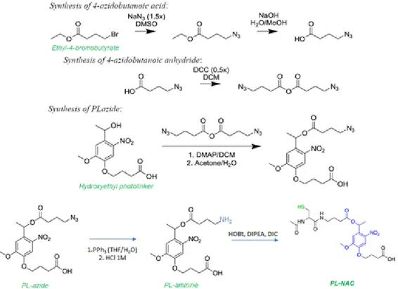
In Chapter 2 a LbL O/W cross-linked photo-responsive nanoemulsion, loaded with a natural drug, curcumin, is explained. As just demonstrated in our group, it is possible to functionalize the polymers deposited on the oil core, such as glycol chitosan and heparin, with a thiol moiety and an allylic moiety respectively, and then create a covalent bond between the polymeric shells via a biocompatible photoinitatior free thiol-ene ‘click’ reaction, to improve the nanosystem’s stability.15 Starting from
this strategy to obtain a stable cross-linked LbL systems, we introduced a photolabile chemical linker, based on a modified N-acetyl cysteine-o-nitrobenzyl moiety,45 between the polymeric materials of
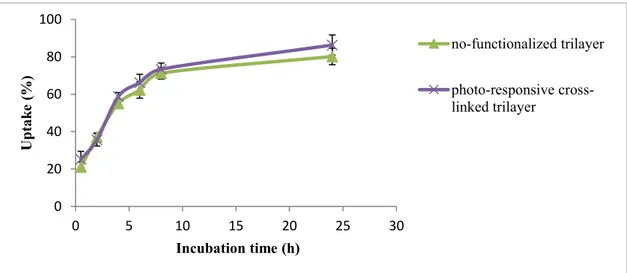
LbL. Functionalized the glycol chitosan with this thiol-photo-responsive moiety and the heparin with an allyl one, we were able to perform the cross-linkage and to stabilize our LbL O/W nanoemulsion multilayer shell by the photoinitiator free thiol-ene ‘click’ reaction. Interestingly, the photo-responsive linker gave us the possibility to trigger a controlled destabilization of the nanocarriers to release the drug by single or multi-photon UV light (365 nm or 740 nm). After the chemical modifications of the nanocarriers and its characterizations in terms of stability and conditions of release, we performed biological studies of uptake and cell viability on melanoma cells.
In Chapter 3 it is reported a recent published article which is part of this PhD project. It regards an oral delivery application of our nanoemulsions encapsulating nutraceutical drugs, such as curcumin and lycopeneand the study of their cardioprotection and anti-inflammatory effects. These properties
27
have been assessed by in vitro tests performed on cardiomyoblasts (H9C2 cells) in presence of doxorubicin.
In Chapter 4 we report another possibility concerning the modification of the oil core formulation of the nanoemulsions, using an inorganic compound, in particular cubic iron oxide nanoparticles as contrast agents. We prepared oil-core-PEG shell nanocarriers encapsulating nanocubic iron oxide nanoparticles to study how these systems respond to in vitro photoacoustic and in vitro and in vivo magnetic resonance imaging.
Final conclusion and future perspectives are presented and discussed in Chapter 5. Each part of this work can be seen distinctly or in a more general point of view. In this case, we can think to engineer a photo-responsive LbL O/W nanoemulsion co-loaded with natural drugs and safe contrast agents to employ as theranostic nanocapsules.
28
1.8 References
1. L.L. del Mercato et al., Advances in Colloid and Interface Science 2014, 207, 139–154. 2. Y. Yan, G. K. Such, A. P. R. Johnston, H. Lomas, and F. Caruso, ACSNano, 2011, 5(6),
4252-4257.
3. A.P.R. Johnston et al., Current Opinion in Colloid & Interface Science, 2006, 11, 203–209. 4. Y. Yan, M. Björnmalm, and F. Caruso, Chem. Mater., 2014, 26, 452−460
5. E. Fleige et al., Advanced Drug Delivery Reviews, 2012, 64, 866-884. 6. Pilar Rivera Gil et al., Nanotoday, 2008, 3(3-4)
7. E. Donath, G. B. Sukhorukov, F. Caruso, S. A. Davis, and H. Möhwald, Angew. Chem. Int., 1998, 37 (16)
8. G. Decher, Science, 1997, 277
9. B. J. Cui, Y. Wang, A. Postma, J. Hao, L. Hosta-Rigau and B. J. Cui, Y. Wang, A. Postma, J. Hao, L. Hosta-Rigau and F. Caruso, Adv. Funct. Mater., 2010, 20, 1625.
10. R. Vecchione, U. Ciotola, A. Sagliano, P. Bianchini, A. Diasproc and P. A. Netti, Nanoscale, 2014, 6, 9300–9307.
11. Prashant K. Deshmukh et al., Journal of Controlled Release, 2013, 166, 294-306. 12. R. Vecchione et al., Small, 2016, 22(12), 3005-3016.
13. J. Fang et al., Advanced Drug Delivery Reviews, 2011, 63, 136–151.
14. G. Bozzuto, A. Molinari, International Journal of nanomedicine, 2015, 10, 975-99.
15. V. Calcagno, R. Vecchione, A. Sagliano, A. Carella, D. Guarnieri, V. Belli, L. Raiola, A. Roviello, P.A. Netti, Colloids and Surfaces B: Biointerfaces, 2016, 142, 281-289.
16. Heng Pho Yap et al., Adv. Mater., 2009, 21, 4348–4352.
17. P. Bawa, V. Pillay, Y. E Choonara and Lisa C du Toit, Biomed. Mater., 2009, 4, 022001. 18. S. Ganta et al., Journal of Controlled Release, 2008, 126, 187–204.
19. S. Mura, J. Nicolas and P. Couvreur, Nature Materials, 2013, 12.
20. V.N Gunjkar, S.L. Patwekar and S.P. Dhage, World Journal of Pharmacy and
Pharmaceutical Sciences, 2015, 4(6).
21. Younghyun Cho, Jaehoon Lim and Kookheon Char, Soft Matter, 2012, 8, 10271–10278. 22. Y. Ping, J. Guo, H. Ejima, X. Chen, J. J. Richardson, H. Sun, and F. Caruso, Small, 2015,
11(17), 2032–2036.
23. K. Liang, G. K. Such, A. P. R. Johnston, Z. Zhu , H. Ejima, J. J. Richardson, J. Cui, and F. Caruso, Adv. Mater., 2014, 26, 1901–1905.
24. Y. Lu et al., Journal of Controlled Release, 2014, 194, 1–19. 25. D. Roy et al., Progress in Polymer Science, 2010, 35, 278–301.
29
26. A. Zhuk and S. A. Sukhishvili, Soft Matter, 2013, 9, 5149–5154.
27. A. G. Skirtach, A. A. Antipov, D. G. Shchukin, and G. B. Sukhorukov, Langmuir, 2004, 20(17), 2004 6989.
28. J. L. Nelson, B. L. Roeder, J. C. Carmen, F. Roloff, and W. G. Pitt, Cancer Research, 2002, 62, 7280–7283.
29. A. Y.Rweia, W. Wanga, D. S. Kohanea, Nano Today, 2015, 10, 451-467.
30. J. U. Menon, P. Jadeja, P. Tambe, K. Vu, B. Yuan, K. T. Nguyen, Theranostics, 2013, 3(3). 31. N. Fomina et al., Advanced Drug Delivery Reviews, 2012, 64, 1005–1020.
32. B. Yan, J.C. Boyer, D. Habault, N. R. Branda, and Y. Zhao, J. Am. Chem. Soc., 2012, 134, 16558−16561.
33. B. Yan, J.C. Boyer, N. R. Branda, and Y. Zhao, J. Am. Chem. Soc,. 2011, 133, 19714–19717. 34. M. He, J. Li, S. Tan, R. Wang, and Y. Zhang, J. Am. Chem. Soc., 2013, 135, 18718−18721. 35. Avijit Jana et al., J. Am. Chem. Soc., 2012, 134, 7656−7659.
36. S. Karthik et al., Chem. Commun., 2013, 49, 10471—10473.
37. I. Tomatsu et al., Advanced Drug Delivery Reviews, 2011, 63, 1257–1266. 38. V. Marturano et al., Polymer, 2015, 70, 222-230.
39. Pauloehrl T. et al., Angew. Chem. Int. Ed., 2012, 51, 9181-9184. 40. Y. Zhao, The Chemical Record, 2007, 7, 286–294.
41. E. Cabane et al., Soft Matter, 2011, 7, 9167–9176.
42. Chiyoung Park et al. Angew. Chem. Int. Ed., 2008, 47, 2959 –2963
43. Yan Li et al., Journal of Polymer Science: Part A: Polymer Chemistry, 2010, 48, 551–557. 44. Juan L. Vivero-Escoto et al., J. Am. Chem. Soc., 2009, 131, 3462–3463.
45. DeForest et al., Angew. Chem. Int. Ed., 2012, 51, 1816-1819.
46. M. Choudhary and V. Tomer, Proc Indian Natn Sci Acad , 2013, 79(4), 985-996. 47. Yuwen Ting et al., Journal of functional foods, 2014, 7, 112-128.
48. J.P. Gleeson et al., Trends in Food Science & Technology, 2016, 53, 90-101. 49. H.B. Nair et al., Biochemical Pharmacology, 2010, 80, 1833–1843.
50. Qingrong Huang et al., Journal of food science, 2010, 75(1).
51. M. Yao, D. J. McClements and H. Xiao, Current Opinion in Food Science, 2015, 2, 14–19. 52. D. Ileš et. al., Strojarstvo, 2011, 53(2), 127-136.
53. Ju-Suk Nam et al., Molecules, 2016, 21, 108.
54. Fasalu Rahiman O.M. et al., International Journal of Pharmaceutical Sciences Review and
Research, 2011, 8(1).
30
56. R. Vecchione et al., Journal of Controlled Release, 2016, 233, 88–100. 57. Xin Wang et al., J Mol Hist, 2014, 45, 113–120.
58. Pankaj Bansal et al., Molecular and Cellular Biochemistry, 2006, 289, 1–9. 59. Gullett A.R. et al., Semin. Oncol., 2010, 37 (3), 258-281.
60. Waliszewski KN. et al., Salud Publica Mex, 2010, 52 (3), 254-265. 61. Trottier G. et al., Nat. Rev. Urol., 2010, 7(1), 21-30.
62. Giovannucci E. et al., Journal of the National Cancer Institute, 2002, 94(5), 391-398. 63. D.P. Cormode et al., Advanced Drug Delivery Reviews, 2010, 62, 329–338.
64. H. Ai, Advanced Drug Delivery Reviews, 2011, 63, 772–788.
65. H. Shokrollahi, Materials Science and Engineering C, 2013, 33, 4485–4497. 66. U. Ayanthi Gunasekera et al., Targ Oncol, 2009, 4, 169–181.
67. E. Tombácz et al.. Biochemical and Biophysical Research Communications, 2015, 468, 442– 453.
68. B. Chertok et al., Biomaterials, 2008, 29, 487–496.
69. G. Prévot et al., International Journal of Pharmaceutics, 2017, 532, 669–676.
70. R. Vecchione, G. Luciani, V. Calcagno, A. Jakhmola, B. Silvestri, D. Guarnieri, V. Belli, A. Costantini and P.A. Netti, Nanoscale, 2016, 8, 8798–8809.
71. R. Vecchione et al., Nanomedicine: Nanotechnology, Biology, and Medicine, 2017, 13, 275– 286
72. X. Ma et al., Accounts of Chemical Research, 2011, 44(10), 1114-1122. 73. J. Xie et al., Advanced Drug Delivery Reviews, 2010, 62, 1064–1079. 74. S.M. Janib et al., Advanced Drug Delivery Reviews, 2010, 62, 1052–1063. 75. Yoo et al., Accounts of Chemical Research, 2011, 44(10), 863–874.
76. Lammers et al., Accounts of Chemical Research, 2011, 44(10), 1029–1038. 77. J.H. Ryu et al., Journal of Controlled Release, 2014, 190, 477–484.
31