Accuracy assessment of computer-guided
Piezocision
TM
: an in-vitro study
TREBALL FINAL DE GRAU
María Lara Muros Convocatoria Juny 2019
UNIVERSITAT DE BARCELONA
Facultat de Medicina i Ciències de la Salut
2 Index
1. Abstract ……….. 4
Resumen……….. 5
2. Introduction ………... 6
3. Objectives and hypothesis …..……… 9
4. Study design ………... 10
5. Material and method……… 10
6. Results ……..………. 20 7. Discussion ………... 23 8. Conclusions ...……… 26 Conclusiones ……….. 27 9. References ………. 28 10. Annexes………... 31
4 1. Abstract
Background: Corticotomies have been described in order to accelerate orthodontic tooth movement, reduce adverse events and/or increase dental arch stability. Original approaches were invasive, with huge morbidity and significant patient discomfort. However, digital workflow has changed its approach. Computer-guided PiezocisionTM has been sprawled as a safer minimally invasive procedure.
Aims: To assess the accuracy and safety of computer-assisted PiezocisionTM comparing its deviation with freehand corticotomies, analyse the effect of location and position, and describe the manufacturing process planning.
Materials and methods: An in-vitro study was made. Four resin mandible models and 52 corticotomies were performed. One investigator made the cuts using either the Computer-guided PiezocisionTM system (guided group) or the conventional freehand method (freehand group). Accuracy assessment was measured by overlapping the virtual presurgical placement of the corticotomy in a Cone-Beam Computed Tomography (CBCT) and the real position in the postoperative CBCT. Descriptive and bivariate analysis of the data was made.
Results: Computer-guided PiezocisionTM accuracy was higher than freehand group corticotomies in all precision variables except for depth discrepancy. However, both groups (freehand and guided) showed some degree of deviation from presurgical planning. Two incisions (7.69%) caused iatrogenic root damage, whereas in freehand 7 cuts were recorded (26.92%) (OR= 4.42; 95% CI: 0.82 to 23.8; p= 0.067). Except for guided angular discrepancy in anterior areas (MD: -6.38 mm; 95% CI: -9.95 to 2.61; p= 0.002), the outcomes were not influenced by position nor location.
Conclusions: The accuracy of computer-assisted PiezocisionTM is higher compared to conventional freehand technique. Thus, iatrogenic root damage is increased 4.42 times when PiezocisionTM is performed without a surgical guide. In accuracy parameters, only angular deviation was influenced by location and position. Technological improvements have led to precise surgical templates with a minimal deviation regarding virtual plan.
5
Resumen
Antecedentes: Las corticotomías nacen con el fin de acelerar el movimiento ortodóntico, reducir sus efectos adversos y/o aumentar la estabilidad de las arcadas. Inicialmente, eran cirugías muy agresivas, con alta morbilidad y poco aceptadas entre pacientes y profesionales. El fujo digital ha revolucionado su abordaje, y así, la PiezocisionTM se ha combinado con la cirugía guiada para ofrecer tratamientos mínimamente invasivos. Objetivos: Analizar la precisión, desviación y seguridad de la PiezocisionTM realizada con cirugía guiada respecto a mano alzada, analizar el efecto de la posición y localización y describir el proceso de diseño y fabricación de una férula quirúrgica.
Materiales y métodos: Se diseñó un estudio in-vitro donde se practicaron un total de 52 corticotomías en cuatro modelos de resina. Un investigador procedió con los cortes tanto de la PiezocisionTM guiada (grupo guiado) como a mano alzada (grupo a mano alzada). La precisión se midió sobreponiendo virtualmente la localización de la corticotomía prestablecida en la Tomografía Computada de Haz Cónico (TCHC) del paciente con la posición real de la TCHC posquirúrgica. Se realizó un estudio descriptivo y bivariante de los resultados.
Resultados: Las corticotomías mínimamente invasivas con cirugía guiada muestran mayor precisión que las realizadas a mano alzada en todos los parámetros, a excepción de la profundidad. Sin embargo, ambos grupos mostraron una cierta desviación respecto la planificación digital. Mientras en el grupo de cirugía guiada, dos incisiones (7.59%) causaron lesión radicular, en el de mano alzada se observaron 7 (26.92%) (OR= 4.42; 95% CI: 0.82 a 23.8; p = 0.067). La angulación en el sector anterior (MD: -6.38 mm; 95% CI: -9.95 a 2.61; p = 0.002) es la única variable que se ve influenciada por la posición y localización.
Conclusiones: La precisión de las corticotomías con cirugía guiada es mayor que las realizadas a mano alzada. Así, el riesgo de lesión radicular aumenta 4.42 veces cuando la PiezocisionTM se realiza sin la férula quirúrgica. Entre todos los parámetros que valoran la precisión, sólo la angulación está influenciada por la localización y posición. El desarrollo tecnológico ha favorecido el perfeccionamiento de las guías quirúrgicas que, cada vez, son más precisas respecto la planificación digital.
6 2. Introduction
Over the last few decades, orthodontics has undergone a considerable development. Provide esthetical and shorter treatment times have become the major goals of daily practice. This tendency is mainly determined by a non-negligible increasing number of adults who are seeking for orthodontic therapy. In this population group, an interdisciplinary approach is often needed. Therefore, in addition to treating malocclusions, orthodontics can be one of the intermediate stages of an integrated treatment plan. Accordingly, by increasing the duration of treatment, acceptance among patients may decrease.
Depending on the therapeutic options and the individual characteristics of the patient, it takes between 18 to 31 months to treat malocclusions in adults. Although orthodontics has shown highly satisfactory results with predictable and safe long-term outcomes, complications may arise. In this sense, gingival recessions, enamel demineralization, bone dehiscence or fenestration, root resorption or malocclusion relapse are some of the most frequent (1).
Several techniques -e.g. local or systemic administration of drugs and mechanical or physical stimulation- and surgical procedures -e.g. gingival fiberotomy, alveolar surgery and distraction osteogenesis- have been described in order to accelerate orthodontic tooth movement, reduce adverse events and/or increase dental arch stability (2).
Corticotomy is an intentional injury to the cortical bone that was first described in 1892 as a surgical approach to correct malocclusion. However, it was not until 1959 that this procedure was modified and popularized by Köle, suggesting the concept of "bony block" movement. The surgical technique involved interradicular cuts in vestibular and palatine/lingual bone surfaces with a horizontal osteotomy to connect them (3).
Wilcko et al. in a series of case reports, described the Accelerated Osteogenic Orthodontics (AOO) or Periodontally Accelerated Osteogenic Orthodontics (PAOO) approach, which combines orthodontic treatment with selective alveolar decortication --and simultaneous bone grafting if needed (4). They hypothesized that the increase in the speed of tooth movement subsequent to corticotomy surgery was due to a demineralization-remineralization process of the alveolar bone rather than a “bony block” movement (4,5). This observation is part of a greater event that is known in the orthopedic literature since Frost (6), in 1989, described the Regional Acceleratory Phenomenon
7 (RAP). In this sense, any bone injury induces a transient demineralization-remineralization phenomenon that corresponds to the initial phase of the physiological healing process. In the initial RAP’s transient osteopenia, there is a dramatic increase in bone turnover on the surface of the trabecular bone, the number of osteoblasts decreases in the medullary bone and the porosity of the cortical bone increases. As a result, bone becomes less dense but maintains its volume, being the degree and duration of the response directly proportional to the intensity and proximity of the surgical insult (6). RAP begins few days after surgery, reaches its peak at 1-2 months and fully recovers between 6 and 24 months (6). The term “Regional” refers to the fact that demineralization extends beyond the stimulus itself, approximately between a tooth or a tooth and a half. On the other hand, the accelerator concept is caused by the propagation of the bone response to the marrow, causing the healing to occur 2 to 10 times faster.
Although effective and highly predictable, PAOO approach is quite invasive because it requires elevation of buccal and lingual/palatal full-thickness flaps with extensive decortications of the buccal and lingual/palatal alveolar bone. Vercellotti & Podesta (7) proposed the use of a piezoelectric knife instead of a high-speed surgical bur to decrease the surgical trauma. Because of its micrometric and selective cut, a piezoelectric device produces safe and precise osteotomies without osteonecrotic damage. However, this technique is also invasive in nature, since it requires extensive flap elevations and osseous surgery, causes a non-negligible postsurgical discomfort as well as postoperative complications. Consequently, because of these shortcomings, these techniques have not been embraced widely by the patient or dental communities.
In 2009, Kim et al.(8) introduced the corticision technique as a minimally invasive alternative to create a surgical injury to the bone without flap reflection. In this procedure, a reinforced scalpel and a mallet -to go through the gingiva and cortical bone- are used. Although the surgical injury created is enough to induce the RAP effect and move the teeth rapidly during orthodontic treatment, corticision has two major drawbacks: the inability to graft soft or hard tissues during the procedure to correct inadequacies and reinforce the periodontium, and the possibility to cause dizziness during the postoperative period due to the repeated malleting.
Recently, minimally invasive flapless procedures have been expanded by Dibart et al.(9) with PiezocisionTM. This approach starts using a blade to perform 5 to 8 mm long vertical buccal incisions, 3-4 mm below the interproximal papilla. Through these microincisions,
8 a piezosurgical knife is placed over to create 3mm depth corticotomy. It also has the advantage of allowing for hard-tissue or soft-tissue grafting via selective tunneling to correct gingival recessions or bone deficiencies in patients (10). In contrast to conventional treatment, higher forces are applied, and orthodontic appliances are regularly adjusted to take advantage of the RAP effect. From a histological point of view, there is evidence that RAP is also present in localized piezoelectric alveolar decortication, and its magnitude is comparable to more traumatic techniques (11).
According to Charavet et al.(12) PiezocisionTM allows to reduce the overall treatment time by 43% without increasing the risk of adverse events. Nevertheless, recent publications have revealed root resorption and iatrogenic root damage associated to piezocision (13,14). Despite being a minimally invasive flapless procedure, interradicular corticotomies are performed in a committed area where teeth crowding, and malocclusions can complicate its management. As a result, some authors have suggested the use of a preoperative Cone-Beam Computed Tomography (CBCT) and other technological tools to increase treatment precision (10).
Guided surgery has been sprawled into dentistry for safer and accurate procedures. After being extensively applied in oral surgery and implantology (15), PiezocisionTM has also benefit from it. Although Milano et al.(16) introduced its use, Cassetta et al.(17) improved it with a three-dimensionally printed Computer-Aided Design/Computer-Aided Manufacturing (CAD/CAM) surgical guide. With computer-guided PiezocisionTM, it is not only possible to reduce patients’ discomfort, but a safer and more accurate design can also be achieved (18). The preoperative analysis includes clinical and radiographic examinations by means of a CBCT for a detailed digital study. Once the individualized surgical guide is printed, PiezocisionTM technique is conventionally performed through the guide slots. It has been stated that treatment times are reduced into a third or a half compared to conventional orthodontics (14,16,18,19).
However, the scientific evidence about computer-guided PiezocisionTM efficacy and precision is scarce (20). Hence, the aim of the present study is to assess the accuracy of PiezocisionTM using a CAD/CAM surgical guide compared with the conventional freehand technique.
9 3. Objectives and hypothesis
Objectives
The main objective of this in vitro study was to assess the accuracy of computer-assisted PiezocisionTM comparing its deviation with freehand corticotomies.
Secondary purposes were to describe surgical guide design and its manufacture, analyse its clinical relevance according to iatrogenic root damage and observe the effect of location and position over corticotomy accuracy.
Hypothesis
Main Hypothesis
The accuracy of computer-guided PiezocisionTM differs from freehand PiezocisionTM. • Null hypothesis
Computer-guided PiezocisionTM does not differ from freehand PiezocisionTM. H0: mean deviation computer-guided PiezocisionTM = mean deviation freehand PiezocisionTM.
• Alternative hypothesis
Computer-guided PiezocisionTM differs from freehand PiezocisionTM.
H1: mean deviation computer-guided PiezocisionTM ≠ mean deviation freehand PiezocisionTM.
Secondary hypothesis
- CAD/CAM surgical guides require a precise design and manufacture. - Iatrogenic root damage rates are higher in freehand group.
10 4. Study design
An in-vitro study was carried out to evaluate the accuracy of a stereolitographic surgical guide in PiezocisionTM technique. With that purpose, a convenience sample of 4 different 3D-printed acrylic casts from one patient data was established: 2 matched with maxilla and 2 with mandible. One of each had an individualized surgical template (computer-guided PiezocisionTM group), and the other, was considered as the control group (freehand PiezocisionTM).
5. Material and method
Patient selection
The candidate must full-fill the following eligibility criteria:
• Be a patient of Oral Surgery and Implantology Master's degree program of the University of Barcelona (Barcelona, Spain).
• Have a previous CBCT for dental purposes.
• Be a PiezocisionTM candidate. This treatment is indicated in (29): o Class I malocclusions with moderate to severe crowding. o Selected class II malocclusions and III.
o Correction of deep or open bite. o Rapid intrusion or extrusion.
o Prevention of mucogingival and osseous defects. o Interdisciplinary treatments.
• Full arch dentition except for third molars. • Absence of periodontitis.
On the other hand, exclusion criteria include patients with dental implants and/or osteosynthesis plates, congenital maxillary malformation or any other disorder.
Case presentation
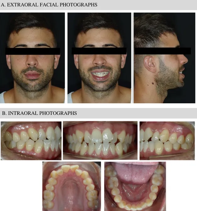
The patient data was extracted from a 30-year-old healthy man (Figure 1). Extraoral examination revealed a symmetric face with an increased height of the lower third. Moreover, it was not proportioned; lower two-thirds were augmented in respect of the first lower third. He had a straight soft tissue profile (165º) with a prominent lower lip, 3.6mm from Ricketts E-Plane.
11 Intraoral and dental cast examination noticed a bilateral class I molar and canine malocclusion with severe anterior crowding (11mm in maxilla and 7mm in mandible). He displayed cross-bite in 1.2 and 2.1, while 1.1 and 2.2 had edge-to-edge bite. Overjet and overbite were 0mm. Lower and upper midlines were centred, but all incisors were crowded and rotated. More detailed information about intraoral and extraoral examination is provided in Supplementary Table A1.
Panoramic radiograph disclosed third molars absence and no dental abnormalities nor pathologic lesions. Ricketts cephalometric analysis revealed Class I skeletal relationship (convexity 0.5mm) with maxillomandibular dentoalveolar protrusion (Figure 2). He presented normodivergent facial pattern with some hyperdivergent values.
.
A. EXTRAORAL FACIAL PHOTOGRAPHS
B. INTRAORAL PHOTOGRAPHS
Figure 1: Case presentation.
12
Patient was given full verbal information. An assignment of all rights to photograph was obtained as well as a written consent. The protocol complied with Declaration of Helsinki guidelines and was approved by the clinical research ethics committee of the Dental Hospital of the University of Barcelona (Protocol: 30/2018).
Measurement Norma SD Value Facial axis 90º ±3.5 92.5º Facial depth* 89º ±3 93.5º Mandibular plane 24º ±4.5 26º Lower Facial height* 47º ±4 52º Mandibular arch 29º ±4 26º
Convexity 1mm ±2 0.5mm
Maxillary Depth* 90º ±3 95º Lower I protrusion* 1mm ±2 6.9mm Lower I inclination* 22º ±4 30º Upper molar position* 21mm ±2 24mm Interincisal angle* 130º ±6 124º
E-Plane* -2mm ±2 3.6mm
C. RADIOGRAPHS
Figure 2: Simplified Ricketts cephalometric analysis.
I: Incisor. SD: Standard Deviation. *Altered parameters.
Figure 1 (continued): Case presentation.
13
Study planning
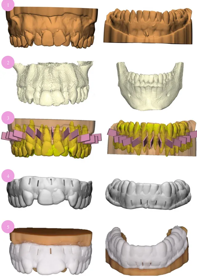
Polyvinylsiloxane impressions of both arches were taken carefully to register vestibular fornix with Aquasil Light body® and Aquasil Soft Putty® (Dentsply Sirona, York, Pennsylvania, USA) following 1-step PuttyWash technique. Using a 3Shape TRIOS® 3D scanner (3Shape A/S® Cophenague, Denmark), casts were digitalized as STereoLithography (STL) files to create a 3D model and sent to Avinent Digital Health® (Avinent Implant System®, Santpedor, Spain). With 3Shape Implant Studio® software (3Shape A/S® Cophenague, Denmark), the Digital Imaging and Communication On Medicine (DICOM) of a previous CBCT of the patient was also transferred into a STL. Thus, both STL files (CBCT and casts) were overlapped to virtually design accurate corticotomies. Following Dibart et al.(9) technique, interradicular incisions were placed 2mm from the papilla to prevent periodontal tissue trauma with a 3mm depth. The width was determined according to piezoelectric knife dimensions as well as the length, which lead to a 0.6mm x 5.3mm cut. Thirteen different interradicular incisions were planned from mesial of right second molar to mesial of the left second molar of each arch. Anterior cuts were set from canine to canine (Figure 3).
Surgical templates were manufactured from corticotomies designs (Figure 4). To get more stability, templates were extended to occlusal surfaces and vestibular fornix. They had 2mm width, which was considered for assessing depth deviation.
Stereolithographic polyamide surgical guides were printed using a Formiga P110® (EOS, München, Germany). On the other hand, the 4 acrylic models were printed using a ProJet® MPF 2500 (3DSystem; South California, USA).
Figure 3: Interradicular incisions design.
Anterior cuts
14
1
3 2
Figure 4: Steps for a CAD/CAM surgical guide design and manufacture.
1) STL casts files. 2) STL form patient’s CBCT. 3) Overlap of both STL files and PiezocisionTM cuts design
4) Surgical guide render according to corticotomies plan. 5) 3D-printed casts and templates.
5 4
15
Surgical procedure
After guide stabilisation in 2 of the casts, one from maxilla and one from mandible corticotomies were performed with an ultrasonic device Piezotome SoloTM (Satelec®; ActeonGroup, Merignac, France) and its PZ1 tip (PiezocisionTM; ActeonGroup, Merignac, France) (0.6mm x 5.3mm), (Figure 5). They were activated in D1 mode following manufacturer’s instructions. Irrigation was constantly perfused.
A B
A2
A1
CC1
C3
B1
B2
C2
Figure 5: Piezocision TM material and procedure
A) Piezosurgery material. A1: Ultrasonic device Piezotome SoloTM. A2PZ1 tip with a 3mm landmark.
B) PiezocisionTM procedure. B1: with a CAD/CAM surgical guide. B2: without template.
16 The first laser mark on the tip was used as the landmark for the corticotomies freehand depth. Computer-guided cuts had not any references. In consequence, every PiezocisionTM cut was checked with a periodontal millimetric probe. In those where surgical guides were placed, 5mm instead of 3mm were measured (Figure 6).
Immediately after corticotomies, the four models underwent a new CBCT (Planmeca ProMax® 3D Mid (Planmeca, Helsinki, Finland) with 90Kv, 10mA,13.9 seconds, 1245 DAP (mGy*cm2), 0,4mm Voxel) and sent to Avinent Digital Health S.L. (Avinent Implant System®, Santpedor, Spain) to be transformed into STL files.
Data sampling
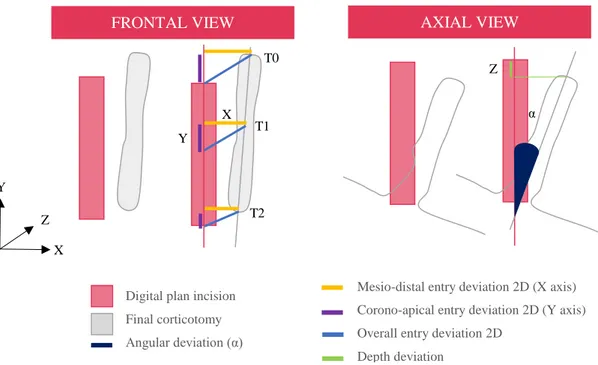
To assess the accuracy, different parameters were considered for each cut (Figure 7): • Iatrogenic root damage (IRD)
• Mean mesio-distal entry deviation 2D: defined as the horizontal deviation in X axis between preoperative plan and performed cut at 0 mm (T0) and at 2.6 mm (T1) and 5.3 mm (T2) in an apical direction of the tip. Expressed in mm and calculated as an absolute value.
Figure 6: Depth assessment.
With a periodontal millimetric probe, 5mm were measured in computer-guided PiezocisionTM
(figure 1 and 2) while in freehand technique 3mmm of depth (figure 3 and 4).
1
2
3
17 • Mean corono-apical entry deviation 2D: defined as the vertical deviation in Y axis between preoperative plan and performed cut at 0 mm (T0) and at 2.6 mm (T1) and 5.3 mm (T2) in an apical direction of the tip. Expressed in mm and calculated as an absolute value.
• Mean overall entry deviation 2D: defined as the sum of mesio-distal and corono-apical deviation between preoperative plan and performed cut. Expressed in mm and calculated as an absolute value.
• Depth deviation: defined as the deviation in Z axis between preoperative plan and performed cut. Expressed in mm and calculated as an absolute value.
• Angular deviation: defined as the angulation discrepancy between the planned and final corticotomy. Expressed as an angle (°) and calculated as an absolute value. Autocad® software (Autodesk®, Sant Rafael, California, USA) and Rinhoceros 3D® (Robert Mc Neal &Associates®, Seattle, Washington, USA) were used to measure the outcomes. Firstly, with Rinhoceros 3D®, presurgical CBCT with corticotomies design STL file was merged with postoperative CBCT. From a 3D view of the render, iatrogenic root damage was identified through visual inspection. Global vision was restricted to the frontal plane to evaluate entry deviation whereas the axial view allowed depth and angular deviation assessment. After that, STL file was transformed into a DraWinG (.DWG) to measure its veritable magnitude (Figure 8).
Y X Z FRONTAL VIEW α AXIAL VIEW X Y Z T0 T2 T1
Figure 7: Three-Dimensions description of the main accuracy outcomes.
- Digital plan incision
- Final corticotomy
- Angular deviation (α)
- Mesio-distal entry deviation 2D (X axis)
- Corono-apical entry deviation 2D (Y axis)
- Overall entry deviation 2D
18 Figure 8: Accuracy assessment
A) Render from Rinhoceros 3D® after overlapping different STL files. B) Autocad ® software screenshots from frontal and axial view to assess deviation veritable magnitude.
A
19
Statistical analysis
Categorical outcomes (IRD) were presented as absolute and relative frequencies for categorical outcomes. Normality of scale variables (deviation parameters) were explored through Shapiro-Wilk’s test and visual analysis of the P-P and box plots. Where normality was rejected, the interquartile range (IQR) and median were calculated. Where the distribution was compatible with normality, the mean and standard deviation (SD) were used.
Unpaired t-tests were used to identify differences in accuracy between the freehand and guided groups at every deviation parameter. Mean differences (MD) with 95% confidence intervals (95% CI) were also estimated. For each variable, multiple linear regression was computed to quantify the effect of position and location.
The association of categorical variables was assessed with either Pearson’s 2 test or Fisher’s exact test. The odds ratio (OR) with 95% CI was calculated for the categorical variable. A multivariate analysis was performed using a nonconditional logistic regression model to explore the effect of position and location over IRD.
To test intraexaminer agreement and consistency, the assessment of 6 randomly selected cuts (48 measurements) was repeated after 2 weeks. The intraclass correlation coefficients (ICC) were 0.97 (95% confidence interval (95%CI) 0.94 to 0.99; p<0.001) and 0.98 (95%CI 0.96 to 0.99); p<0.001), showing excellent reliability and consistency.
The statistical analysis was carried out with Stata14 (StataCorp®, College Station, TX, USA). The level of significance was set at p <0.05, using Tukey’s correction for multiplicity of contrasts.
20 6. Results
A total of 52 PiezocisionTM cuts were analysed without registering any protocol deviation.
Computer-guided PiezocisionTM vs freehand deviation
Descriptive results of the main outcome variables are summarized in Table 1 and Supplementary Table A2. Both groups (freehand and guided) showed some degree of deviation from presurgical planning. However, while in computer-guided PiezocisionTM each deviation parameter was less than 0.5 mm or 5°, in the freehand group all results were above that thresholds except for depth discrepancy (Mean: 0.37 mm; SD: 0.21).
N: Sample size, SD: Standard Deviation.
Computer-guided PiezocisionTM had a significant higher accuracy for all studied variables, except for depth deviation variable (MD: 0.90 mm; 95% CI: -0.44 to 0.22; p=0.185) (Table 2 and Figure 9).
.
Table 1: Descriptive results of the main deviation outcomes for computer-assisted
PiezocisionTM and freehand technique.
Variable N Mean (SD) Range
Freehand Mesio-distal entry deviation 26 0.59 mm (0.38) 0.01 to 1.46 Corono-apical entry deviation 26 1.11 mm (0.69) 0.02 to 2.26 Overall entry deviation 26 1.34 mm (0.63) 0.36 to 2.47 Depth deviation 26 0.37 mm (0.21) 0.05 to 0.82 Angular deviation 26 8.12º (6.20) 0.10 to 21.57 Guided Mesio-distal entry deviation 26 0.17 mm (0.10) 0.00 to 0.38
Corono-apical entry deviation 26 0.31 mm (0.21) 0.00 to 0.74 Overall entry deviation 26 0.37 mm (0.19) 0.09 to 0.83 Depth deviation 26 0.46 mm (0.27) 0.03 to 1.08 Angular deviation 26 3.79º (2.20) 0.00 to 8.28
21 Table 2: Results of bivariate analysis.
MD: mean difference between. CI: confidence interval. *statistically significant difference.
Iatrogenic root damage
Digital preoperative planning only interfered with the distobuccal root of the upper right first molar, where interradicular space was less than 0.6 mm. In computer-guided PiezocisionTM, 2 incisions (7.69%) caused iatrogenic root damage, whereas in freehand 7 (26.92%) lesions were recorded (OR= 4.42, 95% CI: 0.82 to 23.8, p = 0.067). Figure 10 depicts iatrogenic root damage locations.
For each of the study groups, when analysing the mean deviation between the cuts that caused root damage compared to those that did not, no significant differences were observed in any of the variables recorded (all p≥ 0.05 after Tukey’s correction for multiplicity of contrasts).
Variable MD (95% CI) p-value
Mesio-distal entry deviation -0.42 mm (-0.57 to -0.26) <0.001* Corono-apical entry deviation -0.80 mm (-1.09 to -0.51) <0.001* Overall entry deviation -0.96 mm (-1.23 to -0.70) <0.001* Depth deviation 0.90 mm (-0.44 to 0.22) 0.185 Angular deviation - 4.33º (-6.96 to -1.70) 0.002*
* * *
*
22
Effect of location and position
Iatrogenic root damage, entry point or depth deviation were not influenced by location (maxilla or mandible) or position (anterior or posterior) (all p ≥ 0.2). Angular discrepancy, however, was affected by position (t = 2.83; p = 0.01). While in posterior cuts the difference was similar (MD: -2.06mm; 95% CI: -5.43 to 1.31; p = 0.211), computer-guided group exhibited significantly less deviation when the cuts were performed in the anterior position (MD: -6.38mm; 95% CI: -9.95 to 2.61; p = 0.002).
• Teeth injured by freehand technique
• Teeth injured by computer-guided PiezocisionTM
• Teeth injured by both groups
• Teeth virtually affected
23 7. Discussion
Computer-guided PiezocisionTM has been introduced to achieve greater accuracy in minimally invasive corticotomies. In addition, this approach has been reported to accelerate orthodontic treatment, increase safety and reduce morbidity.
Despite its advantages, different sources of bias such as the radiographic technique performed, the material used to make the impressions and their scanning, the impression of the guides, the surgical procedure or the inherent tolerance of the instrument can interfere with the accuracy of the individual CAD/CAM surgical guides. As a result, at least to this day, the perfect transfer of digital design to reality is not possible. More precisely, according to other fields of dentistry, the average linear deviation is around 0.5 mm (21). Our findings seem to support this statement, since all the deviation parameters assessed were close to this value (Table 1).
To the best of the authors’ knowledge, this is the first study that has evaluated the accuracy of computer-guided PiezocisionTM in three-dimensional space as well as its clinical consequences. Cassetta et al.(20) have previously assessed computer-guided PiezocisionTM precision in 6 different points of each incision. The authors reported an overall deviation at entry point of 0.67 mm (Range: 0.0 to 1.44; SD: 0.31) whereas depth deviation was 0.54 mm (Range: 0.17 to 0.80; SD: 0.21). Although depth deviation was comparable to the present outcomes, our overall entry point deviation is approximately reduced into a third (Table 1). A possible explanation of these findings could be related with the fact that while Cassetta’s trial was conducted in ten different patients, the present study was performed in acrylic models from a single subject.
Regarding angular discrepancy, a difference of 3.79º was reported between presurgical planning and final corticotomy. A recent metanalysis conducted by Tahmaseb et al.(22), who assessed the accuracy of static stereolithographic surgical guide in implants, pointed to an angular deviation of 3.5º (95% CI: 3.00 to 3.96), which agrees with our results. To reduce this discrepancy, it has been suggested to create a ledge adhered to slot so that, piezosurgical knife is guided for a long distance (20). It could also help as a depth stop. The maximum deviation values in computer-guided PiezocisionTM do not exceed from 1 mm (Table 1). Nevertheless, these figures might be enough to cause iatrogenic damage in areas where dental crowding and/or malocclusions are present. IRD rates differed from virtual design, where only one incision was originally compromised due to a lack of
24 interproximal space (Figure 10). Preliminary studies suggest that PiezocisionTM's RAP effect extends beyond the stimulus itself, approximately between a tooth or a tooth and a half (23). Moreover, piezosurgical knife vibration frequency may activate more osteoblast and other cells progenitors (24). Therefore, avoiding those narrow interproximal areas and selecting a strategic incision location, less root injury could be reported (12,25). What is more, a minimum interdental bone of 2 mm has been suggested in order to avoid complications and unexpected events (12).
Recent reports have suggested that PiezocisionTM might cause iatrogenic root damage (13,14). In our study, this adverse event was reported in 26.92% of the freehand cuts (n = 7). On the other hand, in the guided group, these figures were reduced to 7.26% (n = 2). Accordingly, freehand surgery increased the risk of IRD by 4.42 times (95% CI: 0.82 to 23.8). However, probably due to the small sample size, this difference did not reach statistical significance (p = 0.067). In addition, clinical studies are needed to clarify the true impact of these lesions, both in the short and long term.
Further investigations are also needed to achieve more reliable precision outcomes. The
in vitro character of the present study urges to interpret all these results with caution.
Accuracy has been analysed under ideal conditions, which may not be adjusted into reality. As an example, root palpation is useful in freehand techniques in order to avoid IRD. However, this feature could not be represented in the acrylic models. In this preliminary study, only 4 casts were assessed from a single patient. Given that, our findings are based on a limited sample size, so that they cannot be extrapolated to every clinical situation. Another limitation is the operator’s lack of experience. Although neglectable differences have been reported between experienced and non-experienced surgeons when using a computer-guided system, the level of experience was positively correlated with precision in freehand procedures (26). For all these reasons, the effect of the intervention observed in our study could be overestimated.
Surprisingly, no previous evidence was found comparing computer-assisted to freehand PiezocisionTM deviation. Except for depth deviation variable, the guided group had a significant higher accuracy for all studied variables (Table 2 and Figure 9).
In an oral implantology in vitro study, Tan et al.(27) reported an angular deviation of 3.91º (IQR: 2.45 to 5.38) and 8.82º (IQR: 4.84 to 9.84) for the computer-assisted and freehand groups, respectively. These results match with present study, since a 3.79º
25 (Range: 0.00 to 8.28º) deviation was found in the test group and 8.12º (Range: 0.10 to 21.57º) in controls. As a result, computer-assisted PiezocisionTM seems to reduce angular deviation in a 46.67%.
Regarding overall deviation and its mesio-distal and corono-apical components, the guided group was closer to presurgical planning. Mean vertical error was found to be slightly higher (MD: -0.80mm; 95% CI: -1.09 to -0.51; p < 0.001) than mesiodistal deviation (MD: -0.42 mm; 95% CI: -0.57 to -0.26; p < 0.001). In computer-guided PiezocisionTM this finding could be partially explained by instrument’s tolerance through the slot. Adjusting this parameter, piezosurgical knife would reduce its friction enhancing tip movement. On the other hand, in freehand corticotomies it might be explained by a lack of references in the acrylic model.
Unlike previous studies (27), depth outcomes were more precise in the freehand group. Even the difference was not statistically significant, it might be attributed to an imprecise assessment of the variable. On one hand, piezosurgical knife had a landmark which was used as a reference for freehand depth stop, a feature not available for computer-guided PiezocisionTM. Although depth was intrasurgically checked with a periodontal probe, the surgical guide offered some resistance against its insertion and its landmark -at 3 mm- differed from planning (i.e. 5 mm). Moreover, the surgical guide was made from an opaque polyamide material which might influence precision. Hou et al.(28) have introduced the use of a translucent resin to enhance visibility. Thus, piezosurgical knife deviation could be easily identified. What is more, small porous in guide’s surface were added to provide greater access to irrigation, thus decreasing the risk of bone and/or soft tissues overheating.
As seen in previous studies, the results were homogenous and consistent when adjusted for location and position for all entry and depth deviation outcomes (20). Nevertheless, in computer-assisted PiezocisionTM, angular deviation in posterior positions was significantly higher than in anterior ones. In clinical research it could be caused by a poorer posterior visibility and difficulties in positioning piezosurgical knife those areas. Future clinical investigations with bigger samples sizes should learn from our limitations and take into account the aforementioned improvements. The impact of operator’s experience as well as other computer-guided systems (i.e. dynamic computer guided surgery) should be also addressed.
26 8. Conclusions
1. The CAD/CAM computer assisted surgery system PiezocisionTM allows a more accurate corticotomy procedure in comparison with the conventional freehand method.
2. Freehand surgery increases the risk of iatrogenic root damage by 4.42 times when compared to computer-guided PiezocisionTM.
3. Regarding position and localization, angular deviation is the one influenced.
4. Digital workflow has let highly precise surgical guides manufacture with a minimal deviation from digital design.
27
Conclusiones
1. La precisión de las corticotomías mínimamente invasivas con férulas guiadas CAD/CAM es significativamente mayor que en técnicas a mano alzada, excepto en la profundidad de corte.
2. La PiezocisionTM realizada a mano alzada multiplica por 4.42 veces el riesgo de lesión radicular comparado con el uso de la férula quirúrgica.
3. A excepción de la desviación angular, ninguna de las variables evaluadas se vio influenciada por la posición o localización de las corticotomías.
4. Gracias al flujo digital podemos obtener guías quirúrgicas con una mínima desviación respecto la planificación virtual preoperatoria.
28 9. References
1. Stöber-Blázquez EK, Genestra P, Molina-Coral A, Puigdollers-Pérez A. La corticotomía alveolar selectiva como coadyuvante al tratamiento de ortodoncia. Revisión de la literatura. Rev Esp Ortod. 2010;40:215–45.
2. Fleming PS, Fedorowicz Z, Johal A, El-Angbawi A PN. Surgical adjunctive procedures for accelerating orthodontic treatment. Cochrane Database Syst Rev. 2015. CD010572.
3. Köle H. Surgical operations on the alveolar ridge to correct occlusal abnormalities. Oral Surg Oral Med Oral Pathol. 1959;12:515-29.
4. Wilcko WM, Wilcko T, Bouquot JE, Ferguson DJ. Rapid Orthodontics with alveolar reshaping: two case reports of decrowding. 2000;21:9-19.
5. Wilcko MT, Wilcko WM, Bissada NF. An Evidence-Based Analysis of Periodontally Accelerated Orthodontic and Osteogenic Techniques: A Synthesis of Scientific Perspectives. Semin Orthod. 2008;14:305-16.
6. Frost HM. The biology of fracture healing. An overview for clinicians. Clin Orthop Relat Res. 1989;248:283-93.
7. Vercellotti T, Podesta A. Orthodontic microsurgery: a new surgically guided technique for dental movement. Int J Periodontics Restorative Dent. 2007;27:325-31.
8. Kim SJ, Park YG, Kang SG. Effects of corticision on paradental remodeling in orthodontic tooth movement. Angle Orthod. 2008;79:284–91.
9. Dibart S, Sebaoun J, Surmenian J. Piezocision: A Minimally Invasive, Periodontally Accelerated Orthodontic Tooth Movement Procedure. Compend Contin Educ Dent. 2009;30:342-50.
10. Sebaoun J, Surmenian J, Dibart S. Traitements orthodontiques accélérés par piézocision : une alternative mini-invasive aux corticotomies alvéolaires. L’Orthodontie Française. 2011;82:311-9.
11. Librizzi Z, Kalajzic Z, Camacho D, Yadav S, Nanda R, Uribe F. Comparison of the effects of three surgical techniques on the rate oforthodontic tooth movement
29 in a rat model. Angle Orthod. 2017;87:717-24.
12. Charavet C, Lecloux G, Bruwier A, Rompen E, Maes N, Limme M et al. Localized Piezoelectric Alveolar Decortication for Orthodontic Treatment in Adults: A Randomized Controlled Trial. J Dent Res. 2016;95:1003-9.
13. Patterson BM, Dalci O, Papadopoulou AK, Madukuri S, Mahon J, Petocz P et al. Effect of piezocision on root resorption associated with orthodontic force: A microcomputed tomography study. Am J Orthod Dentofac Orthop. 2017;151:53-62.
14. Strippoli J, Durand R, Schmittbuhl M, Rompré P, Voyer R, Chandad F et al. Piezocorticision-assisted orthodontics: Efficiency, safety, and long-term evaluation of the inflammatory process. Am J Orthod Dentofac Orthop. 2019;155:662-9.
15. Vercruyssen M, Hultin M, Van Assche N, Svensson K, Naert I, Quirynen M. Guided surgery: accuracy and efficacy. Periodontol 2000. 2014;66:228-46. 16. Milano F, Dibart S, Montesani L, Guerra L. Computer-guided surgery using the
Piezocision technique. Int J Periodontics Restorative Dent. 2014;34:523-9.
17. Cassetta M, Pandolfi S, Giansanti M. Minimally invasive corticotomy in orthodontics: A new technique using a CAD/CAM surgical template. Int J Oral Maxillofac Surg. 2015;44:830-3.
18. Cassetta M, Giansanti M, Di Mambro A, Calasso S, Barbato E. Minimally invasive corticotomy in orthodontics using a three-dimensional printed CAD/CAM surgical guide. Int J Oral Maxillofac Surg. 2016;45:1059-64.
19. Finotti M, Gracco A, Del Torre M, Siviero L, de Stefani A, Bruno G. The use of computer-assisted corticotomy to enhance surgical procedures. Int Orthod. 2017;15:498-514.
20. Cassetta M, Ivani M. The accuracy of computer-guided piezocision: a prospective clinical pilot study. Int J Oral Maxillofac Surg. 2017;46:756-65.
21. Vercruyssen M, Hultin M, Van Assche N, Svensson K, Naert I, Quirynen M. Guided surgery: accuracy and efficacy. 2014;66:228-46.
30 22. Tahmaseb A, Wu V, Wismeijer D, Coucke W, Evans C. The accuracy of static computer-aided implant surgery: A systematic review and meta-analysis. Clin Oral Implants Res. 2018;29:416-35.
23. Dibart S, Yee C, Surmenian J, Sebaoun J, Baloul S, Goguet-Surmenian E et al. Tissue response during Piezocision-assisted tooth movement: A histological study in rats. Eur J Orthod. 2014;36:457-64.
24. Dibart S, Alasmari A, Zanni O, Salih E. Effect of corticotomies with different instruments on cranial bone biology using an ex vivo calvarial bone organ culture model system. Int J Periodontics Restorative Dent. 2016;36:123-36.
25. Keser EI, Dibart S. Sequential piezocision: A novel approach to accelerated orthodontic treatment. Am J Orthod Dentofac Orthop. 2013;144:879-89.
26. Jorba-García A, Figueiredo R, González-Barnadas A, Camps-Font O, Valmaseda-Castellón E. Accuracy and the role of experience in dynamic computer guided dental implant surgery: An in-vitro study. Med Oral Patol Oral y Cir Bucal. 2019;24:76-83.
27. Tan PLB, Layton DM, Wise SL. In vitro comparison of guided versus freehand implant placement: use of a new combined TRIOS surface scanning, Implant Studio, CBCT, and stereolithographic virtually planned and guided technique. Int J Comput Dent. 2018;21:87-95.
28. Hou HY, Li CH, Chen MC, Lin PY, Liu WC, Cathy Tsai YW et al. A novel 3D-printed computer-assisted piezocision guide for surgically facilitated orthodontics. Am J Orthod Dentofac Orthop. 2019;155:584-91.
29. Dibart S, Keser E. PiezocisionTM; Minimally invasive periodontally accelerated orthodontic tooth movement procedure. In: Brugnami F, Caiazzo A. Orthodontically Driven Corticotomy: tissue engineering to enhance orthodontic and multidisciplinary treatment. 1st edition. Ames, Iowa; John Wiley & Sons Inc; 2015.
31 10. Annex
Table A1: Orthodontic analysis
(P)Palatine (L)Lingual (M)Mesial (D)Distal.
TEETH
NUMBER Full arch except for third molars
SHAPE No abnormalities
OTHERS 2.1 Enamel-dentin fracture. 4.6 Decay in vestibular fossa 4.6M Composite restoration. Plaque and gingivitis
ARCHES
UPPER LOWER
SHAPE Ovoid Ovoid
TEETH POSITION 1.2P, 2.2P, 2.4P, 2.5P 3.2L ROTATIONS 1.6MP, 1.2MP, 2.1MP, 2.3MP 2.6MP 3.5ML, 3.2DL, 3.1ML, 4.2DL AXIAL INCLINATION 1.3, 2.1D 2.3M 4.1M, 4.2D, 4.3M OCCLUSION
MALOCCLUSION Bilateral class I molar and Canine
1.2-2.1 Crossbite Overjet = 0 mm 1.1-2.2 Edge-to-edge bite Overbite = 0 mm
EXTRAORAL EXAMINATION
FRONTAL Increased height of the lower third: lower two-thirds are increased respect the upper third.
Inclined bipupilar line Symmetrical face
LATERAL Soft tissue profile angle 165º Nasolabial angle 104º
Lower lip cross 3.6mm Rickets Plane E SMILE Upper and lower midlines centred
Incisal smile. No gingival smile.
Table A2: PiezocisionTM deviation results.
SD: Standard Deviation.
Location and Position Mean(SD)
Mandible Maxilla Total
Deviation Posterior Anterior Subtotal Posterior Anterior Subtotal Posterior Anterior Total n = 6 n = 7 n = 13 n = 6 n = 7 n = 13 n = 12 n = 14 n = 26 Fr ee h a n d Entry 2D x 0.82 mm (0.15) 0.48 mm (0.50) 0.64 mm (0.41) 0.56 mm (0.40) 0.53 mm (0.34) 0.54 mm (0.36) 0.69 mm (0.32) 0.50 mm (0.41) 0.59 mm (0.38) Entry 2D y 0.69 mm (0.75) 1.04 mm (0.67) 0.88 mm (0.70) 1.64 mm (0.53) 1.07 mm (0.61) 1.33 mm (0.63) 1.16 mm (0.79) 1.06 mm (0.62) 1.11 mm (0.69) Depth 0.35 mm (0.19) 0.37 mm (0.17) 0.36 mm (0.17) 0.50 mm (0.25) 0.27 mm (0.22) 0.38 mm (0.25) 0.42 mm (0.22) 0.32 mm (0.20) 0.37 mm (0.21) Angle 2.77 º (2.01) 10.15 º(5.53) 6.74 º (5.63) 7.57 º (6.36) 11.15º(6.91) 9.50 º (6.64) 5.17 º (5.15) 10.65 º (6.03) 8.12 º (6.20) Overall 1.17 mm (0.58) 1.20 mm (0.74) 1.19 mm (0.64) 1.77 mm (0.51) 1.24 mm (0.60) 1.49 mm (0.60) 1.47 mm (0.61) 1.22 mm (0.65) 1.34 mm (0.63) Gu id ed Entry 2D x 0.19 mm (0.07) 0.21 mm (0.10) 0.20 mm (0.08) 0.16 mm (0.12) 0.12 mm (0.09) 0.14 mm (0.10) 0.17 mm (0.10) 0.17 mm (0.10) 0.17 mm (0.10) Entry 2D y 0.51 mm (0.08) 0.42 mm (0.18) 0.46 mm (0.15) 0.17 mm (0.15) 0.14 mm (0.09) 0.15 mm (0.12) 0.34 mm (0.21) 0.28 mm (0.20) 0.31 mm (0.21) Depth 0.48 mm (0.26) 0.60 mm (0.34) 0.54 mm (0.30) 0.24 mm (0.23) 0.50 mm (0.13) 0.38 mm (0.22) 0.36 mm (0.26) 0.55 mm (0.25) 0.46 mm (0.27) Angle 2.54º (1.90) 3.76 º (2.13) 3.19 º (2.04) 3.68 º (1.38) 4.99º (2.79) 4.39º (2.27) 3.11º (1.69) 4.37º (2.47) 3.79º (2.20) Overall 0.55 mm (0.06) 0.47 mm (0.19) 0.51 mm (0.15) 0.26 mm (0.16) 0.21 mm (0.07) 0.23 mm (0.12) 0.41 mm (0.19) 0.34 mm (0.20) 0.37 mm (0.19) n = 12 n = 14 n = 26 n = 12 n = 14 n = 26 n = 24 n = 28 n = 52 To ta l Entry 2D x 0.51 mm (0.35) 0.34 mm (0.37) 0.42 mm (0.36) 0.36 mm (0.35) 0.33 mm (0.32) 0.34 mm (0.33) 0.43 mm (0.35) 0.33 mm (0.34) 0.38 mm (0.35) Entry 2D y 0.60 mm (0.52) 0.73 mm (0.57) 0.67 mm (0.54) 0.90 mm (0.85) 0.60 mm (0.64) 0.74 mm (0.75) 0.75 mm (0.71) 0.67 mm (0.60) 0.71 mm (0.65) Depth 0.41 mm (0.23) 0.48 mm (0.28) 0.45 mm (0.26) 0.37 mm (0.27) 0.38 mm (0.21) 0.38 mm (0.23) 0.39 mm (0.24) 0.43 mm (0.25) 0.41 mm (0.24) Angle 2.65º (1.87) 6.96º mm (5.22) 4.97º(4.53) 5.62º (4.83) 8.07º (5.99) 6.94º (5.52) 4.14º (3.89) 7.51º (5.54) 5.96º (5.10) Overall 0.86 mm (0.51) 0.84 mm (0.64) 0.85 mm (0.57) 1.02 mm (0.87) 0.72 mm (0.68) 0.86 mm (0.77) 0.94 mm (0.70) 0.78 mm (0.65) 0.85 mm (0.67)
Figure A 6: 3D printed lower cast.
Figure A: PiezocisionTM material and procedure.
Figure A 1: 3D-printed upper surgical guide.
Figure A 2: 3D-printed upper cast and surgical guide.
Figure A 3: 3D-printed upper cast.
Figure A 4: 3D-printed lower surgical guide.
1
Figure A 7: Computer-guided PiezocisionTM in upper cast.
Figure A 8: Freehand PiezocisionTM in upper cast.
Figure A 10: Freehand PiezocisionTM in lower cast.