UNIVERSITÀ DEGLI STUDI DI ROMA
"TOR VERGATA"
FACOLTA' DI SCIENZE MATEMATICHE FISICHE E
NATURALI
DOTTORATO DI RICERCA IN IMMUNOLOGIA
XIX CICLO
Characterization of immunologic mechanisms
in rare childhood chronic inflammatory
diseases: Implication for specific therapeutic
intervention
CANDIDATA
Dott.ssa FLAMINIA MURATORIRELATORE
Dott. FABRIZIO DE BENEDETTICOORDINATORE
Prof. PAOLO ROSSIIndex
1. ABSTRACT………..4
2. INTRODUCTION
2.1 Autoimmune and autoinflammatory disorders……….6
2.2 Hereditary periodic fevere syndromes………..7
2.3 Febrile response………11
2.4 The PAPA Syndrome………... 12
2.5 Regulatory T cells and self tolerance……...…...16
2.6 FOXP3………..17
2.7 IPEX Syndrome………....19
2.8 Extracorporeal photochemotherapy………..21
3. MATERIALS AND METHODS
3.1 Mutation analysis………23
3.2 In Vitro Cytokine Production………..24
3.3 Evaluation of the Phenotype and Function of
Regulatory T Cells……….…………..25
3.4 Real-time PCR………...……….26
3.5 Extracorporeal photopheresis procedure………27
4. RESULTS
4.1 PAPA Syndrome case report………..28
4.2 CD2BP1 Mutation analysis……….31
4.3 Alteration of pro-inflammatory cytokine production
in PAPA syndrome………...………...…...33
4.4 IPEX Syndrome case reports……….…………..41
4.5 Additional information……….………...……44
4.6 FOXP3 Mutation analysis……….…………..47
4.7 Phenotype and function of regulatory T cells……….49
4.8 ECP increases FOXP3 expression in CD4+ T
4.9 ECP induces circulating CD4+CD25+
T cells with
regulatory function……….……...….55
5. DISCUSSION
5.1 PAPA Syndrome………..….………...60
5.2 IPEX Syndrome………..……....………… 63
6. CONCLUSION
………68
7. ABBREVIATIONS……….
………..69
8. REFERENCES……….71
ABSTRACT
The molecular defects recently identified in rare childhood immune-dysregulations outline critical steps in the pathways that contribute to the development of normal innate immune responses and the maintenance of self-tolerance. This project is based on the study of the mechanisms involved in the alteration of immune regulation, both in hereditary autoimmune diseases and in chronic inflammation, with the aim of developing a therapeutic target. Our attention has focused on:
i) A rare autosomic dominant inherited disorder of childhood named PAPA syndrome (pyogenic sterile arthritis, pyoderma gangrenosum, acne, OMIM 604410), primarily affecting the joints and skin. This disease falls under the definition of auto-inflammatory diseases, characterized by recurrent “unprovoked” inflammatory events which are not associated with high-titre of auto-antibodies or auto-antigen specific T cells. The disease has been associated with mutations in the CD2 binding protein 1 (CD2BP1). We have observed a family affected with PAPA syndrome and identified an alteration in the tumor necrosis factor- α (TNFα) and early interluekine-1β (IL-1β) production. This laboratory finding was associated with a valuable therapeutic goal represented by the administration of the TNF inhibitor etanercept which resulted in a rapid, sustained clinical remission.
ii) A sever multi-organ autoimmune disease, the IPEX syndrome (Immunodysregulation, Polyendocrinopathy, Enteropathy, X-linked OMIM 304790). The disease is caused by mutations in the FOXP3 gene (transcription factor fork-head box protein 3) which results in absence/alteration of the regulatory T cells (Treg) population. We have
studied two IPEX patients with a milder clinical phenotype and have identified their mutations which lay outside the FOXP3 functional DNA binding domain. The CD4+CD25+ T cell population was studied and analysed. A functional impairment in the suppressive properties of these cells was found. It has been recently demonstrated that extracorporeal photopheresis (ECP) treatment in humans increases the levels of circulating regulatory T cells in vivo [1, 2]. Although the efficacy of this treatment awaits definitive proof in controlled clinical trials, data in several published series suggest that ECP represents an alternative to standard immune-suppression. Based on these findings, with a therapeutic purpose, we tempted to modulate regulatory T cells in vivo in one of the IPEX patient. Our results show that treatment with ECP leads to a significant increase in CD4+CD25+ T cells with suppressive ability associated with a marked clinical response.
2. INTRODUCTION
2.1 Autoimmune and autoinflammatory disorders
Autoimmune disorders, estimated to affect 3–5% of the population, pose a significant public health problem [3]. Although various criteria have been applied to define classic autoimmunity, in general the destruction of the individual’s own tissues seen in these diseases, is thought to result from proliferation of T lymphocytes and/or antibodies directed against self antigens. These antigens may be tissue-specific or ubiquitously expressed, and the resulting destructive inflammation may be localized, as in autoimmune endocrinopaties or systemic, as in lupus erythematosus. Many of these diseases are associated with particular histocompatibility locus antigens (HLA), suggesting that antigen presentation to T cells is important in the pathogenesis of disease [4]. When adaptive immune responses are defective there is frequently a breakdown of self-tolerance mechanisms as well. There are 3 distinct clinical syndromes which have been associated with multiple autoimmune disorders. Each is caused by a mutation in a single gene in which the primary immune defects interfere with tolerance induction and regulatory mechanisms. The autoimmune lymphoproliferative syndrome (ALPS), is characterized by lymphadenopathy, splenomegaly, autoimmune hemolytic anemia, thrombocytopenia, neutropenia, and other autoimmune manifestations. ALPS is caused by mutations in 1 of several genes involved in apoptosis [5]. The 2 other syndromes associated with multiple autoimmune disorders are both caused by mutations of
candidiasis ectodermal dystrophy (APECED) is caused by mutations in the autoimmune regulator (AIRE) gene [6], and the immune dysregulation, polyendrocrinopathy, enteropathy, X-linked (IPEX) syndrome is caused by mutations FOXP3 gene [7].
A separate but potentially related group of disorders comprises the so-called ‘auto-inflammatory’ diseases these include the hereditary periodic fever syndromes (HPFs). Like autoimmune disorders, these conditions involve chronic inflammation; however, auto-antibodies or antigen-specific T lymphocytes are not detected [8].
There has been considerable recent progress in identifying causative factors in auto-immune/auto-inflammatory diseases. However, effective treatments or preventatives are needed for most of these disorders [4]. These studies are based on the identification of the mechanisms involved in the alteration of immune regulation, with the aim of applying mechanism specific therapeutic approaches.
2.2 Hereditary periodic fever syndromes
The first mention of periodic fever disease in medical literature probably goes back two centuries, to 1802 [9], when Herbeden described a disorder characterized by periodic pain in the abdomen and sometimes chest and extremities. Medical knowledge and research into the periodic fever syndromes have progressed over the past 200 years along a characteristic path. The first important step was a detailed clinical description and recognition of several clinical syndromes associated with periodic fever. This was followed by a brief but eventful period of genetic discoveries,
when between 1997 and 2002 each of the major syndromes was linked to mutations in its own gene. We have now reached the era of unraveling the pathophysiology: how do these gene mutations result in increased inflammation and fever ? At present, the unraveling of the pathophysiology of the periodic fever syndromes is yielding new insights into innate immunity and fever: the rare defects in these genetic disorders reveal the workings of the inflammatory response.
There are at least eight major clinical syndromes, which fall under the denomination of hereditary periodic fever syndromes, the most famous being the Familial Mediterranean Fever (FMF). Each member of this group of disorders has it’s specific characteristics clinical phenotypes (Table 1) [10, 11]. They are however united by a central clinical phenotype of recurring episodes of fever and other symptoms of systemic inflammation particularly involving the membranous synovial and serosal linings of the abdomen, chest and joints.
The fever attacks most often start during childhood, sometimes within the first few weeks of life but can make their first appearance in adolescence (as in FMF) or even late in adulthood, especially in TNF-receptor-associated periodic syndrome (TRAPS). Their duration can vary from hardly 1 day [e.g., familial cold auto-inflammatory syndrome (FCAS)] to weeks (e.g., TRAPS), and they can occur as often as every few days to once or twice a year. The symptoms that accompany the fever are also caused by increased inflammation. Symptoms caused by serositis are common, especially in FMF: these can include peritonitis, pleuritis, and pericarditis. Myalgia, arthralgia, or arthritis and erythematous skin lesions are also often observed.
chronic infantile neurological cutaneous and articular syndrome (CINCA)/neonatal onset multisystemic inflammatory disease (NOMID) [11]. The HPFs are also term “auto-inflammatory syndromes” to underline the phenotype of seemingly unprovoked recurrent inflammation, without auto-antibodies or self-reactive T-cells, which distinguish them from autoimmune diseases [12, 13]. Although some fever attacks do have an obvious trigger, e.g., exposure to cold in FCAS or vaccination in HIDS, in most cases, the trigger remains elusive. The general hypothesis is that the inflammatory response in these patients is wrongly tuned and arises from undue or increased activation of pattern recognition molecules of the innate immune system, being either too sensitive to very minor stimuli or not turned off rapidly enough at the appropriate time. Although our knowledge of the triggers involved is still limited, it is reasonable to assume that exogenous, as well as endogenous molecules, may serve as ligands.
2.3 Febrile response
Fever is an adaptive systemic response to an inflammatory stimulus. During fever, body temperature is regulated at a higher level [14]. Pyrogens, substances that trigger fever, are historically divided into two groups [15]. The first group consists of exogenous substances such as components of the gram-negative bacterial cell wall (e.g. lipopolysaccharide, LPS) and other microbial products [16], which share certain molecular patterns (pathogen-associated molecular patterns or PAMPs). PAMPs are recognized by a set of receptors of the innate immune system known as Toll-like receptors (TLRs) [17, 18]. LPS is sensed by the innate immune system, mainly monocytes and macrophages, through TLR4 and is considered to be of major importance in the pathogenesis of Gram-negative sepsis. There are also certain intracellular proteins able to sense pathogenic components and mount an inflammatory response; these include nucleotide-binding oligomerization domain-leucine rich repeat (NOD-LRR) proteins.
The second group of pyrogens comprises the pyrogenic cytokines, known as “endogenous pyrogens”. These include tumor necrosis factor- α (TNFα), interluekin-1β (IL-1β), and interluekin-6 (IL-6) which are among the main mediators induced by LPS. All are able to produce a rapid onset of fever within minutes [18]. These cytokines exert their action through their own specific receptors. Fever is accompanied by a systemic reaction called the acute phase response, which is also driven by these cytokines [19]. Serum concentrations of special hepatic acute phase proteins such as C-reactive protein and serum amyloid A protein can be increased 100- to 1,000-fold, while the synthesis of a variety of other proteins is increased. The exact
function of most of the acute-phase proteins is still unclear, but there are numerous speculations about their role in the immune response. The fever episodes in the hereditary periodic fever syndromes are invariably accompanied by a marked acute-phase response. Even at times between the episodes, when the patient feels well, it is not uncommon to detect raised serum concentrations of acute phase proteins. This seems to indicate that the inflammatory cascade is turned on much more often than the patient notices and does not always reach the level of fever or other symptoms.
2.4 The PAPA Syndrome
Pyogenic sterile arthritis, pyoderna and acne (PAPA) syndrome (OMIM No. 604410), a rare autosomic dominant inherited auto-inflammatory disorder of childhood, is one of the eight major auto-inflammatory syndromes currently identified [20-22]. It’s clinical manifestations include early onset of recurrent and destructive inflammation of joints, skin, and muscle which can be extensive and disfiguring in some cases (Fig. 1). The joint involvement is represented by destructive recurrent arthritis affecting one to three joints at a time, and is characterized by recurring inflammatory episodes that lead to accumulation of neutrophil-rich material within the affected joints (Fig. 1a), with characteristic of septic arthritis. This ultimately results in significant synovial and cartilage destruction. Synovial fluid is purulent with neutrophil accumulation, but cultures are invariably negative. Arthritis and skin lesions have been reported to be responsive to glucocorticoids [20, 23]. Dermatologic manifestations are also episodic and
characterized by debilitating, aggressive, ulcerative skin lesions (Fig. 1b). The other component of this triad is cystic acne, which begins in adolescence and persists into adulthood
b a
Fig.1 (a) Joint involvement, represented by destructive arthritis and
accumulation of neutrophil-rich material within the affected. (b) Pyoderma gangrenosum lesion on right lower extremity. (Dorothee S Stichweh 2005)
The disease is caused by two missense mutations in the CD2 binding protein 1 (CD2BP1) gene, also known as phosphatase- proline serine threonine interacting protein 1 (PSTPIP1) [21], situated on chromosome 15q [22]. The CD2BP1 gene encodes for an adaptor protein known to act as a bridge between PEST-type protein tyrosine phosphatases (PTPs) and both the Wiskott Aldrich syndrome protein (WASP) and cAbl tyrosine kinase [24-26] (Fig.2). CD2BP1 is expressed predominantly in hematopoietic tissues and the lung [27, 28] and is the mammalian homologue of the yeast Schizosaccharomyces pombe CDC15p, a tyrosine-phosphorylated protein involved in the organization of the cytoskeleton [29]. CD2BP1 is found in association with cortical actin and with lamellipodia during most of the cell cycle and migrates to the cleavage furrow during cytokinesis [28]. Ectopic expression of CD2BP1 in mice induces formation of filopodial membrane
extensions, suggesting a role in actin reorganization [28]. In T lymphocytes, CD2BP1 serves as an adaptor protein bringing the cell-surface CD2 molecule into complexes with WASP, thereby coupling the T cell receptor complex with the actin cytoskeleton to promote the formation of the ‘‘immunological synapse’’ [24].
CDC15
SH3
CD2BP1/PSTP1P1
PTP PEST
CD2
WASP
C-Abl
Interacting proteins:
Fig.2 CD2BP1 structure and protein/protein interactions. The
CDC15-like and SH3 domains are shown boxed. Clear circles are PEST-rich sequences, and stars represent the E250Q and A230T mutations. Interacting proteins are shown below their associated domain
Dephosphorylation of CD2BP1 occurs through the interaction of the tryptophan at residue 232 of its coiled-coil domain with the C-terminal proline-rich homology domain of PEST-type PTPs. Both disease-causing missense mutations lie in the coiled-coil domain of CD2PB1 and almost completely abolished the interaction with the PTP PEST-type. The mutant
experiments revealed no detectable differences in the interaction of CD2 or WASP with mutant CD2BP1, relative to wild type CD2BP1[21].
It has also been demonstrated that CD2BP1 protein also binds, and has a similar cellular and sub-cellular distribution, to pyrin, the protein coded by the MEFV (for Mediterranean fever) gene and whose mutations cause FMF [30]. PAPA-associated CD2BP1 mutants exhibit increased binding to pyrin. [30]. This is the result of tyrosine hyper-phosphorylation. In this regard it is notable that the PAPA associated mutations A230T and E250Q, increase the interaction between CD2BP1 and pyrin, whereas the Y344F mutation (not PAPA-associated) reduces the interaction. Pyrin, by inhibiting caspase-1, acts as a inhibitory protein for the inflammasome/proIL-1β complex and subsequent processing of IL-1β [31]. The abnormal interaction of the mutated CD2BP1 with pyrin may thus indirectly modulate inflammasome activity.
To our attention came M.C., a 22 year-old boy whose diagnosis of PAPA syndrome was based on clinical data. We identified the mutation in the CD2BP1 gene. Given the bad clinical conditions of M.C. and the poor response to cortisone and cyclosporine A, we decided to study the pro-inflammatory mediators involved in PAPA syndrome with the therapeutic goal of adjusting the pharmacological treatment.
2.5 Regulatory T cells and self tolerance
The immune system must be capable of mounting an effective immune response against foreign/microbial agents, but must not be self-reactive. Mechanisms at both central and peripheral levels exist to maintain tolerance against self-antigens [32]. Centrally, self-reactive clones are eradicated during thymocyte differentiation, and peripherally, various mechanisms exist to control the self-reactive clones that have escaped central tolerance. Among these, CD4+CD25+ regulatory T cells (Treg) play a critical role in the maintenance of peripheral immunological tolerance by limiting autoimmune processes and inflammatory responses [32, 33]. Neonatal thymectomy in mice and thymic hypoplasia in humans (DiGeorge syndrome) result in impaired Treg generation and in the development of organ-specific autoimmune diseases [34, 35]. Treg generation in the thymus requires interaction with MHC class II molecules expressed by cortical epithelial cells [36]. Other signals have also been implicated, including those delivered by CD28, CD40 ligand, and cytokines (TNFα and TGFβ ) [37]. However, IL-2, which is necessary for Treg survival in the periphery, is dispensable for their development [37]. Treg represent a small subset of CD4+ T cells (5–10%). They express several surface markers including CD25 (the IL-2 receptor α−chain), CTL-associated antigen 4 (CTLA4), and glucocorticoid-induced TNF receptor (GITR). However, these molecules are up-regulated in naive CD4+CD25– T cells following TCR stimulation and cannot therefore serve as absolute Treg markers.
activation of CD4+CD25– T cells to survive and exert their function. The mechanisms mediating the suppression of immune responses in vivo by Treg are still unclear. Several studies support the involvement of contact-dependent inhibition, while others suggest that Treg can exert their function by secreting immunosuppressive cytokines such as IL-10 or TGFβ or by directly killing their target in a perforin-dependent manner [38]. The transcription factor fork head box protein 3 (FOXP3) has been identified as a molecular marker, and a key regulatory gene, for the development and function of Treg cells [39]. FOXP3 expression seems to be largely restricted to a small subset of T cells and defines 2 pools of Treg with suppressive activity: CD4+CD25+high cells and a minor population of CD4+CD25l+ low T cells. Ectopic expression of FOXP3 in vitro and in vivo is sufficient to convert naive murine CD4+ T cells to Treg [40]. In contrast, over expression of FOXP3 in human CD4+CD25– T cells in vitro is not sufficient to generate potent suppressor activity, suggesting that additional factors are required for bona fide Treg activity [41]. In humans, cross-linking of the TCR and CD28 stimulation, or antigen-specific stimulation, can induce CD4+CD25– FOXP3– cells to become Treg, express FOXP3 and exert suppressor function [40]. These results suggest that de novo generation of Treg may be a natural consequence of the immune response in humans.
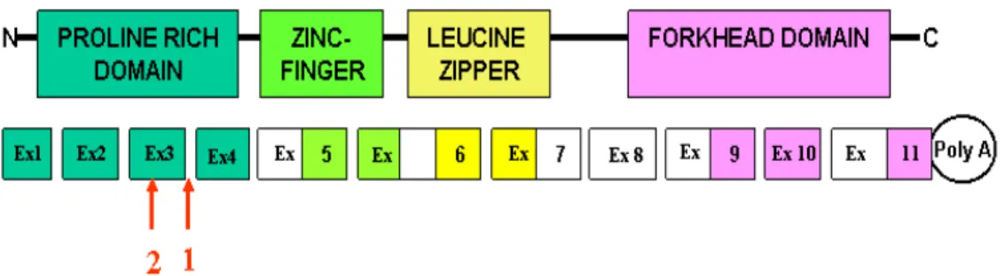
2.6 FOXP3
Human FOXP3 is a 47 kDa protein member of the forkhead/winged-helix family of transcription factors. The members of the fox family are both transcriptional repressors and activators, and have a forkhead (FKH) domain
(Fig. 3), which is critical for DNA binding and nuclear localization [40]. FOXP3 acts as a transcriptional repressor for promoters of key cytokine genes such as IL-2 and GM-CSF [42, 43]. Recent studies suggest that FOXP3 physically and functionally interacts with transcription factors that play key roles in the expression of multiple cytokine genes such as nuclear factor of activated T cells (NFAT), acute myeloid leukaemia 1/Runt-related transcription factor 1 (AML1/Runx1), and possibly nuclear factor-κB (NF-κB) [42, 44, 45].
In general, transcription factors have protein domains that allow them to interact simultaneously with DNA, with other protein cofactors, and potentially with the basal transcription machinery. In the case of FOXP3, the proline-rich N-terminus is a trans-repression domain required for suppression of NFAT mediated gene transcription, a central domain that contains a C2H2 zinc finger, a leucine zipper involved in protein/protein interactions, and a C-terminal region that contains the forkhead DNA-binding domain and nuclear targeting sequences (Fig. 3) [46]. Mutations that affect any of these key functional domains may alter or abrogate the ability of the transcription factor to regulate gene expression.
Fig. 3 Schematic representation of FOXP3 protein showing the locations
2.7 IPEX Syndrome
The IPEX syndrome was described 25 years ago by Powell et al [47] as a syndrome of enteropathy, endocrinopathy, and fatal infections affecting young male patients. The murine equivalent of IPEX is the so called scurfy mutant mouse with an analogous X-linked pathology [48]. The presence of this phenotype in multiple generations of the same kindred suggested that the disorder was genetic and followed an X-linked pattern of inheritance [47] IPEX is characterized by multiple autoimmune and allergic features [47, 49, 50], but it is now recognized that most patients present with a basic clinical triad of enteropathy, endocrinopathy (diabetes or thyroid disease), and dermatitis.
Early onset enteropathy, with villous atrophy and infiltrating inflammatory cells, is the most common manifestation. Clinically, this results in severe watery diarrhoea that is at times mucoid or bloody. The diarrhoea may markedly worsen after an affected infant is switched from being breast-fed to regular formula, and some patients develop severe food allergies [51]. Symptoms may or may not respond to dietary manipulation and consequently, patients often require total parenteral nutrition to reverse severe failure to thrive. Immune-suppressive therapy often improves but may not resolve the gastrointestinal symptoms and in most cases this difficulty is the primary cause of death. Early onset type 1 diabetes (T1D), with auto-antibodies to islet antigens, is usually present and other autoimmune endocrinopathies are common, together with thrombocytopenia, haemolytic anaemia, and arthritis [7, 50, 51].
Asthma and eczema, with increased serum immunoglobulin (Ig)E levels, are also commonly reported [50, 52]. In addition to the basic IPEX triad, patients with FOXP3 mutations typically have other autoimmune phenomena as well. The most common of these are autoimmune hematologic disorders including Coombs-positive haemolytic anaemia, autoimmune thrombocytopenia, and autoimmune neutropenia, which together affect approximately 50% of patients [7, 47, 52]. Relevant auto-antibodies can frequently be demonstrated in the serum.
In human beings, 13 FOXP3 mutations have been reported [7]. Ten disrupt the forkhead domain, 2 affect the leucine zipper domain, and 1 affects the first polyadenylation signal. The forkhead domain is required, both in humans and mice, for FOXP3 function [40]. The impact of mutations in the leucine zipper domain and in the polyadenylation site on expression and function of FOXP3 is still unknown.
We have studied two IPEX patients with a milder clinical phenotype and have identified their mutations which lay outside the FOXP3 functional DNA binding domain. The CD4+CD25+ T cell population was studied and analysed. One of the patients presented an uncontrolled enteropathy that became unresponsive to traditional immune-suppression. We therefore treated him with ECP according to the rational described in the subsequent paragraph.
2.8 Extracorporeal photochemotherapy
Extracorporeal photochemotherapy (ECP) consists in exposing ex-vivo patient’s peripheral blood leucocytes, obtained by a leukapheresis process, to ultraviolet-A (UV-A) irradiation in the presence of the DNA intercalating agent 8-methoxypsoralen (8-MOP) and subsequent reinfusion of autologous treated leukocytes into the patient (Fig. 4). ECP has been used in the treatment of severe refractory graft versus host disease, and to a lesser extent in the prevention of organ graft rejection [53]. Although the efficacy of this treatment awaits definitive proof in controlled clinical trials, data in several published series suggest that ECP represents an alternative to standard immune-suppression. Unlike conventional immune-suppressive therapies, patients undergoing long-term ECP have no reported risk of developing infections or acute side effects [53].
The mechanism of the immune-modulatory action of ECP remains elusive [54]. Exposure to UV-A and 8-MOP leads to apoptosis of the reinfused lymphocytes [55-57]. In animal models it has been shown that these cells localize mainly in the spleen and liver where they are scavenged by resident immature dendritic cells (DCs) which in turn develop into tolerogenic DCs [58]. It has been recently demonstrated that ECP treatment in humans increases the levels of circulating regulatory T cells in vivo [1, 2]. Based on this findings, on the poor control of clinical manifestations by conventional immune-suppression in our patient, on the observation that the mutation carried by the patient allowed the generation of some normal FOXP3 mRNA, we hypothesized that treatment with ECP in this patient could force FOXP3 expression inducing a significant increase in the amount
of normal FOXP3 mRNA leading therefore to an increase in regulatory T cells and to a better control of the clinical manifestations.
Fig.4 Extracorporeal photopheresis consists in: a) collection of PBMC
from patient by leukapheresis, b) ex-vivo exposure of PBMC to UVA in the presence of the DNA intercalating agent 8-methoxypsoralene, c) reinjection of the irradiated PBMC in the patient.
3. MATERIALS AND METHODS
3.1 Mutation analysis
CD2BP1
CD2BP1 exons were amplified from genomic DNA through the use of oligonucleotide primers reported by wise et al. [21]. Screening for mutations was performed by denaturing high-performance liquid chromatography (DHPLC) with the use of Wave analysis (Transgenomic, San Jose, Calif), using conditions recommended by WaveMaker 2.0 software. Polymerase chain reaction amplimers were purified with the Nucleospin kit (Macherey-Nagel, Duran, Germany) and sequenced with CEQ DTCS (Beckman, Fullerton, Calif), with the use of a CEQ 2000 automated sequencer (Beckman).
FOXP3
The 11 exons, including the intron-exon junctions, of the FOXP3 gene were amplified from genomic DNA by polymerase chain reaction (PCR), sequenced by the Big Dye Terminator kit version 1.1 (Applied Biosystems, Warrington, Cheshire, UK) and the sequences were run on an ABI Prism 377 (Applied Biosystems). A panel of 200 control men was analyzed by DHPLC to exclude that the nucleotide variations detected in the 2 patients were polymorphisms.
For the expression analysis in patient 1, the total RNA was obtained from peripheral blood mononuclear cells (PBMC) using TRIzol (Invitrogen,
Carlsbad, CA). Reverse transcription was performed with the Advantage reverse transcription for PCR kit (Clontech, Palo Alto, CA). Complementary DNA (cDNA) spanning from exons 1 to 5 was amplified by PCR, with specific primers. Transcripts were cloned by TOPO TA Cloning kit (Invitrogen, Life Technologies Inc.) and selected colonies, representing all the PCR products observed, were sequenced.
3.2 In Vitro Cytokine Production
Peripheral blood mononuclear cells (PBMC) from 15 healthy control subjects (age range, 6 to 21 years), 15 patients with oligoarticular juvenile idiopathic arthritis (oligo-JIA) (age range, 4 to 18 years), and unaffected and affected individuals of the family with PAPA syndrome were separated by means of Ficoll/Histopaque. Three samples of individual IV-2, one obtained before etanercept treatment, were available; 2 X10⁵ PBMC were cultured in RPMI 1640 (GIBCO, Invitrogen Corp, Carlsbad, Calif) with 10% FBS (HyClone, Logan, Utah) at 37°C in 5%CO2 in a 96-well plate with or without 10 ng/mL of lipopolysaccharide (LPS) (Escherichia coli, 055:B5; Sigma, St Louis, Mo) for the indicated time. A neutralizing monoclonal antibody to human interleukin (IL)-1β (R & D Systems) was added at 10 mg/mL. Otherwise for tolerization experiments, PBMC were pre-activated with LPS for 8 hr and re-stimulated with 10ng/ml LPS for 24 hr. The supernatants were harvested, centrifuged, and frozen at -80°C. IL-1β, IL-6, and TNFα levels were measured with commercial ELISA (R&D
Systems, Minneapolis, Minn, and Biosource Europe, Nivelles, Belgium), according to the manufacturer’s instructions.
3.3 Evaluation of the Phenotype and Function of
Regulatory T Cells
At the time of sampling, the IPEX patient 1 was receiving prednisone (.5 mg/kg/day) and cyclosporin A (7 mg/kg/day), and the IPEX patient 2 was receiving prednisone (2 mg/kg/day) and azathioprine (2 mg/kg/day). Two independent blood samples from the 2 patients were analyzed. PBMCs from the two IPEX patients were prepared by centrifugation on Ficoll-Hystopaque. Samples from 4 patients with autoimmune diseases treated with the same immune-suppressants (Table 3) were used as disease/treatment controls. Ten healthy young adults (age, 22–40 y) served as normal controls. Monoclonal antibodies for flow cytometry and sorting were from Becton-Dickinson (San Jose, CA). For flow cytometry, 5 X10⁵ PBMCs were stained with appropriate antibody combinations of fluorescein, phycoerythrin, antigen-presenting cells, and cytochrome-conjugated monoclonal antibodies; in particular, FN50 CD69), M-A251 (anti-CD25), RPA-T4 (anti-CD4), UCHT1 (anti-CD3), G42-8 (anti-CD8), HIB19 (anti-CD19), and 3G8 (anti-CD16). Cells were analyzed by FACSCalibur (Becton-Dickinson). For sorting, PBMCs were stained with fluorescein isothiocyanate– conjugated anti-CD8, CD16, and CD19 antibodies, and PE-conjugated anti-CD25 antibodies. CD4+ T cells were identified as CD8, CD16, CD19, and CD25 negative. The CD4+CD25+ T cells, containing Treg
cells, were negative for all markers, except for CD25. The cell populations were separated using the FACS Table Vantage SE (Becton-Dickinson).
2x10⁴ cells per well of sorted T cells (total CD4+ or CD4+CD25-) were cultured for 4 days in triplicate in 96-well round bottomed plates (Costar) in the presence of 2x10⁴ irradiated (3000 rad) autologous PBMCs in 200μl/well of serum-free X-VIVO-15 medium (BioWhittaker, Verviers, Belgium). Cells were stimulated with 2 μg/ml of soluble anti-CD3 (clone OKT3, ATCC, Manassas, VA, USA) and 0.15 μg/ml of soluble anti-CD28 (Becton-Dickinson). During the final 16 hours cells were pulsed with 0.5μCi/well of H-thymidine and proliferation was evaluated by thymidine uptake using a liquid scintillation counter.
3.4 Real-time PCR
Total RNA was extracted using TRI-reagent™ (SIGMA®, Saint Louis. MI or RNAeasy Mini Kit™ (Qiagen, Valencia, CA), according to the manufacturer’s instructions. Complementary DNA was synthesized from each RNA sample using 1st Strand cDNA Synthesis Kit for RT-PCR (AMV)+ (Roche Diagnostics, Indianapolis, IN ). Real-time polymerase chain reaction (PCR) was performed on the ABI PRISM™ 7700 Sequence Detector (Applied Biosystems, Foster City, CA) platform, using Taqman universal Master Mix (Applied Biosystems). FOXP3 expression was tested using Assays on Demand reagents (Applied Biosystems, Hs00203958_m1). All reported mRNA levels were normalized using TaqMan® Endogenous Control, GAPDH (Applied Biosystems 4319413E). Each PCR reaction was
conducted in triplicate. Relative quantification was preformed using the comparative CT method, as described [59].
3.5 Extracorporeal photopheresis procedure
ECP consisted of discontinuous leukapheresis with an extracorporeal system (UVARR apparatus Therakos, Westchester, Penn, USA). Approximately 240ml leukocyte-enriched blood was mixed with 8-methoxypsoralen (8-MOP) (final concentration: 200ng/ml) and was exposed to UVA radiation, cells irradiated with 2 J/cm2. The UVA treated leukocytes were then re-infused into the same patients. The protocol was approved by our institution’s ethics committee and the patient gave his informed consent. Ten healthy individuals provided control PBMC. ECP protocol consisted in two procedures in two consecutive days that were performed weekly for the first month and subsequently, every other week.
4. RESULTS
4.1 PAPA Syndrome case report
M.C., a 22-year-old man, was seen at age 2 years because of left shoulder arthritis, abscess of the left arm, and fever. Erythrocyte sedimentation rate, C-reactive protein, and white blood cell counts were elevated (Table 2). He was treated with antibiotics and showed slow improvement. His mother had severe necrotic pustules on her face and trunk during adolescence and had development of insulin-dependent diabetes mellitus (IDDM) at 42 years of age. The maternal uncle had severe joint deformities reportedly resulting from relapsing, intensively inflammatory arthritis involving several joints during childhood and had development of IDDM at 35 years. Until 10 years of age, the patient M.C. had 3 episodes of arthritis with similar clinical and laboratory features (Table 2). Synovial fluid was purulent, but cultures were negative. The synovial biopsy showed marked infiltration of neutrophils. The tuberculin test was negative. The NBT test, serum immunoglobulin levels, lymphocyte subsets, ANA, and RF were normal. He was treated with antibiotics and showed slow improvement. He had no additional episodes for 6 years. From the ages of 16 to 19 years, he had 4 similar episodes treated with non steroidal anti-inflammatory drugs and showed slow resolution (Table 2). PAPA syndrome was diagnosed on clinical data (recurrent pyogenic arthritis with negative synovial cultures) and family history. Prednisone (2 mg/kg per day) was started, resulting in rapid and complete remission. Prednisone was tapered and discontinued in 1 month.
prednisone (2 mg/kg per day). Cyclosporine A (5 mg/kg per day) was added. When prednisone was reduced to 1 mg/kg per day, he had two additional episodes of arthritis of the left knee and a sterile abscess of the quadricep muscle. Radiography showed narrowing of the joint space and a short and irregular femoral head of the right hip. He also had cystic acne and significant glucocorticoid side effects. Etanercept (25 mg/dose twice weekly), a recombinant soluble TNF receptor II/Fc fusion protein that neutralizes TNF was then started. Glucocorticoids were tapered and discontinued in 2 months. Up to now, the patient has been receiving etanercept for 30 months. No additional episodes of arthritis occurred, and cystic acne resolved. Moderate limitation of motion of the right hip persists.
Age (y) Affected joint Temperature at admission (C°) ESR (mm/h) CRP (mg/dL) WBC count (103/mm3) Neutrophils (%) SF leukocyte (x 103/mm3) Neutrophils (%) 2 Left shoulder 39,0 120 18,3 28,000 92 ND ND 7 Left ankle 39,2 80 18,6 11,750 74 80 92 8 Right hip 37,8 130 21,3 12,900 81 ND ND 10 MCP joints (2°,3°,4°) 39,5 80 2,9 8900 61 ND ND 16 Right wrist 36,8 14 1,5 8890 65 ND ND 17 Right ankle 37,8 80 2,9 7300 60 117 96 18 Right ankle 39 50 5,8 11,300 68 ND ND 19 Right hip 36,8 65 3,8 4000 71 97 87 20 Right hip 37 125 7,8 9670 88 ND ND 20 Left Knee 36,8 54 2,6 9330 72 81 90 20 Left knee 37,6 70 5,2 10,200 74 ND ND
Table 2. Clinical presentation and laboratory investigations of arthritis episodes in the proband individual IV-2. ESR, Erythrocyte
4.2 CD2BP1 Mutation analysis
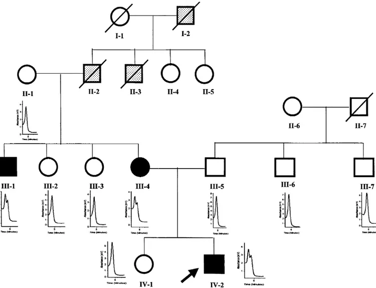
Denaturing high-performance liquid chromatography analysis of exon 10 amplimers showed heterozygosity in the proband and in two affected family members (Fig.4). Sequencing identified a wild-type and a mutant sequence with a G/A transition at position 904 in exon 10 of CD2BP1, identical to one of the reported disease-causing mutations [21].DHPLC analysis did not reveal additional heterozygous sequence variants in both affected and non affected family members.
Fig. 4 Pedigree of the family with PAPA syndrome. DHPLC profiles of
exon 10 of CD2BP1 gene are shown for individuals in whom analysis was performed. Arrow, proband; line through symbol, deceased; closed symbols, affected (III-1: recurrent arthritis from age 7 years and IDDM at age 35 years; III-4: pyoderma gangrenosum at age 12 and IDDMat age 42 years; IV-2: see Case Report); open symbols, unaffected; stippled symbols, history suggestive of PAPA syndrome but unable to confirm (I-2: severely crippled by arthritis at young age; II-2: some episodes of mild, self-remitting arthritis, died at age 32 years in an accident; II-3: recurrent arthritis and diabetes at age 40 years, died at age 45 years).
4.3 Alteration of pro-inflammatory cytokine
production in PAPA syndrome
It has been hypothesized that the increase in pyrin binding seen with the PAPA mutations may alter inflammatory susceptibility by altering the balance between proinflammatory and compensatory anti-inflammatory mechanisms. LPS, sensed by the innate immune system through TRL4, is a well known stimulus for inflammatory responses. The effects of LPS are mediated by multiple signalling cascades. We studied the production of pro-inflammatory cytokines after short, long and repeated exposure to LPS.
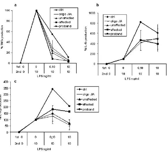
TNFα production after an 18-hour LPS stimulation was higher in affected subjects than in healthy control subjects or in patients with oligoarticular juvenile idiopathic arthritis (oligo-JIA). TNFα production of genetically normal relatives did not differ from that of control subjects (Figure 5a). In the proband (IV-2), TNFα production was high before and during etanercept treatment. Individuals III-1 and III-4 and patients with JIA were not receiving glucocorticoids or TNF inhibitors at sampling. No difference was found in IL-6 and IL-1β production after an 18-hour stimulation (Figure 5, b and c).
a b
c
Fig. 5 Production of proinflammatory cytokines TNFα (a), IL-6 (b), and IL-1β (c) in healthy controls, in patients with Oligo-JIA, and in unaffected and affected individuals of the family with PAPA syndrome. PBMC cells were cultured in the presence of 10 ng/mL LPS for 18 hours. For healthy control subjects and patients with Oligo-JIA, results are shown as whisker box, where limits of boxes represent 25th and the 75th percentile values; lines across boxes are median values and whiskers depict minimum and maximum values. For unaffected and affected family members, results are shown as individual symbols and are the means of two independent experiments. Two samples of PBMC from individual IV-2 were tested, one
In agreement with a recent observation [30],IL-1β release was markedly increased in subjects with PAPA syndrome after a short-term (2-hour) stimulation (Fig. 6a). To assess whether the early increase in IL-1β production could be responsible for the late increase in TNFα production, we evaluated TNFα production after an 18-hour stimulation with LPS in the presence of a neutralizing antibody to IL-1β. Neutralization of IL-1β resulted in reduction of TNFα production in control subjects and in patients. However, in the patients, TNFα production was still markedly higher than that of control subjects (Fig. 6b), suggesting that the increased production of TNFα after a long incubation (18hours) was partially secondary to the early IL-1β release.
b a
Fig. 6 Release of IL-1β after short-term stimulation (2 hours) with 10
ng/mL LPS (a) and production of TNFα after long-term stimulation (18 hours) in the presence or absence of a neutralizing antibody to human IL-1β (b) in healthy control subjects and in individuals with PAPA syndrome. For healthy control subjects, results are shown as whisker box, where limits of boxes represent 25th and the 75th percentile values; lines across boxes are median values and whiskers depict minimum and the maximum values. For individuals with PAPA syndrome, results are shown as individual symbols and are the means of two independent experiments. Two samples of mononuclear cells of individual IV-2 were tested, one obtained before (*) and the other after (**) etanercept treatment.
Chronic repeated exposure to endotoxin or LPS causes a transient increase in the threshold to endotoxin challenge. This is known as “endotoxin tolerance” and represents a negative feedback mechanism to protect from endotoxin shock. While TNFα is a phenotypic marker for LPS tolerance, production of other cytokines in tolerance is less clear.
Our results show that IL-1β, IL-6 and TNFα production, in response to LPS repeated exposure, is differently regulated (Fig. 7). PBMC were pre-stimulated with different doses of LPS for 9 hours and then re-pre-stimulated with 10ng/ml LPS. Cytokine production was observed after this second stimulation. At each dose of LPS, cytokine level was compared to the cytokine levels of PBMC that received only the second LPS stimulation. As shown by previous studies [60-62], TNFα levels showed a very strong reduction in a dose dependent manner, reaching 94% inhibition with the higher LPS dose pre-treatment. IL-6 showed a different regulation pattern since low doses of the 1st LPS stimulation augmented IL-6 levels, a phenomenon known as priming, while higher doses induced a modest (30%) reduction. IL-1β levels in contrast did not undergo any tolerogenic reduction but instead increased in a dose dependant manner.
Fig 7. Different cytokines respond in a different manner to LPS repeated
exposure. PBMC from normal controls were pre-treated with different doses of LPS (1st LPS). After 9 hours incubation cells were washed and restimulated with 10ng/ml of LPS (2nd LPS). After 16 hr TNFα, IL-1β and IL-6 levels were determined by ELISA. The concentrations of cytokine in each sample were normalized to the concentration of the sample that received the second stimulation alone. Values are represented as percentage of the cytokine levels in samples receiving the second stimulation alone. ( ) IL-6, ( ) TNFα, ( ) IL-1β
When we studied PBMC from the PAPA affected patient and his affected relative, a perturbation in endotoxin tolerance was observed only for IL-6. PBMC pattern production for TNFα and IL-1β in response to LPS pre-treatment reflected that observed for normal controls, oligo-JIA and unaffected family member (Fig 8a, 8b). In contrast IL-6 production was altered. No tolerance was observed for the higher LPS dose pre-treatment (Fig 8c). The proband also showed a very high priming, the percent increase of IL-6 levels for the lower dose of LPS was twice that of normal controls or oligo-JIA.
Although our patient showed a complete therapeutic response to TNF inhibition, these data suggest that mutation in CD2BP1 leads to a complex perturbation of the regulation of inflammatory cytokine production. The possibility exists that other cytokine inhibitor (i.e. anti-IL-6 receptor) may be of therapeutic benefit in these patients
Fig 8. PBMC were treated as already described with different doses of
LPS (1st LPS). After 9 hours incubation cells were washed and restimulated with 10ng/ml of LPS (2nd LPS). Cytokines levels were determined by elisa after 16 hr incubation. a) TNFα b) IL-1β and c) IL-6. The concentrations of cytokine in each sample were normalized as already described. ( ) controls, ( )oligo-JIA, ( )unaffected family members, ( ) affected family member, ( ) proband.
4.4 IPEX Syndrome case reports
Patient 1
A 22-year-old man was referred because of recurrent diffuse joint pain
associated with the presence of antinuclear antibodies (ANAs). The mother died at 42 years of age of intestinal lymphoma. The patient had 3 brothers. One brother died at 1 month of age from intractable diarrhoea; diabetes was not reported. The other 2 brothers, both asymptomatic, had biopsy examination proven celiac disease and responded to a gluten-free diet. One brother died at 14 years of age in an accident; the other brother, aged 26 years, was lost to follow-up evaluation. At 18 days patient 1 developed diarrhoea and severe malnutrition. Parenteral nutrition and a subsequent diet with a peptide formula reportedly were associated with improvement and he was maintained on a gluten-free diet. At 5 years, after reintroduction of a full gluten containing diet, abdominal pain, diarrhoea, and failure to thrive relapsed. An intestinal biopsy examination showed subtotal villous atrophy and mononuclear cell infiltrate in the lamina propria (Fig.9a). Immunohistochemistry subsequently was performed and revealed an intraepithelial CD3+CD8+ cell count of 42/100 epithelial cells (upper limit of normal, 40) (Fig.9b). High titers of antigliadin IgG and antiendomysial antibodies were found. Celiac disease was diagnosed. After an initial clinical improvement with a gluten-free diet, diarrhoea rapidly relapsed and failure to thrive persisted. Two years later a second intestinal biopsy examination showed similar findings (not shown). IgE levels were high (200 mU/mL; normal values for age, <60 mU/mL), reportedly in the absence of allergy. Diarrhoea and failure to thrive persisted. At 15 years, the patient
developed arthralgia and transient arthritis. Antienterocyte antibodies were detected for the first time (immunofluorescence on human duodenum; Dr. E. Bosi, San Raffaele Research Institute, Milan, Italy). A diagnosis of autoimmune enteropathy was made and treatment with glucocorticoids and azathioprine was started with partial improvement. At 18 years the patient developed well-defined, scaly, erythematous plaques on the trunk and limbs. A skin biopsy examination showed paracheratosis, Poitrier-like epidermal microabscesses, and mononuclear cell infiltrate in perivascular aggregates, consistent with psoriasiform dermatitis (Fig.9e). Chronic enteropathy remained active until admission to our unit despite continuous treatment with glucocorticoids, immune-suppressants (cyclosporin A, tacrolimus), and infliximab, alone or in combination, and intermittent parenteral nutrition. At admission, the patient was receiving prednisone, cyclosporin A, and nightly parenteral nutrition. His height was 164 cm (25th–50th percentile), and his weight was 48 kg (10th– 25th percentile). He was in fair general condition, presenting with weakness and mild signs of dehydration. Maculopapular lesions were present over the limbs and trunk. The liver was mildly enlarged. The white blood cell count was 10,570/mm3, hemoglobin level was 12.1 g/dL, platelet count was 219,000/mm3, ESR was 28 mm/h (normal, <20 mm/h), and the C-reactive protein level was 39 mg/L (normal, <5 mg/L). Metabolic acidosis (pH 7.30; bicarbonate, 13.3 mEq/L) and renal failure (serum creatinine level, 2.59 mg/dL; urea nitrogen level, 42 mg/dL) were present. Blood electrolyte, glucose, and liver enzyme levels were normal. ANAs were positive (1:160; homogeneous pattern). Antitransglutaminase antibodieswere negative. The patient was treated with 3 intravenous methylprednisolone pulses (1 g/day) and parenteral
prednisone (1 mg/kg/day in 1 daily dose), tacrolimus (.04 mg/kg/day), azathioprine(2 mg/kg/day), and nightly parenteral nutrition. His weight was 50 kg, creatinine level was 1.45 mg/dL, and bicarbonate level was 22.0 mEq/L.
Patient 2
A 5 year-old boy was referred to our unit because of pain in the wrist and the left proximal interphalangeal joint of the second finger and diffuse arthralgia. His 12-year-old brother was healthy. Severe chronic diarrhoea had appeared at 14 months of age. An intestinal biopsy examination showed dysmorphic villi with no atrophy and a marked mononuclear cell infiltrate in the lamina propria (Fig.9c) and an intraepithelial CD3+CD8+ cell count of 60/100 epithelial cells (upper limit of normal, 40) (Fig.9d). Antigliadin and antiendomysial antibodies were negative. The patient did not improve on a gluten-free diet. IgE levels were 74 mU/mL (normal value for age, <10 mU/mL). Antienterocyte antibodies were negative. ANAs were present (1:5120, homogenous pattern). The patient was diagnosed with an unspecified autoimmune enteropathy and treatment with prednisone, azathioprine, and exclusive intravenous nutrition was started. Substantial clinical improvement and weight gain ensued. At 18 months of age the patient developed pneumonia with pleuritis, and pericarditis a month later. He was treated with prednisone and azathioprine; treatment was tapered and eventually withdrawn at 3 years and 5 months. Since then he had been growing satisfactorily on a full gluten-containing diet. At admission, he was in good clinical condition. His height was 110 cm (50th–75th percentile) and his weight was 16 kg (10th–25th percentile). Physical examination
showed moderate swelling of the wrists and of the left proximal interphalangeal joint of the second finger. No other signs or symptoms were present. Blood cell counts and liver and kidney functions were normal. ESR was 34 mm/h, the C-reactive protein level was 3.3 mg/L. ANAs were confirmed at a titer of 1:5120 with a homogenous pattern. He was treated with naproxen (15 mg/kg/day). In the subsequent weeks, arthritis and arthralgia persisted and he presented with occasionally watery diarrhoea. Treatment with prednisone and azathioprine was started and was followed by a rapid resolution of arthritis and diarrhoea. After 2 years, the patient is presently a 7-year-old boy in good health, regularly attending school, who has not presented with recurrences of his enteropathy, and is being treated with azathioprine (2 mg/kg/day) and prednisone (presently .2 mg/kg/day on an alternate-day regimen). ANAs still are present (1:2560). The IgE level is 7.6 IU/mL (normal values for age, <100 IU/mL).
4.5 Additional information
The following investigations were normal or negative in both patients: oral glucose tolerance test, HbA1c, fruttosamine, insulin-like growth factor, DHEA-S, thyroid functions, including a thyroid ultrasound,antibodies to glutamic acid decarboxylase, thyroid (thyroglobulin and thyroid-peroxidase), smooth muscle, liver-kidney microsomes, mitochondria, extractable nuclear antigens, cardiolipin, and neutrophil cytoplasm.
In patient 1 the intestinal biopsy examination showed subtotal villous atrophy, an intraepithelial lymphocyte count greater than 40%, and
modified Marsh classification [63], and the persistently high titers of antigliadin and antiendomysial antibodies were consistent with a diagnosis of celiac disease. However, patient 1 did not respond to a gluten-free diet. Whether he is affected with both IPEX and celiac disease, or whether the features compatible with celiac disease are manifestations of the immune dysregulation of IPEX, is difficult to ascertain. From a practical point of view, this case indicates that in patients with early onset enteropathy and celiac disease–related antibodies, a failure to respond to a gluten-free diet should prompt the suspicion of IPEX syndrome, even in the presence of histological findings highly suggestive of celiac disease.
a
d b
c
e
Fig.9 Intestinal biopsy specimen of (a, b) patient 1 and (c, d) patient 2,
and a skin biopsy specimen of (e) patient 1. (b, d) Immunohistochemistry of the intestinal biopsy specimen with an anti-CD3 antibody.
4.6 FOXP3 Mutation analysis
Patient 1 carries an A>G transition at +4 in intron 3 (454 + 4 A>G), very
close to the splice site (Fig. 10) . Reverse-transcription PCR was performed on RNA from PBMCs to obtain cDNA corresponding to exons 1–5. Three cDNA transcripts of 431, 326, and 187 base pairs (bp) were detected (Fig. 11). Cloning and sequencing of the 3 fragments showed that the 431-bp fragment corresponds to the full-length cDNA produced by healthy subjects; the 326-bp transcript also is found in some normal controls and represents an alternative splicing with skipping of exon 2 and conserved frame (GenBank AJ005891); in the 187-bp transcript skipping of exons 2 and 3 was seen, resulting in a frame shift. The putative resulting protein has an abnormal amino-acid composition and it is truncated owing to a premature stop codon in exon 7.
Patient 2 is hemizygous for a C>T transition in exon 3 at nt 323 (Fig.
10), causing the substitution of a threonine with methionine at position 108 (T108M). This missense mutation affects the proline-rich domain and the resulting protein has a predicted normal forkhead domain.
The mutations of the 2 patients were not found in the screening of the 200 healthy adult men.
Fig. 10 Schematic representation of Foxp3 protein showing the locations
of the mutations identified in IPEX patient 1 (1: A>G np+4 intron 3) and IPEX patient 2 (2: C323T (T108M)).
Fig. 11 FOXP3 transcripts in patient 1 and in 1 healthy control. The
431-bp fragment corresponds to the full-length cDNA produced by healthy patients; the 326-bp transcript found in the patient and in some of the normal controls represents an alternative splicing with skipping of exon 2 and conserved frame. The 187-bp transcript skips exons 2 and 3 and results
4.7 Phenotype and function of regulatory T cells
The 2 IPEX patients had a CD4+ T cell subpopulation that constitutively expressed CD25. Flow cytometry analysis showed that the percentage of CD4+CD25+ was normal compared with that found in healthy controls (Table 3). Children with other chronic autoimmune diseases who were treated with the same immune-suppressants had similarly normal percentages of CD4+CD25+ T cells (Table 3). Treg cells are enriched in the population expressing high levels of CD25 (CD4+CD25+bright T cells). Evaluation of CD4+CD25+bright T cells showed no differences from healthy controls (Table 3). In both IPEX patients, similar to disease/treatment control patients, a negligible percentage of the CD4+CD25+ T cells expressed the activation marker CD69 (Table 3), therefore suggesting that antigen-activated T cells did not contribute significantly to the subset of CD4+CD25+ T cells. To investigate the function of the CD4+CD25+ population, we evaluated the ability of CD4+CD25+ to suppress the proliferation of autologous CD4+CD25+ T cells. CD4+CD25+ T cells from healthy controls suppressed the proliferation of autologous effector T cells by 15%–40% (Fig.12). On the contrary, co culture of CD4+CD25+ T cells from IPEX patients with autologous T cells did not result in any suppression, but rather in increased proliferation (Fig.12). CD4+CD25+ T cells from disease/treatment controls suppressed proliferation of autologous T cells in a range similar to that of healthy controls, excluding that the lack of suppression by the CD4+CD25+ cells of IPEX patients was secondary to treatment (Fig. 12).
sample
CD25
+ total(% CD4
+)
CD25
+ bright(% CD4
+)
CD69
+(%CD4
+CD25
+)
Healthy controls (range) 6.5 - 10.7 .48 - 1.72 .6 – 4.2 IPEX (patient 1) 7.3 .91 1.95 IPEX (patient 2) 6.6 1.01 1.79 Autoimmune/treatment Control 1 8.53 .62 2.41 Control 2 9.2 1.99 1.89 Control 3 6.92 1.05 1.29 Control 4 10.38 2.40 .51Autoimmune/treatment: control 1, systemic juvenile arthritis treated with prednisone (.4mg/kg/day) and cyclosporine A (4 mg/kg/day); control 2, juvenile dermatomyositis treated with cyclosporine A (5mg/kg/day); control 3, systemic juvenile idiopathic arthritis treated with prednisone (.1mg/kg/day) and cyclosporine A (5mg/kg/day); control 4; systemic lupus erythematosus treated with prednisone (.4mg/kg/day) and azathioprine (2mg/kg/day).
Table 3. CD4+ CD25+ T Cells in Patients with IPEX Syndrome, Healthy
Controls, and in Patients with Autoimmune Diseases Treated with Glucocorticoids and Immune-suppressants
Fig.12 Proliferation of effector CD4+CD25- T lymphocytes in the absence
or in the presence of CD4+CD25+ Treg from healthy controls, from autoimmune/treatment controls, and from the 2 IPEX patients. CD4+CD25 -and CD4+CD25+ populations were purified by sorting and were stimulated with anti-CD3 and anti-CD28 as described in the Materials and Methods section. Results are expressed as a percent of thymidine uptake of stimulated effector CD4+CD25- T lymphocytes cultured in the absence of
CD4+CD25+ T cells. ( ) Healthy controls; ( ) patient 1 ( ) patient 2;; (●) disease/treatment controls.
4.8 ECP increases FOXP3 expression in CD4+
T lymphocytes
Patient 1 showed a milder clinical phenotype and long-term survival. His FOXP3 splicing mutation (454+4A>G in intron 3) causes the generation of a splicing variant and the predicted protein lacks the fork-head domain, which is essential for FOXP3 function. We also found that this mutation allows the generation of low amounts of normal full length FOXP3 mRNA [64]. This genetic background, the poor control of clinical manifestations by conventional immune-suppression, and the previous observation that ECP treatment induces Treg in vivo [54] made us hypothesize that treatment with ECP in this patient could force FOXP3 expression inducing a significant increase in the amount of normal FOXP3 mRNA leading therefore to an increase in regulatory T cells and to a better control of the clinical manifestations. Incidentally, the primers used for the Real-time PCR assay amplify a 69bp amplicon which lies between exon 10 and 11 of the FOXP3 gene and code for the functional forkhead domain present only in full length FOXP3 mRNA and not in the abnormal splicing variant produced by the IPEX patient 1.
Indeed in preliminary experiments we found that treatment with ECP caused an increase in FOXP3 mRNA expression in vivo in otherwise healthy individuals who underwent organ transplantation (Fig.13a), therefore suggesting that the in vivo increase in regulatory T cells following ECP treatment involves induction of FOXP3 expression. As expected, before treatment with ECP normal full-length FOXP3 mRNA levels in the IPEX patient 1 were markedly lower than those of the healthy controls
levels increased markedly to levels comparable to those of healthy controls (Fig.13b). The increase in full-length FOXP3 mRNA was present in the CD4+CD25+ population, as well as in the CD4+CD25- T cell population (Fig.12c). Consistently, flow cytometry analysis revealed a significant increase in the percentage of CD4+CD25+ T cells expressing the FOXP3 protein (Fig.13d). The latter observation suggests that ECP can induce de novo expression of FOXP3 in non-regulatory T cells.
Fig.13 Extracorporeal photopheresis up-regulates FOXP3 expression.
Normal full-length FOXP3 mRNA expression assessed by real-time PCR in: (a) PBMC from organ transplanted patients before ( ) and 1 month after beginning ECP treatment ( ), (b) PBMC from healthy controls ( n=3) and from the IPEX patient before ( ) and 1 month after beginning ECP treatment ( ), (c) CD4+CD25+ and CD4+CD25- T cells purified by cell sorting from a healthy control ( ) and from the IPEX patient before ( ) and 1 month after beginning ECP treatment ( ). Results are shown relative to pre-treatment levels using the comparative CT method as described in Materials and Methods. (D) Intracellular FOXP3 protein expression by flow cytometry in CD4+CD25+ T cells from two representative (out of 10) healthy controls and from the IPEX patient before and 1 month after
4.9 ECP induces circulating CD4+CD25+
T cells
with regulatory function
Flow cytometry analysis showed that the treatment with ECP induced in the IPEX patient 1 a substantial increase in the percentage of circulating CD4+CD25+CD69- T cells (Fig.14a). When CD4+CD25+high CD69- T cells were evaluated, a marked increase was also observed (Fig.14a). The patient refused to follow a continuous every other week ECP treatment protocol, and therefore the treatment was administered in three different periods separated by interruptions of 6 to 8 weeks. We have sequentially measured the levels of CD4+CD25+CD69- T cells for 1 year. Following the beginning of each ECP treatment period we observed an increase in CD4+CD25+CD69- T cells (Fig.14b). During ECP the percentage of CD4+CD25+ (mean 20,2 ± 10,1) and of CD4+CD25+high (mean 8,9 ± 2,2) was significantly higher (p= 0.04 and p= 0.006, respectively) than that observed when the patient was not treated with ECP (mean 6,7 ± 3 and 0,23 +/-0,07, respectively).
To verify whether the CD4+CD25+ T cells induced by ECP treatment showed regulatory function, we evaluated the ability of CD4+CD25+ T cells, present in the total CD4+ T cell population, from the IPEX patient 1 to suppress the proliferation of activated autologous effector CD4+CD25- T cells stimulated with anti-CD3 and anti-CD28. CD4+CD25+ T cells from healthy controls suppressed the proliferation of autologous effector T cells by 15%–40% (mean 29,3+/- 9,3). As previously mentioned, CD4+CD25+ T cells from patients with IPEX have defective suppressive function in vitro. Co-culture of CD4+CD25+ T cells from IPEX patient 1 with autologous T cells before ECP treatment did not result in any suppression, but rather in
increased proliferation (Fig.14c). In contrast, following ECP treatment CD4+CD25+ T cells from IPEX patient 1 suppressed proliferation of autologous effector CD4+CD25- T cells to an extent similar to that of healthy controls (Fig.14c). This suppressive activity was lost when ECP was suspended.
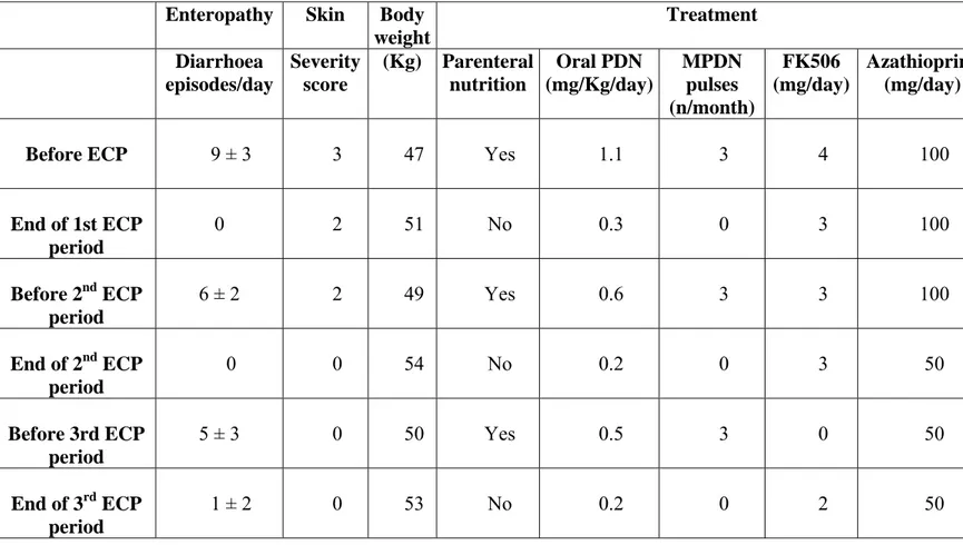
IPEX patient 1 was treated with three cycles of ECP (every two weeks for the first month, then every other week) as an add-on to standard treatment with prednisone, azathioprine and FK506 (Table 4). Following ECP treatments his clinical manifestations disappeared, renal function and metabolic acidosis normalized, a 5 Kg weight increase occurred, nightly parenteral nutrition was withdrawn and immunosuppressive treatment was tapered (Table 4). As previously mentioned the patient refused a continuous every other week ECP treatment protocol. Following the interruption of treatment intestinal symptoms reappeared together with a decrease in regulatory T cell numbers and a loss of suppressive function.
Fig.14 ECP treatment increases CD4+CD25+ with a regulatory function in the IPEX patient. (a) Representative plots of CD4+CD25+ T
cells from the IPEX patient before and after ECP treatment. The percentage of total CD4+CD25+ and of CD4+CD25+high are indicated (b) Percentage of CD4+CD25+ T cells during the different periods of ECP treatments, indicated by the shaded boxes. (c) The functional activity of CD4+CD25+ T cells was evaluated by the ability to suppress proliferation of stimulated autologous CD4+CD25- effector T lymphocytes as described in Material and Methods. CD4+CD25- and total CD4+ T cells, containing the CD4+CD25+ T cell population, were purified by cell sorting from healthy controls ( n=10) and from the IPEX patient 1 before ( ) and during ( ) period 1 and 2 of ECP treatment. After 72 hours of culture, 3H-thymidine incorporation was determined. Results are expressed as percent proliferation of stimulated effector CD4+CD25- T lymphocytes cultured in the presence of autologous total CD4+ T cells. Indicate the period during which blood samples were collected.
Enteropathy Skin Body weight Treatment Diarrhoea episodes/day Severity score (Kg) Parenteral nutrition Oral PDN (mg/Kg/day) MPDN pulses (n/month) FK506 (mg/day) Azathioprine (mg/day)
Before ECP 9 ± 3 3 47 Yes 1.1 3 4 100
End of 1st ECP period 0 2 51 No 0.3 0 3 100 Before 2nd ECP period 6 ± 2 2 49 Yes 0.6 3 3 100 End of 2nd ECP period 0 0 54 No 0.2 0 3 50 Before 3rd ECP period 5 ± 3 0 50 Yes 0.5 3 0 50 End of 3rd ECP period 1 ± 2 0 53 No 0.2 0 2 50
5. DISCUSSION
5.1 PAPA Syndrome
The PAPA associated mutations in CD2BP1 do not affect it’s interaction with either CD2 or WASP [21], and up to now no defects in adaptive immunity have been reported in PAPA syndrome patients. The severe neutrophil infiltration in affected tissues of PAPA patients suggests a model of disease pathogenesis in which inflammation results directly from dysfunction of the innate immune response. In this scenario, disease-causing mutations in CD2BP1 somehow exaggerate the signal for proliferation and infiltration of inflammatory initiator cells, alter apoptotic pathways and/or inhibit their clearance, leading to the observed abnormal accumulations of neutrophil-rich material.
Our study of a subject with PAPA syndrome, of his affected mother and maternal uncle, the third family reported in the literature, support this idea. We show that PBMC isolated from the affected subjects produce markedly increased amounts of TNFα and treatment of the proband with the TNF inhibitor etanercept resulted in clinical long-lasting remission. This also underscores the possible role of TNFα in the inflammatory process of this disease. The high in vitro TNFα production of patients with genetically confirmed PAPA syndrome is consistent with this hypothesis.
At high concentrations, TNFα can lead to excess inflammation and organ injury. Activated macrophages are the primary source of TNFα in inflamed synovial tissue, and both the number of macrophages and the degrees of
release of very large amounts of TNFα during sepsis may result in septic shock. In disease states, TNFα is generally considered to be a pro-inflammatory cytokine, along with IL-1β, IL-6, IL-17, and other cytokines. TNFα is a pleiotropic cytokine, in that it mediates a variety of direct pathogenic effects and induces the production of other mediators of inflammation and tissue destruction, placing it at the head of an inflammatory cascade within an inflammatory network. [66]. A variety of activities of TNFα such as cell recruitment, cell proliferation, cell death and immune regulation [66, 67] are thought to be inhibited by TNF antagonists [68].
As a result of tyrosine hyperphosphorylation, PAPA-associated CD2BP1 mutants bind with higher and stronger affinity pyrin, the protein encoded by the MEFV gene responsible for FMF [30]. This observation, together with our findings, suggest that mutation in CD2BP1 leads to deranged regulation of the inflammatory response. Indeed changes produced by CD2BP1 missense mutations are sufficient to confer susceptibility to inflammatory episodes, suggesting that CD2BP1 is critical in the homeostasis of inflammation. FMF is a rare, recessively inherited auto-inflammatory disease characterized by self-limited episodes of fever with arthritis, and sterile inflammation of peritoneal and pleural membranes [69]. Although clinically distinct from PAPA syndrome, FMF is also characterized by neutrophil infiltration into target tissues, and pyrin itself is expressed in neutrophils, but not T or B cells [70].
Pyrin binds to and inhibits ASC, a protein involved in recruitment and activation of caspase-1, which in turn is responsible for processing pro– IL-1β in its mature form [71]. Pyrin-deficient mice show increased