SCUOLA DI DOTTORATO IN SCIENZE E TECNOLOGIE CHIMICHE INDIRIZZO: SCIENZE FARMACEUTICHE
CICLO XXVIII
Polysaccharides as Drug
Delivery Systems for
different Administration
Routes
Andrea Salis
Tutor:
Prof. Paolo Giunchedi
Direttore della Scuola di Dottorato:
2
CONTENTS:
Abstract 4
Introduction 7
Chapter 1: Relationships Between the Properties of Self-Emulsifying Pellets and of the Emulsions Used as Massing Liquids for Their Preparation
29
Chapter 2: Development of thermosensitive chitosan/glicerophospate injectable in situ gelling solutions for potential application in intraoperative fluorescence imaging and local therapy of hepatocellular carcinoma: a preliminary study
55
Chapter 3: Nasal Chitosan Microparticles Target A Zidovudine Prodrug To Brain Hiv Sanctuaries
85
Chapter 4: Composite Chitosan/Alginate Hydrogel For Controlled Release Of Deferoxamine: A System To Potentially Treat Iron Dysregulation Diseases
118
Chapter 5: Engineered microparticles based on drug–polymer coprecipitates for ocular-controlled delivery of Ciprofloxacin: influence of technological parameters
147
Licence agreements 172
4
ABSTRACT
Polysaccharides are biocompatible and biodegradable long carbohydrate molecules organized in repeated monosaccharide units interconnected with glycosidic bonds. These polymers constitute a large source of biomaterials for drug vehicles, controlled drug delivery, tissue engineering etc. The ionic character and the presence of reactive functional group led them to be good candidates for the development of controlled release formulations. They are well tolerated by living tissues and body cavities due to their biocompatibility and biodegradability. In fact, polysaccharides have been widely proposed as drug delivery devices for many administration routes such as oral, parenteral, ophthalmic, nasal and transdermal route.
Furthermore, polysaccharides can be considered appropriate for tissue engineering thanks to their mechanical and biochemical functions and ability to deliver and foster cells.
The aim of the present work was the development of polysaccharides based platform formulations suitable for the delivery of drugs into specific anatomical districts.
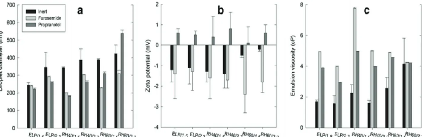
Self-emulsifying pellets can be considered a multiple unit dosage form capable of transferring a lipophilic drug in the gastric fluids dissolved in fine oil droplets. The combined advantages of this composite formulation are improved stability and absorption of lipophilic drugs, lower dose dumping in gastric transit time tank to a smother movement in the gastrointestinal track. Self-emulsifying mixtures (oil/surfactant/drug), in the form of o/w emulsions, consisting of caprylic/capric triglyceride, and three Cremophors (ELP, RH40, and RH60) at 1.5 and 2.3 weight ratios, and two drugs (furosemide and propranolol) of different lipophilicity were prepared and used as wetting liquids for the preparation of microcrystalline cellulose (MCC) pellets.
Properties of emulsions such as droplet size, zeta potential (ζ),viscosity and evaluation of reconstituted emulsions were determined. Furthermore, the characteristics of pellets such as pellet size, shape, friability, tensile strength, disintegration, and drug migration in pellets were evaluated. An experimental factorial design and 3-way ANOVA was performed in order to assess the effect of drug, surfactant and oil/surfactant ratio on the above-mentioned properties of emulsions and pellets. The results of these studies revealed that the 3 factors affected droplet size, viscosity and ζ of emulsions and size, shape, and friability of MCC pellets. Drug migration was more evident for the less lipophilic drug (furosemide) and lower oil/surfactant ratio.
Hepatocellular carcinoma (HCC) is a high chemoresistant tumour and localized anticancer therapies play a key role inthe treatment of advanced stages of HCC. Among them, transarterial embolization (TAE) implies the injection of embolic materials in the artery feeding the tumour, which results in ischemia and subsequent tumour necrosis.
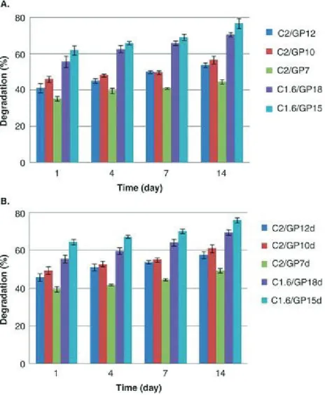
Indocyanine green (ICG) loaded chitosan/glycerophosphate (C/GP) thermogelling solutions were prepared with the aim to develop a new platform for the imaging and loco-regional treatment of HCC. ICG was incorporated in these formulations in order to evaluate their potential for the detection of tumours by fluorescence. The technological properties of these systems such as gel formation, gelling time, injectability, gel strength, gelling temperature, biodegradability and in vitro dye release studies were assessed. Ex vivo embolization studies were carried out for a preliminary embolic evaluation by using an isolated bovine liver.
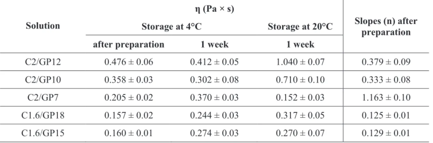
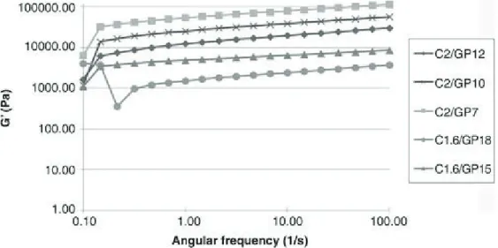
It was detected an increase of gel strengths and gelation rate for formulations with higher cross-link density between C and GP. These behaviours are unexpectedly more evident for C/GP solutions where a gel-like precipitation at 4°C occurred. Solutions with the lowest cross-link density between C and GP were more injectable thanks to a lower resistance to flow. Dye loading did not affect the hydrogel properties. The release of ICG from all C/GP systems analysed was not significant due to a strong electrostatic interaction between C and ICG. Ex vivo studies showed the fast gelation of these formulations in correspondence of the injected site. In conclusion, ICG-loaded C/GP thermo sensitive solutions have the potential for loco regional treatment of HCC following intraoperative fluorescence detection and resection of tumour nodules.
Antiretroviral therapy against HIV infection shows limited efficacy due to poor bioavailability of the corresponding drugs, such as zidovudine (AZT), at HIV reservoir sites. This drawback is quite evident in cerebrospinal fluid (CSF) subarachnoid spaces where macrophages are the only site of HIV replication in the brain. The rationale is that the active efflux transporters extrude zidovudine from the central nervous system (CNS) and macrophages. The prodrug called UDCA-AZT made with the conjugation of AZT to ursodeoxycholic, previously synthesized by our research group, is able to elude the AET systems. Chitosan chloride-based microparticles (CP) loaded with UDCA-AZT were developed for the nasal administration of this molecule as a potential strategy to enhance its uptake into the CNS. This formulation was characterized in terms of size, morphology, density, water uptake and the dissolution rate of UDCA-AZT. In addition, the CP sample was administered to rats via the nasal route. Drug release studies showed the capability of this formulation to release the drug in a relatively short time. Thus, a rapid absorption of the
6 prodrug after the nasal administration of the CP can be potentially achieved. In vivo studies demonstrated that nasal administration of UDCA-AZT loaded CP microparticles produced detectable amount of this prodrug in the CSF of the rats that could be sufficient to achieve an anti-HIV activity.
Iron excess is considered a key factor in the etiology of neurological disorders, cancer, stroke, muscle diseases, and aging. This toxicity is the consequence of the release of reactive oxygen species (ROS) whose are responsible of oxidative damage to various cell components such as lipid membranes, DNA, and proteins. Iron dysregulation can be overcame using iron chelators such as deferoxamine (DFO). Nevertheless, DFO displays reduced absorption in the gut and short plasma half-life. DFO loaded chitosan/alginate hydrogels were developed with the aim to overcome DFO limitations by using prolonged delivery systems. DFO loaded hydrogel alone or composite systems of chitosan/alginate hydrogels and DFO loaded poly(d,l-lactide-co-glycolide) microspheres were investigated in vitro. The influence of the preparation methods on the performance of composite hydrogels on controlled DFO release was assessed. Among the formulations studied, only the composite hydrogels were able to sustain the release of DFO. In particular, it was observed that the inclusion of microspheres into pre-formed chitosan/alginate hydrogel led to the best platform for the release of this iron chelator: DFO released from microspheres is strongly entrapped in the hydrogel network and, consequently, it is slowly released from the gel matrix by diffusion.
Ocular infections from Gram-positive and Gram-negative bacteria are commonly treated with topical eye drops loaded with ciprofloxacin. This type of formulation displays some drawbacks such as the rapid ocular clearance and frequent administrations are required. To overcome this limit, ciprofloxacin loaded chondroitin sulphate or lambda carrageenan microparticulate controlled release systems were engineered. The ionic interactions between ciprofloxacin and these polysaccharides led to coprecipitates that can act as vehicles for the sustained release of this drug after being displaced by the mediums ions. Technological parameters such as drug and polymer concentration, use of surfactant (kind and concentration), temperature and stirring of the solutions were evaluated in order to achieve coprecipitates with a particle size suitable for ophtalmic administration. In addition, mucoadhesion tests were performed on the leader formulations in order to understand if these systems are capable to be retained in the precornal region. Chondroitin sulphate coprecipitates exhibited the best properties in terms of particle size and mucoadhesivity, in particular with the use of surfactants during the process of preparation of these formulations.
INTRODUCTION
Biomaterials are considered biocompatible resources with the ability to interact with biological systems to evaluate, treat, augment or replace any function, tissue or organ of the body [1].
Polysaccharides are well-known carbohydrate polymers that constitute one of the most abundant supply of biomaterials for drug vehicles, controlled drug delivery, tissue engineering etc.
Polysaccharides have several natural resources from microbial, algal, plant and human origin [2]. The based-molecular structure is composed of one or more monosaccharide repeated units interconnected with o-glycosidic bonds. The backbone can be composed of only one kind of repeating monosaccharide, named homopolysaccharides or homoglycans (e.g. starch, cellulose), or formed by two or more different monomeric units, named heteropolysaccharides or heteroglycans (e.g. agar, alginate, carrageenan).
The general formula is Cx(H2O)y where 200≤ x ≤2500. Taking into account that the repeating units in the polymer backbone are usually monosaccharides with six carbons, the general formula can also be symbolized as (C6H10O5)n where 40≤ n ≤3000.
Polysaccharides possess linear or branched chains with the presence of reactive moieties such as hydroxyl, amino, and carboxylic acid groups. The presence of this reactive functional group indicates the prospect for chemical modifications [3]. Molecular weight range of this class of polymeric materials is from hundreds to thousands of Daltons [3].
Polysaccharides are widely applied in the drug delivery field due to their following merits:
1) they can be isolated in a well defined and reproducible way from natural sources including plants, animals, and microorganisms [4];
2) they are biocompatible and biodegradable due to their biochemical similarity with human extracellular matrix components. In particular, they can be immediately recognized and accepted by the human body with low immunogenic properties and they can go through enzymatic and/or hydrolytic degradation into non-toxic compounds [5]
3) they are capable of chemical and enzymatic modifications to give origin to different derivatives potentially suitable as drug delivery systems [6];
4) the ionic character and the presence of reactive functional group led them to be good candidates for the development of controlled release formulations;
8 5) ionic polysaccharides, which exhibit sensitiveness to environmental stimuli (chemical or physical), are an efficient alternative to synthetic polymers in the development of stimuli responsive drug delivery systems [7];
6) ionic polysaccharides have mucoadhesive properties [8];
7) they are capable to make conjugates and complexes with many macromolecules, such as proteins and peptides for achieving active targeting and controlled release systems [9];
8) large number of polysaccharides are capable to form interpenetrated hydrogel networks as matrices to encapsulate drugs and living cells, as biocompatible scaffolds for tissue engineering and for the controlled release of macromolecules like proteins. These hydrogels usually show physic-chemical characteristics more different from their macromolecular constituents [8,10,11].
The purpose of the present introduction is to describe five different polysaccharides that were used for the development of my thesis project.
Cellulose and derivatives
Cellulose is the most abundant compound of the cell walls of plants, bacteria, fungi and algae, which possesses a fundamental structural function. It is primary composed of glucose units interconnected with o-glycosidic bonds, organized in a fibrillary structure. This polymer shows high rigidity due to the various inter and intra-molecular hydrogen bonding between the hydroxyl groups of glucose units into elementary microfibrils which aggregate forming thick macrofibrils [12]. Cellulose presents different composition and morphology depending on the source from which it is obtained. No type of natural celluloses dissolves in common aqueous and organic solvents. The presence of hydroxyl groups in the glucose units of cellulose permits to synthesise many cellulose derivatives [13]. Among them, cellulose esters, cellulose ethers, microcrystalline cellulose, and cross-linked or graft copolymers are widely used in the pharmaceutical field as controlled drug delivery systems. Their applications include formulation of membrane controlled drug release systems or monolithic matrix systems [14]. The manufacture of membrane controlled release can be obtained by film coating with the aim to develop enteric coated dosage forms and osmotic pump delivery systems by using semi-permeable membranes [14].Monolithic matrix systems are amongst the most popular technologies for controlled drug delivery because of their simplicity of formulation, ease of manufacture, low cost, acceptance and applicability to drugs with a wide range of solubility [14].
Cellulose ethers and esters have been widely used for the development of oral sustained and controlled release dosage forms as coating agents that respond to variations in physiological environment, as matrices to enhance the bioavailability and solubility of drugs, to realise a pre-determined release profile from the final delivery system, and to increase the stability of the formulation [15].
Cellulose acetate phthalate and phthalated hydroxypropylmethyl cellulose are derivatives used as entering coating materials for the development of controlled intestinal targeting drug release formulations. Indeed, these polymers are stable in acidic conditions but soluble in a slightly alkaline environment [15].
Carboxymethyl cellulose and hydroxypropyl cellulose are considered mucoadhesive polymers applied for the delivery of drugs sublingually, orally, intravaginally, rectally, nasally and ophthalmically. Formulations made with this polymer prolongs the residence time of the drug at the site of absorption, allowing to high concentration gradient of the released compound, its protection from enzymatic degradation and enhanced absorption [16].
Typical cellulose esters used as hydrophobic matrices for sustained release of drugs (including highly water-soluble drugs) are cellulose acetate, cellulose acetate propionate, and cellulose acetate butyrate. The matrix of these polymers remain essentially intact in the GI tract while drug is released [17]. Cellulose esters are also used to construct the semi-permeable membrane of osmotic pump delivery systems. When the device is in contact with body fluids, water is able to permeate the cellulosic membrane and dissolves the osmotic agent resulting in a water activity gradient across the membrane [17].
Recently, Plackettet al. [18] have been developed nanocellulose which including cellulose nanocrystals, nanofibrilated cellulose, and bacterial cellulose as novel cellulose-based systems capable to entrap water soluble drugs and poorly water soluble drugs. These formulations exhibited long lasting drug release profile over weeks or even months.
Another important cellulose derivative is microcrystalline cellulose (MCC). MCC is a partially depolymerised cellulose obtained by treating high quality cellulose with hydrochloric acid at high temperature. MCC is one of the most important pharmaceutical excipients for pellet and tablet formulations. For example, it is used for the preparation of pellet formulations via extrusion– spheronisation process [19]. The rationale is its efficient binding properties that allow cohesiveness to a wetted mass containing MCC. In addition, its large surface area and high internal porosity permit to adsorb and retain a big amount of water, enhancing extrusion, wetted mass plasticity and
10 spheronisation [20]. Phase separation during extrusion or spheronisation is avoided because MCC is capable to control the movement of water through the plastic mass [21].
As a consequence of these advantages, MCC-pellets obtained via extrusion–spheronisation display good spherical shape, low friability, high density and smooth surface properties [20].
A possible explanation of the mechanism of action of MCC during extrusion–spheronisation process is reported by Dukic-Otta et al. [20]: “the MCC particles are able to retain water in a manner similar to a sponge. During extrusion these sponges are compressed, and water that is squeezed from the internal structures acts as a lubricant. After extrusion, the volume of the sponges expands and they appear dry and brittle, which facilitates the breaking of the extrudates during the initial phase of spheronisation. During the spheronisation phase, the sponges are densified due to collisions between particles and the spheronizer plate and wall, and water facilitates spheronisation of pellets”.
MCC is used in the pharmaceutical industry as a diluent/binder in tablets for granulation and direct compression processes. MCC tablets display fast aqueous penetration even at low porosities, caused by breaking of the hydrogen bonds and subsequent widening of the pores [22].
MCC has also been studied as an absorption enhancer of the intranasal powder forms for peptides such as insulin [23], leuprolide and salmon calcitonin [24].
Cellulose and its derivatives have also been applied for bone tissue engineering because of their high mechanical and porous properties and osteoinductivity [5]. This polymer has structural and functional similarity with collagen, the major extra cellular matrix component of animal tissue. Several 3D porous systems of cellulose derivatives such as hydrogels, sponges, fiber matrices have been developed as scaffolds with high mechanical characteristics able to support large compressive loads.
Chitosan
Chitosan is a cationic polysaccharide composed of β-(1-4)-linked D-glucosamine and N-acetyl-D-glucosamine units obtained by partial N-deacetylation of chitin, a structural element in the exoskeleton of crustaceans and insects [25].
The cationic character of chitosan, based on its amino groups, rends it useful for the development of drug delivery systems [26]. In particular, these amino groups confer well defined properties on chitosan such as controlled drug release, mucoadhesion, in situ gelation, transfection, permeation enhancement, and efflux pump inhibitory properties [26]. Chitosans have been used to engineer
drug vehicles for various administration routes such as oral, buccal, nasal, transdermal, parenteral, vaginal, cervical, intrauterine and rectal [27]. These drug carriers include membranes, sponges, hydrogels, rods, nanoparticles and microspheres [27].
The controlled drug release properties of chitosan are mainly based on retardation mediated by ionic interactions [26]. At this point, chitosan is the best choice for sustained anionic drugs release. Anionic drugs such as naproxen [28], enoxaparin [29], indomethacin [30] etc., can interact with chitosan resulting in cross-linking between chitosan molecules and prolonged release profile.
In addition, polyanion polymers and/or multivalent inorganic anions were used to make polyelectrolyte complexes with chitosan as excipients in drug delivery systems [31]. Polyanions such as alginate, carrageenan, pectin, xantan gum, hyaluronic acid, gelatine, polymethacrylate copolymer, tripolyphosphate, exhibit favourable physicochemical properties with preservation of biocompatible characteristics of chitosan. Furthermore, drugs incorporated in these complexes are released in a sustained manner by diffusion and erosion processes [26,31].
Chitosan exhibits mucoadhesive characteristics due to the ionic interactions between the cationic amino groups of this polymer and the anionic substructures of the mucus gel layer. Better mucoadhesion was observed for chitosans with high-molecular weight (approximately 1400 kDa) [32]. These properties were used to improve the bioavailability of drugs such as amoxicillin, diclofenac sodium, nifedipine, buserelin as well as protein and peptides [32]. The muco-adhesion of chitosan is negatively influenced if it is used to formulate polyelectrolyte complexes because the ionic interactions between chitosan and mucus are in this way limited [33]. Several strategies have been proposed to improve the muco-adhesion properties of chitosan. For instance, Jintapattanakit et al. [34] developed trimethyl chitosan nanocomplexes exhibiting more cationic character compared to chitosan alone.
The major issue concerning the applicability of chitosan formulations is the impossibility of maintaining chitosan in solution up to physiological pH due to its pKa(~6.3). Above a pH of 6, chitosan has no charge and forms a gel-like precipitate [35]. Chenite and co-workers [35] developed a thermosensitive neutral aqueous solution based on chitosan/glycerophosphate which exhibit sol-gel transition around body temperature. They found that the addition of polyol salts, such as glycerophosphate, leads to the transformation of purely pH-dependent chitosan solutions into thermally sensitive pH-dependent solutions, without any chemical modification or crosslink. In the presence of glycerophosphate, chitosan solutions remain liquid below room temperature, even with pH values within a physiologically acceptable neutral range from 6.8 to 7.2 [35]. These nearly
12 neutral chitosan/glycerophosphate aqueous solutions gel when heated [36] (Chenite et al.2001). The neutralization behaviour of glycerophosphate is the main characteristic which determines chitosan/glycerophosphate solubility and phase transition phenomena. Glycerophosphate presents a mild alkalinity (pKa about 6.34) providing correct buffering without inducing immediate precipitation or gelation when the temperature provided is between 4 and 15ºC [36]. These properties indicate the ability of this liquid chitosan-based material of gelling within the desired tissue or body cavity in response to body temperature. Consequently a degradable gel implant in situ is formed [37]. Chitosan/glycerophosphate hydrogels are adapted to the delivery of drugs protecting them from hostile environments and controlling the release profile. In vitro release experiments revealed that these systems could deliver macromolecules and poor-water soluble drugs over a period of several hours to a few days [38]. Recently, it was also proposed to use these thermosensitive hydrogels for the local administration of paclitaxel at tumour resection sites to prevent local tumour recurrence [39].
Chitosan is also considered a polymer with transfection enhancing properties [26]. Non-viral vectors of chitosan have been engineered for the delivery of DNA and siRNA exploiting the ionic interaction between the cationic amino groups of chitosan and these polyanionic molecules that lead to the spontaneous formation of nanoparticles [40]. The variation of formulation parameters such as the molecular weight (Mw) of chitosan, degree of deacetylation (DD), stoichiometry of the chitosan/DNA complex (N/P ratio, charge ratio of amine (chitosan) to phosphate (DNA)), plasmid concentration, serum concentration, pH of the transfection medium, cell type affect the affinity between chitosan and DNA, size, cell uptake and release of the chitosan/DNA complex as well as their transfection efficiency [40]. The clinical application of these chitosan-based complexes was not recommended due to a low transfection efficiency of the conventional chitosan [26,40]. On the contrary, the use of thiolated chitosan and the trimethylation of amino groups led to plasmidic nanoparticles with a higher transfection rate [41].
The cationic amino groups of chitosan give permeation enhancing properties to this polymer. In particolar, thi polymer is able to improve absorption of hydrophilic and hydrophobic drugs through monostratified and pluristratified epithelia of intestine, nose, vagina and cornea mucosae that are rich in tight junctions [42]. The permeation enhancing result of chitosan is due to a transient widening of the junctions between cells [43]. The efficacy of chitosan as penetration enhancer is limited to its insolubility at pH > 6. The synthesis of partially quaternized derivative N-trimethyl chitosan chloride led to more efficient penetration enhancement properties opening the tight
junctions between cells and favouring the paracellular transport of hydrophilic and hydrophobic drugs [43].
Chitosan belongs to the family of polysaccharide with colon targeting properties thanks to its preservation in the stomach and small intestine and degradation in the colon by the bacterial flora. Thus, it was proposed as coating agent for tablets [26].
Taking into account all the above-mentioned properties chitosan-based drug vehicles for various administration routes were developed. Oral drug delivery formulations were prepared in the form of microspheres for the delivery of amoxicillin and diclofenac sodium to the gastric and intestinal region respectively [44,45]. Takeuchi et al. [46] developed insulin-loaded liposomes coated with chitosan to increase the enteral absorption of insulin exploiting the muco-adhesion properties of chitosan. Lueßen et al. [33] used a chitosan hydrochloride solution to improve the bioavailability of buserelin in the duodenum taking advantage of the intestinal transport-enhancing effect of chitosan. Chitosan glutamate was used as coating agent for pellets and tablets exhibiting swelling and permeability characteristics, which were dependent on pH and on the concentration of the crosslinking agent [43]. A vesicle system composed of palmitoyl glycol-chitosan was developed for gene delivery and as a system for oral delivery of proteins and peptides [47]. Colon-specific insulin delivery was obtained through capsules coated with chitosan that were stable in the stomach and in the small intestine but they exhibited degradation by the bacterial flora in the colon [48].
Chitosan is considered a good material for the development of ocular drug delivery systems thanks to its permeation enhancing properties. Chitosan-based in situ gelling systems were developed by Gupta et al. [49] with the aim to improve the retention and biodistribution of drugs applied topically onto the eye [26]. Chitosan-based nanoparticles loaded with cyclosporine A [50] and chitosan-based microparticles loaded with acyclovir [51] were prepared in order to improve the delivery of these drugs to the ocular mucosa.
The mucoadhesive and permeation enhancer properties of chitosan were considered for the development of nasal drug delivery carriers such as nanoparticles for the delivery of insulin [52], spray solutions loaded with fentanyl [53] and in situ gelling thermosensitive solutions [26].
Chitosan highly purified and with low molecular weight, was also used for the development of injectable preparations for the controller delivery of drugs. For instance, Kim et al. [54] prepared chitosan-based nanoparticles for the delivery of RGD peptide (Arg-Gly-Asp) to improve the antitumoral activity of this peptide intravenously. Ouchi et al. [55] developed a macromolecular system consisted of chitosan conjugated to 5-fluorouracil that was able to induce growth-inhibitory
14 effects on Met-A fibrosarcoma and MH-134Y hepatoma. Furthermore, the 5FU side effects were drastically reduced.
Alginates
Alginates or alginic acids, are polysaccharides isolated from brown seaweed and marine algae. These compounds are linear unbranched copolymers consisted of homopolymeric blocks of (1,4)-linked β-D-mannuronic acid and α-L-guluronic acid, which are (1,4)-linked together, by covalent bonds, in different blocks [56]. This type of polymer is considered a biomaterial with excellent biocompatibility and non-immunogenicity. Alginates have been proposed as stabilizers in emulsions, suspending agents, binders and disintegrants in tablets [14]. A critical property is its gelling behaviour: the carboxylic groups of two guluronic blocks of adjacent polymer chains can be cross-linked with multivalent cations, such as Ca2+, allowing to the formation of a gel network [56]. The gelling ability of alginates depends on their selective cation binding. Hydrogels with high gel strength can be achieved from alginates with high content of guluronic acid residues [57].
The gelling properties of alginates were widely employed in the preparations of drug delivery systems such as beads, nanoparticles, microparticles, pellets, films and matrices able to encapsulate various substances [14].
Alginate has been applied in oral dosage forms. In particular, sodium alginate is a well known tablet binding agent, while alginic acid is considered a tablet disintegrant for immediate drug release tablets [57]. The suitability of alginates as oral dosage forms derives from the ability of this polymer to create a hydrocolloidal layer under hydration. This layer exhibits high viscosity making a diffusion barrier able to control the release of drugs. For this reason, alginate has been used for the preparation of gel capsules microspheres and tablets. Joseph et al. [58] developed capsules incorporating indomethacin. The release of the drug was controlled by the alginate-based encapsulating membrane following zero-order kinetics. In the case of microspheres and tablets, the drug is dispersed in the alginate-based matrix. The release of the drug depends on “diffusion through matrix swelling and dissolution/erosion at the matrix periphery” [57]. Drug release is more prolonged for high concentrations of alginate and when its guluronic content is predominant due to the formation of gels with high strength in which the swelling and erosion processes are less evident [59]. In the case of selective interactions between drug and alginate residues, higher content of the residue that interact with the drug lead to a more sustained release [60]. The drug release profile is also influenced by the type of cross-linker. For instance, Takka e al. [61] demonstrated that Ca2+
alginate beads exhibited more prolonged release compared to those prepared by using cross-link agents such as Ba2+ and Sr2+.
The release of drugs from alginate-based marices can be also modulated by using other cross-linking agents such as glutarladehyde or by using combination of alginates with other polymers [57]. Murata et al. [62] developed alginate beads coated with chitosan by strong ionic interaction between these polymers. The gel matrix erosion was markedly delayed due to the formation of the strong complex chitosan-alginate. Chitosan-alginate beads loaded with diclofenac were prepared by Fernandez-Hervas and co-workers [63]. It was demonstrated that a complex between both polymers subsisted in a gel form at low pH due to the protonation of chitosan in acid conditions. The release profile was dependent on the pH conditions and on the degree of cross-linking between chitosan and alginate. At neutral pH, the gel swells and a prolonged release of the drug is achieved by slow disintegration of this gel complex. Alginate-based matrices were also used for the preparation of compressed tablets. The release of both hydrophilic and hydrophobic drugs is influenced by the creation of a hydrated viscous layer surrounding the tablet. This layer is a sort of barrier avoiding the penetration of water into the matrix and the leakage of solutes [57].
An in situ forming hydrogel of sodium alginate for oral administration of theofilline was suggested by Miyazaki et al [64]. The in situ gelation was achieved by the sequential administration of two solutions, the first containing the polymer and the second containing Ca2+ions. The bioavailability of the drug was higher compare to conventional theofilline-based formulations.
In-situ-forming ophthalmic drug delivery systems prepared from alginates without the addition on ionic cross-linker were developed by Cohen et al. [65] An aqueous solution of sodium alginate loaded with pilocarpine nitrate was able to gel in the eye with a longer precorneal residence time compared to conventional liquid ophthalmic formulations. As a consequence, an increase of ocular bioavailability and duration of the pilocarpine action were achieved. The best results in terms of gelling time and sustained release were observed for alginates with high contents of guluronic residues.
Another important characteristic of alginate is the ability to incorporate biomacromolecules thanks to the following reasons:
• no use of organic solvents during the gelation process [57];
• controlled released due to the porous structure of alginate-based gels [66];
• stability of alginate-based gels in the temperature range from 0 to 100°C, allowing sterilization [67];
16 • biocompatibility and low immunogenicity [57].
For these reasons, many proteins have been encapsulated in beads consisted of alginate matrices. Promising results were also observed for oral and nasal administrations of antigens, DNA and cells[57].
Alginate-based gels are also able to incorporate other types of formulations. For istance, Josef et al [68] developed alginate hydrogels incorporating an o/w emulsion. The emulsion was suitable for the delivery of hydrophobic drugs, while the gel matrix was useful to achieve a sustained drug release profile.
Carrageenan
Carrageenan (CG) are sulphated polysaccharides isolated from red seaweeds and composed of D-galactose and 3,6-anhydro-D-D-galactose units linked together by glycosidic bonds [69.]. These residues are generally substituted with ester sulphates which are neutralized by cations, such as Na+, K+ and Ca2+. The major varieties of CGs are κ, λ- and τ-CG, which differs from the number and position of sulphate groups. The κ , λ-, and τ-CG dimers have one, two and three sulphate ester groups, respectively [70]. Furthermore, 3, 6-anhydro bridges are present only in κ- and τ-CG. These differences affects the properties of CGs, such as solubility temperature and gel strength [69]. Indeed, κ-CG can be dissolved in hot water, while λ-CG is soluble in hot and cold water [71]. The presence of 3, 6-anhydro bridges confers gelling properties on CGs.
As a consequence, κ- and τ-CG are able to form brittle gels and softer ones, respectively. On the contrary λ-CG cannot form gels due to the lack of 3, 6-anhydro bridges [72]. The gel formation of these CGs occurs in a two step process. At high temperatures, the CGs chains exist as random coils and upon cooling, a coil-to-helix transition takes place. Then, these helices start to aggregate in double helices in which sulphate groups are positioned outside of the helices. The gel network is composed of three dimensional double helices stabilised by hydrogen bonds between the two chains [73].
CGs are widely employed in the field of pharmaceutical technology with the aim to (1) improve drug formulation, (2) to develop prolonged drug release systems and (3) to create pH-/temperature-sensitive delivery systems, which respond to physiological environmental stimuli [72].
CG-based tablets were developed for the oral administration of drugs thanks to the appropriate properties of these polymers such as high molecular weight, high viscosity and gelling. Tablets can be prepared with a unique matrix composed of CG. When the water come in contact with this type
of matrix, its outer layer swells and become a gel layer or a viscous one, depending on the type of CG. The gel/viscous layer is able to control the release of the loaded drug with a marked resistance to erosion [74]. Furthermore, the type of CG influences the release rate. Ghanam et al. [75] demonstrated that κ-CG-based tablets had faster erosion compared to the same formulations prepared with the other two CG. In addition, the degree of water-uptake is affected by the number of sulphate groups on CGs: λ> τ> κ [76]. Prolonged drug release tablets were also obtained combining different types of CGs. Tripelennamine-HCL-loaded tablets made with equal amounts of λ-CG and τ-CG exhibited a near-zero-order release profile [77].
Tablets composed of a polymer mixture of CG and other types of polymers such as hydroxypropyl cellulose [78], hydroxypropyl methyl cellulose [79] were engineered in order to adjust drug release profiles.
CGs are able to form polyelectrolyte complexes with positively charged polymers such as chitosan. For instance, Li et al. [80] developed chitosan-CGs-based tablets that were capable to form an in situ polyelectrolyte film in the simulated intestinal fluid and this film could further control drug release.
Taking into consideration the above-mentioned capacity of CGs to interact with positive charged compounds, many researchers applied this property for the development of CGs-based formulations loaded with a positive charged drug. Among them, Bonferoni et al. [81] observed that λ-CG was able to prolong the release of salbutamol sulphate independently of the pH of the dissolution medium.
Polyelectrolyte complexes formed by CGs and chitosan were also used for the development of capsules, beads and nanoparticles in order to improve the controlled or prolonged release of drugs. The rationale of the preparation of beads with CGs is the thermo reversibility of the gel network and the adequate viscoelastic properties of these polymers [69]. For instance, Tomida et al. [82] prepared κ-CG-chitosan beads evaluating the release of theophylline as a model drug. It was observed that this drug displayed a zero-order release profile. Polyelectrolyte complex beads based on chitosan and CG have been also employed as a controlled release platform for colon-specific delivery of sodium diclofenac [83]. It was demonstrated that the release of this drug was slow in pH 1.2 and pH 6.8 fluids but much higher in pH 7.4.
Pinheiro et al.[84] engineered nanoparticles based on polyelectrolyte complex based on chitosan and CG as a potential platform either for the controlled release of drugs and for tissue engineering.
18 These nanoparticles displayed non cytotoxicity in L929 fibroblasts, and a controlled release for up to 3 weeks with ovalbumin, as a model protein,was observed.
CGs were also employed for the preparation of pellets by extrusion/spheronization process. It was observed that CG could be used as an extrusion aid due to its elastic and brittle properties [69]. κ-CG-based pellets exhibited lower tensile strength, faster disintegration and drug release compared to pellets prepared with microcrystalline cellulose. Furthermore, the release profile of κ-CG-based pellets is less influenced by the drug’s solubility [85]. Thus, κ-CG-based pellets could be considered a useful approach to surmount the bioavailability deficiency of poorly soluble drugs. Neverthless, considering the fast disintegration of these pellets, an adequate coating is necessary to obtain a sustained release [69].
Another important application of CGs is in the field of microspheres as delivery systems of drugs in a controlled and targeted manner. An important contribution in this field was performed by Bonferoni and coworkers [85], which developed λ-CG-gelatin-based microspheres loaded with timolol maleate for the ocular route, taking into consideration the adequate properties of CGs such as gelling, film forming and mucoadhesive ones. The results showed high timolol maleate concentration and bioavailability in the aqueous humour and a more efficient alternative to commercial formulations.
As previously reported, CGs such as κ-CG and τ-CG are able to gel. This property is widely used for the development of sustained delivery systems for various administration route. Miyazaki et al. [86] proposed a τ-CG gel for the oral delivery of drugs to dysphagic patients. CG-based hydrogel loaded with microbicides were developed by Li et al.[87] for vaginal administration.
Many researchers focus their attention on the utilization of gelling CGs added to other polymers in order to improve the controlled-release profiles of gels composed solely from conventional polymers. For instance, κ-CG-agarose-based hydrogels were able to decrease the release of drugs due to an increase of gel strength compared to agarose gels [88].
Starch-CG-based gels were prepared by Lefnaoui et al. [89] to improve the prolonged buccal delivery of miconazole. In addition, gels composed of methyl cellulose and CGs were able to prolong the retention of macromolecules for transscleral delivery [90].
Chondroitin sulphate (CS) is a negatively charged linear polysaccharide composed of alternate sequences of β-glucuronic acid and N-acetylgalactosamine unites, linked by β bonds,
presenting sulfates, carboxylic acid and hydroxyl moieties. It is an important component of the extracellular matrix of articular cartilage linked to the core of proteins. This linkage makes proteoglycans, which are resistant to compression stress [91]. CS is synthesised by chondrocytes and its primary function is to preserve normal cartilage activities [5]. In particular, CS gives
cushioning properties to the cartilage by retaining water and acts like a lubricant at the
articulating surfaces [5]. Furthermore CS is indispensable for cartilage repair because it can synthesise the most important component of cartilage such as hyaluronan, collagen type II, and glucosamine and it is able to inhibit the enzymes involved in the degradation of extracellular matrix [5]. CS in an attractive biomaterial extracted from animal sources, which exhibit biodegradability, non-immunogenicity and non toxicity. Cs have been used for tissue engineering as well as for drug delivery or controlled drug release.
As it is reported by Doppaladupi et al. [91], “CS can bind with core protein to produce highly absorbent aggregan, which is a major structure inside cartilage and acts as a shock absorber; or, it can produce sydecan, a cell receptor that can interact with adhesion proteins, cells and the extracellular matrix”.
From the delivery system’s point of view, the negative charges of these polysaccharides are capable to make electrostatic interactions with positively charged polymers or drugs, allowing to the development of matrices-based hydrogels, nanoparticles and microparticles.
CS could be considered a potential carrier for colon-targeted delivery of bioactive agents. Since natural CS is water soluble, its persistence as a solid dosage form in the stomach and small intestine would not be successful. To overcome this limit, CS cross-linked with other polymers could be a better alternative decreasing CS hydrophilicity [92].
Wang et al. [93] developed hydrogels by crosslinking CS with poly(ethylene glycol) diglycidyl ether (CS-EX) or forming an interpenetrating polymer network (CS-EX-IPN). It was observed that CS-EX-IPN hydrogel exhibited better three-dimensional structures with smaller pore sizes than the CS-EX hydrogel. The release profiles of diclofenac sodium and bovine serum albumin displayed a diffusion from these platforms mechanism. Furthermore, the release for macromolecular drugs is more sustained than small molecular drugs. Jensen and co-workers [94] prepared CS-based hydrogels for the electro-controlled delivery of peptides and proteins. The negative charges of CS
20 allow loading efficiently proteins and peptides positively charged. Moreover, drug loading and the electrically-stimulated drug release were influenced by the size of the compound.
An interesting approach for targeting orally administered drugs to the colon was proposed by Amrutkar et al. [95]. Matrix tablets were obtained by forming a polyelectrolyte complex between chitosan and CS. Selective delivery of indomethacin to the colon was achieved and the release profile was influenced by time of cross-linking and concentration of the polymers used.
Chitosan-CS polyelectrolyte complex was also exploited for the development of microcapsules loaded with the anti-cancer drug 5-fluorouracil [96]. The drug release was affected by the degree of crosslinking. the release of 5-fluorouracil was lower for higher cross-link density due to a decrease of the swelling ability of the matrix.
Furthermore, Yeh et al. [97] prepared nanoparticles by using this chitosan-CS polyelectrolyte complex. This systems showed high degree of cross-linking and it was able to incorporate proteins and peptides. Cellular uptake studies performed on Caco-2 and HEK-293 cells showed edndocytose of these nanoparticles into the cells. As a consequence, the developed nanocarrier had the potential to deliver hydrophilic compounds such as vaccines.
On the basis of the multiple advantages and versatility of the above mentioned polysaccharides as materials for the preparation of pharmaceutical carriers, the aim of the present work was the development of polysaccharides-based platform formulations as novel and suitable delivery systems of specific drugs into determined anatomical districts.
References:
1. Nair LS, Laurencin CT. Polymers as Biomaterials for Tissue Engineering and Controlled Drug Delivery. Adv BiochemEngin Biotechnol. 2006;102:47–90.
2. Liu Z, Jiao Y, Wang Y, Zhou C, Zhang Z. Polysaccharides-based nanoparticles as drug delivery systems. Adv Drug Deliv Rev. 2008;60:1650–62.
3. Saravanakumar G, Jo DG, Park .H. Polysaccharide based nanoparticles: A versatile Platform for Drug Delivery and Biomedical Imaging. Curr Med Chem. 2012;19:3212–9
4. Pawar HA, Kamat SR, Choudhary PD. An Overview of Natural Polysaccharides as Biological Macromolecules: Their Chemical Modifications and Pharmaceutical Applications. Biol Med. 2015;7:1.
5. Shelke NB, James R, Laurencin CT, Kumbar SG. Polysaccharide biomaterials for drug deliveryand regenerative engineering. Polym Adv Technol. 2014;25:448–60.
6. Tian H, Tang Z, Zhuang X, Chen X, Jing X. Biodegradable synthetic polymers: Preparation, functionalization and biomedical application Prog Polym Sci. 2012;37:237–80.
7. Posocco B, Dreussi E, de Santa J, Toffoli G, Abrami M, Musiani F, Grassi M, Farra R, Tonon F, Grassi G, Dapas B. Materials. 2015;8:2569-615.
8. Kontogiorgos V, Smith AM, Morris GA. The parallel lives of polysaccharides in food and pharmaceutical formulations. Curr Opin Food Sci. 2015;4:13–8.
9. Reyes-Ortega F, Parra-Ruiz FJ, Averick SE, Rodríguez G, Aguilar MR, Matyjaszewski K, San Román J. Smart heparin-based bioconjugates synthesized by a combination of ATRP and click chemistry. Polym Chem. 2013;4:2800-14.
10. Coviello T, Matricardi P, Marianecci C, Alhaique F. Polysaccharide hydrogels for modified release formulations.J Control Release. 2007,119:5–24
11. Matricardia P, Di Meoa C, Covielloa T, Henninkb WE, Alhaique F. Interpenetrating Polymer Networks polysaccharide hydrogels for drug delivery and tissue engineering. Adv Drug Deliver Rev. 2013;65:1172–87.
12. Ding SY, Liu YS, Zeng Y, Himmel ME, Baker JO, Bayer EA. How does plant cell wall nanoscale architecture correlate with enzymatic digestibility? Science. 2012;338:1055–60. 13. Heinze,T, Petzold-Welcke K.(2012).Recent advances in cellulose chemistry. In Y.Habibi, &
L. Lucia (Eds.), Polysaccharide building blocks: A sustainable approach to the development of renewable biomaterials (pp.1–50). John Wiley & SonsInc.
22 14. Beneke CE, Viljoen AM, Hamman JH. Polymeric Plant-derived Excipients in Drug
Delivery. Molecules. 2009;14:2602-20.
15. Liu J, Willför S, Xu C. A review of bioactive plant polysaccharides: Biological activities, functionalization, and biomedical applications. Bioactive Carbohydrates and Dietary Fibre 2015;5:31–61.
16. Karolewicz B. A review of polymers as multifunctional excipients in drug dosage form technology. Saudi Pharm J. Available online 7 March 2015.
17. Edgar KJ, Buchanan CM, Debenham JS, Rundquist PA, Seiler BD, Shelton MC, Tindall D. Advances in cellulose ester performance and application. Prog. Polym. Sci. 2001;26:1605-85.
18. Plackett DV, Letchford K, Jackson JK, Burt HM. A review of nanocellulose as a novel vehicle for drug delivery. Nord Pulp Pap Res J. 2014; 29:105–18
19. Newton JM. Extrusion and extruders J. Swarbrick, J.C. Boylan (Eds.), Encyclopedia of Pharmaceutical Technology, Marcel Dekker Inc., New York and Basel (2002), pp. 1220– 1236
20. Dukić-Otta A, Thommesb M, Remona JP, Kleinebuddeb P, Vervaeta C. Production of pellets via extrusion–spheronisation without the incorporation of microcrystalline cellulose: A critical review. Eur J Pharms Biopharm, 2009;71:38–46.
21. Fielden KE, Newton JM, Rowe RC. The influence of lactose particle size on spheronization of extrudate processed by a ram extruder. Int. J. Pharm. 1992;81:205–24
22. Lerk CF, Bolhuis GK, de Boer AH. Effect of Microcrystalline Cellulose on Liquid Penetration in and Disintegration of Directly Compressed Tablets. J Pharm Sci. 1979;68:205-11.
23. Nagai T, Nishimoto Y, Nambu N, Suzuki Y, Sekine K. Powder dosage form of insulin for nasal administration. J Control Release. 1984;1:15–22.
24. Suzuki Y, Makino Y. Mucosal drug delivery usingcellulose derivatives as a functional polymer. J Control Release 1999;62:101–7.
25. Bhattarai N, Gunn J, Zhang M. Chitosan-based hydrogels for controlled, localized drug delivery. Adv Drug Deliv Rev. 2010;62:83–99.
26. Bernkop-Schnürch A, Dünnhaupt S. Chitosan-based drug delivery systems. Eur J Pharm Biopharm. 2012;81:463–9.
27. Denkbas EB, Ottenbrite RM. Perspectives on: Chitosan Drug Delivery Systems Based on their Geometries J Bioact Compat Polym. 2006;21:351-68.
28. Bhise K, Dhumal R, Paradkar A, Kadam S. Effect of drying methods on swelling, erosion and drug release from chitosan–naproxen sodium complexes, AAPS Pharm Sci Tech. 2008;9:1–12.
29. Sun W, Mao S, Wang Y, Junyaprasert VB, Zhang T, Na L, Wang J. Bioadhesion and oral absorption of enoxaparin nanocomplexes, Int J Pharm. 2010;386:275–81.
30. Mi FL, Sung HW, Shyu SS. Release of indomethacin from a novel chitosan microsphere prepared by a naturally occurring crosslinker: Examination of crosslinking and polycation– anionic drug interaction. J Appl Polym Sci. 2001;81:1700–11.
31. Hamman JH. Chitosan Based Polyelectrolyte Complexes as Potential Carrier Materials in Drug Delivery Systems. Mar Drugs. 2010;8:1305-22.
32. Dodane V, Vilivalam VD. Pharmaceutical applications of chitosan. Pharm Sci Technol Today. 1998;1:246–53.
33. Lueßen HL, de Leeuw BJ, Langemeÿer MWE, de Boer AG, Verhoef JC, Junginger HE. Mucoadhesive polymers in peroral peptide drug delivery. VI. Carbomer and chitosan improve the intestinal absorption of the peptide drug buserelin in vivo. Pharm Res. 199613:1668–72.
34. Jintapattanakit A, Junyaprasert VB, Kissel T. The role of mucoadhesion of trimethyl chitosan and PEGylated trimethyl chitosan nanocomplexes in insulin uptake. J Pharm Sci. 2009;98:4818–30.
35. Chenite A, Chaput C, Wand D, Combes C, Bushmann MD, Hoemann CD, Leroux JC, Atkinson BL, Binette F, Selmani A, Novel injectable neutral solutions of biodegradable gels in situ. Biomaterials. 2000;1:155-61.
36. Chenite A, Buschmann M, Wang D, Chaput C, Kandani N. Rheological characterisation of thermogelling chitosan / glycerol-phosphate solutions. Carbohyd Polym. 2001;46:39–47 37. Chenite A, Chaput C, Combes C, Jalal C, Selmani A. Patent WO09907416A1 (1999).
38. Ruel-Garièpy E, Chenite A, Chaput C, Guirguis S, Leroux JC, Characterization of thermosensitive chitosan gels for the sustained delivery of drugs. Int J Pharm. 2000;203:89– 98.
39. Ruel-Gariepy E, Shive M, Bichara A, Berrada M, Le Garrec D, Chenite A, Leroux JC. A thermosensitive chitosan-based hydrogel for the local delivery of paclitaxel. Eur J Pharms
24 Biopharm. 2004;57:53–63.
40. Mao S, Sun W, Kissel T. Chitosan-based formulations for delivery of DNA and siRNA. Adv Drug Deliv Rev. 2010;62:12–27.
41. Varkouhi AK, Verheul RJ, Schiffelers RM, Lammers T, Storm G, Hennink WE. Gene silencing activity of siRNA polyplexes based on thiolated N,N,N-trimethylated chitosan. Bioconjugate Chem. 2010;21:2339–46.
42. Sandri G, Rossi S, Bonferoni MC, Ferrari F, Zambito Y, Di Colo G., Caramella C. Buccal penetration enhancement properties of N-trimethyl chitosan: Influence of quaternization degree on absorption of a high molecular weight molecule. Int J Pharm. 2005;297: 146–55. 43. Dodane V, Amin Khan A, Mervin JR. Effect of chitosan on epithelial permeability and
structure. Int J Pharm. 1999;182:21–32.
44. Roberts GAF, 1997. Chitosan production routes and their role in determining the structure and properties of the product. In Domard A, Roberts GAF, Varum KM (eds), Advances in Chitin Science. Volume II. Jacques André Publisher, Lyon: 22–31
45. Lorenzo-Lamosa ML, Remuñán-López C, Vila-Jato JL, Alonso MJ. Design of microencapsulated chitosan microspheres for colonic drug delivery. J Control Release 1997;52:109–18.
46. H. Takeuchi, Yamamoto H, Niwa T, Hino T, Kawashima Y. Enteral Absorption of Insulin in Rats from Mucoadhesive Chitosan-Coated Liposomes. Pharm Res. 1996;13:896–901. 47. Uchegbu IF, Schätzlein AG, Tetley L, Gray AI, Sludden J, Siddique S, Mosha E. J Pharm
Pharmacol. 1998;50:453–8.
48. Tozaki H, Komoike J, Tada C, Maruyama T, Terabe A, Suzuki T, Yamamoto A, Muranishi S. Chitosan capsules for colon-specific drug delivery: improvement of insulin absorption from the rat colon. J Pharm Sci. 1997;86:1016–21.
49. Gupta H, Velpandian T, Jain S. Ion- and pH-activated novel in-situ gel system for sustained ocular drug delivery, J. Drug Targeting. 2010;18:499–505.
50. De Campos AM, Sánchez A, Alonso MJ. Chitosan nanoparticles: a new vehicle for the improvement of the delivery of drugs to the ocular surface. Application to cyclosporin A, Int J Pharm. 2001;224:159–68.
51. Genta I, Conti B, Perugini P, Pavanetto F, Spadaro A, Puglisi G. Bioadhesive microspheres for ophthalmic administration of acyclovir, J Pharm Pharmacol. 1997;49:737–42.
52. Zhang X, Zhang H, Wu Z, Wang Z, Niu H, Li C. Nasal absorption enhancement of insulin using PEG-grafted chitosan nanoparticles, Eur J Pharm Biopharm. 2008;68:526–34.
53. Fisher A, Watling M, Smith A, Knight A. Pharmacokinetic comparisons of three nasal fentanyl formulations; pectin, chitosan and chitosan–poloxamer 188. Int J Clin Pharmacol Ther. 2010;48:138–45.
54. Kim JH, Kim YS, Park K, Kang E, Lee S, Nam HY, Kim K, Park JH, Chi DY, Park RW, Kim IS, Choi K, Chan Kwon I. Self-assembled glycol chitosan nanoparticles for the sustained and prolonged delivery of antiangiogenic small peptide drugs in cancer therapy, Biomaterials 2008;29:1920–30.
55. T. Ouchi, T. Banba, H. Masuda J. Design of Chitosan-5Fu Conjugate Exhibiting Antitumor Activity. Macromol Sci-Chem A. 1991;28:959–75.
56. Augst AD, Kong HJ, Mooney DJ. Alginate Hydrogels as Biomaterials. Macromol Biosci. 2006;6:623-33.
57. Tønnesen HH, Karlsen J. Alginate in Drug Delivery Systems. Drug Dev Ind Pharm. 2002;28:621–30.
58. Joseph I; Venkataram S. Indomethacin sustained release from alginate-gelatin or pectin-gelatin coacervates. Int J Pharm. 1995;126:161–8.
59. Østberg T, Vesterhus L, Graffner C. Calcium alginate matrices for oral multiple unit administration: II. Effect of process and formulation factors on matrix properties. Int J Pharm. 93;1997:183–93.
60. Iannuccelli, V.; Coppi, G.; Cameroni, R. biodegradable intraoperative system for bone infection treatment .2. in-vivo evaluation. Int J Pharm. 1996;143:195–201.
61. Takka S, Acarturk F. Calcium alginate microparticles for oral administration : III. The effect of crosslink agents and various additive polymers on drug release and drug entrapment efficiency. Pharmazie. 1999;54:137–9.
62. Murata Y, Maeda T, Miyamoto E, Kawashima S. Preparation of chitosan-reinforced alginate gel beads — effects of chitosan on gel matrix erosion. Int J Pharm. 1993;96:139–45.
63. Fernandez-Hervas MJ, Holgado MA, Fini A, Fell JT. In vitro evaluation of alginate beads of a diclofenac salt. Int J Pharm. 1998;163:23–34.
64. Miyazaki S, Kubo W, Attwood D. Oral sustained delivery of theophylline using in-situ gelation of sodium alginate J Contr Rel. 2000;67:275–80.
26 65. Cohen S, Lobel E, Trevgoda A, Peled Y. A novel in situ-forming ophthalmic drug delivery
system from alginates undergoing gelation in the eye J Control Release 1997;44:201–8. 66. Bhardwaj TR, Kanwar M, Lal R, Gupta A. Natural Gums and Modified Natural Gums as
Sustained-Release Carriers. Drug Dev Ind Pharm. 2000;26:1025–38.
67. Daigle DJ, Cotty PJ. The Effect of Sterilization, pH, Filler and Spore Inoculum
Concentration on the Preparation of Alginate Pellets. Biocontrol Sci Technol. 1997;7:3–10. 68. Joseph I; Venkataram S. Indomethacin sustained release from alginate-gelatin or
pectin-gelatin coacervates. Int J Pharm. 1995;126:161–8.
69. Li L, Ni R, Shao Y, Mao S. Carrageenan and its applications in drug delivery. Carbohydr Polym. 2014;103:1–11.
70. Nanaki S, Karavas E, Kalantzi L, Bikiaris D. Miscibility study of car-rageenan blends and evaluation of their effectiveness as sustained releasecarriers. Carbohydr Polym.
2010;79:1157–67.
71. Bixler HJ. The carrageenan connection IV. Br Food J. 1994;96:12–7.
72. Liu J, Zhan X, Wan J, Wang Y, Wang C. Review for carrageenan-based pharmaceutical biomaterials: Favourable physical features versus adverse biological effects. Carbohydr Polym. 2015;121:27–36.
73. Stone AK, Nickerson MT.). Formation and functionality of whey proteinisolate-(kappa-, iota-, and lambda-type) carrageenan electrostatic complexes. Food Hydrocolloid.
2012;27:271–7.
74. Pavli M, Baumgartner S, Kos P, Kogej K. Doxazosin-carrageenan interac-tions: A novel approach for studying drug-polymer interactions and relation tocontrolled drug release. Int J Pharm. 2011;421:110–9.
75. Ghanam D, Kleinebudde P. Suitability of kappa-carrageenan pellets forthe formulation of multiparticulate tablets with modified release. Int J Pharm 2011;409:9–18.
76. Pavli M, Vrecer F, Baumgartner S. Matrix tablets based on carrageenanswith dual controlled release of doxazosin mesylate. Int J Pharm. 2010;400:15–23.
77. Hariharan M, Wheatley TA, Price JC. Controlled-release tablet matri-ces from
carrageenans: Compression and dissolution studies. Pharm Dev Technol. 1997;2:383–93. 78. Nerurkar J, Jun HW, Price JC, Park MO. Controlled-release matrixtablets of ibuprofen using
cellulose ethers and carrageenans: Effect of for-mulation factors on dissolution rates. Eur J Pharm Biopharm. 2005;61:56–68.
79. Bonferoni MC, Rossi S, Tamayo M, Pedraz JL, Dominguez-Gil A, Caramella C. On the employment of λ-carrageenan in a matrix system. II. Carrageenan and
hydroxypropylmethylcellulose mixtures. J Control Release. 1994;30:175–82.
80. Li L, Wang L, Shao Y, Tian Y, Li C, Li Y, Mao S. Elucidation ofrelease characteristics of highly soluble drug trimetazidine hydrochloride fromchitosan-carrageenan matrix tablets. J. Pharm. Sci. 2013;102:2644–54.
81. Bonferoni MC, Rossi S, Tamayo M, Pedraz JL, Dominguez-Gil A, Caramella, C. On the employment of carrageenan in a matrix system. I. Sensitivityto dissolution medium and comparison with Na carboxymethylcellulose andxanthan gum. J Control Release. 1993;26: 119–27.
82. Tomida H, Nakamura C, Kiryu S. A novel method for the preparation of controlled-release theophylline capsules coated with a polyelectrolyte complex of κ-carrageenan and chitosan. Chem Pharm Bull (Tokyo).1994;42:979-81.
83. Piyakulawat P, Praphairaksit N, Chantarasiri N, Muangsin N. Prepa-ration and evaluation of chitosan/carrageenan beads for controlled release ofsodium diclofenac. AAPS Pharm Sci Tech. 2007;8:E1–E11.
84. Thommes M, Kleinebudde P. Use of kappa-carrageenan as alternativepelletisation aid to microcrystalline cellulose in extrusion/spheronisation. II.Influence of drug and filler type. Eur J Pharm Biopharm. 2006;63:68–75.
85. Bonferoni MC, Chetoni P, Giunchedi P, Rossi S, Ferrari F, Burgalassi S., Caramella C. Carrageenan–gelatin mucoadhesive systems for ion-exchange basedophthalmic delivery: In vitro and preliminary in vivo studies. Eur J Pharm Biopharm. 2004;57:465–72.
86. Miyazaki S, Ishitani M, Takahashi A, Shimoyama T, Itho K, Attwood D. Carrageenan gels for oral sustained delivery of acetaminophen to dysphagicpatients. Biol Pharm Bull. 2011;34:164–6.
87. Li B, Zaveri T, Ziegler GR, Hayes JE. User preferences in a carrageenan-based vaginal drug delivery system. PLoS One. 2013;8:e54975.
88. Caram-Lelham N, Sundelof LO. The effect of hydrophobic character ofdrugs and helix-coil transition of kappa-carrageenan on the polyelectrolyte-drug interaction. Pharm Res.
28 89. Lefnaoui S, Moulai-Mostefa N. Formulation and in vitro evaluation of
kappa-carrageenan-pregelatinized starch-based mucoadhesive gels containingmiconazole. Starch-Starke. 2011;63:512–21.
90. Thrimawithana TR, Young SA, Bunt CR, Green CR, Alany RG. In-vitro and in-vivo evaluation of carrageenan/methylcellulose polymeric systemsfor transscleral delivery of macromolecules. Eur J Pharm Sci. 2011;44:399–409.
91. Doppalapudi S, Katiyar S, Domb AJ, Khan W. Chapter: Biodegradable Natural Polymers, Advanced Polymers in Medicine eds:Francesco Puoci 2014 pp 33-66.
92. Jain A, Gupta Y, Jain SK. Perspectives of Biodegradable Natural Polysaccharides for Site-Specific Drug Delivery to the Colon. J Pharm Pharmaceut Sci. 2007;10:86-128.
93. Wang SC, Chen BH, Wang LF, Chen JS. Characterization of chondroitin sulfate and its interpenetrating polymer network hydrogels for sustained-drug release. Int J Pharm. 2007;329:103–9.
94. Jensena M, Hansena PB, Murdanb S, Frokjaera S, Florence AT. Loading into and electro-stimulated release of peptides and proteins from chondroitin 4-sulphate hydrogels. Eur J Pharm Sci. 2002;15:139–48
95. Amrutkar JR, Gattani SG. Chitosan–Chondroitin Sulfate Based Matrix Tablets for Colon Specific Delivery of Indomethacin. AAPS Pharm Sci Tech. 2009;10:
96. Liangliang Huang a, Weiping Sui a,*, Yuanxiu Wang b, Qiang Jiao Preparation of
chitosan/chondroitin sulfate complex microcapsules and application in controlled release of 5-fluorouracil. Carbohydrate Polymers 80 (2010) 168–173.
CHAPTER 1:
Relationships Between the Properties of Self-Emulsifying Pellets and of the Emulsions Used as Massing Liquids for Their Preparation
Adapted from:
Relationships Between the Properties of Self-Emulsifying Pellets and of the Emulsions Used as Massing Liquids for Their Preparation
Ioannis Nikolakakis, Athanasia Panagopoulou, Andrea Salis, Stavros Malamataris. AAPS PharmSciTech February 2015, Volume 16, Issue 1, pp 129-139
30
INTRODUCTION
Self-emulsifying pharmaceutical pellets combine the advantages of emulsions and multi-particulate solid dosage forms, namely improved absorption of lipophilic drugs with lower variability in gastric transit time (independently of nutrition state) and better stability in the gastric fluids and easier application of coatings for GI track targeting [1–7]. It has been reported that self-emulsifying pellets do release the drug in the dog GI tract, as if they were the emulsion liquid itself [8]. Furthermore, it has been found that bioavailability of poorly water soluble drugs is improved when they are administered as self-emulsifying pellets, although there are no such products yet on the market [9,10].
Self-emulsifying pellets can be prepared by incorporating self-emulsifying mixtures (oil/surfactant/drug) in the form of o/w emulsions in microcrystalline cellulose (MCC) during the process of wet massing, before extrusion–spheronization. It has been reported that with surfactants of higher hydrophilicity the massing liquid requirements for pelletization increase and faster disintegration of the self-emulsifying MCC pellets is achieved, while the reconstitution of emulsion is facilitated with higher oil/surfactant ratios [11,12].
Until now, the effect of the drug nature on the properties of self-emulsifying drug delivery systems has received relatively little attention and even fewer literature reports appear for self-emulsifying pellets [13,14]. From the available reports it appears that the presence of drug in the oily phase of o/w emulsions affects their droplet size and stability, with maximum destabilization occurring near drug saturation [15,16]. Furthermore, the presence of the drug may affect the viscosity of the emulsions through its effect on the droplet diameter and its presence on the oil/water interface [17] which in turn may affect agglomeration during wet massing of MCC with the emulsion, the rheological properties of the resulting wet mass, and the properties of the final pellets [18]. In addition, the presence of any free drug in the aqueous phase of the emulsion and its migration during drying, towards the surface of the pellets, may alter the distribution of the drug in the pellet [19–21]. So far, there is little bibliographic information on this point and no data at all for the distribution (migration) of drugs in self-emulsifying pellets, although previously published results on the kinetics of drug release and emulsion reconstitution, have indicated the operation of a bi-phasic release process, a fast initial and a slow terminal release attributed to migration of oil/surfactant/drug mixture during drying of pellets [22].
Therefore, the aim of the present work was to expand the already studied effects of formulation variables (drug lipophilicity, surfactant type, and oil/surfactant ratio) on the properties of the emulsions used for the preparation of MCC pellets (droplet size, ζ, and viscosity) and elucidate possible relationships between them and the properties of the produced self-emulsifying pellets (micromeritic, mechanical, and drug migration). Furosemide and propranolol that belong to BCS class IV and II, respectively, and of similar aqueous solubility (0.063 and 0.051 mg/ml at 25°C) but different lipophilicity (log P 2.0 and 3.5) were the drugs, medium chain triglycerides the oil and Cremophors ELP, RH40, and RH60, the surfactants, employed at oil/surfactant ratios of 1.5 and 2.3 previously found to give good reconstitution of emulsions from the self-emulsifying pellets [12,22].
MATERIALS AND METHODS
Materials
Microcrystalline cellulose (Avicel® PH-101, lot 6950C, FMC Ireland) was used as the pellet forming material. Furosemide (FS) and propranolol base (PR) were selected as the active pharmaceutical ingredients. Furosemide (Batch 9033HRII) from Ipca (Mumbai, India) was donated by Help Hellas (Athens, Greece) and propranolol base was prepared by reacting 20% w/w propranolol hydrochloride with 3% w/w sodium bicarbonate water solutions, followed by filtration and drying [23]. Medium chain triglycerides of caprylic/capric esters; C8: 59.6%, C10: 39.9%, C14 0.4% (Radia 7104, Oleon N.V., Oelegen, Belgium), and glyceryl polyethylene glycol ricinoleate (Cremophor ELP) or glyceryl polyethylene glycol oxystearate (Cremophors RH 40 and RH 60), donated by BASF (Ludwigshafen, Germany), were used as the oil and the surfactant components of the self-emulsifying liquid mixture. Distilled water was used as the external phase of the emulsions employed as massing liquids during pellet preparation.
Emulsion Preparation and Characterization
Appropriate amounts of oil and surfactant were mixed at ratios 1.5 and 2.3 and then drug (2% w/w) was added to them. The mixtures were heated at 50°C, until clear solutions were formed. Then, o/w emulsions used as massing liquids for the preparation of pellets were made by 1/4 dilution containing 25% (w/w) of oil/surfactant/drug mixture and 75% deionized water and their viscosity was measured. For the determination of droplet size and ζ of the emulsions, a higher dilution of oil/surfactant mixture in water (1/10) was applied to avoid multiple scattering effects.
32
Droplet Size
The droplet size was measured in triplicate using the Dynamic Light Scattering sizer (Zetasizer, ZEN3600, Malvern Instruments Ltd, Worcestershire, UK) and expressed as the average hydrodynamic diameter derived from the change of intensity of scattered light with time (laser 633 nm, detection angle 173°).
Zeta Potential (ζ)
Emulsions were placed in the electrophoretic cell of the above-mentioned Zetasizer and ζ, which represents the charge on the surface of the droplets, was obtained from the electrophoretic mobility using the M3-PALS technique which is a combination of Malvern’s laser Doppler velocimetry method (M3 measurement technique) and the Phase Analysis Light Scattering technique (PALS).
Viscosity
Kinematic viscosity was measured with an Ubbelohde glass capillary viscometer (Schott Geräte, Mainz, Germany) of internal diameter 0.63 mm, taking as reference the viscosity of water at 25°C (ηԜ=Ԝ0.89 cST (centistokes, mm2/s)) and the efflux time for water (81Ԝ±Ԝ0.1 s). Measurement was applied to inert emulsions without drug, which were not used for pellet preparation, and, for the twelve emulsions containing drug. To convert the values to cP (centipoise, g•s/cm2), multiplication by the average density, calculated from the density of the oil (0.95 g/cm3) and that of the surfactants (1.05 g/cc, Handbook of Pharmaceutical Excipients), was applied.
Pellet Preparation and Characterization
For the preparation of pellets by extrusion/spheronization, 30 g batches of MCC powder were mixed with 30 g emulsions (75% water and 25% w/w oil/surfactant/drug self-emulsifying mixture) for about 5 min in a 1-L cylindrical mixing vessel, fitted with a three-blade impeller [22]. The emulsions had milky appearance except for those containing FS and oil/surfactant ratio 1.5, which gave clear (RH40) or slightly turbid, translucent emulsions. Further addition of small amounts of 2 to 5 g of water was required to obtain most spherical pellets. The resulting wet mass was extruded in a radial extruder (Model 20, Caleva Process Solutions, Dorset, UK) operated at 25 rpm and fitted with a 1-mm circular orifice and 1.75 mm thickness screen. The extrudate was immediately processed for 10 min at 1,360 rpm corresponding to a linear perimeter speed of 8.55 m/s in a spheronizer (Model 120, Caleva Process Solutions, Dorset, UK) fitted with a cross-hatch friction
![Table 3. Droplet diameter, polydispersity index, and zeta potential of added and reconstituted emulsions together with the viscosity of added emulsions [mean, (SD), nԜ=Ԝ3]](https://thumb-eu.123doks.com/thumbv2/123dokorg/8345979.133288/40.892.81.802.372.875/droplet-diameter-polydispersity-potential-reconstituted-emulsions-viscosity-emulsions.webp)