“Sapienza” University of Rome
PhD in
Biotechnology In Clinical Medicine XXX Cycle
The transcription factor Foxm1 controls pro-stemness microRNAs in cerebellar neural stem cells (NSCs)
PhD Student: Luana Abballe Tutor: Prof. Elisabetta Ferretti Matricola: 1243805
1
SummaryAbstract ... 3
Introduction ... 4
Neural stem cells ... 4
Hedgehog pathway in NSCs ... 7
Next Generation Sequencing ... 9
RNA-Sequencing ... 10
MicroRNAs ... 12
Scope of the study ... 14
Results ... 15
High-throughput transcriptome profiling of cerebellar NSCs ... 15
Hedgehog–Gli pathway components enriched in NSCs ... 19
Foxm1 mediates Hh–Gli-driven self-renewal of the NSCs ... 22
Foxm1 modulates stemness through the activation of specific microRNAs in NSCs ... 25
A role for Nanog in Foxm1 regulation ... 29
Discussion ... 33
Methods ... 36
Murine cerebellar NSC cultures ... 36
Experimental and analysis design ... 36
Overview of study design... 36
mRNA- sequencing ... 38
Library preparation and RNA sequencing ... 38
Mapping and differential expression analysis of RNA-seq reads ... 38
Transcriptome mapping with Genomatix Mining Station and differential expression analysis with Genomatix Genome Analyzer (Method 1) ... 38
Differential expression analysis with Genomatix Genome Analyzer... 38
Transcriptome mapping with TopHat and differential expression analysis with Cuffdiff (Methods 2 and 3) ... 38
Mapping of RNA-Seq reads ... 38
RNA-Seq transcriptome assembly ... 39
Comparison of the differential expression results from all different methods ... 39
Functional Analysis ... 40
Clustering analysis ... 40
miRNA-sequencing ... 40
2
Mapping of microRNA-seq reads and differential expression analysis with DESeq ... 40
Mapping of microRNA-seq reads ... 40
microRNA-seq reads and differential expression analysis ... 40
Identification and characterization of binding sites in promoter regions ... 41
mRNA-Seq mapping statistics ... 41
Differentially expressed transcripts ... 44
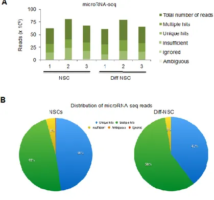
microRNA-seq mapping statistics ... 44
Differentially expressed microRNAs ... 45
Neurosphere-forming assay ... 45
Immunofluorescence ... 46
Immunoblotting assay ... 46
RNA isolation and quantitative RT-PCR ... 46
PCR for Foxm1 isoforms ... 47
Statistical analysis of in vitro experiments ... 47
Luciferase-reporter assays ... 48
Site-directed mutagenesis ... 48
Chromatin immunoprecipitation (qPCR-ChIP assay) ... 48
Knockdown studies ... 51
Validated targets of miRNAs ... 51
3
AbstractBackground: Cerebellar neural stem cells (NSCs) maintenance is of great interest since NSCs can be used to treat impaired cells and tissues or improve regenerative power of degenerating cells in neurodegenerative diseases or spinal cord injuries. Under maintenance conditions, NSCs express a number of Hedgehog-Gli (Hh-Gli) linked and stemness genes (e.g. Nanog, Oct4, Sox2) whose mechanisms of regulation have been under investigation. However, the interplay between transcription factors and microRNAs in NSCs is still being charted.
Aim: Identification of new molecular players involved in NSCs’ maintenance with particular interest in the major regulatory pathway Hedgehog-Gli.
Materials and Methods: Cells used for the study were NSCs isolated from postnatal day 4 (P4) wild type (C57BL/6) mice cultured both as neurospheres in selective medium and as differentiated NSCs when cultured in medium with serum. NSCs and their differentiated counterparts were analysed by high-throughput technologies. Bioinformatics analysis was used for the identification of the Foxm1-regulated miRNAs; knock-down experiments and clonogenic assays were used for functional studies. Chromatin immunoprecipitation experiments (ChIP) were used to investigate the binding between Foxm1 and its targets and between Foxm1 and its regulators.
Results: NSCs and their differentiated counterparts were analysed using next-generation mRNA- and miRNA-sequencing. The transcriptional analysis allowed the identification of Foxm1 as one of the highest transcripts in NSCs and the miRNA-sequencing provided a number of highly expressed miRNAs. The use of bioinformatics analysis resulted in the Foxm1-regulated miRNAs, miR-15 ~ 16 cluster, miR-17 ~ 92 cluster, miR- 130b and miR-301a. Functional experiments, such as knock-down experiments and clonogenic assays enabled the identification of Foxm1 as a downstream mediator of the Hh-Gli signalling and with the ability to regulate the above mention miRNAs.
Conclusion: The study presented reveals a new Foxm1-microRNAs network with a major role in the maintenance of NSCs. These results add a previously unidentified important molecular aspect that could be used in future neurodegenerative disease studies, thus enriching the field of translational medicine.
4
IntroductionNeural stem cells
Stem cells have been a major focus of research because of their unique capacities of self-renewal and differentiation capacity. These capacities are defined by the expression of transcription factors and epigenetic modulations (Montalbán-Loro, R. et al. 2015).
They reside in specific environment or niches of the adult mammalian brain, the subgranular zone of the dentate gyrus of the hippocampus (SGZ), the subventricular zone (SVZ) lining the lateral ventricles, and the white matter of the cerebellum (Gage, F.H. 2000; Lee, A. et al., 2005), in which NSCs support neurogenesis and gliogenesis during adult life. A small number of NSCs may still be in other areas of the brain (Fig. 1).
In SGZ there are two types of NSCs, particularly type 1 that are quiescent characterized by the expression of specific molecular markers such as glial fibrillary acidic protein (GFAP), Nestin and Sox2. Type-1 NSCs (true stem cells) are the neurogenic entities that generate type-2 cells that proliferate actively and express Nestin, Sox2 but not GFAP.
In the SVZ area three types of NSCs can be distinguished (Fig. 2): A, B, C. Type-A is composed of migratory neuroblasts, type-B and -C correspond to type 1 and 2 in SGZ (Yao J. et al., 2012). The cell lineage differentiation goes from type-B, through type-C to type-A cells (Doetsch F. et al., 1999).
5
Figure 2. Cytoarchitecture of the subventricular zone of the healthy adult brain (Martino G. and Pluchino S. 2006).Neural cerebellar Stem Cells (NSCs) have the ability to self-renewal and to give rise to neurons, astrocytes and oligodendrocytes (Fig. 3) (Davis, A. A. and Temple, S. 1994; Gage, F.H. 2000). They express stemness markers such as Sox2, Nestin, Nanog and Prom1 (Palm, T. et al., 2013; Po, A. et al., 2010).
6
Figure 3. Self-renewal of Neural cerebellar Stem Cells and differentiation lineage (Wakabayashi, T. et al., 2014).NSCs are in physical contact with the basal lamina, which regulates cytokines and growth factors derived from local cells (Campos, L. S. et al. 2006).
Moreover, the proliferation and differentiation are finely regulated by both intrinsic and extrinsic factors (Imayoshi, I. et al., 2010; Pierfelice, T. et al., 2011) consisting of morphogens, growth factors, tissue micro-environment (germinal niche), transcriptional factors and epigenetic mechanisms. Among the main determinants of differentiation, Notch is responsible for neuronal differentiation (Imayoshi, I. et al., 2010; Pierfelice, T. et al., 2011), fibroblast growth factor (FGF), WNT that
7
promotes differentiation in the subventricular zone (Lie, D.C. et al., 2002) and Sonic Hedgehog directs NSCs to the glial lineage (Ahn, S., and Joyner, A.L. 2005; Balordi, F., and Fishell, G., 2007; Han, Y.-G. et al. 2008).NSCs are characterized by a low degree of epigenetic silencing, resulting in the activation of a multitude of genes that maintain self-renewal (Yao J. et al.,2012).
Up-regulated pathways consistent with NSCs self-renewal have been identified, such as G1-S cell cycle regulation, nucleic acid synthesis, DNA replication, packaging, and repair genes (Gage, F.H. 2000).
Advancing the understanding of the signalling molecules that are responsible for the transition of NSCs from proliferation to differentiation will further the potential use of NSCs as therapeutic agents (Gage, F.H. 2000; Harris, L. et al., 2016).
There are cues that NSCs can reach the target organ and differentiate into the appropriate cell lineage but the molecular mechanisms that sustain functional integration and repair capabilities are not clear (Martino G., Pluchino S., 2006).
The main goal is to use NSCs to treat impaired cells and tissues or improve regenerative power of degenerating cells in neurodegenerative diseases (for example Parkinson’s disease, Huntington’s disease, multiple sclerosis) or spinal cord injuries.
Hedgehog pathway in NSCs
A key signal sustaining NSCs is Hedgehog (Hh) signalling (Po, A. et al., 2010). Hh pathway has a central role in development and tumorigenesis in a wide variety of tissues, both processes being supported by stem cells (Ahn, S., and Joyner, A.L., 2005; Lai, K. et al., 2003; Palma, V. and i Altaba, A.R. 2004; Palma, V. et al., 2005).
The pivotal role of this signal pathway has already been described in embryonic stem cells and NSCs of SVZ, hippocampal regions (Ahn, S., and Joyner, A.L., 2005; Lai, K. et al., 2003; Palma, V. et al., 2005; Palma, V. and i Altaba, A.R. 2004) and cerebellum (Po, A. et al., 2010).
The canonical Hh pathway requires the presence of a transmembrane receptor, Smoothened (Smo), which can be inhibited by the Patched 1 (Ptc) receptor. Hh ligands (Desert Hedgehog (DHH), Indian Hedgehog (IHH), and Sonic Hedgehog (SHH) bind Ptc which is internalized, degraded, and thus not able to inhibit the activator receptor Smo. Smo, then, interacts and inhibits the Suppressor of fused (SUFU), this results in the activation and nuclear translocation of the only known transcriptional mediators of the Hh response, zinc-finger proteins of the glioma-associated oncogene (Gli) family. These are bifunctional transcription factors that can both activate or inhibit transcription. In detail,
8
Gli1 and Gli2 (GliA) are activated and transported in the nucleus where they activate the transcription of Gli1 itself, Gli2 and Ptc, thus amplifying the Hh signaling pathway. Gli3 (GliR) is a suppressor of the Hh pathway and is degraded after inhibition of Sufu by Smo (Fig. 4) (Ng, J.M., and Curran, T., 2011).Figure 4. Hedgehog signaling pathway.
In NSCs Nanog and Hh/Gli are co-expressed and it has been demonstrated that both are essential in driving self-renewal. Nanog is a transcriptionl factor essential for maintaining the pluripotency of the inner cell mass during embryonic development and its expression is down regulated in differentiated cells. Nanog is a downstream factor of the Hh signal transduction, in fact Gli1 and Gli2 bind to specific consensus cis-regulatory responsive elements on Nanog promoter enhancing its transcription. This way, Gli1 and Gli2 mediate the Hh-dependent control of Nanog and downstream stemness genes, which promote self-renewal of NSCs (Po, A. et al., 2010).
9
Next Generation SequencingIn 1977 F. Sanger and colleagues proposed a new innovative method for sequence DNA by chain termination and fragmentation techniques (first generation), that was used for the next 30 years, introducing the possibility to study genomes and for fast and low-cost DNA sequencing.
The Human Genome Project (HGP) (Consortium, I.H.G.S. 2004; Lander, E.S. et al., 2001), was an international, publicly funded consortium of scientists at universities and research institutes with the aim to provide a virtually complete sequence of human DNA, the genome.
The Sanger methods allowed the realization of the first human genome sequence in 2004. HGP was still very expensive and needed plenty of resources, so, in the same year the National Human Genome Research Institute (NHGRI) started a program to reduce the cost of the sequencing to US$1000 in ten years. This has led to the development of next-generation sequencing (NGS) technologies. These new technologies offered improvements respect to Sanger methods. First, in the preparation of libraries in a cell free system to fragment the DNA; second, they could generate gigabases of genomic data in a single run because the sequencing reactions are produced in parallel; and lastly, they didn’t require the electrophoresis step (Van Dijk, E.L. et al., 2014). In this way, they largely reduced the cost and the complexity of the experiments.
NGS systems are based on shotgun sequencing approach which consists of random fragmentation from an entire genome, transcriptome, or smaller targeted regions and sequencing of DNA in a single run (Morozova, O. et al. 2009).
There are different commercial platforms, the widely utilized ones are Roche/454, Illumina, Applied Biosystems SOLiD, Oxford Nanopore, Ion Torrent, Pacific Biosciences and Helicos. They differ from each other on the type of chemistry used, in fact they can be based on either “sequencing by synthesis” or “sequencing by ligation” detection methods (Metzker, M.L. 2010).
The advantage of New-Generation Sequencing Methods can be applied to different areas of research and for different applications. These include a complete genome annotation, for example the knowledge of all regulatory sequences, splice variants and exon-intron structures (Morozova, O. et al., 2009). Omics technologies provided us data that allowed us to better understand the genotype–phenotype interaction. NGS has a wide application in the identification and quantifying of transcripts in cells, tissue and organisms (RNA-seq) (Morozova, O. et al., 2009; Voelkerding, K. V. et al., 2009).
In genomics they allow us to study whole genomes from microbes to humans and its products, and the communication between genes and environment. In the medical field NGS advances have improved the understanding of the relationship between genetic modification and phenotype,
10
moreover the re-sequencing of target genomic regions is useful to identify polymorphisms in genes to study rare variants in genetic diseases (Koboldt, D.C. et al., 2013; Voelkerding, K. V. et al., 2009).RNA-Sequencing
The RNA sequencing, through NGS, allows for the quantification and characterization of transcripts, producing an adequate representation of transcriptome in both prokaryotes and eukaryotes. These technologies avoid several problems connected to the hybridization-based microarrays, previously used to gene expression, such as cross-hybridization artefacts, limited range of detection and a priori knowledge of gene sequences.
There are several applications of RNA-seq in transcriptomics that have an important impact on the study of diseases. For example, it is used for the characterization of splicing variants that could help us to understanding the contribution of alternative splicing in the development of human diseases. In the field of cancer research, it also can be used to find gene fusion events, such as a translocation or another genomic arrangements that produce aberrant RNAs, identifying potential targets for therapeutic approach (Ozsolak, F., and Milos, P. M., 2011).
It is largely used to map transcription start sites, to identify nucleotide variations and mutations and noncoding RNAs (ncRNAs) expression profiling in many species. RNA-seq is effective not only in the discovery of novel microRNAs and siNAs, but also in the detection of variants of microRNAs and editing events (Morozova, O. et al. 2009).
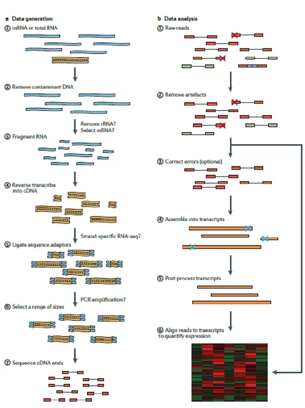
RNA-seq workflow (Fig. 5) starts with the conversion of total RNA in a library of cDNA containing sequencing adaptors. Then, each molecule is sequenced to obtain short sequences (reads) from one end (single-end sequencing) or both ends (pair-end sequencing). The size of the resulting reads obtained are very short, between 35 and 500 bp (Wang, Z. et al., 2009), so it is necessary to reassembly the full-length RNAs, except in the case of small classes of RNA (miRNAs, piRNAs snoRNAs and siRNAs). Following sequencing, there are three different strategies to perform the assembly of the transcriptome: a reference-based strategy, a de novo strategy or a combined strategy that merges the two. The “reference-based” is easier to perform, it aligns the reads to a
11
reference genome and the advantage is that can assemble transcripts of low abundance (Martin, J.A. and Wang, Z., 2011).12
MicroRNAsThe discovery of microRNAs twelve years ago (Ambros, V., 2004) brought to the forefront of epigenetic research the post-transcriptional gene regulation.
MicroRNAs are known to be involved in the maintenance of stem cell self-renewal and promotion of differentiation (Blakaj A, Lin H. 2008; Tay, Y. et al. 2008).
MicroRNAs are a class of small (~22 nt) non-coding RNAs that bind through a 5´ “seed region” to the 3´untranslated region (3´ UTR) of target mRNAs driving them to translation repression and/or mRNA degradation (Fig. 6) (Bartel, D.P. 2009).
It has been estimated that since microRNAs only need as few as 7 nucleotides of complementarity to bind to their target, computational and experimental approaches indicate that more than 60% of human protein coding genes are predicted to contain microRNA-binding sites. This fact highlights the necessity for microRNA profiling in order to acquire a more complete understanding of their identity and role in different biological contests. The small size of mature microRNAs renders them suitable for characterization using RNA-seq technologies by implementing the appropriate modifications in sequencing and bioinformatics analysis.
13
Figure 6. Biogenesis and function of microRNAs in mammals.14
Scope of the studySince the discovery of NSCs, researchers have focused on maintenance mechanisms of these cells. The main goal is to use them to treat impaired tissues or to improve regenerative power of degenerating cells in neurodegenerative diseases or spinal cord injuries. Under maintenance conditions, NSCs express several stemness genes (e.g. Nanog, Oct4, Sox2) whose mechanisms of regulation have been investigated (Garg, N. et al., 2013; Po, A. et., 2010; Kim, J. B., 2009; Zhang S, Cui W., 2014). However, the interplay between other transcription factors and NSCs maintenance is still being charted.
Therefore, the aim of the study was to understand stemness molecular mechanisms, investigating the role of transcription factors and microRNAs, in NSCs compared to their differentiated counterparts. With this aim next-generation RNA sequencing was used that allowed the identification of transcripts and microRNAs characterizing NSCs. The highest expressed transcript in NSCs implicated in the Hh signaling was Forkhead Box m1 (Foxm1), part of the FOX superfamily of transcriptional regulators that play a pivotal role in cell cycle progression. Therefore, the study focused on identifying a Foxm1-microRNA network involved in maintaining NSCs.
15
ResultsHigh-throughput transcriptome profiling of cerebellar NSCs
Cerebellar NSCs from postnatal day 4 (P4) mice were grown in stem-cell-selective medium, as described elsewhere (Po, A. et al., 2010). As expected, under these conditions, the cells displayed high-level expression of stemness genes (Nanog, Nestin) and of Gli1 (Fig. 7A). Transfer of these NSCs to differentiation medium (Po, A. et al., 2010) was followed by significant increases in the expression of genes encoding astrocytic, neuronal, Purkinje, and oligodendrocytic cell markers (Fig. 7B). 0 2 4 6 8 10 12 14 R e la ti v e m R N A e x p re s s io n NSC Diff-NSC ** * *
Supplementary Figure 1.
Diff-NSCs
S100/Hoechst Pvalb/HoechstTubb3/Hoechst Cspg4/Hoechst
Astrocyte Purkinje Neuron Oligodendrocyte
A
NSCs
Δ C t 20 -2 D if f. S te m n e s s / H h Gli1 Nestin Nanog Neurod1 Math1 N a n o g / H o e c h s t N e s ti n / H o e c h s t G li1 / H o e c h s tB
Tubb3 S100 Pvalb Cspg4 **16
Figure 7. Characterization of P4 murine cerebellar NSC cultures before and after differentiation. A Heatmap: Levels of mRNA for markers of stemness (Nanog, Nestin), Hh-Gli signaling (Gli1), and neuronal differentiation (Neurod1, Math1) in NSCs grown in stem-cell-selective medium. Transcript levels are represented on a green-red color scale based on ΔCt values. Immunofluorescence images: Representative results of NSC staining for markers of stemness and Hh-Gli signalling (green); nuclei are counterstained with Hoechst (blue).B Left: Levels of mRNA for genes encoding neuronal differentiation markers in NSCs grown for 48 h in differentiation medium (Diff-NSCs), as measured by RT-qPCR single assays. P values vs. pre-differentiation NSC controls: **P<0.01: 0.0019 (S100 P), 0.0083 (Pvalb); *P<0.05: 0.0298 (Tubb3), 0.0316 (Cspg4) (unpaired T-test). Right: Representative results of immunofluorescence staining of Diff-NSCs for neuronal differentiation markers (green); nuclei are counterstained with Hoechst (blue). Scale bar: 5 µm for all panels.
Paired-end polyA+ RNA-sequencing was used to profile the transcriptomes of NSCs grown in stem cell and differentiation media (NSCs and Diff-NSCs, respectively; three replicates of each). A total of 988 genes were differentially transcribed by the cells under these two conditions. NSCs and Diff-NSCs were clearly segregated, as observed in the hierarchical clustering of the 988 differentially expressed transcripts (DETs) (Fig. 8 and Supplementary Table 5).
17
Figure 8. Clustering of differentially expressed transcripts (DETs) in NSCs and Diff-NSCs.Hierarchical clustering of the 988 transcripts differentially expressed (adj. P < 0.05) (Bray-Curtis method with average linkage).
Functional analysis of the DETs using the DAVID platform (Database for Annotation, Visualization and Integrated Discovery) revealed significant enrichment (Bonferroni-corrected P<0.05) for the four Gene Ontology categories reported in Table 1 and detailed in Figure 9. The most interesting clue that emerged from this analysis was the over-representation of genes involved in p53 signalling. This pathway is a well-known negative regulator of NSCs self-renewal (Garg, N. et al., 2013; Solozobova, V. & Blattner, C., 2011), whose activity is modulated by signalling through the Hh-Gli-Nanog axis (Lin, T. et al., 2005; Po, A. et al., 2010).
18
Figure 9. Functional analysis of DETs in NSCs and Diff-NSCs.Functional analysis with DAVID (see Table 1). Clusters are shown with heat maps indicating transcript abundance (based on normalized FPKM values) for genes belonging to each Gene Ontology (GO) category.
Cell cycle DNA replication NSC1 NSC2 NSC3 Diff-NSC1 Diff-NSC2 Diff-NSC3 p53 NSC1 NSC2 NSC3 Diff-NSC1 Diff-NSC2 Diff-NSC3 ECM receptor interaction NSC1 NSC2 NSC3 Diff-NSC1 Diff-NSC2 Diff-NSC3
Supplementary Figure 2.
19
Hedgehog–Gli pathway components enriched in NSCsIn order to identify other molecular players with potential roles in Hh-Gli-driven self-renewal of cerebellar NSCs, a compiled list of 53 genes known to be regulated by Hh-Gli signalling in settings (physiologic or pathologic) other than NSCs (Supplementary Table 1) was used. Nine of these genes were differentially transcribed in NSCs before and after differentiation (Supplementary Table 2, Figure 10). Six of the nine genes encode cyclins (Ccnb2, Ccnb1, Ccna2, Ccnd2, Ccne1, Ccnd1) known to be involved in the regulation of cell cycle and cell division in NSCs. The seventh, Sema6a, is involved in nervous system development, in particular, in axon guidance (Rivron, N. C. et al., 2012), and the eighth gene, Insm1, has been reported to be involved in mouse cerebellar development (De Smaele, E. et al., 2008).
Analysis of this list revealed that Foxm1 was the Hh-Gli-regulated transcription factor most markedly expressed in NSCs prior to differentiation. Foxm1 is a transcriptional activator (Wierstra, I., 2013) whose role as a downstream mediator of Hh-Gli signalling has been documented exclusively in human cancer cells (Teh, M.T. et al., 2002; Katoh, Y. and Katoh, M. 2006; Shi, C. et al., 2016). The relation between Foxm1 and Hh-Gli signalling was evident in our NSC model. Consistent with their high-level expression of Gli1 (Fig. 7A), the NSCs displayed strikingly higher levels of Foxm1—at both the transcriptional (Fig. 11A) and protein levels (Fig. 10B)—prior to their differentiation. Review of our mRNA-seq data confirmed this NSC-associated upregulation for two of the four known protein-coding Foxm1 transcript isoforms (ENSMUST00000073316 [Foxm1-201] and ENSMUST00000112148 [Foxm1-202]). As shown in Figure 11B, this finding was validated by PCR performed with isoform-specific primers and by immunoblot analysis, which revealed clear predominance in the NSCs of the 757-amino-acid Foxm1-201 protein isoform.
20
Figure 10. The Hh-signalling mediator Foxm1 is differentially expressed in NSCs and Diff-NSCs. A Heatmap and dendrogram of the nine DETs whose genes are regulated by Hh signalling.B Immunofluorescence staining of endogenous Foxm1 (green) in NSCs and in Diff-NSCs. Nuclei were counterstained with Hoechst (blue). Scale bar: 5 m.
NSCs Diff-NSCs Merge Foxm1 Nucleus 2 1 3 1 2 3 Diff-NSC NSC
A
B
Supplementary Figure 3.
21
Figure 11. Upregulated expression of Foxm1 in P4 cerebellar NSCs and its effect on self-renewal.A
C
B
N S C 1 D if f-N S C 1 N S C 2 N S C 3 D if f-N S C 3 D if f-N S C 2 Foxm1 Tubb3 Actin Foxm1 Hsp70 s iF o x m 1 s iC tr l 1 0.5 siCtrl siFoxm1 400bp 300bp Foxm1 isoform 201 Foxm1 isoform 202 83 kDa 81 kDa 55 kDa 42kDa 83 kDa 81 kDa 70 kDa Foxm1 NSC Diff-NS C 0.0 0.5 1.0 1.5 NSC Diff-NSC m R N A l e v e ls ***** NSC Diff-NSC NSC Di ff-NSC 0 5 10 15 NSC Diff-NSC Tubb3 m R N A l e v e ls * NSC Diff-NSC siCtrl siFox m1 0 20 40 60 siCtrl siFoxm1 N e u ro s p h e re -f o rm in g c e ll s ( % ) *Fig 2.
22
A RT-qPCR data showing differential expression in pre- and post-differentiation NSCs of mRNA for Foxm1 (*****P<0.0001: 0.0000089) and the neuronal differentiation gene Tubb3 (*P<0.05: 0.029) (Mann–Whitney U test).B Left: PCR assay of Foxm1 expression using isoform-specific primers. Agarose (2%) gel separation of the amplified product yielded two bands corresponding to Foxm1 isoforms 201 (400 bp) and 202 (300 bp). Right: Immunoblots showing endogenous levels of Foxm1, Tubb3, and Actin (loading control) in three NSC cultures before and after induced differentiation.
C Left: Immunoblots showing endogenous levels of Foxm1 and Hsp70 (loading control) in NSCs transfected with siRNA against Foxm1 or non-targeting siRNA controls (siCtrl). Densitometric values appear below blots.
Right: Representative bright-field images of neurospheres formed by NSCs transfected with siCtrl
and siFoxm1. Scale bar: 100μm. Graphs show percentages of seeded cells that formed neurospheres (*P<0.05: 0.036) (Two-tailed paired t-test).
Bars in panels A and C represent the mean (SD) of three independent experiments.
Foxm1 mediates Hh–Gli-driven self-renewal of the NSCs
To explore the functional relevance of this upregulation, NSCs were transfected with siRNA directed against Foxm1 (siFoxm1) and evaluated for their self-renewal capacity, as reflected by their ability to form neurospheres. As shown in Figure 11C, Foxm1 knock-down was associated with significantly impaired neurosphere formation.
Comparison of the human FOXM1 and murine Foxm1 promoter regions revealed a high percentage of identical base pairs (36%-45%), which indicated substantial similarity. Consistent with recent findings on its human ortholog (Shi, C. et al., 2016; Wang, D. et al., 2017), the murine Foxm1 promoter was found to harbour eight putative Gli-binding sites (s1–s8) (Figure 12A and Supplementary Information-Section 1). Quantitative PCR-ChIP assays were performed in order to determine whether both transcriptional activators of the Hh-Gli pathway could occupy the Foxm1 promoter in these putative binding sites. Experiments were performed in NSCs both before and after differentiation to quantitatively assess Gli recruitment and histone H3 acetylation (AcH3, a marker of transcriptional activation). Transcriptional activation was reported in all putative Gli binding sites (s1-5) and (s6-8) as evidenced by the higher percentage of promoter occupancy in NSCs when compared to Diff-NSCs. In particular, Gli2 reported a significantly higher percentage of occupancy in the Gli (s1-5) binding sites of the Foxm1 promoter in NSCs in respect to Diff-NSCs (Fig. 12B). Similarly, Gli1 promoter occupancy in the Gli (s6-8) binding sites of the Foxm1 promoter was
23
significantly higher in NSCs when compared to Diff-NSCs (Fig. 12C). These data concluded that Gli-binding sites were bound by both transcriptional activators of the Hh-Gli pathway in NSCs.Luciferase reporter assays showed significant activation of Foxm1 by Gli2 binding and to a somewhat lesser extent by the binding of Gli1 (Fig. 12D). Collectively, these findings strongly support the importance of Foxm1 as a major mediator of Hh–Gli-driven self-renewal of the NSCs phenotype in the post-natal murine cerebellum.
24
Figure 12. Foxm1 promoter occupancy by Gli1 and Gli2.Fig 3.
Foxm1 +1 -2649 -2574 -2523 s2 s3 Gli Binding SitesCh6, GRCm38.p2 s4 -2668 s5 -2692 -2498 s6 s1 s7 s8 -3066 -3398 -3151 -3954 TSS
A
B
C
PCDN A GLI1 GLI2 0 1 2 3 4 ** F -L u c if e ra s e :R -L u c if e ra s ePCDNA Gli1 Gli2
*
D
Gli1 Gli2 AcH3 0.0000 0.0025 0.0050 0.15 0.20 0.25 0.30 P ro m o te r o c c u p a n c y ( % ) NSC Diff-NSC Gli s1-5 Gli s1-5 * * P ro m o te r o c c u p a n c y ( % ) NS Gli1 Gli2 AcH3 0.00 0.02 0.04 0.5 1.0 1.5 2.0 2.5 Gli s6-8 P ro m o te r o c c u p a n c y ( % ) NSC Diff-NSC Gli s6-8 ** * P ro m o te r o c c u p a n c y ( % ) NS25
A Schematic of the Foxm1 promoter showing locations of the 8 putative Gli-responsive elements (s1–s8).B-C qPCR-ChIP assay of endogenous Gli1 and Gli2 occupancy of the Foxm1 promoter region in NSCs and Diff-NSCs. Immunoprecipitation with anti-acetyl-H3 antibodies was used to detect Foxm1 transcriptional activation. Eluted DNA was qPCR-amplified using primers for putative Gli binding sites [s1–s5 (B) and s6–s8 (C)]. Results are expressed as fold induction values relative to ChIP input controls. Bars represent means (SD) of three independent experiments. P values vs. Diff-NSCs (Mann-Whitney U test): (B) *P<0.05: 0.04797 (s1-5, Gli2), 0.03271 (s1-5, AcH3); NS (not significant): 0.2514 (s1-5, Gli1). (C) *P<0.05: 0.0490 (s6-8, AcH3); **P<0.01: 0.001374 (s6-8, Gli1); NS: 0.296763205 (s6-8, Gli2).
D Luciferase activity induced in the Foxm1 promoter region in 293T cells by Gli1, Gli2, and PCDNA (negative control). Results are normalized to pRL-CMV-Renilla luciferase (R-Luciferase). Bars represent means (SD) of at least three independent experiments, each performed in triplicate. P values vs. control cells (One-way ANOVA test): *P<0.05: 0.02 (Gli1); **P<0.01: 0.005 (Gli2).
Foxm1 modulates stemness through the activation of specific microRNAs in NSCs
As previously noted, the Hh-Gli-regulated stemness marker, Nanog, modulates the proliferation and self-renewal of murine cerebellar NSCs via miRNA-mediated suppression of genes promoting cell-cycle arrest and differentiation (Garg, N. et al., 2013; Po, A. et al., 2010). This observation lead to the question whether miRNAs might also play a role in Foxm1’s effects on NSC self-renewal. As shown in Figure 13A, miRNA-sequencing studies identified 80 microRNAs that were differentially expressed in NSCs and Diff-NSCs.
To identify miRNAs likely to be direct targets of Foxm1, the promoter regions of the 40 miRNAs that were upregulated in NSCs (Supplementary Table 3) were examined and putative Foxm1 binding sites were found in 20. To increase the chances of identifying targets with biological relevance to NSC self-renewal, subsequent analyses were restricted to the 15 miRNAs on this list with the most statistically significant upregulated expression in the NSCs (Table 2, Fig. 14). (See Supplementary Information-Section 2 for the promoter regions of the miRNAs).
27
Figure 13. Foxm1 controls the transcription in P4 murine cerebellar NSCs of multiple miRNAs and miRNA clusters that affect NSC neurosphere formation.A Heat map and dendrogram depiction of the 80 miRNAs displaying significant differential expression in NSCs before and after induction of differentiation.
B-C qPCR-ChIP assays of NSCs and Diff-NSCs using anti-Foxm1 antibody and anti-acetyl-H3 antibody. Eluted DNA was PCR-amplified with primers annealing to promoter regions of the microRNA genes of interest. Findings for miRNA candidates belonging to a cluster are based on assays of one representative cluster member. Results are expressed as fold induction versus input controls. Bars represent the mean (SD) of three independent experiments. P values NSCs vs. Diff-NSCs (Mann–Whitney U test):
Statistically significant (B) Foxm1: **P<0.01: 0.002204 17~92); ***P<0.001: 0.000 (miR-15b~16-2), 0.0003471 (miR-130b), 0.00004 (miR-15a~16-1), 0.0003906 (miR-301a).
AcH3: *P<0.05: 0.049416827 (miR-15b~16-2); **P<0.01: 0.008868 (miR-130b); ***P<0.001: 0.0008656 (miR-17~92), 0.00000069 (miR-15a~16-1), 0.00000920 (miR-301a). Not Significant (C) Foxm1: *P<0.05: 0.01467 335), 0.01021 106b~25); NS: not significant: 0.07214 (miR-130a). AcH3: *P<0.05: 0.02903 (miR-130a); NS: 0.5259 (miR-335), 0.4417 (miR-106b~25).
D LNA anti-miR-130b, -miR-301a, miR-19a (to inhibit miR-17-92 cluster members) and miR-15b (to inhibit miR-15-16 cluster members) were used separately and combined. [3 LNA combination: anti- miR-130b, -miR-301a, and miR-19a; 4 LNA combination: anti-miR-130b, -miR-301a, miR-19a, and miR-15b]. Bars represent means (SD) of at least three independent experiments performed in triplicate. P values vs. scrambled LNA control (One-way ANOVA test): *P<0.05: 3 LNA combination: 0.0424 ; 4 LNA combination: 0.0500).
28
Figure 14. Validation of miRNAs displaying significant differential expression in NSCs vs. Diff-NSCs. Q-PCR single assay validation of NSC expression of the top 15 DE miRNAs listed in Table 2. Results for each miRNA are expressed as the log2 fold change relative to NSC expression of the endogenous control gene U6. Bars represent the mean (SD) of three independent experiments. P values vs. U6 control (Mann–Whitney U test): **P<0.01: 0.0058 (miR-15b-3p), 0.0092 (miR-335-3p), 0.0069 (miR-15b-5p), 0.0074 (miR-16-2-3p), 0.0078 (miR-16-1-3p), 0.0099 (miR-15a-3p); *P<0.05: 0.029 92a-1-5p), 0.0204 130b-5p), 0.022 130a-5p), 0.031 25-5p), 0.037 (miR-93-3p), 0.042 (miR-301a-5p), 0.021 (miR-130b-3p), 0.019 (miR-106b-5p), 0.032 (miR-19a-3p).Quantitative PCR-ChIP assays were then performed on NSCs before and after differentiation to quantitatively assess Foxm1 recruitment and histone H3 acetylation (AcH3) at the promoter region of each miRNA gene putatively targeted by Foxm1. The 15 microRNAs were either transcribed singularly or as part of a cluster (Table 2), in case of a cluster for further experiments expression levels of one representative miRNA are reported. For all miRNAs tested Foxm1 recruitment was higher in the NSCs when compared to Diff-NSC. Attention was focused on the cases where transcriptional activation of these promoters, as evidenced by AcH3, was significantly more intense in NSCs than in Diff-NSCs (Fig. 13B-C). Foxm1 recruitment indeed was statistically significant for miR-130b, miR-301a, and miRNAs belonging to the miR-15~16 and miR-17~92 clusters (Fig. 13B). The results of the previous experiments pointed to miR-130b, miR-301a, and miRNAs of miR-15~16 (n=4) and miR-17~92 clusters (n=2) as particularly important mediators of Foxm1’s effects in NSCs. This conclusion was supported by the effects observed in the cells after locked nucleic acid (LNA)-mediated depletion of these miRNAs. As shown in Figure 13D, the NSCs’ capacity for neurosphere formation was not significantly reduced by anti-miR knockdown of any single miRNA or miRNA cluster. However, it was significantly impaired by combined depletion of miR-130b, miR-301a, and miR-19a (3 LNA combination). The additional depletion of miR-15b (4 LNA combination) resulted in an equally significant impairment of neurosphere formation.
To investigate mechanisms underlying the stemness-promoting effects of this miRNA network, genes targeted by the ChIP-confirmed miRNAs listed in Table 2 were explored. A miRTarBase (http://mirtarbase.mbc.nctu.edu.tw/) search returned validated murine targets for only three of these miRNAs: miR-15b-5p, miR-130b-3p, and miR-92a-3p (Supplementary Table 4). Thus requiring, the extension of the search to the literature on each miRNA, focusing specifically on validated or putative target genes (in any species) whose downregulation could explain the combined effect of these miRNAs in NSC self-renewal.
29
The results that emerged reiterated the importance of p53 signalling, whose loss/suppression is essential for the maintenance of embryonic stem-cell pluripotency (Hong, H. et al. 2009; Kawamura, T. et al. 2009). Of particular interest was a report showing that miR-130b-3p regulates CD133+ tumour-initiating cells in human hepatocellular carcinoma by targeting TP53INP1 (Ma, S. et al. 2010), which encodes a downstream component of the p53 signalling pathway. Previous work of the laboratory showed that Trp53inp1 expression in murine cerebellar NSCs is also suppressed by microRNAs of the miR-17~92 cluster, and the upregulated expression of these miRNAs was attributed to signalling through the Hh-Gli-Nanog axis (Garg, N. et al., 2013). In light of the current findings, p53 signalling in these cells also appears to be under the control of a second miRNA network, this one regulated by Foxm1. In support of this hypothesis, mirSVR prediction scores provided by microRNA.org (http://www.microrna.org/microrna/home.do) indicate that murine Tp53inp1 is a very likely target of 130b (mirSVR score: -0.0029). The same applies to miR301a, another Foxm1regulated microRNA belonging to the miR130b family (mirSVR score: -0.0030). The Foxm1-regulated miRNA network that modulates p53 signalling might also comprise miR-92a-3p, whose validated targets include Trp63, another member of the p53 family of transcription factors. Because of the high-level sequence homology that characterizes these transcription factors, p63 and p73 are capable of transactivating p53-responsive genes, thereby causing cell cycle arrest and apoptosis (NCBI).A role for Nanog in Foxm1 regulation
Interestingly, members of the miR-17~92 cluster are components of both the Nanog-regulated (Garg, N. et al., 2013) and Foxm1-regulated miRNA networks, but this was the only commonality observed. This evidence indicates that Nanog and Foxm1 activate largely non-overlapping cohorts of miRNAs to ensure suppression of p53 signaling in cerebellar NSCs. How these two networks interact to achieve this goal is unclear. Interestingly, Nanog has been identified as a target of Foxm1 (Wang, Z. et al. 2011; Xie, Z. 2010). To determine whether this relation might be reciprocal, the Foxm1 promoter region was re-examined for evidence of Nanog binding sites. As shown in Figure 15A, four putative binding sites for Nanog were found -3790 to -3277 bp upstream from the Foxm1 TSS. (For details, see Supplementary Information-Section 1).
ChIP experiments (Fig. 15B) demonstrated endogenous Nanog at these four sites in both NSCs and Diff-NSCs. However, pre- and post-differentiation occupancy rates were significantly different only at s2–s3, where the higher Nanog occupancy in NSCs was associated with greater activation of Foxm1. To further elucidate the relation between these two transcription factors, dual luciferase reporter assays was performed in 293T cells transfected with Foxm1 wild-type promoter or an s2- or s3-defective mutant promoter (Fig. 15C). Ectopic expression of Nanog in these cells resulted in
30
substantial activation of the wildtype promoter, whereas luciferase activity was appreciably reduced in the absence of s2 in the promoter and even more so by the deletion of s3, suggesting that this site is required for Nanog binding onto the Foxm1 promoter.Taken together, these results indicate that the Hh-Gli-driven miRNA networks regulated by Nanog and Foxm1 are characterized by bidirectional crosstalk, which might conceivably allow more finely tuned, combinatorial regulation of cerebellar NSC self-renewal.
31
Figure 15. Foxm1 promoter occupancy by Nanog.A
P ro m o te r o c c u p a n c y ( % )B
NanogC
+1 Foxm1 Nanog Binding SitesCh6, GRCm38.p2 -3543 -3451 -3277 s1 s2 s3 s4 -3790 -2498 -3954 TSS Nan og w t Mut Nan og s 2 Mut Nan og s 3 0 2 4 6 PCDNA Nanog F -L u c if e ra s e :R -L u c if e ra s e Na no g w t Mu t N an og s2 Mu t N an og s3 * *** NS Nano g NSC Nano g NSC -Diff AcH3 N SC AcH3 NSC -Diff 0.0 0.2 0.4 0.6 2 3 4 5 Nanog s1 NS Cs Dif f-N SC s NS Cs Dif f-N SC s NS **** Nano g NSC Nano g NSC -Diff AcH3 N SC AcH3 NSC -Diff 0.0 0.2 0.4 2 3 4 5 Nanog s2-3 NS Cs Dif f-N SC s NS Cs Dif f-N SC s ** **** Nano g NSC Nano g NSC -Diff AcH3 N SC AcH3 NSC -Diff 0.0 0.2 1 2 Nanog NSC Nanog NSC-Diff AcH3 NSC AcH3 NSC-Diff Nanog s4 NS Cs Dif f-N SC s NS Cs Dif f-N SC s **** NS Nanog NSC Nanog Diff-NSC AcH3 NSC AcH3 Diff-NSC
Fig 5.
32
A Schematic of the Foxm1 promoter showing putative Nanog-responsive elements (s1; s2 and s3 which are fairly close to one another and s4).B qPCR-ChIP assay of endogenous Nanog occupancy of the Foxm1 promoter region in NSCs and Diff-NSCs. Immunoprecipitation with anti-acetyl-H3 antibodies was used to identify Foxm1 transcriptional activation. Eluted DNA was PCR-amplified with primers for Nanog binding sites s1, s2-s3, s4. Results are expressed as fold induction values relative to input controls. Bars represent means (SD) of three independent experiments. P values vs. Diff-NSCs (Mann-Whitney U test): **P<0.01: 0.002572 (s2-3, AcH3); ****P<0.0001: 0.00000718 (s2-3, Nanog), 0.0001051 (s1, AcH3), 0.00004356 (s4, AcH3); NS: 0.4531 (s1, Nanog), 0.7118 (s4, Nanog).
C Luciferase activity induced by ectopic expression of Nanog and PCDNA (negative control) in 293T cells transfected with luciferase vector carrying the wild-type Foxm1 promoter (wt) and its mutant lacking the Nanog binding sites s2 and s3 (mutants s2, s3). Results are normalized to pRL-CMV-Renilla luciferase (R-Luciferase). Bars represent means (SD) of at least three independent experiments, each performed in triplicate. P values vs. indicated controls (Mann–Whitney U test): *P<0.05: 0.01849; ***P<0.001: 0.0002265; NS: 0.08604.
33
DiscussionThe main goal of this study was to characterize new molecular Hh-related mechanisms responsible for maintaining stemness features in cerebellar NSCs. These results permit the proposal of a model where Hh-Gli signalling controls the self-renewal of murine NSCs through the transcription of a series of microRNAs. As shown in Figure 16, two stemness miRNAs networks are described, via Nanog and via Foxm1, that partially overlap in our system and shed light to the role of Foxm1 in NSCs self-renewal. Hereby identifying the transcription factor Foxm1 as transcriptionally regulated by Hh through Gli 1 and 2 and Nanog. These results expand the list of known downstream effectors in the canonical Hh-Gli signalling pathway, as well as the list of molecules capable of activating Foxm1. Foxm1 is an activating transcription factor that plays a role in several different cellular contexts. In human epidermal stem cells, Foxm1 sustains the balance between self-renewal and terminal differentiation (Gemenetzidis, E. et al., 2010). Foxm1 is known to regulate neuronal precursor induced mitosis (Gemenetzidis, E. et al. 2010), as well as several stem cell-like properties. It is required for proper execution of mitosis and this is highlighted by the fact that Foxm1 knockout is embryonically lethal (Wierstra, I. 2013). It has been reported that Foxm1 upregulates the expression of the neural stemness marker Nestin1 in NSCs from the embryonic cerebral cortices of mouse and is critical for their self-renewal (Wierstra, I. 2013). Mutants conducted with loss-of-function Foxm1 suggest that in the murine cerebellum its main function is the adjustment of the transition in G2 / M phase (Schuller U. et al, 2007; Gage, F.H. 2000). All previous studies did not however provide information regarding Foxm1’s regulation or other functions in cerebellar NSCs.
Previous works of the laboratory reported Hh-related miRNAs involved in the maintenance of cerebellar NSCs (Ferretti, E. et al, 2008, Garg, N. et al, 2013). The present study has allowed to identify, through mRNA- and miRNA-sequencing, a series of Foxm1-controlled miRNAs highly expressed in NSCs. As a result, miR-130b, miR-301a and the clusters miR-17~92 and miR-15~16 were identified as direct transcriptional targets of Foxm1. Combined knock-down of these microRNAs leads to a significant decrease in terms of NSC abilities to form neurospheres. Such results show how microRNAs are fundamental for the self-renewal of these cells, through mediating the effects of Hh signal.
Subsequently, Foxm1-miRNAs were analysed. In support of this hypothesis, one of these miRNAs, miR-130b, has already been correlated with pro-proliferative effects in NSCs (Gong, X. et al., 2013) and that one of its target is Trp53inp1, a molecule involved in p53 pathway.
Also, the miR-17~92 cluster is part of the miRNA network, whose expression is required for the expansion of cortical NSCs in vivo (Bian, S. et al., 2013). Previous investigations of the group identified miR-17~92 cluster members as direct targets of signaling through the Hh-Gli-Nanog axis
34
and important regulators of NSC self-renewal (Garg, N. et al, 2013). This study demonstrated that these miRNAs are also components of the Foxm1-miRNA network.In previous works it has been reported that Foxm1 regulates Nanog expression (Wang and Park et al., 2011; Xie, Z. 2010, Wierstra, I. 2013), and the presented data of this thesis have shown that Foxm1 itself is transcriptionally activated by Nanog, leading to a positive feedback regulation. It is evident from the proposed model that a crosstalk between Gli, Nanog and Foxm1 exists.
Moreover, Garg et al reported the maintenance of NSC self-renewal via the Gli-Nanog-miR-17~92 axis by counteraction of p53 inhibition of Gli and Nanog, through Trp53inp1 regulation. The dual suppression of Trp53inp1 via 17~92 cluster (Hh-Nanog- and Hh-Foxm1-microRNAs) and miR-130b (Hh-Foxm1-microRNAs alone) suggests a convergence, also partially overlapping on p53 pathway control. This convergence could suggest the possibility of some functional redundancy that could be part of a cellular protection mechanism in case of a gene mutation affecting key components of the regulatory network.
In the present work, it is demonstrated that miR-15~16 is part of the miRNAs regulated by Foxm1 and it cooperates to maintain the undifferentiated status of NSCs. In support to this hypothesis, a role for miR-15a has been described in neuronal context. It has been shown that the high expression of miR-15 appears to be involved in neuronal maturation in MeCP2-deficient adult-born new neurons (Gao, Y. et al., 2015). So, in cerebellar NSCs context it could act as a regulated of the balance between pro- and anti-stemness cues, mediating the effect of Hh-Gli-Foxm1 axis.
In conclusion, this project presents an in depth investigation of the Hh pathway in the context of the cerebellar NSCs. In addition, a new molecular mechanism Gli-Nanog-Hh-related that participates in the maintenance of the stemness features of these cells was described. The identified network is composed of a downstream effector of the Hh-Gli signalling, Foxm1, and a series of microRNAs transcribed by itself. It has been demonstrated how the Hh-Foxm1-microRNAs axis is involved in the fine-tuning of pro-proliferative and pro-apoptotic cues in these cells.
Based on the results of this study that identified Foxm1 as a downstream effector of the Hh-Gli signalling, and due to the high similarity of the human and mouse promoter regions of Foxm1, Foxm1 could be a major player and should be taken into consideration in future investigations regarding not only mouse but also human cells.
35
Figure 16. Regulation of cerebellar NSCs self-renewal by the Hh-Foxm1-miRNA axis.The tumour-suppressor p53 checks NSC self-renewal directly, by activating Trp53inp1, and by inhibiting Gli and Nanog. Increased Hh-Gli signalling promotes NSC self-renewal by inhibiting p53 and upregulating Nanog expression. Our data show (bold-face type) that it also upregulates the expression of Foxm1 (directly and indirectly via Nanog). These two transcription factors activate the expression of several miRNAs that target Trp53inp1. Silencing of Trp53inp1 allows expansion of the NSC pool by promoting proliferation and diminishing apoptosis. Its loss also disrupts a feedback loop that maintains high p53 levels, thereby diminishing the tumour suppressor’s ability to repress Gli and Nanog expression. Hh-Gli Nanog
Foxm1
cluster miR 17~92
Trp53inp1 p53cluster miR 15
~16
miR-130b
miR-301a
Neural stem cell
self-renewal
In blue the results of our study; in grey and black data from literature
36
MethodsMurine cerebellar NSC cultures
Cerebellar NSCs were isolated from P4 wild type black 6 /C56 (C57BL/6) mice (n=8 per group) and cultured, as previously described 3. In brief, freshly dissected cerebella were placed in HBSS supplemented with 0.5% glucose and penicillin-streptomycin and dissociated, mechanically and enzymatically. The cells were maintained as mycoplasma-free neurosphere cultures in selective stem-cell medium consisting of serum-free DMEM-F12 supplemented with 0.6% glucose, insulin 25 mg/ml, N-acetyl-L-cysteine 60 mg/ml, heparin 2 mg/ml, B27 supplement without vitamin A, EGF 20 ng/ml, and bFGF 20 ng/ml. To induce differentiation, the cells were transferred for 48 h to differentiation medium consisting of the medium described above, prepared without the EGF/bFGF and supplemented with platelet-derived growth factor (PDGF; 10 ng/ml) (Sigma, P3076). Mycoplasma contamination was excluded by routine screening with the PCR Mycoplasma Detection Kit (ABM, Cat. No. G238). The neurospheforming cells from these cultures were selected and re-plated in the same medium, where they gave rise to new neurospheres, thereby confirming the capacity for self-renewal. Animal experiments were performed according to the European Community Council Directive 2010/63/EU and were approved by the local Ethical Committee for Animal Experiments of the Sapienza University of Rome.
Experimental and analysis design
Overview of study design
NSCs (n=3) and Diff-NSCs (n=3) were subjected to RNA (mRNA-seq) and small-RNA (microRNA-seq) sequencing. A consensus-based approach was used to analyse the mRNA-seq dataset, in order to overcome any methodological bias inherent in specific alignment and differential expression tools. Two different tools were used for mapping and alignment, and three different approaches were used for the differential expression analysis. The intersection of all three methods resulted in common genes with similar expression changes (Supplementary Table 5). The expression values reported by the DESeq algorithm were used for the remaining analysis as representative of all methods. The common genes were used as input for the pathway enrichment analysis using DAVID (david.ncifcrf.gov). Potential downstream targets of the Hedgehog (Hh) pathway were taken under further consideration (Fig S1).
In parallel, the microRNA-Seq dataset was analysed using the DESeq2 algorithm to detect differential expression of microRNAs between NSCs and Diff-NSCs (Fig S1). Subsequently, from the list of up regulated microRNAs in NSCs, the binding sites of the highest expressed gene
37
implicated in the Hh pathway were searched in the promoter region of the microRNAs. Then, the candidate Transcription Factors (TFs) were validated and the network connecting the TFs and microRNAs was created (Fig S1).Figure S1. Experimental and analysis design.
Experimental and analysis design mRNA-seq microRNA-seq Method 1: DESeq Alg Differential expression analysis with log2 fold change>1 and adj. p-value<0.05 DESeq2 Algorithm: Differential expression analysis
with log2 fold change>1 and adj.
p-value<0.05 between NSC vs.
Diff-NSC
From the list of up regulated microRNAs:
Search of microRNAs that have binding sites of the selected genes
in their promoter region Selection of genes that are implicated in the
Hedgehog Pathway
Validation of candidate TFs and microRNAs
Network creation of TFs and microRNAs
N S C 1 D if f-N S C 1 N S C 2 D if f-N S C 2 N S C 3 D if f-N S C 3
Nanog protein Level
+ − + − + − 1 2 3 Method 2: Cuffdiff Alg Differential expression analysis with default parameters- UCSC annotation FPKM>0.3
Enriched Pathways of genes, using DAVID
S e le c te d g e n e s Fig S1. Crosscheck of differentially expressed genes
from Methods 1-3 Mapping and alignment
with Genomatix Mining Station (GMS)
Mapping with TopHat and assembly with
Cufflinks
Mapping and alignment with Genomatix Mining Station (GMS) Method 3: CuffDiff Alg Differential expression analysis trimmed reads trimmed to 55mers –same parameters with Method 2
38
mRNA- sequencingLibrary preparation and RNA sequencing
RNA extraction. Total RNA was extracted from NSCs and Diff-NSCs using Trizol reagent (Life Technologies, USA). Quality-control assays were performed on an Agilent BioAnalyzer using the RNA Nano Kit (Agilent # 5067-1511). Only RNAs with RIN values > 9 were used for library preparation. The standard Illumina protocol and TruSeq RNA Sample Prep Kit were used to construct the mRNA-seq libraries. The mRNA in 400 ng of total RNA was converted into a library of template molecules suitable for deep sequencing analysis. All samples were sequenced using the Illumina Genome Analyzer IIx.
Mapping and differential expression analysis of RNA-seq reads
Transcriptome mapping with Genomatix Mining Station and differential expression analysis with Genomatix Genome Analyzer (Method 1)
All 70mer paired-end sequenced reads were aligned to the latest version of the Mus musculus genome available at the time, (NCBI38/mm10), using the Genomatix Mining Station (GMS, Sesame 2.4, https://www.genomatix.de/). All samples were quality-controlled with the FastQC tool (http://www.bioinformatics.babraham.ac.uk/projects/fastqc/) before mapping, and an average of 75% of reads with a quality score >30 was obtained. In terms of annotation, the read distributions for NSCs and Diff-NSCs were similar (Fig. S2B), confirming the absence of biases in sequencing and normalization.
Differential expression analysis with Genomatix Genome Analyzer
The RNA-Seq sequenced samples were subjected to differential expression analysis using the DESeq method in the Genomatix Genome Analyzer platform (GGA, v3.30126, https://www.genomatix.de/). Differentially expressed transcripts were selected as those exhibiting a minimum log2 fold change of 1 and adj. P-value < 0.05 (P-value adjusted with multiple testing correction using the Benjamini and Hochberg method).
Transcriptome mapping with TopHat and differential expression analysis with Cuffdiff (Methods 2 and 3)
Mapping of RNA-Seq reads
All sequenced 70mer paired-end reads were aligned to the Mus musculus genome (NCBI37/mm9) using the spliced read aligner TopHat version v1.3.1 3. Two iterations of TopHat alignments have been made to maximize the use of splice site information derived from all samples.
39
RNA-Seq transcriptome assembly
The transcriptome of each sample was assembled separately from the mapped reads using Cufflinks V1.2.1 5 with default parameters and the UCSC reference annotation. Transcript abundance was estimated as fragments per kilobase of exon per million fragments mapped (FPKM).
Analyses of the transcript distributions after TopHat mapping and Cufflinks assembly among different samples, biological replicates and cell types exclude the presence of biases in sequencing and normalization (Fig S3B, S3C and S3D). The samples displayed similar proportions of alternatively spliced genes. Transcribed isoforms not annotated in the reference version of the UCSC were found in all samples (1100-1500 per sample). Clustering analysis of the expression data confirmed clear separation of NSCs from their respective Diff-NSCs and the similarity of biological replicates within each group (Fig S3F).
The RNA-Seq reads were processed using two variations of the same protocol: the standard whole-read protocol (Cuffdiff-WR, Method 2), as well as the variation of this protocol based on the analysis of trimmed reads (Cuffdiff-TR, Method 3). The motivation for the trimmed reads approach is based on the observation that some NSC samples had comparatively lower average base quality towards the end of the reads. By trimming all datasets to 55bp reads, mappability has been enhanced by eliminating low-quality bases, and avoiding mapping artifacts across samples, by ensuring a consistent read length. Additionally, the follow-on impact on differential expression analysis was tested by using these trimmed reads.
Comparison of the differential expression results from all different methods
After analysing our samples with three different methods, the intersection of the results was selected, which yielded 988 DETs (Supplementary Table 5). Comparing the expression among the three different algorithms, the Pearson correlation was confirmed between the DESeq method and Cuffdiff, and it was extremely high. Additionally, the ANOVA test was performed to verify that the values obtained using the DESeq method are representative of the other two methods (p-value=0.9567, no statistically significant differences among means and standard deviations). The Pearson correlation and ANOVA test were performed using GraphPad Prism version 6 (La Jolla, CA, USA, http://www.graphpad.com/). In conclusion, the three approaches show a high degree of synergy and that results are not affected by methodological bias, alignment artefacts, or sequence quality. For downstream analyses, the expression values obtained from the DESeq algorithm were selected as representative of all methods.
40
Functional Analysis
The Database for Annotation, Visualization and Integrated Discovery (DAVID, david.ncifcrf.gov) was used for functional annotation, Gene Ontology categories with a Bonferroni correction adjusted p-value < 0.05 were reported10.
Clustering analysis
Clustering and heat maps were generated in Gene-E (version 3.0.238, http://www.broadinstitute.org/cancer/software/GENE-E/) (Supplementary Figure 2 for better visualization) and in R (http://www.r-project.org/) (the rest of the figures) using differentially expressed transcript levels as input. In Gene-E, the one minus Pearson correlation method was used for clustering and the complete linkage as a linkage method whereas in R the Bray-Curtis method and the average linkage were used in hclust to cluster the samples and heatmap.2 to generate the heat maps.
miRNA-sequencing
microRNA library preparation and sequencing
Small RNA-seq libraries were prepared in accordance with Illumina's TruSeq Small RNA Sample Prep Kit protocol.
Mapping of microRNA-seq reads and differential expression analysis with DESeq
Mapping of microRNA-seq reads
All sequenced single-end reads (31 bases) of the three biological replicates for each cell type (NSC
and Diff-NSC) were controlled with FastQC
(http://www.bioinformatics.babraham.ac.uk/projects/fastqc/) before mapping and they showed an average of 89% of reads with a quality score >30. All samples were aligned to the Mus musculus small RNA library (12-2013), (miRBase V20) using the Genomatix Mining Station (GMS, Sesame 2.4, https://www.genomatix.de/). The microRNA-seq read distributions for the NSCs and Diff-NSCs are shown in Figure S5B.
microRNA-seq reads and differential expression analysis
The microRNA-seq reads were analysed for differential expression in NSCs vs. Diff-NSCs in the Genomatix Genome Analyzer (GGA, v3.30126, https://www.genomatix.de/) using the DESeq2 method. Differences characterized by a minimum log2 fold change of 1 and an adjusted p-value of < 0.05 (Benjamini–Hochberg correction for multiple testing) were selected for further analysis.
41
Identification and characterization of binding sites in promoter regionsThe MatInspector tool in the Genomatix Genome Analyzer was used to identify putative binding sites in the Foxm1 promoter for Gli and Nanog (Supplementary Information – Section 1. The Foxm1 promoter region) and putative Foxm1 binding sites in the promoters of miR-130b, miR-301a and representative members of miR-17~92 and miR-15~16 clusters (Supplementary Information – Section 2. The miRNA promoter regions). Occupancy of the Foxm1 promoter by endogenous Gli1, Gli2, and Nanog in NSCs and Diff-NSCs was assessed by real-time qPCR-ChIP assay. Anti-acetyl-H3 antibodies were used to identify transcriptional activation of Foxm1.
mRNA-Seq mapping statistics
mRNA-seq data were analysed with the Genomatix Mining Station (GMS) (Genomatix GmbH, Münich, Germany) and TopHat/Cufflinks. Mapping statistics for the two methods are summarized in Figures S2A and S3A, respectively. The number of reads per biological replicate that could be aligned to the mouse genome and the transcript distributions were similar with the two methods, thereby excluding the presence of biases in sequencing and normalization (Fig S2-3).
42
Figure S2. mRNA-seq mapping statistics with Genomatix Mining Station.A. Mean (SD) number of reads successfully aligned to the mouse genome, transcriptome, and exon junctions for each NSC and Diff-NSC sample.
B. Average distribution of reads mapped to the genome for NSC and Diff-NSC samples.
Fig S2.
0 50 100 150 200 1 2 3 1 2 3 Sequenced reads Aligned to genome Aligned to transcriptome Aligned to junctions NSC Diff-NSCA
B
R e a d s ( x 1 0 6)Diff-NSC Read Distribution NSC Read Distribution
43
Figure S3. mRNA-seq mapping statistics with TopHat and Cufflinks assembly.A. Mean (SD) number of reads successfully aligned to the mouse genome, transcriptome, and exon junctions for each NSC and Diff-NSC sample.
B. Box plots showing FPKM values (median [IQR]) for NSCs and Diff-NSCs.
C-D. M-A plots for the (C) NSC and (D) Diff-NSC replicates showing the expression ratio (M) of each transcript as a function of its average abundance.
E. Compositions of the NSC and Diff-NSC transcriptomes.
B
E
NSCs Diff-NSCs
A
D
M-A plot NSC1/NSC2 M-A plot NSC1/NSC3 M-A plot NSC2/NSC3
FPKM mean lo g 2 F o ld c h a n g e lo g 2 F o ld c h a n g e lo g 2 F o ld c h a n g e FPKM mean FPKM mean
C
N S C 1 N S C 2 N S C 3 D if f-N S C 3 D if f-N S C 1 D if f-N S C 2M-A plot Diff-NSC1-Diff-NSC2 M-A plot Diff-NSC1/Diff-NSC3 M-A plot Diff-NSC2/Diff-NSC3
FPKM mean lo g 2 F o ld c h a n g e lo g 2 F o ld c h a n g e lo g 2 F o ld c h a n g e FPKM mean FPKM mean
F
Fig S3.
NSC R e a d s ( x 1 0 6) 50 100 150 200 Diff-NSC NSCs Diff-NSCs T ra n s c ri p ts ( x 1 0 3) 5 10 15 20 044
F. Unsupervised hierarchical clustering of all samples using all expressed transcripts. Nodal numbers indicate bootstrap values obtained by resampling the data 10,000 times.Differentially expressed transcripts
A total of 988 DETs were identified with all three methods (Fig. S4). Pearson correlation analysis disclosed strong correlation between the DET expression values obtained with DESeq and those generated with the other two methods (DESeq vs. Cuffdiff-WR: r=0.985, P value=0.00; DESeq vs. Cuffdiff-TR: r=0.997, P value=0.00). ANOVA confirmed that the DESeq expression values were reliably representative of the others (P value=0.9567), and they were therefore used for subsequent analyses of the 988 DETs.
Figure S4. Transcripts displaying differential expression (DETs) in NSCs and Diff-NSCs. Venn diagram of DETs identified with DESeq, Cuffdiff-WR, and Cuffdiff-TR.
microRNA-seq mapping statistics
The mapping and read distribution results allowed us to proceed with the differential expression analysis without fear of bias from the sequencing and mapping (Fig S5).
Fig S4. 1535 369 102 54 284 988 1300 DESeq 2994 CuffDiff-WR 2941 CuffDiff-TR 1428 DETs in NSC vs Diff-NSCs