PhD Programme in Medical Sciences and Biotechnology Department of Science and Health
PhD program in
“Neoplastic, metabolic and age-related diseases” XXXII cycle
Biological Effects of Vitamin D3 Mediated by the
Interaction with VDR in Different Tissues
TUTOR:
Prof. Claudio G. Molinari
PhD STUDENT: Dott.ssa Vera Morsanuto
1
Index
Summary……….pag. 3 Riassunto……….pag. 5 1. Introduction………...………pag. 7 1.1 Seasonal Variation of Vitamin D Levels……….…………...pag. 9 1.2 Vitamin D Deficiency………....…pag. 10 1.3 The Genomic Mechanism of Action of Vitamin D………...pag. 10 1.4 The Nongenomic Mechanism………...pag. 11 1.5 Physiological Effects of Vitamin D………...pag. 12 1.6 Vitamin D and Ovarian Tissue……….………....….pag. 16 1.6.1 Vitamin D3 and Resveratrol……….………..…pag. 17 1.7 Vitamin D and Gastric Disorder………....pag. 19 1.7.1 Vitamin D and Alginates………..……...………pag. 21 1.8 Vitamin D and Cardiovascular System……….…….…pag. 22 1.8.1 Vitamin D and Protective Molecules………..…...…….pag. 23 1.9 Vitamin D in Nervous System……….…..pag. 25 1.9.1 Vitamin D3 and Lipoic Acid……….……...…...pag. 27 2. Aims of the Thesis………..…pag. 30 3. Materials & Methods
3.1 Cell Culture………...…....………..…...pag. 32 3.2 Animal Model………..………...pag. 35 3.3 Experimental Protocol………..…...…...pag. 36 3.4 Cell Viability……….………...pag. 40 3.5 Agents Preparation………....………...pag. 41 3.6 Blood-Brain Barrier Experimental Model…………...………...pag. 42 3.7 ROS Production………...…....………...…....pag. 42 3.8 SOD Activity Assay………...…....………..…..……pag. 43 3.9 Griess Assay………...pag. 44 3.10 Akt/ERK Activation Assay………...…...…...pag. 44 3.11 Mitochondrial Membrane Potential………...……....….. pag. 45 3.12 ATP Assay……….……...pag. 45 3.13 p53 Activity………....……...pag. 45 3.14 RES Quantification ………...………...…...pag. 46 3.15 Vitamin D Quantification...pag. 47 3.16 Lipoic Acid Determination………..…………...pag. 48 3.17 Amyloid Precursor Protein (APP) Quantification………….………...pag. 48 3.18 Iron Quantification Assay………...…....pag. 49 3.19 Western Blot ……….………...…...pag. 49 3.20 Statistical Analysis………...…...pag. 52
4. Results
4.1 Project 1 (ovarian line of research)………...…………..……...pag. 53 4.2 Project 2 (gastric line of research)……….……..…...pag. 68
2
4.3 Project 3 (cardiovascular line of research)………...pag. 81 4.4 Project 4 (brain line of research)...…….…………...pag. 96 5. Discussion...……….……..…....pag. 114 6. Future perspective………...…….……..…...pag. 128 7. References...……….……….……...pag. 130
3
SUMMARY
Vitamin D plays an important role in calcium homeostasis and bone metabolism, and it is able to modulate proliferation and differentiation of cells, cardiovascular function and also innate and adaptive immune function. The active form of vitamin D (vitamin D3, 1,25(OH)2D), exerts most of its effects through the vitamin D receptor (VDR). Indeed, in recent years the interest in vitamin D’s studies is increased, especially due to the presence of its receptor in many tissues and organs. VDR forms a heterodimer with the retinoid X receptor (RXR) upon stimulation by 1,25(OH)2D. In turn, VDR/RXR binds to DNA sequences termed “vitamin D response elements” in target genes, regulating gene transcription. In order to exert its biological effects, VDR signaling interacts with other intracellular signaling pathways. Sometimes, vitamin D exerts its biological effects without regulating either gene expression or protein synthesis. Although the regulatory role of vitamin D in many biological processes is well understood yet, many studies indicate that the role of vitamin D is not limited to regulation of bone and mineral homeostasis, but it is also important for its extraskeletal actions. Indeed, it has become the major driving force behind the significant increase in research articles on vitamin D published over the past several decades.
For this reason, this research aimed to examine the role of vitamin D signaling in a number of extraskeletal tissues (ovarian, gastric, cardiac and brain), and to assess the feasibility of translating these findings into treatments of human diseases affecting those extracellular tissues.
It has been demonstrated that in ovarian tissues a high density of VDR is present as well and vitamin D3 acts through intracellular mechanisms similar to what observed for resveratrol, a natural antioxidant polyphenol able to exert a wide range of biological effect on several tissues. The aims of this research were to evaluate the cooperative effects of resveratrol combined with vitamin D3 on CHO-K1 cells studying cell viability, ROS production, activated pathways in in vitro study and in in vivo study to quantify vitamin D3 and resveratrol, to analyze the concentration of radical oxygen species and to study activated pathways. Moreover, the modulation of specific intracellular pathways involving ER and VDR receptors has been studied. Results show that both in in vitro and in in vivo experiments, resveratrol exerts a greater effect when administered in combination with vitamin D3. In particular, the role of vitamin D3 in maintaining and supporting the biological activity of resveratrol has been clearly observed. Moreover, resveratrol plus vitamin D3 in blood concentrations showed a biphasic absorption rate. Such results could be used as a fundamental data for the development of new therapies for gynecological conditions, such as hot-flashes.
Another widespread problem is the abnormal use of nonsteroidal anti-inflammatory drugs in order to reduce pain. There is a high risk of developing gastricand enteric damages. These patients usually receive anti-acid treatment, but a number of clinical studies provided evidence of theineffectiveness of proton-pump inhibitors. Vitamin D, on the other hand, appears to have highpreventive and therapeutic potential.Recently, it has been introduced a commercial product that, in addition to anti-acid properties of alginates, claimsto possess gastroprotectiveproperties deriving from vitamin D3 and from plant
4
extracts.In this research the effectiveness of vitamin D3 combined with alginates to preventthe damage induced in culturedgastric cells by Diclofenac during acidic or hyperacidic exposition have been studied measuring cell viability, radicaloxygen species productionalong with apoptotic and survival pathways. Analyzing the effects on cardiac tissue, the study demonstrate that vitamin D3 has a cooperative effect with Q10 and L-arginine on both cardiac and endothelial cells to prevent the damage of ischemic condition. In particular, the effects of vitamin D3, Q10 and L-arginine alone or combined on cell viability, nitric oxide and reactive oxygen species productions in endothelial and cardiac cells were studied. Moreover, the involvement of PI3K/Akt and ERK/MAPK pathways leading to eNOS activation as well as the involvement of vitamin D receptor were also investigated. The same agents were tested in an animal model to verify vasodilation, nitric oxide and reactive oxygen species production. The data obtained demonstrate for the first time the beneficial and cooperative effect of stimulation with vitamin D3, Q10 and L-arginine. Indeed, in cardiac and endothelial cells, vitamin D3, Q10 and L-arginine combined were able to induce a nitric oxide production higher than the that induced by the 3 substances alone. The effects on vasodilation induced by cooperative stimulation have been confirmed in an in vivo model as well. The use of a combination of vitamin D3, Q10 and L-arginine to counteract increased free radical production could be a potential method to reduce myocardial injury or the effects of aging on the heart.
As regards the research focused on vitamin D3 in brain ageing, the ability of vitamin D3 combined with lipoic acid to prevent or repair the damage caused by oxidative stress and iron accumulation, two different biological aspects involved in brain ageing and neurodegeneration, has been studied in astrocytes. Cell viability, mitochondrial membrane potential and pathways activated in oxidative condition have been investigated. In addition, catalytic iron was used to assess the protection exerted by the combination of vitamin D3 and lipoic acid in induced neurodegeneration. In these experiments, cell viability, ROS production, iron concentration and activation of intracellular pathways have been studied. In this research, the combination of vitamin D3 and lipoic acid showed beneficial effects on viability of astrocytes, since the substances are able to cross the brain barrier. In addition, vitamin D3 plus lipoic acid was able to attenuate the H2O2-induced apoptosis through the mitochondrial-mediated pathway. The combination was also able to counteract the adverse effects caused by iron, preventing its accumulation. All these data support the hypothesis of a cooperative activity exerted by vitamin D3 and lipoic acid in astrocytes, indicating a possible new strategy to slow down brain ageing.
Therefore, the ability of vitamin D3 and lipoic acid to counteract brain aging has resulted in the creation of a commercial product, Cebral®.
5
RIASSUNTO
La vitamina D svolge un ruolo fondamentale nell'omeostasi del calcio e nel metabolismo osseo, modula la proliferazione e la differenziazione cellulare, l’immunità innata ed adattativa e contribuisce a regolare la funzione cardiovascolare. La forma attiva della vitamina D (vitamina D3, 1,25 (OH) 2D), esercita la maggior parte dei suoi effetti attraverso l’interazione con il suo recettore specifico (VDR). Negli ultimi anni l'interesse nello studio della vitamina D è aumentato, vista la presenza del suo recettore in molti organi e tessuti. VDR forma un eterodimero con il recettore X retinoide (RXR) dopo il legame con la vitamina D3. A sua volta, VDR/RXR si lega alle sequenze di DNA chiamate “elementi responsivi alla vitamina D” nei geni bersaglio, regolando la trascrizione genica. Al fine di esercitare i suoi effetti biologici, la segnalazione innescata da VDR interagisce con altre vie di segnalazione intracellulari. Sebbene il suo ruolo regolatorio in molti processi biologici sia ben noto, molti studi indicano che il ruolo della vitamina D non si limita alla regolazione dell'omeostasi ossea e minerale, ma è anche importante per le sue azioni extrascheletriche. Per questo motivo, questo studio è andato ad indagare il ruolo della segnalazione della vitamina D in numerosi tessuti extrascheletrici (ovarico, gastrico, cardiaco e cerebrale) per valutare la traslazionalità di questi risultati in trattamenti di malattie che colpiscono l’uomo. È stato dimostrato che nei tessuti ovarici è presente un'alta densità di VDR e la vitamina D3 agisce attraverso meccanismi intracellulari simili a quelli osservati per il resveratrolo, un polifenolo antiossidante naturale in grado di esercitare una vasta gamma di effetti biologici su diversi tessuti. Gli obiettivi di questa ricerca sono stati valutare gli effetti cooperativi del resveratrolo combinato con la vitamina D3 su cellule CHO-K1 (cellule ovariche) studiando la vitalità cellulare, la produzione di ROS, i percorsi attivati in vitro ed in vivo, la quantificazione di vitamina D3 e resveratrolo. Inoltre, è stata studiata la modulazione di specifici pathways intracellulari che coinvolgono i recettori ER e VDR. I risultati mostrano che sia in esperimenti in vitro, sia in vivo, il resveratrolo esercita un effetto maggiore quando somministrato in combinazione con vitamina D3. In particolare, è stato chiaramente osservato il ruolo della vitamina D3 nel mantenimento e nel supporto dell'attività biologica del resveratrolo. Inoltre, il resveratrolo più la vitamina D3 hanno mostrato un tasso di assorbimento bifasico nelle concentrazioni ematiche. Tali risultati potrebbero essere utilizzati come dati fondamentali per lo sviluppo di nuove terapie per condizioni ginecologiche, come ad esempio le vampate di calore. Un altro problema diffuso è l'utilizzo anormale di farmaci antinfiammatori non steroidei al fine di ridurre il dolore che porta però ad un alto rischio di sviluppare danni gastrici ed enterici. Questi pazienti di solito ricevono un trattamento anti-acido, ma una serie di studi clinici ha fornito prove dell'inefficacia degli inibitori di pompa protonica. La vitamina D, d'altra parte, sembra avere un alto potenziale preventivo e terapeutico. Recentemente, è stato introdotto un prodotto commerciale che, oltre alle proprietà anti-acide degli alginati, afferma di possedere proprietà gastroprotettive derivanti dalla vitamina D3 e dagli estratti vegetali. In questa ricerca, l'efficacia della vitamina D3 combinata con gli alginati è stata studiata analizzando la vitalità cellulare, la produzione di specie reattive dell’ossigeno
6
insieme a percorsi apoptotici e di sopravvivenza per prevenire il danno indotto da Diclofenac in cellule gastriche in condizioni di acidità o iperacidità.
Analizzando gli effetti anche sul tessuto cardiaco, lo studio dimostra che la vitamina D3 ha un effetto cooperativo con Q10 e L-arginina su cellule cardiache (cellule H9c2) ed endoteliali (cellule PAE) per prevenire il danno in condizioni ischemiche. In particolare, sono stati studiati gli effetti di vitamina D3, Q10 e L-arginina da soli e combinati sulla vitalità cellulare, sull'ossido nitrico e sulla produzione di specie reattive dell'ossigeno in cellule endoteliali e cardiache. Inoltre, sono stati studiati anche il coinvolgimento dei percorsi PI3K/Akt ed ERK/MAPK che portano all'attivazione di eNOS ed il coinvolgimento del recettore della vitamina D. Gli stessi agenti sono stati testati in un modello animale per verificare la produzione di specie reattive dell’ossigeno e di ossido nitrico. I dati ottenuti dimostrano per la prima volta l'effetto benefico e cooperativo della stimolazione con vitamina D3, Q10 e L-arginina. In effetti, nelle cellule cardiache ed endoteliali, vitamina D3, Q10 e L-arginina combinati sono stati in grado di indurre una produzione di ossido nitrico superiore a quella indotta dalle 3 sostanze singolarmente. Gli effetti sulla vasodilatazione indotti dalla stimolazione cooperativa sono stati confermati anche in un modello in vivo. L'uso della combinazione di vitamina D3, Q10 e L-arginina potrebbe essere un potenziale metodo per ridurre le lesioni del miocardio o gli effetti dell'invecchiamento sul cuore.
Per quanto riguarda la parte dello studio incentrato sulla vitamina D3 nell'invecchiamento cerebrale, è stata studiata la capacità della vitamina D3 combinata con l'acido lipoico di prevenire o riparare i danni causati dallo stress ossidativo e dall'accumulo di ferro, due diversi aspetti biologici coinvolti nell'invecchiamento cerebrale e nella neurodegenerazione astrocitaria. Sono state studiate la vitalità cellulare, il potenziale di membrana mitocondriale ed i pathways molecolari attivati in condizioni di stress ossidativo. Inoltre, il ferro catalitico è stato utilizzato per valutare la protezione esercitata dalla combinazione di vitamina D3 ed acido lipoico nella neurodegenerazione indotta. In questi esperimenti, sono stati studiati la vitalità cellulare, la produzione di ROS, la concentrazione di ferro e l'attivazione di pathways intracellulari. In questo studio, la combinazione di vitamina D3 e acido lipoico ha mostrato effetti benefici sulla vitalità degli astrociti. Inoltre, la vitamina D3 e l'acido lipoico sono stati in grado di attenuare l'apoptosi indotta da H2O2 attraverso la via mediata dai mitocondri. La combinazione è stata anche in grado di contrastare gli effetti negativi causati dal ferro, impedendone l'accumulo. Tutti questi dati supportano l'ipotesi di un'attività cooperativa esercitata dalla vitamina D3 e dall'acido lipoico negli astrociti, indicando una possibile nuova strategia per rallentare l'invecchiamento cerebrale. Pertanto, la capacità della vitamina D3 e dell'acido lipoico di contrastare l'invecchiamento cerebrale ha portato alla creazione di un prodotto commerciale, Cebral®.
7
1. Introduction
Vitamin D refers to a group of fat-soluble corticosteroids hormone responsible for enhancing intestinal absorption of calcium, iron, magnesium, phosphate and zinc. In humans, the most important compounds in this group are vitamin D3 (also known as cholecalciferol) and vitamin D2 (ergocalciferol) [1].
They are presents naturally in very few foods, for example in fish (vitamin D3) and in some plants (mainly vitamin D2) and for this reason a number of foods have been fortified by the food industries.
Vitamin D3 and vitamin D2 are inactive in many biological systems and have to undergo a series of metabolic transformations before exerting effects in target tissues [2-4]. Vitamin D is also popularly known as “sunshine vitamin” because in the skin sun exposure produces vitamin D. Indeed, the prohormone vitamin D3 is produced in the skin through ultraviolet irradiation of 7-dehydrocholesterol, than to make it biologically active the prohormone vitamin D is transported in the blood by the vitamin D binding protein (DBP) to the liver, where it is metabolized to 25-hydroxyvitamin [5]. Vitamin D is hydroxylated at C-25 by cytochrome P450 vitamin D 25-hydroxylases in the liver, forming 25-hydroxyvitamin D (25(OH)D3). This is the major circulating metabolite of vitamin D in plasma, and its measurements are used to provide an index of vitamin D nutritional status [3, 4]
However, 25(OH)D3 itself is metabolically inactive and must be modified before function [4]. On the next metabolic step, 25(OH)D3 is transported by DBP to the kidney, where in the proximal convoluted tubule of the nephron at the position C1 is hydroxylated, resulting in the hormonally active form of vitamin D 1,25-dihydroxyvitamin D3 (1,25(OH)2D3), which is responsible for most of the biological actions of vitamin D [5]. As a result of 1- and 25-hydroxylation, the prohormone vitamin D is being structurally transformed into an active hormone.
8
The best known target organs or tissues for 1,25(OH)2D3 are the intestine to stimulate absorption of calcium and phosphate and the bone to cause release of calcium and phosphate. Beside 1,25(OH)2D3, the kidney can also produce 24,25 dihydroxyvitamin D3 (24,25(OH)2D3), a relatively inactive metabolite comparing to 1,25 (OH)2D3. The metabolite 24,25 (OH)2D3 is produced by 24 hydroxyvitamin D3 24 hydroxylase from both substrates: 25(OH)D3 and 1,25 (OH)2D3 [5, 6]. This enzyme 24(OH)D3 24 hydroxylase limits the amount of 1,25(OH)2D3 in target tissues by accelerating the catabolism of 1,25(OH)2D3 and by decreasing the amount of 25(OH)D3 available to 1 hydroxylation [5]. Vitamin D and its nuclear receptor (VDRs) affect the expression of many genes acting as a transcription factor [7] but also non‐genomic effects of vitamin D have also been described [8-15].
9
1.1 Seasonal Variation of Vitamin D Levels
Seasonal variation of vitamin D is a well‐documented phenomenon [16]. The endogenous production of vitamin D in winter is drastically reduced because of the reduced exposure to UVB radiation, while in summer, exposure to UVB radiation is adequate for vitamin D synthesis by the skin [17].
Studies have also shown that this seasonal variation might depend on latitude, since it has been found that vitamin D production is greater on latitudes close to the equator [18].
In northern latitudes, production of vitamin D is problematic during the winter and there is an expected seasonal variation in vitamin D status.
The use of sun blocking agents, that is a ubiquitous issue at all latitudes, reduced the endogenous vitamin D production. SPF 8 will block 95% of UVB light while SFP 15 will block 98%, leading to decreased production of vitamin D3 from its precursor 7-dehydrocholesterol. Further, vitamin D is a fat soluble vitamin with a half-life of approximately 15 days [19]. However, vitamin D deficiency has even been reported in sunny regions [20]. This deficiency has been linked to many factors. For instance, skin pigmentation has a strong effect on vitamin D status, since it reduces the UVB radiation that effectively reaches the skin [21]. In the same way, sunscreen use decreases vitamin D production [22]. Another factor capable of influencing the metabolism of vitamin D is obesity. It has been proposed that this is due to fatty tissue uptake of vitamin D, reducing its bioavailability. BMI has been negatively associated with vitamin D levels and greater prevalence of deficiency [23-25]. Regarding age, it has been proposed that vitamin D deficiency in the elderly can be attributed to a decrease in the skin capacity to produce vitamin D due to ageing, from a lack of exposure to sunlight, or from a deficient dietary intake [26].
10
1.2 Vitamin D Deficiency
A surprisingly high prevalence of vitamin D insufficiency has been recently reported world-wide regardless of their insolation rate [27-30]. In Canada, 30–50 % of children and adults are vitamin D deficient and need vitamin D supplementation [31]. Similar data have been reported in Australia, Brazil, Middle East, Mongolia, Africa and New Zealand documenting a high risk for vitamin D deficiency in adults and children [31-33]. Cross‐sectional studies of vitamin D status in adolescents have found deficiency in 17–47% with an increased risk in black and Hispanic teenagers [31-33].
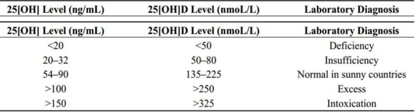
Vitamin D insufficiency is defined as levels <30 ng/ml (80 nmol/L), whereas levels below 20 ng/ml (50 nmol/L) and 10 ng/mL (25 nmol/L) represent deficiency and severe deficiency, respectively (Table 1) [34]. Levels between 40 and 60 ng/mL are the preferred range, while vitamin D intoxication usually does not occur until 25(OH)D3 reaches levels higher that 150 ng/ml [31].
Table 1: Blood levels of vitamin D.
1.3 The Genomic Mechanism of Action of Vitamin D
All genomic actions of vitamin D are mediated by VDR. VDR is a transcription factor and a member of the steroid hormone nuclear receptor family, related to the retinoic acid receptors and as most of the receptors has a DNA-binding domain (C-Domain), a ligand-binding domain (E-domain) and finally activating domain (F-domain) [2]. It has been proved that a single
11
receptor mediates all of the functions of vitamin D. This receptor is a 427 amino acid peptide and acts through vitamin D-responsive elements (VDREs), a complexes required for its genomic activity which are placed on the start site of the target gene [2]. These complexes can be both gene and cell specific, cells research enabling the selectivity of vitamin D3 action from cell type to cell type [4]. VDR was proved to be found in almost all tissues and cells. It has been established that the diverse biological actions of 1,25-dihydroxyvitamin D3 are initiated through precise changes in gene expression, which are mediated by an intracellular VDR. Activation of the VDR through direct interaction with 1,25(OH)2D3 prompts the receptor’s rapid binding to regulatory regions of target genes, where it acts to nucleate the formation of large protein complexes whose functional activities are essential for directed changes in transcription. When VDR interacts with the ligand, the repressor is no longer able to bind the receptor and the receptor changes conformation, forming heterodimer at the VDREs [2, 35]. At the same time, it binds several other proteins required in the transcription complex and acquires an activator [2, 35]. Once the complex is formed, DNA bends, biochemical processes take place, and transcription is either initiated or suppressed depending on the gene [2, 35, 36]. These responses are tissue-specific and range from highly complex actions essential for homeostatic control of mineral metabolism to focal actions that control the growth, differentiation and functional activity of numerous cell types including those of the immune system, skin, the pancreas, and bone.
1.4 The Nongenomic Mechanism
Vitamin D3 is also able to activate very rapid nongenomic mechanisms [4], lasting from seconds to 10 minutes [37]. This mechanism involves signal transduction pathways including activation of adenylyl cyclase-cAMP-protein kinase A and phospholipase C-diacylglycerol-inositol (1,4,5)-trisphosphate-protein kinase C signal transduction pathways [38]. Particularly,
12
the second messengers Raf (rapidly accelerated fibrosarcoma)/MAPK play an important role because they may engage in cross-talk with the nucleus to modulate gene expression [37]. This firstly identified nongenomic activation is associated with the rapid stimulation of intestinal calcium transport called “transcaltachia” [39]. Successively, this effect was identified in the chondrocytes of the bone growth plate [39] and in keratinocytes of the skin [40]. Identification of the receptor for vitamin D3 has focused on the VDR itself albeit in a different configuration to identify agonists able to induce nongenomic effects [41]. Interaction between vitamin D3 and membrane-associated rapid response steroid binding protein (MARRS) has been studied as well. These receptors are located in the membrane within caveolae/lipid rafts [42] where they are poised to activate kinases, phosphatases, and ion channels.
1.5 Physiological Effects of Vitamin D
Vitamin D is involved in mineral metabolism, bone growth and it also stimulates absorption of phosphate and magnesium ions; indeed, its main effect is to facilitate intestinal absorption of calcium. In cases when there is a lack of vitamin D, dietary calcium is not absorbed efficiently. The best studied of these calcium transporters is calbindin, an intracellular protein that carries calcium across the intestinal epithelial cell [43].
There is evidence in the literature on the effects of vitamin D on bone tissue: it induces the expression of osteocalcin and suppresses synthesis of type I collagen and in cell cultures, vitamin D stimulates differentiation of osteoclasts. The crucial effect of vitamin D on bone is to provide the proper balance of calcium and phosphorus to support mineralization [43]. Some experiments, using cultured cells, have demonstrated that vitamin D has potent effects on the growth and differentiation of many types of cells. These findings suggest that vitamin D has physiologic effects much broader than that of a role in mineral homeostasis and bone
13
function. This is an active area of research and a much better understanding of this area will likely be available in the near future [43].
Of interest, VDRs are not only present in different tissues, such as bone, skin, intestine and kidneys, but also non-classical organs like brain, eyes, heart, pancreatic islets (β-cells), immune cells, muscle, adipose tissue, thyroid, parathyroid and adrenal glands [44].
14
Importantly, many of these non-classical tissues also express vitamin D-activating enzymes, hence, allowing these non-classical actions to occur via local activation of vitamin D.
Recently, a crucial role for vitamin D also in digestive system health has been described [45]. Vitamin D links to several target tissues in the digestive system, in the oral region, in the salivary glands, in the stomach and in the small and large intestine [46].
In gastric mucosa, vitamin D appears to have high preventive and therapeutic potentials [47] and it is able to regulate endocrine and paracrine secretion of gastrin with secondary effects, for instance, on parietal cell HCl and pepsinogen secreting chief cells [46].
A growing body of data suggests that both vitamin D and VDR, play an important role also in the regulation of cardiovascular homeostasis and suggest a protective effect for vitamin D against cardiovascular disease [48-51]. Vitamin D3 deficiency is significantly associated with cardiomyopathy and increased risk of cardiovascular disease with consequent mortality [4]. The Intermountain Heart Collaborative Study revealed that vitamin D3 blood levels below 15 ng/mL compared to vitamin D3 above 30 ng/mL are associated with significant increases in the prevalence of type 2 diabetes mellitus, hypertension, hyperlipidemia and peripheral vascular disease, coronary artery disease, myocardial infarction, heart failure, and stroke, as well as with incident death, heart failure, coronary artery disease/myocardial infarction and stroke [51]. The ability of this hormonal system to inhibit the renin-angiotensin system [52], control blood pressure, inhibit cellular proliferation and hypertrophy, reduce fibrosis and suppress immune function suggests a variety of plausible mechanisms that could contribute to these palliative effects. However, the cardioprotective “hypothesis” has not been without controversy and results to date have been inconclusive.
Some, but not all, observational studies in humans provide support for these experimental findings, raising the possibility that vitamin D or its analogs might prove useful therapeutically in the prevention or treatment of cardiovascular disease. For example, VDR knockout mice
15
have elevated circulating levels of renin and angiotensin II and develop hypertension [52]. A similar phenotype occurs in mice lacking the 1α-hydroxylase gene [53]. In mice, injection of 1,25(OH)2D3 analogs suppresses renin production in vivo and negatively regulates the expression of the angiotensinogen gene [54-56].
Vitamin D is also important in brain homeostasis; the role of vitamin D in the brain development and function of neurons was suggested when the first evidence of 1α-hydroxylase, an enzyme responsible of an active form of vitamin D and VDR, were found to be present in the brain [57]. The specific mechanisms that mediate the neuroprotective effect of vitamin D are still unclear; however, vitamin D may act in many pathways including antioxidant pathways, neuronal calcium regulation, immunomodulation and glutamatergic systems [58-61].
A meta-analysis examining the association between vitamin D3 status and the risk of cerebrovascular events, including more than 1200 stroke cases, found that the pooled relative risk for stroke was 52% higher when comparing 25(OH)D levels ≤12.4 ng/mL with 25(OH)D levels >18.8 ng/mL [62]. As concerned to cerebral effects, the observed widespread distribution of 1α-hydroxylase and the nuclear VDR in both neurons and glial cells suggest that vitamin D3 may have autocrine and paracrine properties in the brain [63, 64]. VDR is highly expressed in multiple brain regions [65] in animal [66] and human [67] brain, particularly in the pontine-midbrain area, cerebellum, thalamus, hypothalamus, basal ganglia, hippocampus, olfactory system, and the temporal, orbital and cingulate areas of brain cortex [68]. Mounting evidence indicates that vitamin D3 and its receptors play an important role in the brain, ranging from neuroprotection to immunomodulation [69], cells proliferation and differentiation [70], and plays an important role both in developing [71] and adult brain [65]. Vitamin D3 can exert these effects since it is able to cross the blood-brain barrier and can bind to VDR within the brain [72, 73].
16
Moreover, vitamin D3 protects neurons against NMDA, glutamate, 6-hydroxydopamine and reactive oxygen species [74, 75]. It has been hypothesized that vitamin D3 exerts its neuroprotective effects via the modulation of neuronal Ca2+ homeostasis, in particular through the downregulation of the L-type voltage-sensitive Ca2+ channel in hippocampal neurons against excitotoxic insults [65], accompanied by an increase in VDR density. Vitamin D3 is able to inhibit proinflammatory cytokine and NOS [76] typically increased during various insults or disorders, such as ischemia and reperfusion, Alzheimer’s disease, Parkinson’s disease, multiple sclerosis and experimental autoimmune encephalomyelitis. Early vitamin D3 deficiency may be considered as a risk factor for a number of neurological disorders including schizophrenia, autism [73, 77], multiple sclerosis, Parkinson’s disease and stroke [62, 78]. Vitamin D3 deficiency is associated with reduced cognitive function, which is an important issue for stroke patients [79]. Moreover, in the rat model of stroke, vitamin D3 supplementation has been found to reduce brain damage [80] and consequent seizures [62] and oxidative stress [14]. For this reason, it should be very important to study the role of vitamin D3 in counteracting negative effects of oxidative stress in vitro brain. So, vitamin D3 may be considered as a potential drug for the treatment of neurodegenerative disorders.
1.6 Vitamin D specific actions on Ovarian Tissue
In recent years, scientists have increased interest in the study of vitamin D thanks to the discovery of the presence of its receptor VDR in many tissues. It has been demonstrated that in ovarian tissues a high density of VDR is present as well [81, 82]. In female reproduction, the importance of vitamin D was initially appreciated in vivo, as mice who were either deficient in vitamin D or lacked the vitamin D receptor (VDR), suffered from underdeveloped uterus and an inability to form normal mature oocytes, which in turn lead to infertility [83]. In humans, VDR is expressed in many female organs, including the ovaries (particularly granulosa cells),
17
uterus (endometrium and myometrium), and placenta [84]. These receptors are targeted by the active form of vitamin D and produce an array of effects in female reproduction. For example, vitamin D3 regulates genes involved in estrogen synthesis [84] and it also controls several genes involved in embryo implantation [85].
1.6.1 Vitamin D3 and Resveratrol
In this part of research (Project 1), the effects of vitamin D3 (vitD) and resveratrol (RES) were tested in ovarian cell culture and tissue. Resveratrol (3,5,4′-trihydroxy-trans-stilbene) is a natural antioxidant polyphenolic compound belonging to stilbene phytoalexins, a subgroup of non-flavonoid phenolic compounds [86, 87]. RES is contained in various vegetables such as berries, grapes, peanuts, besides red wine [88]. Particularly, red wine is the main source of RES, but a recent study discovered that peanut sprouts contain abundant RES both in cis and trans isoforms [89, 90]. Cis- and trans-isomers of RES coexist in plants and in wine. RES is rapidly metabolized in vivo and has a low water solubility, which reduces the rate-absorption in cells [91] reducing oral bioavailability [92]. The effectiveness of orally administered RES depends on its absorption, metabolism and tissue distribution. At intestinal level, RES is absorbed by passive diffusion or through the formation of complexes with membrane transporters, whereas in the bloodstream it can be found as glucuronide, sulfate, or free as well [93]. In some studies performed on animal models, the peak concentrations of trans-RES occur in blood and serum very rapidly, about 15 min from the beginning of the administration [94]. In different studies, RES solubility has been increased by the use of ethanol (50mg/mL) or other organic solvents [94]. Moreover, researchers have recently attempted to improve RES chemical stability, water dispersibility, bioavailability, permeability through blood–brain barrier (BBB) and therapeutic efficacy by using nanostructure-based drug delivery systems [95-97]. Epidemiologic studies have shown that RES has beneficial effects in preventing
18
various pathologic conditions ranging from cardiovascular diseases to cancer [98]. As reported by in vitro studies, RES can inhibit cell proliferation, induce apoptosis and block cell cycle progression in numerous types of human cancer cell lines, such as those of the colon, skin, breast, lung, prostate and liver, as well as pancreas [99]. In addition, in a few in vivo experimental models of colon and esophagus cancers the effectiveness of oral doses of RES was shown [100, 101]. RES acts as a phytoestrogen modulating estrogen receptor (ER)-mediated transcription [102]. There is a variety of RES-sensitive tissues that are ER positive and the two ER subtypes in mammals, ERα and ERβ, exhibit different tissue-specific expression profiles [103]. Specifically, effects of RES on ER include anti-inflammatory effects such as protection from trauma hemorrhage-induced injury and suppression of Interleukin-6 expression in the liver, intestine and cardiovascular system [104]. However, RES does not induce proliferation of mammary or uterine tissues, allowing it to be taken as a dietary supplement. RES binds and increases the transcriptional activity of estrogen receptors (ERα and ERβ) at 50–100 μM [103, 104-106]. RES displays a great affinity for ER behaving as either agonist or antagonist in a cell- and tissue specific manner [107]. This is important to explain the effectiveness of RES in reducing the number of vasomotor episodes and the intensity of hot flashes (HF), with the transition from moderate/severe to mild symptoms in 78.6% of patients [108]. The common incidence of hot flashes is around 75% and presently hormone replacement therapy is the gold standard in the management of moderate to severe vasomotor symptoms associated with menopause. RES has also been associated with anti-inflammatory effects, particularly in tissues that contain a large number of estrogen receptors, through this connection has been studied, but there are few studies on the mechanisms activated [109, 110].
It has been demonstrated that in ovarian tissues a high density of VDR is present as well [81, 82] and vitD acts through intracellular mechanisms similar to what observed for RES [111]. The role of vitD in cellular growth regulation is demonstrated by its ability to arrest cells in the
19
G1/G0 phase of the cell cycle, and by up-regulating p21, a powerful tumor suppressor gene. Thus, vitD can control cell division and proliferation [112]. VitD also has important anti-proliferative, anti-angiogenic and pro-differentiative effects in a wide range of cancers [113]. Many of the bioeffects of RES overlap with reported benefits from high circulating levels of vitD. Thus, given the ability of vitD to elicit a wide range of bio-effects via transcriptional regulation, it is interesting study RES in the context of VDR signaling to help in elucidating the molecular pathways involved by these two dietary lipophilic substances in optimizing healthspan and well aging [112]. The potential for RES to modulate vitamin D receptor signaling has recently been postulated [114, 115]. There is an overall structural symmetry and parallel configuration of RES and known VDR ligands, which could suggest that RES might serve as a low-affinity VDR ligand with the ability to activate VDR. Intriguingly, several targets emerge such as eNOS, cyclooxygenase and Akt kinase, all of which are likewise regulated by vitD [116]. Finally, vitD exerts beneficial effects on ovarian tissues preventing ROS-derived cellular injury [82]. Therefore, since these two substances have similar effects on ovarian cells, some form of interaction in exerting effects can be hypothesized. This could lead to interesting results for future clinical use in menopause-related conditions like hot flashes.
1.7 Vitamin D and Gastric Disorders
Recently, a crucial role for vitamin D in digestive system health has been described [45]. In gastric mucosa, vitamin D appears to have high preventive and therapeutic potentials [47] and is able to regulate endocrine and paracrine secretion of gastrin with secondary effects, for instance, on parietal cell HCl and pepsinogen secreting chief cells [46]. A problem related to the gastric system is the disproportionate use of nonsteroidal anti-inflammatory drugs (NSAID); indeed, around 30 million people consume NSAID to manage pain, inflammation and fever [117, 118]. The action mechanism of these drugs consists in the inhibition of the
20
biosynthesis of prostaglandins, the inactivation of cyclooxygenase and an increase in leukotrienes production [119], but adverse events, such as alterations in renal function, effects on blood pressure, hepatic injury and platelet inhibition, are a challenge in clinical treatment optimization [120]. However, severe gastrointestinal disorder accompanied by gastric mucosal perforation and bleeding is a major concern as well as the worst outcome of prolonged NSAID-therapy [121]: indeed, they induce gastric mucosal lesions because of their acidic properties [119]. The mechanism behind gastric damage involves a highly acidic gastric environment that favours the migration of nonionized lipophilic NSAID into the epithelial cells and at the cell surface these are dissociated into ions, trapping hydrogen ions and inducing mucosal injury. This action is further enhanced by the decrease mucosal blood flow, secretion of mucous and bicarbonates, and the defensive factors of the gastric layer [119]. Indeed, these side effects lead to reduced intestinal mucus formation, disturbed micro-circulation causing increased intestinal motility and increased mucosal permeability to many inflammatory mediators including neutrophils and cytokines [122]. The consequences can range from dyspepsia to severe peptic ulcer and bleeding; endoscopically the range from subepithelial haemorrhages and erosions to total destruction of epithelial membrane and full thickness ulcer [123]. Diclofenac, an anthranilic acid derivative with pKa of 4.0 [117], is the most widely prescribed NSAID for treating several forms of pain and inflammation [124-126]. The main clinical problem of Diclofenac is toxicity induced by oxidative tissue injury in the intra-mitochondrial environment, which appears to play a prominent role in the pathophysiology of digestive ulceration [117, 127, 128]. For these reasons, NSAID users are subjected, often empirically by doctors, to anti-acid treatments such as the proton-pump inhibitors (PPI) [129], thus, not surprisingly, NSAID and PPI are among the most frequently co-prescribed drugs worldwide [130]. Moreover, a number of clinical studies have provided the support for the ineffectiveness of PPI in preventing or reducing the NSAID-damages [131-133]. Indeed, current evidence
21
suggests that PPI are also associated with numerous side effects such as hypergastrinemia [134], enteric infections [135], adverse cardiovascular events [136] and increased mortality rates [137]. The need for NSAID clinical use despite their side effects encourages continuous research to create novel agents able to counteract their adverse effects with better safety profile, in particular using natural compounds. Recently, a product has been introduced that, in addition to having the antacid properties of alginates, claims to possess gastroprotective properties deriving from vitamin D3 and from plant extracts. It has been tested alone and combined with Diclofenac in order to provide an effective and safer strategy for the management of NSAID-induced gastroenteric lesions (Project 2).
1.7.1 Vitamin D and Alginates
There is a new group of widely used molecules that includes alginates, a polymer of alginic acid derived from the cell wall of various brown algae [138]. Alginates are polysaccharides composed of two β-d-mannuronic acid (M) and α-1-glucuronic acid (G) monomers which are held together by β 1,4 bonds [139, 140]. Recently, there is a growing interest in alginates as a therapy for Gastro-Esophageal Reflux Disease (GERD) [141]. Alginates are able to block the HCl reflux in a mechanic manner; indeed, they do not have any pharmacologic property [142]. The mechanism of action of alginates has been called “rafting”, which means that in the presence of gastric acid, they form a gel in which carbon dioxide (resulting from the splitting of bicarbonate) is trapped. Then the gel is carried to the top of the gastric contents neutralizing the acidity and preventing the ascent of acid material into the esophagus [143]. The advantage of alginate-based reflux suppressants over antacid alone is that they provide rapid and longer-lasting symptom relief [144].
It was seen that it is useful to associate alginates with natural substances. This association has been found to be effective in promoting stomach health, reducing inflammation and supporting
22
the immune system. Moreover, alginate/antacid system has been used as a carrier of probiotic, drugs and plant extracts [145]. For example, alginate/bicarbonate combined with two herbal gastroprotective extracts (Opuntia ficus-indica and Olea europaea) has been successfully evaluated in patients with GERD [145].
Recent studies have explored a possible role of vitD on gastroprotection [45, 146]. VitD binds its receptor VDR which is present in several tissue targets in the digestive system, in particular in the oral region and in epithelial cells of the oral cavity, tongue and gums. In addition, vitD appears to have a therapeutic role in gastric mucosa as well, stimulating cell proliferation and differentiation [147] and regulating endocrine/paracrine gastrin and pepsinogen secretions [148]. Finally, vitD acts on smooth muscle cells in the pyloric region and in different areas of the small intestine [148]. Furthermore, the low plasmatic level of vitD is found to be responsible for an insufficient emptying of the stomach, swelling, constipation and intestinal irritation [148]. After the binding between vitD and its receptor, several intracellular events involved in different mechanism start, including the protective role against oxidative stress [149], the regulation of autophagic pathways through the regulation of ATG16L1, a protein complex necessary for autophagy [35], and the ability to inhibit apoptosis by increasing the expression of endothelial nitric oxide synthase (eNOS) leading to the nitric oxide (NO) production [149]. In this context, vitD can exert some beneficial effects on gastric tissue. For this reason, its use in association with other gastroprotective agents such as alginates can increase therapeutic efficacy of the formulation in respect to the efficacy that would be obtained only with gastroprotective drugs.
1.8 Vitamin D and Cardiovascular System
VitD is also a modulator of vascular wall growth as well [150] and induces a decrease in the expression and/or secretion of proinfammatory and proatherosclerotic factors in the
23
endothelium [151]. The role of endothelium as a target of vitD is demonstrated by the study published by Zehnder et al. [152], in which the expression of mRNA and protein for lα-hydroxylase in human endothelium was shown for the first time. These findings demonstrated the direct effects of vitD on endothelial function, whose alteration plays an important role in the development of atherosclerosis. Indeed, vascular endothelial function is a crucial element as regards cardiovascular function and its impairment is an early manifestation of atherothrombotic disease [153, 154]. Endothelium acts as a barrier and a regulator of blood vessel activity [154] mainly through NO, which can stimulate the relaxation of the underlying smooth muscle, leading to vasodilation [154]. NO is produced by vascular endothelial cells in response to different stimuli and acts as a messenger molecule [154], activating guanylate cyclase to enhance cGMP, which in turn causes relaxation of smooth muscle and vasodilation [155]. NO is produced by nitric oxide synthases (NOS) which utilize L-arginine as their principal substrate, oxidizing it into L-citrulline and NO [153, 156]. The endothelial isoform of NOS (eNOS) is constitutively expressed in endothelial cells and is responsible for the basal release of nitric oxide from the endothelium and for the rapid change into nitric oxide flux in response to physical stimuli and molecular agonists [153, 157]. A decrease in endothelial NO production appears in the early phases of atherosclerosis development [158, 159]. Moreover, NO synthesis impairment is involved in a number of cardiovascular diseases such as peripheral vascular disease, congestive heart failure and cerebrovascular events [154, 160, 161]. The impaired NO status may cause an overproduction of reactive oxygen species in the vasculature, representing a risk factor for cardiovascular disease.
1.8.1 Vitamin D and Protective Molecules
Numerous studies have shown that acute and chronic supplementation with L-arginine improves NO production by endothelium in individuals with risk factors for atherothrombosis,
24
as well as in individuals with established atherothrombotic disease [153]. For this reason, extracellular L-arginine concentration is the principal determinant of intracellular L-arginine availability for eNOS [153]. Another important element to maintain ROS production at basal level is the use of “natural” substances able to modulate metabolic pathways and to treat pathological conditions. Many studies suggest that a number of natural compounds such as coenzyme Q10, vitD and other products, included in the nutraceuticals group, have the potential to target multiple pathways under these pathological conditions [162-164]. For example, with regard to cardiovascular diseases, there is strong evidence that demonstrate the link between oxidative stress and impaired mitochondrial function [165, 166]. CoQ10 is located in the mitochondria, lysosomes, Golgi apparatus and plasma membranes and provides an antioxidant action either by direct reaction with free radicals or by regeneration of tocopherol and ascorbate from their oxidised state [167, 168]. Endogenous synthesis of CoQ10 in the body decreases with age. It has been observed that up to 75% of ischemic heart disease patients exhibit low levels of CoQ10 in the plasma and in the heart in relation to the course of the disease [169, 170]. A long-term therapy with CoQ10 has been shown to decrease heart failure symptoms, to reduce major adverse cardiovascular events and mortality and to be safe and well tolerated [171]. CoQ10 has been reported to have many biological effects both in vitro and in vivo [172, 173]. CoQ10 is commonly used for prevention and treatment of many cardiovascular diseases such as myocardial infarction, congestive heart failure and other drug-induced or disease-induced cardiomyopathies [168, 174, 175]. Moreover, Q10 showed the ability to prevent oxidative stress, apoptotic cell death and monocytes cell adhesion in HUVEC [176]. On the basis of the results obtained in this research field, it is possible to speculate on the possibility of preparing a mixture of antioxidants and vitamins able to prevent cardiovascular diseases. Some studies demonstrate that a mix of micronutrients, including vitD, significantly reduces markers of lipid peroxidation and at the same time increases antioxidant potential of the
25
molecules contained. The importance of vitamin D in the cardiovascular system is evidenced by the observation that the deletion of VDR from the heart results in hypertrophy [177, 178] and contributes to the acceleration of atherosclerosis [4, 179]. Both experimental and clinical studies support the protective role of vitamin D in vascular system and in cardiac function through lowering blood pressure, improving endothelial function, inhibiting oxidative stress and reducing the activity of renin-angiotensin system [180]. Additional studies demonstrate the possible autocrine/intracrine mechanisms exerted by vitamin D as a modulator of endothelial functions [181]. In addition it has been demonstrated that the control on NO production exerted by vitD is mediated by VDR and related to intracellular pathways leading to eNOS activation [181]. Therefore, the VDR modulation could represent a new treatment option for all cardiovascular diseases in the future [182].
Thus, in the present study (Project 3) it was examined the effects of combination of vitD, Q10 and L-arginine on cell viability and NO/ROS production in endothelial and cardiac cells in order to assess a potential cooperative effect of these substances on cardiovascular function at the cellular level.
1.9 Vitamin D in Nervous System
The effects of vitamin D also affect the central and peripheral nervous system. Vitamin D receptors are in fact present throughout the brain including the primary motor cortex, the region that coordinates movement [183-185]. Specifically, VDRs have been identifIed in neuronal and glial cells in several brain areas including the cortex, deep gray matter, cerebellum, brainstem nuclei, spinal cord and the ventricular system [185.]. Furthermore, the enzyme 1α‐ hydroxylase, the activator of vitamin D precursors, is also present in the brain [183-187]. Vitamin D levels are associated with the conductance velocity of motor neurons and neurotransmission mediated by dopamine, serotonin, acetylcholine, GABA and the
26
catecholamines [186, 187]. The combination of in vitro, ex vivo and animal model data provides evidence that vitD has a crucial role in neuronal proliferation, differentiation, neurotransmission, neuroplasticity and neuroprotection and its levels correlating to the levels of several neurotrophic factors including nerve growth factors (NGF) and that of neurotrophins, which play crucial roles in the maintenance and growth of neurons [188, 189].
Some evidence implicates vitD as a candidate in influencing susceptibility to a number of psychiatric and neurological diseases, such as schizophrenia, autism, Parkinson disease (PD), amyotrophic lateral sclerosis, epilepsy, Alzheimer disease (AD) and is especially strong for multiple sclerosis (MS) [190-192].
The neuroprotective effect of vitD has been recently reported in cognitive decline of aging rats [193], and it has been extensively studied in the animal model of MS and the experimental allergic encephalomyelitis. The hormone prevents onset and reversibly blocks progression of clinical signs, but such a protective effect is absent in VDR knockout mice [191]. The effect of vitD might not be due exclusively to its neuro-immunomodulatory properties [191] since recently it has been reported that the hormone enhances neural stem cell proliferation and differentiation into neurons and oligodendrocytes, the myelinating cells of central nervous system [194, 195]. Neural stem cells constitutively express VDR, which can be upregulated by vitD [194]. VitD regulates the expression of many AD-related genes. It attenuates Aβ peptide accumulation by stimulating phagocytosis of Aβ peptide probably by modulating transcription of Toll-like receptors and cytokines together with enhancing brain-to-blood efflux transport by increasing P-glycoprotein expression [196]. Alterations in adult neurogenesis appear to be a common hallmark in different neurodegenerative diseases including PD and AD [197]. Therefore, factors that stimulate neurogenesis have been indicated as possible treatments of neurodegenerative disorders.
27
Moreover, the combination of anti-neurodegenerative drugs with vitD supplementation might be useful. Indeed, the supplementation of the combination nemantidine plus vitD has been shown to prevent cognitive decline more efficiently than that of the single compounds [198]. In addition, vitamin D exerts direct neuroprotective effects via the synthesis of proteins binding calcium (Ca2+) ions; it is important in neuronal function and in neuronal transmission. Proper levels of neuronal calcium are critical because their excess may result in the formation of ROS with consequent neuronal damage. Indeed, vitamin D levels are inversely associated with oxidative stress which damages the brain leading to neuronal apoptosis or necrosis [199]. Vitamin D also affects neuroplasticity, a process in which neural synapses and pathways are adapted to the needs of environmental and behavioral demands adjusting the brain to noxious stimuli diseases or environmental cues [185, 200, 201]. The VDRs in glial cells play in the uptake and release of neurotransmitters, including that of GABA neurotransmission within the motor cortex [202]. GABAergic function is the principal “brake” within the brain affecting muscle relaxation via the corticospinal neurons [203].
1.9.1 Vitamin D3 and Lipoic Acid
Alpha-lipoic acid (LA) is a fatty acid containing 8 carbons and 2 sulfur molecules in a dithiolane ring [204]. Initially discovered as a bacterial growth factor in potato extract, LA was later isolated and characterized from bovine liver [205, 206]. It is naturally synthesized in the liver and other tissues and obtained from various animal and plant sources in the diet. LA is a potent antioxidant, acting as a cofactor for mitochondrial enzymes, pyruvate dehydrogenase, α-keto-glutarate dehydrogenase activity and branched chain alpha-keto acids. In addition, LA affects numerous inflammatory pathways by modulating the NF-κB-dependent gene expression [207-210]. LA’s antioxidant properties were identified in 1959 when Rosenberg and Culik found that the administration of LA to vitamin C-deficient guinea pigs relieved
28
scurvy symptoms [211]. LA has been used for decades in Germany to treat diabetic neuropathy, cirrhosis and mushroom and heavy metal poisonings [212]. Endogenous LA occurs in the R chiral conformation (R-LA) at the C6 carbon atom. Manufacturing LA produces racemic LA, an even mix of the R-LA and S-LA chiral conformations. The bioavailability and bioactivity of racemic vs R-LA is debated [208, 213]. The functional activity of LA derives from the disulfide bond between the 2 thiol groups that cycles between the oxidized (LA) and reduced conformations (dihydrolipoic acid, DHLA). LA is highly reactive to free radicals, preventing oxidative damage by directly scavenging and neutralizing ROS [204, 210].
Several randomized clinical trials have demonstrated that LA can act as a therapeutic agent in chronic diseases such as diabetes mellitus [214], cardiovascular diseases [215] and cancers [216] by decreasing chronic inflammation and improving glucose-insulin homeostasis and endothelial function [217]. It has also been shown that LA exerts beneficial effects on the redox state of the plasma and endothelium-dependent vasodilation [218]. Previous studies have demonstrated that LA can decrease lipopolysaccharide-induced inflammatory responses and act as an anti-inflammatory agent through affecting the cyclooxygenase-2 [219, 220] and inducible nitric oxide synthase (iNOS) [221].
LA binds to Cu and Fe preventing their production of free radicals. In mitochondria, the LA/DHLA redox couple serves as a key cofactor of the pyruvate dehydrogenase complex and α-ketoglutarate dehydrogenase of the Krebs cycle, and aids synthesis of nucleic acids, thereby improving mitochondrial efficiency and reducing ROS generation [210, 212]. LA modulates also the PKB/Akt signaling pathway important for vascular endothelial integrity and the redox-sensitive transcription factor Nrf2 and NF-κB pathways, thereby indirectly promoting an antioxidant environment [222-225].
There is no doubt that it is a strong antioxidant, but due to certain reasons its use for medicinal purposes is prohibited; however, in some states it is used as a supplement and in others as a
29
remedy [226, 227]. These restrictions are due to some endogenous characteristics of substance by itself, such as the changeableness due to the disclosing of dithiolane ring and the emergence of disulfide bond between molecules. Other properties that limit the oral use of LA are its decreased ability to become dissolved in the gastrointestinal tract and increased rate of hepatic metabolism. In addition, besides it is widely known antioxidant potential, LA has also many other functions, as it is its involvement in mitochondria producing energy, by acting as cofactor for various enzymes involved in metabolism [226]. For its antioxidant effect, LA has been used in combination with vitD to study the capacity to prevent or repair the damage caused by oxidative stress and neurodegeneration (Project 4).
30
2. Aims of the Thesis
In recent years, vitamin D has seen growing interest among researchers, especially due to the presence of its receptor VDR in many tissues and organs. For this reason the biological effects of vitamin D3 (the active form of vitamin D, 1,25-dihydroxyvitamin D3, vitD) mediated by the interaction with VDR were studied in different tissues (ovarian, gastric, cardiac and brain).
➢ It has been demonstrated that in ovarian tissues a high density of VDR is present as well and vitD acts through intracellular mechanisms similar to what observed for resveratrol, a natural antioxidant polyphenol able to exert a wide range of biological effect on several tissues. Despite its important beneficial properties, it has a low water solubility, which limits its therapeutic applications in humans. The aims of this part of research (Project 1) were to evaluate the cooperative effects of resveratrol (RES) combined with vitD on ovarian cells studying cell viability, ROS production, activated pathways in in vitro study and in in vivo study to quantify vitD and RES, to analyze the concentration of radical oxygen species and to study activated pathways. Moreover, the modulation of specific intracellular pathways involving ER and VDR receptors has been studied to hypothesize a future clinical use in menopause related conditions like hot flashes.
➢ Since vitD appears to have gastroprotective properties, the effects of vitD have been studied also in stomach combined with alginates (Project 2); it has been tested alone and combined with Diclofenac in order to provide an effective and safer strategy for the management of NSAID-induced gastroenteric lesions. Hence, the primary objective of this research was to evaluate whether it is possible to improve the protection of the gastric mucosa during therapy with Diclofenac. Thus, the present study was designed to examine the protective effects of alginates combined with vitD assessing its effects on factors related to gastric protection such as viability, radical productions and intracellular mechanism involved.
➢ As regards the research part focused on vitD in cardiovascular system (Project 3), the effects of combination of vitD, Q10 and L-arginine on cell viability and NO/ROS production in endothelial and cardiac cells were evaluated in order to assess a potential cooperative effect of these substances on cardiovascular function at the cellular level. Moreover, the involvement of PI3K/Akt and ERK/MAPKs pathways leading to eNOS activation has been investigated. To confirm data, the same agents were also tested in an animal model to assess vasodilation, NO and ROS production.
31
➢ As regards the research part focused on effects of vitD in brain ageing (Project 4), the ability of vitD combined with lipoic acid (LA) has been studied to prevent or repair the damage caused by oxidative stress and iron accumulation, two different biological aspects involved in brain ageing and neurodegeneration. Cell viability, mitochondrial membrane potential and pathways activated in oxidative condition has been studied. In addition, to investigate the protection exerted by combination of vitD and LA in induced neurodegeneration using catalytic iron, cell viability, ROS production, iron concentration and activation of intracellular pathways have been studied in order to hypothesize a new human anti-ageing treatment.
32
3. Materials & Methods
3.1 Cell Culture
In order to investigate the effects of vitD mediated by VDR receptor in different tissues, we used several cell types which of them some directly isolated by organ of interest.
● Project 1 (ovarian line of research)
CHO-K1 cells (Chinese Hamster Ovary cell), an epithelial cell line derived as a subclone from the parental CHO cell line initiated from a biopsy of an ovary of an adult Chinese hamster [228], have been purchased from Lonza (Basel) and cultured in Dulbecco’s modified Eagle’s medium: Nutrient Mixture F-12 (DMEM-F12; Sigma, Milan, Italy) supplemented with 10% fetal bovine serum (FBS, Sigma, Milan, Italy), 2mM glutamine and 1% penicillin/streptomycin (Sigma, Milan, Italy) and incubated at 37°C, 5% CO2, and 95% humidity [229]. When the cells
reached 80–90% of confluence were seeded for different experiments; 1×10⁴ and 2.5×10⁴ cells were plated in a 96-well for MTT test and ELISA activation assay, respectively; 1×10⁵ cells plated on 24-well plates to analyzed ROS production; for Western blot analysis and SOD activity the cells were seeded in 6 wells and maintained until 85% of confluence.
● Project 2 (gastric line of research)
Primary epithelial gastric cells were obtained from the stomach of anaesthetized prepubescent pigs as previously described [45]. Briefly, the gastric cells were isolated using enzymatic solution in agitation (collagenase/dispase solution, Sigma-Aldrich, Milan, Italy) for 60 minutes at 37°C, centrifuged at 1500 rpm for 5 minutes at 4°C and then the pellet resuspended in complete medium (Ham’s F12 supplemented with 10% FBS, Sigma-Aldrich, Milan, Italy) on collagen coated dishes. The cells used for the experiments were obtained from passage 3 to passage 5. The cells were used to perform different experiments; to study cell viability, ATP level and NO production 1x10⁴ cells were plated on 96 well-plates; to study ROS production 1x10⁵ cells were plated on 24 well-plates; to study the intracellular pathways by Western blot
33
and to analyse the activities by kit ELISA the cells were plated on 60mm dishes until confluence. To synchronize the cells, before stimulations they were maintained in DMEM without red phenol and FBS and supplemented with 1% penicillin/streptomycin, 2mM L-glutamine and 1mM sodium pyruvate in an incubator at 37°C, 5% CO2 and 95% humidity for
18h and then to create a similar condition of human stomach, the cells were maintained for 2h on acidified medium (HCl was added at DMEM without red phenol and FBS and supplemented with 1% penicillin/streptomycin, 2mM L-glutamine and 1mM sodium pyruvate to obtain a medium with pH 4) before the stimulation. This acidified medium was maintained during the successive stimulations.
● Project 3 (cardiovascular line of research)
Experiments were performed on both PAE and H9c2 cells.
Porcine aortic endothelial cells (PAE) were purchased from Cell Applications, Inc. (San Diego, CA, USA) and maintained in Dulbecco’s modified Eagle’s medium (DMEM; Sigma, Milan, Italy) supplemented with 10% fetal bovine serum (FBS; Sigma), 2mM L-glutamine (Sigma), 1% penicillin-streptomycin (Sigma) at 37°C with 5% CO2, as reported in literature [230]. The
cells were plated to 1x10⁴ in 96-well plates to analyze cell viability using MTT test, to evaluate NO production and ROS production using Griess assay and a rate of superoxide release, respectively. To study the intracellular pathways using Western blot, they were plated in a 6-well until confluence (about 90%).
Rat cardiac cells (H9c2) were obtained from the American Type Culture Collection (Rockville, MD) and cultured in Dulbecco modified Eagle Medium (DMEM; Sigma, Milan, Italy) supplemented with 10% fetal bovine serum (FBS; Sigma), 1% penicillin–streptomycin (Sigma) and 2mM L-glutamine (Sigma) in an incubator at 5% CO2, and 37°C, as reported in literature
[231]. The experiments were performed with cells from passages 14–17. The cells were plated to 8x10³ in 96-well plates to analyze cell viability using MTT test; to evaluate NO and ROS
34
production using Griess assay and a rate of superoxide release, respectively; finally, to study the intracellular pathways using Western blot, cells were plated in a 6-well until confluence (about 90%). Both cell lines before the experiments were maintained in DMEM 0% serum supplemented with L-glutamine, penicillin-streptomycin without red phenol (starvation medium) for 4-18h.
● Project 4 (brain line of research)
Experiments were performed using both primary mouse astrocyte and HUVEC cells.
Primary mouse astrocyte cultures were prepared from both male and female C57BL/6 mouse pups, following a classical technique described elsewhere [232] according to the National Guideline for the Use and Care of Laboratory Animals. Briefly, within 24 h of birth, pups were euthanized and cortices were dissected, minced, mechanically digested, and let settle for 30 min at room temperature. Then the cell suspension was centrifuged at 800 rpm for 5min. Pelleted cells were resuspended in Neuronal Basal Medium (Sigma-Aldrich), supplemented with 5% fetal bovine serum (FBS, Gibco), 1% penicillin/streptomycin (Sigma-Aldrich), and 2mM L-glutamine (Sigma-Aldrich), plated in multiwells and maintained in culture for 6 days before treatment. Astrocytes should be separated from microglia and oligodendrocyte precursor cells by shaking, as reported in literature [233]. For the experiments, 1×10⁴ cells on a 96-well plate were plated to study cell viability by the MTT test, amyloid precursor protein (APP) by the ELISA test, and ROS production by the colorimetric test; 1×10⁴ cells on a black 96-well plate to analyze the oxygen consumption by a fluorescence kit; 1×10⁶ on a 6-well plate to determine the iron concentration by the colorimetric assay; 1×10⁶ on a 6-well plate to analyze the intracellular pathways activated by Western blot analysis; 1×10⁶ on a 6-well plate to analyze p53 activity and ERK/Akt activation; and 4×10⁴ on Transwell support to study the
permeability, to quantify vitD and LA. Before stimulations, the cells were maintained in Dulbecco's modified Eagle's medium (DMEM, Sigma-Aldrich) without red phenol and fetal