Treatment of Degenerative Mitral Regurgitation
Antonio M. Calafiore,
MD,
Michele Di Mauro,
MD,
Angela L. Iacò,
MD,
Valerio Mazzei,
MD,
Giovanni Teodori,
MD,
Sabina Gallina,
MD,
Luca Weltert,
MD,
Mauricette Samoun,
MD,
and Gabriele Di Giammarco,
MD
Division of Cardiac Surgery, University Hospital, Torino, Division of Cardiac Surgery, “G D’Annunzio” University, Chieti, and Division of Cardiac Surgery, Papardo Hospital, Messina, Italy
Background. The concept of overreduction of the pos-terior annulus was applied in surgical treatment of de-generative mitral valve disease.
Methods. From April 1993 to November 2004, 141 patients underwent overreduction of the posterior an-nulus of the mitral valve in mitral valve repair for degenerative disease. Mean scallop involvement per patient was 2.3 and increased to 3.0 in the last period. Correction of the prolapse of the posterior leaflet included resection with focal sliding (n ⴝ 100), or application of artificial chordae (nⴝ 28), with (n ⴝ 11) or without (nⴝ 17) plication of one or more scallops. The anterior leaflet prolapse was corrected with edge-to-edge technique (nⴝ 20) or chordal replacement (n ⴝ 28). An overreducting ring, 40 (n ⴝ 81) or 50 (n ⴝ 60) mm long (autologous pericardium in 64 cases and Sovering Miniband [Sorin, Saluggia, Italy] in 77) was used in all the patients.
Results. Three patients died in the early period (2.1%) and 3 (2.1%) were reoperated on from 3 to 24 months due to endocarditis (2 cases) and failure of repair (1 case). Ten-year freedom from death any cause was 91.6%, from reoperation 96.4%, from death any cause and reoperation 87.7%, from death any cause, reoperation, and New York Heart Association class III-IV 79.8%. Sixty-four patients out of 68 who survived more than 2 years (94.1%) at a mean follow up of 4.2ⴞ 2.5 years had no or 1ⴙ residual mitral regurgitation.
Conclusions. Although the complexity of mitral valve repair for degenerative disease increased, results of sur-gery remained stable. Apposition of a posterior overre-ductive ring was useful to cover any mistake performed during the correction.
(Ann Thorac Surg 2006;81:1310 – 6) © 2006 by The Society of Thoracic Surgeons
S
urgical treatment of degenerative mitral regurgita-tion (MR) is becoming more and more popular as the number of patients referred to surgery increases. The natural history of untreated patients with severe MR and good left ventricular function is widely recog-nized to be better if the treatment of the disease occurs when the patient is asymptomatic [1]. This tendency forced many surgeons to apply different techniques, less or more difficult, targeted mainly to the treatment of prolapse of the middle scallop of the posterior leaflet (PL), P2.In the great majority of the patients a ring, complete or incomplete, flexible, semiflexible, or rigid, was used. The purpose was to maintain the length of the insertion of the anterior leaflet (AL) as it was, and to reduce the length of insertion of the posterior annulus to a length equal to two times the length of the AL.
In the 1990s the concept of overreduction of the mitral annulus (MA) was introduced by Bolling and colleagues [2] to correct functional MR in dilated cardiomyopathy and postischemic MR. Our group
lim-ited the ring insertion to the posterior annulus using, in the beginning, a pericardial strip, glutaraldehyde-treated, 52 mm[3]and later 40 mm long[4]. Since 1993, we applied the same concept in the surgical treatment of degenerative MR. The purpose of this study is to describe the surgical techniques used by us to correct the different anatomical aspects of degenerative MR and to evaluate the results of overreduction of the posterior mitral annulus.
Material and Methods
From April 1993 to November 2004, 141 patients with prolapsing mitral leaflet(s) underwent mitral valve (MV) repair and overreduction of the posterior annu-lus. Patients with aortic valve disease were excluded. All the procedures were performed by one surgeon (AMC). They represent the 53.8% of all MV repair for nonfunctional MR performed during the same period (141/262).Table 1shows the preoperative clinical and echocardiographic characteristics of the patients.
Isolated PL prolapse was present in 49.9% of the patients (n⫽ 69) and isolated AL prolapse in 8.5% (n ⫽ 12), whereas both leaflets were involved in the remaining 42.6% (n⫽ 60). Prolapse of a single scallop was present in Accepted for publication Aug 22, 2005.
Address correspondence to Dr Calafiore, Division of Cardiac Surgery, “S Giovanni Battista” Hospital, c.so Dogliotti 86, 10126, Torino, Italy; e-mail: [email protected].
© 2006 by The Society of Thoracic Surgeons 0003-4975/06/$32.00
Published by Elsevier Inc doi:10.1016/j.athoracsur.2005.08.031
40.0% of the patients. Mean scallop involvement per patient was 2.3.
Surgical Indication
Presence of severe MR represented a surgical indication for mitral valve repair, independently from the left ven-tricular function. Eleven patients were asymptomatic (in 9 the left ventricular ejection fraction [EF], was equal or higher [and in 2 lower] than 60%, mean 62.4⫾ 6.4) and 130 were symptomatic (in 77 EF was equal or higher [and in 53 lower] than 60%, mean 58.9 ⫾ 10.4). Coronary bypass grafting was complementary and not substantial in 16 patients. In 3 patients with angina, MR was due to degenerative disease and was not functional. In one of them a septal reshaping[5]was also performed.
Surgical Techniques
All the patients were operated on through a median sternotomy; all but one who specifically asked for a small right lateral thoracotomy. Arterial inflow was always the ascending aorta and both vena cavae were directly can-nulated. The approach to the MV was always transseptal. A perioperative transesophageal echocardiogram was obtained in all the patients.
Prolapse of the Posterior Leaflet
The PL was involved in 129 patients. The prolapsing scallop was only one in 75 cases (P2 in 73 and P3 in 2). A combination of 2 or 3 scallops was present in 54 patients (P1 and P2 in 10, P1 and P3 in 4, P2 and P3 in 14, and all 3 scallops in 26). The correction of the prolapse was achieved by means of the following guidelines.
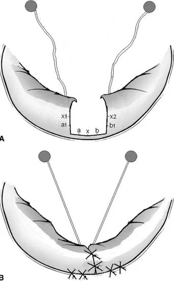
When P2 was involved, a quadrangular resection was performed, but only if the length of its insertion was not longer than 1/3 of the posterior annulus[6]. Reduction of the height of the posterior leaflet was obtained sewing the borders of P1 and P3 directly to the annulus[7]. This maneuver, which we call focal sliding, allows to reduce
the height of the posterior leaflet in a simpler and faster way (Fig 1). Furthermore, it has some important side effects. If P1, P3, or both are involved together with P2 (the chordae have to be elongated and not ruptured), the focal sliding moves medially the chordae of the adjacent scallop(s), eliminating the mechanism of the prolapse (Fig1B). If the chordae of P1 and/or P3 are ruptured, insertion of artificial chordae (Gore-Tex, W L Gore & Assoc, Flagstaff, AZ) will avoid any further scallop dis-placement after the focal sliding. In no case was the annulus itself plicated.
When P2 was involved, but its insertion was longer than 1/3 of the posterior annulus, we applied 4 or more artificial chordae (one suture is considered 2 chordae). If necessary, other chordae were applied to P1 and/or P3. If the height of P2 was roughly 20 mm or less, we did not believe it necessary to reduce its height; if higher, P2 (but if necessary also P1 and/or P3) was plicated with multiple U sutures using 4-0 Prolene (Ethicon, Somerville, NJ) to reduce its height to less than 20 mm (Fig 2)[8]. When P1
Fig 1. Focal sliding. (A) The suture of the lateral edges of P1 and P3 on the annulus, a1 to a, b1 to b, and x1 and x2 on x, increases the tethering on the elongated chordae of P1 and P3, counteracting the mechanism at the basis of the prolapse (B).
Table 1. Preoperative Clinical and Echocardiographic Characteristics
n⫽ 141
Age (years) 63⫾ 12 (28–85)
Female gender 50 (35.5%)
NYHA class (I to IV) 2.9⫾ 0.9
I 11 (7.8%) II 26 (18.6%) III 66 (46.4%) IV 38 (27.2%) Angina 3 (2.1%) Atrial fibrillation 32 (22.8%) EDv (mL/m2) 85⫾ 19 ESv (mL/m2) 37⫾ 14 EF (%) 59⫾ 10 MR (1/1 to 4/4) 3.8⫾ 0.5
EDv⫽ end-diastolic volume; EF⫽ ejection fraction; ESv⫽ end-systolic volume; MR⫽ mitral regurgitation; NYHA⫽ New York Heart Association.
or P3 were involved, but not P2, we used the artificial chordae or an edge-to-edge suture[9], according to the involvement, or noninvolvement, of the AL.
Prolapse of the Anterior Leaflet
The AL was involved in 69 patients. The prolapsing scallop was A2 in 38 patients, A1 in 2, and A3 in 3. Combination of 2 or 3 scallops was present in 26 patients (A1 and A2 in 5, A2 and A3 in 3, and all 3 scallops in 21).
In the first part of our experience we preferred to use the edge-to-edge technique to correct A2. This strategy is still used only when A1 or A3 are the only scallop(s) involved. We now prefer to correct the prolapse of the AL applying the artificial chordae.
Guidelines for Artificial Chordae Application
The new chordae are passed in the fibrous tip of the papillary muscle and are tied twice. The choice of the correct length of the new chordae is crucial for the success of MV repair. Many authors have faced this problem[10 –15]and we have reported a technique based on echocardiographic perioperative data [16]. With in-creasing experience we can reach our goal, keeping in mind some basic concepts.
First, the new chordae do not have the same elasticity of the native chordae and this extra length to be given has to be considered during the decision making. Second, when the heart is arrested and flaccid the tips of the papillary muscles are near the plane of the MA. When a leaflet is pulled up with a hook its excursion is often limited by its height and not by the length of the native elongated chordae that are generally longer than the height of the leaflet involved in the prolapse. The excur-sion of the AL can also be limited by the second-order chordae. If we tie the new chorda at the level of the maximal excursion of the leaflet, the chordal length will be undersized and a restricted motion will follow. As a consequence, we have to tie the new chorda at a higher level (3–5 mm) than the maximal excursion of the leaflet involved.
Overreduction of the Posterior Annulus
At the end of the procedure, an overreductive ring is applied in the posterior annulus. In the first part of our experience we used the autologous pericardium; glutar-aldehyde-treated, 40 or 50 mm long. More recently, we have been using the Sovering Mini Band with the same length (SMB40 or SMB50, Sorin, Saluggia, Italy). It is a flexible ring, made by a radiopaque silicone core covered with knitted polyester fabric coated with Carbofilm (Sorin Biomedica Cardio, Via Crescentino, Italy). The ring is inserted from trigone to trigone, using many embrocated U sutures, to reduce the stress on the annu-lus. The purpose of the ring is to move the posterior annulus toward the anterior one, which remains fixed. The PL is then attracted posteriorly; this mechanism increases the tethering on its chordae eliminating any further possibility of prolapse of any scallop of the PL. It is functionally eliminated, becoming only a doorstop for the anterior leaflet (Fig 3), to which it offers a huge surface of apposition.
As the mitral valve becomes a unileaflet valve the correction of the AL prolapse becomes crucial. Never-theless, if new chordae are applied, the use of an overreductive ring is able to cover some mistake done when the correct length of the new chorda is decided. As the length of leaflet’s coaptation is huge, if the new chordae are 2 mm too long or 2 mm too short, this aspect is not a determinant for the final result (Fig 3). The size of the ring is chosen according to the height of AL measured during the transesophageal echocardio-grams. If the height is 25 mm or higher, a 50 mm ring is used; if lower, a 40 mm ring is positioned.
Fig 2. Plication of P2. Intraoperative view. (A) P2 has a large annu-lar insertion (⬎ 50%) and is 35 mm high. (B) P2 is plicated longitu-dinally with 6 interrupted U sutures. Its height is now 18 mm.
Follow-Up
The great majority of the patients were followed up at our outpatient clinic. Further information was obtained from the patients themselves or by the referring physician by telephone or by mail. A postoperative echocardiogram was performed at our institution in 102 patients, whereas in the remaining patients the records were sent by fax. The follow-up ended on November 2004 and was 100% completed.
Statistical Analysis
Results are expressed as mean value⫾ SD unless other-wise indicated. Statistical analysis comparing two inde-pendent groups was performed with unpaired two-tailed t testing for the means or2
test for categorical variables. Preoperative and postoperative data, expressed as mean value ⫾ SD, were compared with paired two-tailed t testing. Actuarial curves were obtained with the Kaplan-Meier method. The SPSS software (SPSS, Chicago, IL) was used; p values less than 0.05 were considered significant.
Results
Surgical details are listed in Table 2. A postoperative echocardiogram was obtained in all the patients in the operating theatre. At the end of the procedure 135 patients had no MR, whereas the remaining 6 had a residual MR with a regurgitant area of less than or equal to 2 cm2
in 5 and higher in 1. In 3 patients the aorta was cross-clamped and an additional procedure was per-formed, with reduction or elimination of the residual MR. The single patients where a regurgitant area of 2.5 cm2
was tolerated underwent a redo a few months later for progressive worsening of the regurgitation.
The amount of residual MR to be accepted changed during our experience. Recently, we do not accept regur-gitant areas higher than 1 cm2
even though the complex-ity of the procedure increased (the number of prolapsing scallops per patient changed from 1.6⫾ 1.3 in the first 50 patients to 3.0⫾ 1.6 in the last 50, p ⬍ 0.001).
Early mortality was 2.1% (3 patients); causes of death were cerebrovascular accident, sudden death, and pneu-monia. Nonfatal cerebrovascular accidents occurred in 2 cases (1.2%). No patients experienced any acute myocar-dial infarction. The mean intensive care unit and postop-erative in-hospital stay were, respectively, 33⫾ 30 hours and 6.1⫾ 5.7 days.
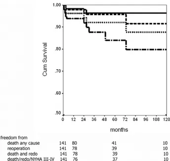
Mean follow-up of the survivors was 55⫾ 36 months. Patients at risk after 1, 5, and 10 years were, respectively, 80, 41, and 10. After a mean period of 75⫾ 49 months, 3 patients died due to acute myocardial infarction (1) and malignancy (2). Freedom from death any cause after 1, 5, and 10 years was 97.9%, 95.7%, and 91.6% (Fig 4).
After a mean of 10⫾ 12 months, 3 patients underwent a new procedure on the mitral valve (3 months in 2 cases, and 24 months in the last one). Cause of the reoperation was endocarditis in 2 and failure of the repair in 1. Freedom from reoperation after 1, 5, and 10 years was 98.4%, 96.4%, and 96.4% (Fig 4). Freedom from death any cause and reoperation was 96.2%, 92.1%, and 87.7%.
Globally, New York Heart Association (NYHA) class
Fig 3. Postoperative transesophageal echocardiogram. (A) The new chordae of the anterior leaflet are at least 2 mm too short; neverthe-less there is no residual mitral regurgitation and the length of coap-tation between the leaflets is normal. (B) The length of the new chordae is correct and no residual regurgitation is detected.
Table 2. Surgical Details
n⫽ 141 Techniques of valvuloplasty Edge-to-edge 20 PL rectangular resection 100 PL plication 11 Chordal replacement 41 Ring sizes 40 mm 81 50 mm 60 Associated procedures
Tricuspid valve repair 30
Coronary bypass grafting 16
Ablation of atrial fibrillation 10
Atrial septal defect repair 1
Septal reshaping 1
PL⫽ posterior leaflet.
improved from 2.9⫾ 0.9 to 1.5 ⫾ 0.6 (p ⬍ 0.001) and 128 patients (94.8% of the survivors) were in NYHA class I or II. Freedom from death any cause, reoperation, and NYHA class III or IV was 93.8%, 87.6%, and 79.8%.
Table 3summarizes the clinical and echocardiographic results in the 68 patients who survived at least 2 years (2 to 7 years; mean, 4.2⫾ 2.5). All were directly controlled in an outpatient clinic. Sixty-four patients (94.1%) had no or residual MR less than or equal to 1/4.
Comment
Mitral valve repair became the procedure of choice in nonrheumatic MV disease. In particular, when MR was due to degenerative disease (fibroelastic or myxomatous disease), the possibilities of repair were explored suc-cessfully by many surgeons.
Carpentier an colleagues, in the 1970s[17, 18], identi-fied different mechanisms of regurgitation and planned different surgical techniques for them. According to the different anatomical aspects, repair was obtained using different methods. If the prolapse involved PL, resection of the prolapsing scallop (generally P2) was performed together with plication of the corresponding annulus. If the prolapse involved AL, chordal shortening or transpo-sition (from P2 to A2 in case of A2 prolapse) or triangular resection (or plication) of A2 were performed. With time, other alternatives were added: chordal replacement[19]
and edge-to-edge [9]. These techniques made simpler mitral valve repair and increased the surgeon’s armamentarium.
Usually, a ring is added to restore the normal geometry of the annulus of the MV. The AL insertion is measured and a ring, complete or incomplete, flexible, semiflexible, or rigid, is positioned with a ratio between the posterior
and anterior annulus of 2:1. Even if this aspect of mitral repair is diffused it is not the rule as some surgeons prefer to avoid a ring implantation after the correction of mitral lesions[20].
Overreduction of the MA was introduced by Bolling and colleagues[2]in the early 1990s to correct functional MR in dilated cardiomyopathy. The ring used in this technique was a #26 in every patient or a ring undersized by two sizes. The same concept was applied to MR that follows an acute myocardial infarction [21]. For a long time we used an autologous, glutaraldehyde-treated, pericardium strip. Its length, at the beginning 52 mm, was reduced to 40 mm[4]and was successfully used in functional MR[3, 21].
In 1993 we started to apply the concept of overreduc-tion in mitral repair for degenerative disease, using a pericardium ring 40 or 50 mm long. The concept to change a bileaflet valve in an unileaflet one, if the AL was moving correctly, was stimulating, as the functional ori-fices obtained in patients where overreduction was per-formed for functional MR were satisfying (from 2.6 to 4.0 cm2
). The functional elimination of PL achieved with an overreducting ring allowed to cover any possible subop-timal correction of the PL. With time, mitral repair became more complex as the mean number of involved scallops increased. The use of an overreducting ring became more important as the huge surface of coaptation between the two leaflets was able to cover both a reduced and an excessive motion of the anterior leaflet (Fig 3).
The use of artificial chordae, both in the anterior and the posterior leaflets, was crucial in our surgical strategy as it allowed us to follow precise guidelines for mitral repair that include all the possible anatomical aspects, also if all the mitral scallops are involved. The use of edge-to-edge correction was then abandoned in favor of a mitral reconstruction that was easy to perform and reproduced the normal mitral anatomy. Long-term re-sults of artificial chordae are favorable[12]and seem to
Fig 4. Ten-year freedom from death any cause (—), from reopera-tion (- - -), from death and reoperareopera-tion ( · · · ), and from death, re-operation, and New York Heart Association class III-IV (– · –).
Table 3. Midterm Clinical and Echocardiographic Results
Pre Post p
n 68 68
NYHA class (I to IV) 2.8⫾ 1.0 1.3⫾ 0.6 ⬍0.001
EDv (mL/m2) 83⫾ 20 73⫾ 26 0.013 ESv (mL/m2) 34⫾ 12 32⫾ 18 0.447 EF (%) 60⫾ 9 59⫾ 9 0.197 MR (1/1 to 4/4) 3.9⫾ 0.4 0.3⫾ 0.7 ⬍0.001 Residual MR None 55 (80.9%) 1⫹ 9 (13.2%) 2⫹ 4 (5.9%) MV functional area (mm2) 40 mm 3.3⫾ 0.6 50 mm 3.7⫾ 0.9 Follow-up (years) 4.2⫾ 2.5
EDv⫽ end-diastolic volume; EF⫽ ejection fraction; ESv⫽ end-systolic volume; MR⫽ mitral regurgitation; MV⫽ mitral valve; NYHA⫽ New York Heart Association.
be superior to chordal shortening[22]. Histologic evalu-ation of explanted new chordae shows also a perfect integration of the new chorda in the biological environ-ment of the human host[23]. We have to keep in mind that MR for degenerative mitral disease is a disease of the valve, but it is because of chordal pathology (elongation or rupture) that the patient needs surgery. Replacement of pathologic chordae with artificial ones is a better option than shortening or transposing diseased native chordae.
The use of artificial chordae for PL, more recently used by Nigro and colleagues [24], is, in our opinion, the preferable method of correction when the height of the posterior scallop is normal, the insertion of the scallop to be resected is excessively long, or the remaining borders of P1 and/or P2 are thin or excessively short. As sug-gested by David and colleagues[12], we do not resect any posterior scallop (generally P2) if longer than 1/3 of the posterior annulus. In that situation, however, we evaluate the height of the prolapsing scallops. Plication of the tissue in excess allows to restore a normal height in a short time, followed by positioning of new chordae.
When P2 can be resected, we prefer to suture directly the borders of P1 and P3 directly to the annulus, in order to reduce the height of the remaining posterior leaflet[7]. This procedure can be done rapidly and is able to correct, if present, any chordal elongation of P1 and/or P3. The use of an overreducting ring is able to stabilize further the PL, eliminating any possibility of prolapse. When the focal sliding is performed, we do not plicate the corre-sponding annulus, as described by others[25], to elimi-nate any possible complication[26].
Even if artificial chordae are used for PL, a correct evaluation of its height is crucial, as the tissue in excess, if present, has to be eliminated. As suggested by Maslow and colleagues[27], the presence of a long PL can be one of the determinants of the presence of the systolic ante-rior motion (SAM), a complication of mitral repair that occurs in 1% to 16% of the cases[28]. The reduction of the height of the posterior leaflet (resection with focal sliding or plication) and the limitation of its movements avoids a coaptation of the two leaflets inside the mitral area and eliminates one of the mechanisms at the basis of the SAM. This strategy cannot avoid the possibility of SAM due to the Venturi effect in the presence of septal hyper-trophy and/or hypovolemia[28], but it at least eliminates some technical causes.
More recently, we replaced the autologous pericar-dium with a new ring that followed the same concepts, the SMB40 and the SMB50. We prefer to use the ring only in the posterior annulus, as its forward displacement is a target of our strategy.
In conclusion, we describe a strategy of MV repair that includes the concept of annular overreduction. The in-creasing complexity of the mechanisms of MR in degen-erative disease forces us to use all the tools we have to improve the quality of the result and to reduce the consequences of any underestimation of a lesion. In our opinion, overreduction can be the extra value we can use to reach a satisfactory mitral repair. Of course, the
strategy described by us is not the only one possible, but it can be helpful especially when the mitral lesions are more complex and diffused.
References
1. Ling LH, Enriquez-Serano M, Steward JB, et al. Early sur-gery in patients with mitral regurgitation due to flail leaflets; a long-term outcome study. Circulation 1997;96:1819 –25. 2. Bolling SF, Deeb GM, Brunsting LA, Bach DS. Early outcome
of mitral valve reconstruction in patients with end-stage cardiomyopathy. J Thorac Cardiovasc Surg 1995;109:676 – 83. 3. Calafiore AM, Gallina S, Di Mauro M, et al. Mitral valve procedure in dilated cardiomyopathy: repair or replace-ment? Ann Thorac Surg 2001;71:1146 –53.
4. Calafiore AM, Di Mauro M, Gallina S, Canosa C, Iacò AL. Optimal length of pericardial strip for posterior mitral overreductive annuloplasty. Ann Thorac Surg 2003;75: 1982– 4.
5. Calafiore AM, Di Mauro M, Di Giammarco G, et al. Septal reshaping for exclusion of anteroseptal dyskinetic or akinetic areas. Ann Thorac Surg 2004;77:2115–21.
6. David TE, Omran A, Armstrong S, Sun Z, Ivanov J. Long-term results of mitral valve repair for mixomatous disease with and without chordal replacement with expanded poly-tetrafluoroethylene. J Thorac Cardiovasc Surg 1998;115: 1279 – 86.
7. Grossi EA, Galloway AC, Kallenbach K, et al. Early results of posterior leaflet folding plasty for mitral valve reconstruc-tion. Ann Thorac Surg 1998;65:1057–59.
8. Calafiore AM, Di Mauro M, Actis-Dato GL, et al. Longitudi-nal plication of the posterior leaflet in mixomatous disease of the mitral valve. Ann Thorac Surg 2006 (in press).
9. Alfieri O, Maisano F, De Bonis M, et al. The double-orifice technique in mitral valve repair: a simple solution for complex problems. J Thorac Cardiovasc Surg 2001;122: 674 – 81.
10. von Oppell UO, Mohr FW. Chordal replacement for both minimally invasive and conventional mitral valve surgery using premeasured Gore-Tex loops. Ann Thorac Surg 2000; 70:2166 – 8.
11. Sarsam MAL. Simplified technique for determining the length of artificial chordae in mitral valve repair. Ann Thorac Surg 2002;73:1659 – 60.
12. David TE, Omran A, Armstrong S, Sun Z, Ivanov J. Long-term results of mitral valve repair for myxomatous disease with and without chordal replacement with expanded poly-tetrafluoroethylene sutures. J Thorac Cardiovasc Surg 1998; 115:1279 – 86.
13. Kasegawa H, Kamata S, Hirasa S, et al. Simple method for determining proper length of artificial chordae in mitral valve repair. Ann Thorac Surg 1989;97:98 –103.
14. Duebener LF, Wendlera O, Nikoloudakisa N, Georgb T, Friesc R, Schäfers HJ. Mitral-valve repair without annulo-plasty rings: results after repair of anterior leaflet versus posterior leaflet defects using polytetrafluoroethylene su-tures for chordal replacement. Eur J Cardiothorac Surg 2000;17:206 –12.
15. Adams DH, Kadner A, Chen RH. Artificial mitral valve chordae replacement made simple. Ann Thorac Surg 2001; 71:1377–9.
16. Calafiore AM. Choice of artificial chordae length according to echocardiographic criteria. Ann Thorac Surg 2006;81: 375–7.
17. Carpentier A, Chauvaud S, Fabiani JN, et al. Reconstructive surgery of mitral valve incompetence: ten-year appraisal. J Thorac Cardiovasc Surg 1980;79:338 – 48.
18. Carpentier A. Cardiac valve surgery: the French correction. J Thorac Cardiovasc Surg 1983;86:323–37.
19. Zussa C, Frater RWM, Polesel E, Galoni M, Valfrè C. Artificial mitral valve chordae: experimental and clinical experience. Ann Thorac Surg 1990;50:367–73.
20. Eisenmann B, Charpentier A, Popescu S, Epailly E, Billaud P, Jirari A. Is a prosthetic ring required for mitral repair of mitral insufficiency due to posterior leaflet prolapse? Long-term results in 96 patients submitted to repair with no ring. Eur J Cardiothorac Surg 1998;14:584 –9.
21. Calafiore AM, Di Mauro M, Gallina S, et al. Mitral valve surgery for chronic ischemic mitral regurgitation. Ann Tho-rac Surg 2004;77:1989 –97.
22. Phillips MR, Daly RC, Schaff HV, Dearani JA, Mullany CJ, Orszulak TA. Repair of anterior leaflet mitral valve prolapse: chordal replacement versus chordal shortening. Ann Thorac Surg 2000;69:25–9.
23. Kobayashi J, Sasako Y, Bando K, Minatoya K, Niwaya K, Kitamura S. Ten-year experience of chordal replacement with expanded polytetrafluoroethylene in mitral valve re-pair. Circulation 2000;102[suppl 3]:III-30 –III-34.
24. Nigro JJ, Schwartz DS, Bart RD, et al. Neochordal repair of the posterior mitral leaflet. J Thorac Cardiovasc Surg 2004; 127:440 –7.
25. Nakajima M, Tsuchiya K, Inoue H, Kobayashi K, Mizutani E, Takizawa K. Leaflet folding plasty combined with annular placation for mitral valve repair. Ann Thorac Surg 2004;77: 1103– 4.
26. Tavilla G, Pacini D. Damage to the circumflex coronary artery during mitral valve repair with sliding leaflet tech-nique. Ann Thorac Surg 1998;66:2091–3.
27. Maslow AD, Regan MM, Haering JM, Johnson RG, Levine RA. Echocardiograhic predictors of left ventricular tract obstruction and systolic anterior motion of the mitral valve after mitral valve reconstruction for myxomatous valve dis-ease. J Am Coll Card 1999;34:2096 –104.
28. Civelek A, Szalay Z, Roth M, et al. Post-mitral valve repair systolic anterior motion produced by non-obstructive septal bulge. Eur J Cardio-Thorac Surg 2003;24:857– 61.
The Society of Thoracic Surgeons Policy Action Center
The Society of Thoracic Surgeons (STS) is pleased toannounce a new member benefit—the STS Policy Action Center, a website that allows STS members to participate in change in Washington, DC. This easy, interactive, hassle-free site allows members to:
● Personally contact legislators with one’s input on key
issues relevant to cardiothoracic surgery
● Write and send an editorial opinion to one’s local media
● E-mail senators and representatives about upcoming
medical liability reform legislation
● Track congressional campaigns in one’s district—and
become involved
● Research the proposed policies that help—or hurt—
one’s practice
● Take action on behalf of cardiothoracic surgery
This website is now available at www.sts.org/takeaction.
© 2006 by The Society of Thoracic Surgeons Ann Thorac Surg 2006;81:1316 • 0003-4975/06/$32.00 Published by Elsevier Inc