Dear Author,
Here are the proofs of your article.
• You can submit your corrections online, via e-mail or by fax.
• For online submission please insert your corrections in the online correction form. Always indicate the line number to which the correction refers.
• You can also insert your corrections in the proof PDF and email the annotated PDF. • For fax submission, please ensure that your corrections are clearly legible. Use a fine black
pen and write the correction in the margin, not too close to the edge of the page.
• Remember to note the journal title, article number, and your name when sending your response via e-mail or fax.
• Check the metadata sheet to make sure that the header information, especially author names and the corresponding affiliations are correctly shown.
• Check the questions that may have arisen during copy editing and insert your answers/ corrections.
• Check that the text is complete and that all figures, tables and their legends are included. Also check the accuracy of special characters, equations, and electronic supplementary material if applicable. If necessary refer to the Edited manuscript.
• The publication of inaccurate data such as dosages and units can have serious consequences. Please take particular care that all such details are correct.
• Please do not make changes that involve only matters of style. We have generally introduced forms that follow the journal’s style.
Substantial changes in content, e.g., new results, corrected values, title and authorship are not allowed without the approval of the responsible editor. In such a case, please contact the Editorial Office and return his/her consent together with the proof.
• If we do not receive your corrections within 48 hours, we will send you a reminder. • Your article will be published Online First approximately one week after receipt of your
corrected proofs. This is the official first publication citable with the DOI. Further changes
are, therefore, not possible.
• The printed version will follow in a forthcoming issue.
Please note
After online publication, subscribers (personal/institutional) to this journal will have access to the complete article via the DOI using the URL: http://dx.doi.org/[DOI].
If you would like to know when your article has been published online, take advantage of our free alert service. For registration and further information go to: http://www.link.springer.com.
Due to the electronic nature of the procedure, the manuscript and the original figures will only be returned to you on special request. When you return your corrections, please inform us if you would like to have these documents returned.
Metadata of the article that will be visualized in
OnlineFirst
Please note: Images will appear in color online but will be printed in black and white.
ArticleTitle In vitro and in vivo evaluation of BMAP-derived peptides for the treatment of cystic fibrosis-related pulmonary infections
Article Sub-Title
Article CopyRight Springer-Verlag Wien
(This will be the copyright line in the final PDF) Journal Name Amino Acids
Corresponding Author Family Name Scocchi
Particle
Given Name Marco
Suffix
Division Department of Life Sciences Organization University of Trieste
Address Via L. Giorgieri 5, 34127, Trieste, Italy
Email [email protected]
ORCID
Author Family Name Mardirossian
Particle
Given Name Mario
Suffix
Division Department of Life Sciences Organization University of Trieste
Address Via L. Giorgieri 5, 34127, Trieste, Italy Email
ORCID
Author Family Name Pompilio
Particle
Given Name Arianna
Suffix
Division Department of Medical, Oral, and Biotechnological Sciences Organization “G. d’Annunzio” University of Chieti-Pescara
Address Via dei Vestini 31, 66100, Chieti, Italy Division Center of Excellence on Aging
Organization “G. d’Annunzio” University Foundation Address Via L. Polacchi 11, 66100, Chieti, Italy Email
ORCID
Author Family Name Crocetta
Particle
Suffix
Division Department of Medical, Oral, and Biotechnological Sciences Organization “G. d’Annunzio” University of Chieti-Pescara
Address Via dei Vestini 31, 66100, Chieti, Italy Division Center of Excellence on Aging
Organization “G. d’Annunzio” University Foundation Address Via L. Polacchi 11, 66100, Chieti, Italy Email
ORCID
Author Family Name Nicola
Particle De
Given Name Serena
Suffix
Division Department of Medical, Oral, and Biotechnological Sciences Organization “G. d’Annunzio” University of Chieti-Pescara
Address Via dei Vestini 31, 66100, Chieti, Italy Division Center of Excellence on Aging
Organization “G. d’Annunzio” University Foundation Address Via L. Polacchi 11, 66100, Chieti, Italy Email
ORCID
Author Family Name Guida
Particle
Given Name Filomena
Suffix
Division Department of Life Sciences Organization University of Trieste
Address Via L. Giorgieri 5, 34127, Trieste, Italy Email
ORCID
Author Family Name Degasperi
Particle
Given Name Margherita
Suffix
Division Department of Life Sciences Organization University of Trieste
Address Via L. Giorgieri 5, 34127, Trieste, Italy Email
ORCID
Author Family Name Gennaro
Particle
Given Name Renato
Suffix
Division Department of Life Sciences Organization University of Trieste
Address Via L. Giorgieri 5, 34127, Trieste, Italy Email
ORCID
Author Family Name Bonaventura
Particle Di
Given Name Giovanni
Suffix
Division Department of Medical, Oral, and Biotechnological Sciences Organization “G. d’Annunzio” University of Chieti-Pescara
Address Via dei Vestini 31, 66100, Chieti, Italy Division Center of Excellence on Aging
Organization “G. d’Annunzio” University Foundation Address Via L. Polacchi 11, 66100, Chieti, Italy Email ORCID Schedule Received 26 February 2016 Revised Accepted 25 May 2016
Abstract Patients with cystic fibrosis require pharmacological treatment against chronic lung infections. The alpha-helical antimicrobial peptides BMAP-27 and BMAP-28 have shown to be highly active in vitro against planktonic and sessile forms of multidrug-resistant Pseudomonas aeruginosa, Staphylococcus aureus, and
Stenotrophomonas maltophilia cystic fibrosis strains. To develop small antibacterial peptides for
therapeutic use, we tested shortened/modified BMAP fragments, and selected the one with the highest in vitro antibacterial activity and lowest in vivo acute pulmonary toxicity. All the new peptides have shown to roughly maintain their antibacterial activity in vitro. The 1–18 N-terminal fragment of BMAP-27, showing MIC90 = 16 µg/ml against P. aeruginosa isolates and strain-dependent anti-biofilm effects, showed the
lowest pulmonary toxicity in mice. However, when tested in a murine model of acute lung infection by P.
aeruginosa, BMAP-27(1–18) did not show any curative effect. If exposed to murine broncho-alveolar
lavage fluid BMAP-27(1–18) was degraded within 10 min, suggesting it is not stable in pulmonary environment, probably due to murine proteases. Our results indicate that shortened BMAP peptides could represent a starting point for antibacterial drugs, but they also indicate that they need a further optimization for effective in vivo use.
Keywords (separated by '-') Antimicrobial peptide - Cathelicidin - BMAP - Cystic fibrosis - Biofilm - Multidrug-resistance - In vivo degradation
Footnote Information Handling Editor: J. D. Wade.
M. Mardirossian and A. Pompilio equally contributed to the work.
Electronic supplementary material The online version of this article (doi:10.1007/s00726-016-2266-4)
Metadata of the article that will be visualized in
OnlineAlone
Electronic supplementary
UNCORRECTED PR
OOF
Journal : Large 726 Dispatch : 27-5-2016 Pages : 8
Article No : 2266 ¨ LE ¨ TYPESET
MS Code : AMAC-D-16-00104 þ CP þ DISK
1 3
DOI 10.1007/s00726-016-2266-4 Amino Acids
ORIGINAL ARTICLE
In vitro and in vivo evaluation of BMAP-derived peptides for the
treatment of cystic fibrosis-related pulmonary infections
Mario Mardirossian1 · Arianna Pompilio2,3 · Valentina Crocetta2,3 ·
Serena De Nicola2,3 · Filomena Guida1 · Margherita Degasperi1 · Renato Gennaro1 ·
Giovanni Di Bonaventura2,3 · Marco Scocchi1
Received: 26 February 2016 / Accepted: 25 May 2016 © Springer-Verlag Wien 2016
BMAP-27(1–18) did not show any curative effect. If exposed to murine broncho-alveolar lavage fluid BMAP-27(1–18) was degraded within 10 min, suggesting it is not stable in pulmonary environment, probably due to murine proteases. Our results indicate that shortened BMAP pep-tides could represent a starting point for antibacterial drugs, but they also indicate that they need a further optimization for effective in vivo use.
Keywords Antimicrobial peptide · Cathelicidin · BMAP ·
Cystic fibrosis · Biofilm · Multidrug-resistance · In vivo degradation
Introduction
Although understanding of the pathophysiology of cystic fibrosis (CF) has been increased in the recent years, pul-monary infections remain the major cause of morbidity and mortality in CF patients. In most cases, Pseudomonas
aeruginosa and Staphylococcus aureus are the pathogens responsible of these complications (Dasenbrook et al.
2010; Emerson et al. 2002), although other pathogens, such as Stenotrophomonas maltophilia, are increasingly isolated from CF airways (Emerson et al. 2010; Mil-lar et al. 2009), probably as a result of the selective effect due to the antipseudomonal therapy (Emerson et al. 2002). Another detrimental consequence of repeated antimicrobial treatments is the spreading of multidrug-resistant (MDR) pathogens. Moreover, even though bacteria do not acquire specific resistance to therapeutically important antibiotics, the microbial adaptation to the CF pulmonary environ-ment results in an increased ability to form biofilms, ses-sile communities intrinsically resistant to many antimicro-bial drugs, such as aminoglycosides, fluoroquinolones, and
Abstract Patients with cystic fibrosis require
phar-macological treatment against chronic lung infections. The alpha-helical antimicrobial peptides BMAP-27 and BMAP-28 have shown to be highly active in vitro against planktonic and sessile forms of multidrug-resistant
Pseu-domonas aeruginosa, Staphylococcus aureus, and
Steno-trophomonas maltophilia cystic fibrosis strains. To develop small antibacterial peptides for therapeutic use, we tested shortened/modified BMAP fragments, and selected the one with the highest in vitro antibacterial activity and low-est in vivo acute pulmonary toxicity. All the new peptides have shown to roughly maintain their antibacterial activ-ity in vitro. The 1–18 N-terminal fragment of BMAP-27, showing MIC90 = 16 µg/ml against P. aeruginosa isolates and strain-dependent anti-biofilm effects, showed the low-est pulmonary toxicity in mice. However, when tlow-ested in a murine model of acute lung infection by P. aeruginosa,
Handling Editor: J. D. Wade.
M. Mardirossian and A. Pompilio equally contributed to the work.
Electronic supplementary material The online version of this
article (doi:10.1007/s00726-016-2266-4) contains supplementary material, which is available to authorized users.
* Marco Scocchi [email protected]
1
Department of Life Sciences, University of Trieste, Via L. Giorgieri 5, 34127 Trieste, Italy
2 Department of Medical, Oral, and Biotechnological Sciences,
“G. d’Annunzio” University of Chieti-Pescara, Via dei Vestini 31, 66100 Chieti, Italy
3
Center of Excellence on Aging, “G. d’Annunzio” University Foundation, Via L. Polacchi 11, 66100 Chieti, Italy AQ1 1 2 3 4 5 6 7 8 9 10 11 12 13 14 15 16 17 18 19 20 21 22 23 24 25 26 27 28 29 30 31 32 33 34 35 36 37 38 39 40 41 42 43 44 45 46 47 48 49 50 51 52 53 54 55 56 A1 A2 A3 A4 A5 A6 A7 A8 A9 A10 A11 A12 A13 A14 A15
Author Proof
UNCORRECTED PR
OOF
Journal : Large 726 Dispatch : 27-5-2016 Pages : 8
Article No : 2266 ¨ LE ¨ TYPESET
MS Code : AMAC-D-16-00104 þ CP þ DISK
M. Mardirossian et al.
1 3
tetracyclines (Di Bonaventura et al. 2007; Hoffman et al.
2005; Linares et al. 2006; Molina et al. 2008; Singh et al.
2000). The sum of these intrinsic and acquired resistances depicts an alarming picture for the treatment of CF pulmo-nary infections. Novel antimicrobial agents are, therefore, needed to flank or replace current antibiotic therapies to overcome chronic infections in CF patients.
Antimicrobial peptides (AMPs) could represent a prom-ising answer to this request. These natural molecules are important component of the innate immunity of animals and plants, representing a first line-defense against infec-tions (Lai and Gallo 2009; Yang et al. 2002; Zanetti 2004; Zanetti et al. 2000, 2002). The eligibility of AMPs as start-ing point to develop new antibiotics is based on their broad spectrum of activity, on the efficacy against bacteria resist-ant to commonly used resist-antibiotics, and on their poor ability to select for antibiotic resistance (Hancock and Sahl 2006; Zanetti 2004). The pulmonary infections related to CF rep-resent, therefore, a suitable field of application for these molecules. The therapeutic potential of some AMPs in the management of CF lung infections started to be explored (Zhang et al. 2005). To this aim, among the galaxy of known AMPs, some peptides belonging to the cathelici-dins family are interesting candidates given their good antimicrobial activity also against CF-related pathogens (Pompilio et al. 2011). The mammalian α-helical catheli-cidins BMAP-27 and BMAP-28, and the artificial peptide P19(9/B), show a potent and rapid bactericidal and anti-biofilm activity against MDR S. aureus, P. aeruginosa, and
S. maltophilia strains collected from CF patients (Pompilio et al. 2011). Under physico-chemical conditions simulat-ing those observed in CF lung environment (Palmer et al.
2007; Worlitzsch et al. 2002) the activity of these AMPs is comparable, or even higher, to Tobramycin (Pompilio et al.
2012). It has also been shown, that shortened fragments or derivatives of BMAPs peptides maintain a good antimicro-bial potency in spite of decreasing their cytotoxicity and their cost of synthesis (Benincasa et al. 2003; Skerlavaj et al. 1996).
Aim of this study was to assess the acute pulmo-nary toxicity of BMAPs shortened forms in mice and to
characterize their in vitro and in vivo activity to make them applicable in the future for early prophylactic and therapeu-tic treatment of CF lung disease.
Results
Antimicrobial activity of BMAPs fragments/analogues against MDR bacterial strains
Some synthetic peptides—comprising the N-terminal 18 residues of the α-helical BMAP-27 and BMAP-28 pep-tides, and a less hydrophobic BMAP-28 analogue—had indeed been shown to have reduced cytotoxicity against human neutrophils and erythrocytes when compared to their natural longer forms (Benincasa et al. 2003). On these bases, we evaluated the antimicrobial activity of these shortened peptides against a panel of previously charac-terized S. aureus, P. aeruginosa, and S. maltophilia strains isolated from CF patients (Pompilio et al. 2012). All pep-tides showed a good antimicrobial activity against P.
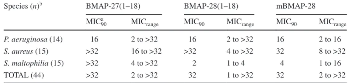
aer-uginosa and S. maltophilia, while a reduced activity was observed against S. aureus. Overall, these shortened or modified forms of cathelicidins substantially retained anti-microbial activity (Table 1), even compared with their par-ent forms (Pompilio et al. 2012).
In vivo acute toxicity of BMAPs fragments/analogues
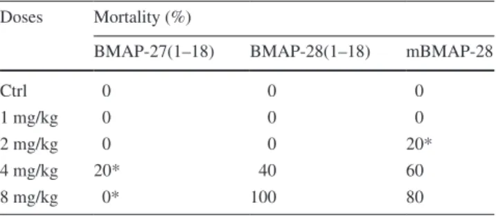
Once assessed that shortened forms maintained a relevant antimicrobial potential, in vivo toxicity of BMAP-27(1– 18), BMAP-28(1–18) and mBMAP-28 was comparatively evaluated in C57BL/6NCrl mice. Exposure to BMAP-27(1–18) caused the death of one mouse when admin-istered at 4 mg/kg, a mortality rate significantly lower than that observed for the other two peptides, regardless of doses used (Table 2). Changes in body weight of mice treated with BMAP-27(1–18) were always less than those observed in mice treated with the other two peptides (see supplementary Fig. S1). Macroscopic C57BL/6NCrl mouse lung pathology was assessed on day 5 post-exposure (p.e.)
Table 1 In vitro activity of
BMAPs analogues vs strains from CF patients
a MIC values are expressed as µg/ml. MIC
90, Minimum Inhibitory Concentration required to inhibit the
growth of 90 % of the strains tested
b Number of strains tested
Species (n)b BMAP-27(1–18) BMAP-28(1–18) mBMAP-28 MICa
90 MICrange MIC90 MICrange MIC90 MICrange
P. aeruginosa (14) 16 2 to >32 16 2 to >32 16 2 to 16 S. aureus (15) >32 16 to >32 >32 4 to >32 32 8 to >32 S. maltophilia (15) >32 4 to >32 2 1 to 4 4 1 to 16 TOTAL (44) >32 2 to >32 32 1 to >32 32 2 to >32 57 58 59 60 61 62 63 64 65 66 67 68 69 70 71 72 73 74 75 76 77 78 79 80 81 82 83 84 85 86 87 88 89 90 91 92 93 94 95 96 97 98 99 100 101 102 103 104 105 106 107 108 109 110 111 112 113 114 115 116 117 118 119 120 121 122 123 124 125 126 127 128 129 130 131 132
Author Proof
UNCORRECTED PR
OOF
Journal : Large 726 Dispatch : 27-5-2016 Pages : 8
Article No : 2266 ¨ LE ¨ TYPESET
MS Code : AMAC-D-16-00104 þ CP þ DISK
In vitro and in vivo evaluation of BMAP-derived peptides for the treatment of cystic…
1 3
using a “four-point scoring” system. BMAP-27(1–18) at 1, 4 and 8 mg/kg caused a macroscopic pulmonary damage comparable to that observed in control mice (macroscopic score: 2 vs 1, respectively; p > 0.05) (Table 3). Further-more, lung injury caused by BMAP-27(1–18) was gener-ally less than those observed for the other peptides tested at the same concentrations. These results clearly indicated BMAP-27(1–18) as the less toxic peptide among those tested. Since BMAP-27(1–18) exhibited the best antibacte-rial activity/pulmonary cytotoxicity ratio among the pep-tides tested, it was selected for subsequent studies.
Bactericidal and anti-biofilm activities of BMAP-27(1– 18)
We evaluated the minimal bactericidal concentration (MBC) of BMAP-27(1–18) against the previously tested
P. aeruginosa and S. maltophilia strains. S. aureus strains were not further tested because, despite the MIC90 for this species was identical to that of S. maltophilia, overall the MIC values for the single strains were higher (see Table
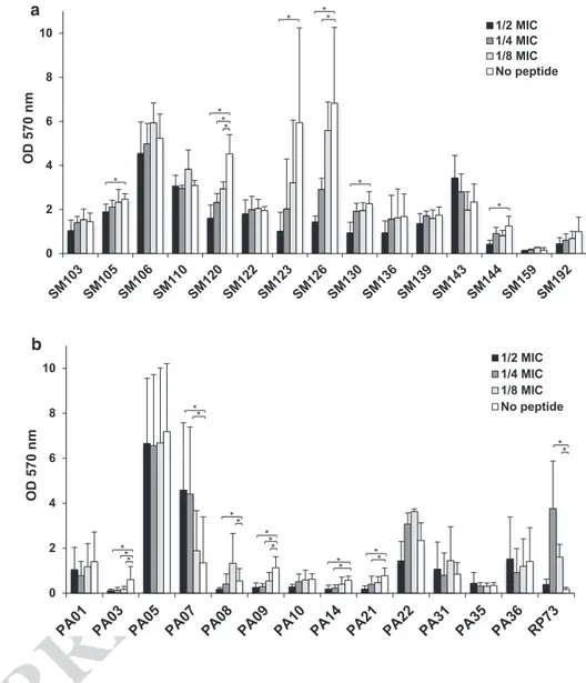
S1). BMAP-27(1–18) showed a MBC50 of 16 µg/ml for more than one-half of P. aeruginosa and S. maltophilia strains tested, thus confirming its bactericidal mecha-nism of action. We also assayed the capability of BMAP-27(1–18) to affect the biofilm formation by S. maltophilia (Fig. 1a) and P. aeruginosa (Fig. 1b) CF strains, and its potential to re-start an infection when tested at sub-inhibi-tory concentrations. To this aim, we evaluated the viability of sessile cells by the tetrazolium salt assay (MTT), rather than evaluating the biofilm biomass by crystal violet assay. Six out of 15 (40 %) S. maltophilia strains were signifi-cantly affected in new biofilm viability in the presence of the peptide. This effect was not concentration-dependent, except for Sm120 strain. The remaining nine S. maltophilia strains did not show significant variations in biofilm viabil-ity. Higher variability was observed for P. aeruginosa bio-films: BMAP-27(1–18) showed a significant anti-biofilm activity against 4 out of 14 (28 %) strains, while no effect was observed for 7 (43 %) strains. On the contrary, expo-sure to peptide even stimulated biofilm production in 3 (21 %) strains. In the case of PA08 strain both inhibiting and enhancing activity were observed, depending on the concentration considered. Taken together, our results indi-cate that the effect of sub-lethal concentrations of peptide on biofilm formation is strain-specific.
In vivo activity of BMAP-27(1–18) in a murine model of P. aeruginosa acute lung infection
The protective role of BMAP-27(1–18) was then evalu-ated in a murine model of acute pulmonary infection by
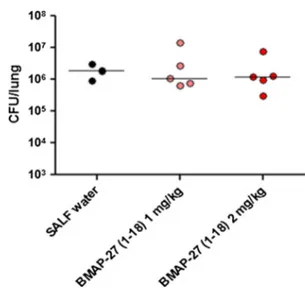
P. aeruginosa RP73. C57BL/6NCr mice were intratrache-ally challenged with 106 CFU, immediately followed by the administration of the peptide at different doses via the same route. No significant differences in CFU/lung values were observed between treated and control mice, regard-less of doses used (Fig. 2). Overall, our results suggest that BMAP-27(1–18), despite the antimicrobial activity exhib-ited in vitro, does not show a protective in vivo activity at safe concentrations and under our experimental conditions.
In vitro degradation of BMAP-27(1–18) by murine bronchoalveolar lavage fluid
To assess whether the absence of in vivo activity would be explained by the scarce stability of the peptide in the pul-monary environment, we incubated BMAP-27(1–18) with bronchoalveolar lavage (BAL) fluid collected from healthy mice. The peptide was rapidly degraded, already after 5 min of exposition to BAL, and the corresponding band disappeared within 20 min of incubation (Fig. 3). Follow-ing exposure of an identical amount of peptide to 0.9 % NaCl instead of BAL fluid, no degradation was observed
Table 2 Mortality rate observed in C57BL/6NCrl mice (n = 5/
group) following a single administration of each AMP tested at dif-ferent doses
Control mice (Ctrl) received vehicle (SALF water) only * p < 0.05 vs other groups, Chi square test
Doses Mortality (%)
BMAP-27(1–18) BMAP-28(1–18) mBMAP-28
Ctrl 0 0 0
1 mg/kg 0 0 0
2 mg/kg 0 0 20*
4 mg/kg 20* 40 60
8 mg/kg 0* 100 80
Table 3 Macroscopic damage of C57BL/6NCrl mouse lungs
fol-lowing a single exposure to BMAP-27(1–18), BMAP-28(1–18) and mBMAP-28, each tested at different doses
Control mice (Ctrl) received vehicle (SALF water) only. Macroscopic lung pathology was assessed on day 5 p.e. using a “four-point scoring system” (Johansen et al. 1993): 1, normal; 2, swollen lungs, hyper-emia, and small atelectasis; 3, pleural adhesion, atelectasis, and mul-tiple small abscesses; and 4, large abscesses, large atelectasis, and hemorrhages
Doses BMAP-27(1–18) BMAP-28(1–18) mBMAP-28
Ctrl 1 1 1 1 mg/kg 2 3 2 2 mg/kg 3 3 4 4 mg/kg 2 3 4 8 mg/kg 2 4 4 133 134 135 136 137 138 139 140 141 142 143 144 145 146 147 148 149 150 151 152 153 154 155 156 157 158 159 160 161 162 163 164 165 166 167 168 169 170 171 172 173 174 175 176 177 178 179 180 181 182 183 184 185 186 187 188 189 190 191 192 193 194 195 196 197 198 199 200
Author Proof
UNCORRECTED PR
OOF
Journal : Large 726 Dispatch : 27-5-2016 Pages : 8
Article No : 2266 ¨ LE ¨ TYPESET
MS Code : AMAC-D-16-00104 þ CP þ DISK
M. Mardirossian et al.
1 3
(Fig. S2), thus excluding instability per se of the molecule. These results strongly suggest that BMAP-27(1–18) is cleaved by mice pulmonary proteases into inactive frag-ments before exerting measurable antibacterial activity in the lungs, therefore, providing an explanation for the poor activity observed in vivo.
Discussion
In this study, we evaluated the potential of some ration-ally designed AMPs as compounds for the development of novel antibacterial to treat lung disease in CF patients. We looked for peptides active against multidrug-resistant bacterial strains and, at the same time, showing reduced in vivo toxicity. To this aim two peptides, BMAP-27(1–18) and BMAP-28(1–18)—corresponding to the N-terminal fragments of their natural peptides lacking the hydropho-bic C-terminal tail—and mBMAP-28—a modified hydro-philic analogue of BMAP-28—have been assayed. All
these α-helical peptides have a reduced hydrophobicity in comparison to the natural peptides, a key factor to increase the selectivity towards prokaryotic cells and, therefore, to decrease their toxicity. Previous in vitro cytotoxicity assays performed on these molecules confirmed this hypothesis (Skerlavaj et al. 1996).
Our results showed that all these shortened or modified peptides have a good antimicrobial activity against most of the isolates of P. aeruginosa and S. maltophilia, despite differences in specificity among the BMAPs, and in gen-eral a lower activity against S. aureus strains (Table S1). In spite of the low in vitro toxicity showed by BMAPs frag-ments, the acute pulmonary toxicity tests indicated that all the peptide analogues begin to be toxic already at a dose of 2 mg/kg. This quite surprising result is in contrast with our previous studies where no toxicity was observed when the same peptides were intraperitoneally administered up to 32 mg/kg (Benincasa et al. 2003). To explain this appar-ent discrepancy, we hypothesized that the higher toxicity reported in this study may be dependent on the pulmonary
Fig. 1 Effects of sub-inhibitory
concentrations of BMAP-27(1– 18) on biofilm formation by a S.
maltophilia (SM; n = 15) and
b P. aeruginosa (PA; n = 14)
strains from CF patients. Biofilm viability was evalu-ated by MTT assay. Results are mean (of three independent experiments and performed as internal duplicate) + SD (n = 6). *p < 0.05, unpaired t test. BMAP-27(1–18) affected the biofilm viability of SM 105, SM 120, SM 123, SM 126, SM 130, SM 144, PA03, PA09, PA14, and PA21 strains, but also stimulated biofilm growth of RP73, PA07 and PA08 strains
0 2 4 6 8 10 OD 570 nm 1/2 MIC 1/4 MIC 1/8 MIC No peptide * * * * * ** * * 0 2 4 6 8 10 OD 570 nm 1/2 MIC 1/4 MIC 1/8 MIC No peptide ** * ** ** ** * ** ** ** a b 201 202 203 204 205 206 207 208 209 210 211 212 213 214 215 216 217 218 219 220 221 222 223 224 225 226 227 228 229 230 231 232 233 234 235 236 237
Author Proof
UNCORRECTED PR
OOF
Journal : Large 726 Dispatch : 27-5-2016 Pages : 8
Article No : 2266 ¨ LE ¨ TYPESET
MS Code : AMAC-D-16-00104 þ CP þ DISK
In vitro and in vivo evaluation of BMAP-derived peptides for the treatment of cystic…
1 3
administration route. Cytotoxicity could be due to a lytic effect on epithelial cells performed by the very concen-trated peptide solution in the first minutes immediately after its intra-tracheal instillation before its spread into the lungs. Further studies will be necessary to explain these differences and, if possible, to reduce toxicity of these
peptides in pulmonary applications. Anyhow, among the peptides, BMAP-27(1–18) was selected as the best candi-date for its lower cytotoxicity and also for its good bacteri-cidal activity against both S. maltophilia and P. aeruginosa (MBC50, 16 µg/ml), suitable features to develop a peptide antibiotic that could be used to eradicate, and not only to contain, infections.
The prophylactic use of BMAP-27(1–18) at doses lower than MIC was investigated by the MTT assay on new biofilm formation. The rationale of this approach was to evaluate the potential of newly formed biofilm to trig-ger a new infection, and not simply to estimate the bio-mass production (e.g. by crystal violet assay). The peptide did not exert impressive results on S. maltophilia strains, significantly inhibiting the biofilm viability in 40 % of the tested strains. Regarding P. aeruginosa, markedly strain-specific data were collected. BMAP-27(1–18) reduced the viability of the biofilm in 28 % of the strains tested, did not exert any effect on the 43 % of the strains, and surprisingly enhanced the viability on the biofilm in 21 % of the iso-lates. The enhancement of biofilm viability in the presence of sub-inhibiting concentrations of BMAP27(1–18) might be due to the up-regulation of specific pathways using anti-microbial compound as activating signals, or to the trig-gering of a bacterial stress response inducing the bacteria to develop biofilm as a resistance form as had been previ-ously observed also for antibiotics and bacterial species (Kaplan 2011) (Hsu et al. 2011; Wu et al. 2014) and for the unique human cathelicidin LL-37 (Limoli et al. 2014). This pathogens’ behavior indicates the need to finely modulate the amount of antibiotic compound to be administered in therapy, to avoid detrimental side effect during antimicro-bial therapy.
Unfortunately, BMAP-27(1–18) did not significantly reduce the bacterial load in the mice lungs infected with
P. aeruginosa RP73. This result can be explained by the scarce stability of the peptide in murine BAL fluid, as suggested by the results of degradation assays performed in vitro. We showed that BMAP-27(1–18) is prone to a rapid degradation by host proteases, a problem already reported in CF sputum for the histatin derivative AMP P-113, (Sajjan et al. 2001). Despite no strict sequence similarity exists between P-113 and BMAP-27(1–18), both AMPs underwent a rapid and non-specific degradation in mice or human pulmonary environments.
The use of an enantiomeric D-BMAP-27(1–18) could represent a good strategy to avoid degradation as already suggested (Sajjan et al. 2001) and, as a consequence, to enhance its antibacterial in vivo activity. Moreover, a pep-tide form resistant to enzymatic digestion and potentially more active could allow its administration at lower doses, possibly reducing its toxicity. For these reasons, a D-form
Fig. 2 In vivo protection assay. C57BL/6NCr mice were
intratrache-ally challenged with 1 × 10⁷ CFU of P. aeruginosa RP73, then 5 min
after infection mice were intratracheally administered with SALF water, as a negative control, or with BMAP-27(1–18) at 1 and 2 mg/ kg dissolved in SALF water. Following 24 h from exposure, con-trol (infected but not treated) mice displayed classical clinical signs observed during an acute infection by P. aeruginosa RP73 strain
0’ 1’ 5’ 10’ 20’ 30’ 60’ 120’ 97 60 45 30 20.1 14.4
Fig. 3 Evaluation of the stability of BMAP-27(1–18) in the presence
of murine bronchoalveolar lavage (BAL) fluid. The peptide is indi-cated by the arrow. Samples were collected at indiindi-cated times and analyzed by SDS-PAGE (gel 16 %, tricine) following staining with Coomassie Brilliant Blue. As controls, 2.4 µg of BMAP-27(1–18) [referred as B27(1–18)] and BAL alone [referred as CTRL] were loaded, corresponding to the original respective concentration of both compounds at the beginning of the time-course
238 239 240 241 242 243 244 245 246 247 248 249 250 251 252 253 254 255 256 257 258 259 260 261 262 263 264 265 266 267 268 269 270 271 272 273 274 275 276 277 278 279 280 281 282 283 284 285 286 287 288 289 290 291 292 293 294 295
Author Proof
UNCORRECTED PR
OOF
Journal : Large 726 Dispatch : 27-5-2016 Pages : 8
Article No : 2266 ¨ LE ¨ TYPESET
MS Code : AMAC-D-16-00104 þ CP þ DISK
M. Mardirossian et al.
1 3
of BMAP-27(1–18) has already been synthesized and its characterization is in progress (manuscript in preparation).
In conclusion, this study shed new insights on the in vitro and in vivo antibacterial properties of BMAP α-helical peptides, allowed the selection of that with the best properties to cope with lung pathogens associated to CF and highlighted the impact that pulmonary proteases can have on AMPs in the treatment of lung infections. Pep-tide resistance to pulmonary proteases is a key factor that should be evaluated in the design of peptides for pulmo-nary applications. Further work is, therefore, needed allow-ing BMAP-27(1–18) application also in vivo and pavallow-ing the way for its use in the future for early prophylactic and therapeutic treatment of CF-related lung infections.
Materials and methods
Bacterial strains
Previously characterized S. aureus, P. aeruginosa, and S.
maltophilia strains were tested (Pompilio et al. 2012). All strains were previously isolated from respiratory speci-mens of CF patients admitted to the “Bambino Gesù” Chil-dren Hospital of Rome. P. aeruginosa RP73, and PAO1 reference strains were also tested. Isolates were stored at −80 °C in a Microbank System (Biolife Italiana srl, Milan, Italy) until use, when each isolate was subcultured in Trypticase Soy broth for 24 h at 37 °C, followed by two passages on Mueller–Hinton agar (MHA; Oxoid S.p.A., Milan, Italy).
Design and synthesis of BMAP-derived antimicrobial peptides
Peptides (BMAP-271–18:GRFKRFRKKFKKLFKKLS-am, BMAP-281–18: GGLRSLGRKILRAWKKYG-am, mBMAP-28: GGLRSLGRKILRAWKKYGPQAWPAWRQ-am) were synthesized using solid-phase Fmoc chemistry method on a CEM Liberty automated microwave peptide synthesizer (USA) as described in (Benincasa et al. 2006). The peptides have been purified by reversed phase HPLC and their quality and purity verified by ESI–MS (API 150 EX Applied Biosys-tems). Peptide concentrations of stock solutions, have been confirmed independently by three methods: by the determi-nation of tryptophan absorbance (ε280 = 5500/M/cm), by measuring the 215/225 absorbance and by spectrophotomet-ric determination of peptide bonds (ε214) and then lyophi-lized. For the in vivo toxicity experiments, which required high amounts of peptides, the peptides were purchased (JPT Peptide Technologies, Germany) and checked for their qual-ity, purity and concentrations as described above.
Evaluation of antimicrobial activity of optimized AMP analogues against MDR bacterial strains
MIC values were determined by microdilution technique. Briefly, serial two-fold dilutions of each peptide were pre-pared in Mueller–Hinton broth (MH; Oxoid S.p.A., Milan, Italy) using a 96-well U-bottom microtiter plates (Bibby-Sterilin Italia srl; Milan, Italy). Each well was inoculated with a standardized inoculum to achieve a final test concen-tration of about 5 × 10⁵ CFU/ml. After incubation at 37 °C for 24 h, the MIC was measured as the lowest concentra-tion of the peptide that completely inhibited visible bacte-rial growth. For MBC evaluation, following 24 h-incuba-tion, 100 µl of broth from clear wells were plated on MHA plates, and incubated at 37 °C for 24 h. MBC was defined as the lowest concentration of the peptide killing at least 99.99 % of the original inoculum.
Biofilm formation assay
Serial two-fold dilutions of BMAP-27(1–18) were, respec-tively prepared in MH broth at a volume of 50 µl per well in 96-well U-bottom microtiter plates (Bibby- Sterilin Ita-lia srl; Milan, Italy). Each well was inoculated with 50 µl of the standardized inoculum, corresponding to a final test concentration of about 5 × 10⁵ CFU/ml. After incubation at 37 °C for 24 h, medium and non-adherent bacteria were discarded. Wells were washed three times using 150 µl of fresh MH broth, then 100 µl of MH broth containing 1 mM MTT (Sigma) were added to the wells. The plate was incu-bated for 4 h in the dark, then the MTT-containing medium was discarded and the wells washed with 150 µl PBS. Sub-sequently, 100 µl of a re-suspending solution (20 % wt/v SDS in 50 % v/v H2O and 50 % v/v N,N-dimethylforma-mide) (Hansen et al. 1989) were added to the wells. The plate was incubated overnight for 16 h in the dark, and then the optical density at 570 nm was measured using a multi-well plate reader (Tecan Trading AG, Switzerland). The signal was directly proportional to the viability of the bio-film under the tested conditions.
In vivo toxicity of optimized AMPs in a murine model
C57BL/6NCrl mice (n = 5/group) (male; 22 g; 6 ± 2 week-old) were obtained from Charles River Laboratories Italia srl (Calco, Milan, Italy). The in vivo toxicity of each AMP was investigated following intratracheal administration of increasing doses (1, 2, 4 and 8 mg/kg) prepared in ster-ile distilled water. The control mice received vehicle only (sterile distilled water). Animal behavior, general health (ruffled coats, huddled position, lack of retreat in handler’s presence), weight loss, and survival were monitored daily
296 297 298 299 300 301 302 303 304 305 306 307 308 309 310 311 312 313 314 315 316 317 318 319 320 321 322 323 324 325 326 327 328 329 330 331 332 333 334 335 336 337 338 339 340 341 342 343 344 345 346 347 348 349 350 351 352 353 354 355 356 357 358 359 360 361 362 363 364 365 366 367 368 369 370 371 372 373 374 375 376 377 378 379 380 381 382 383 384 385 386 387 388
Author Proof
UNCORRECTED PR
OOF
Journal : Large 726 Dispatch : 27-5-2016 Pages : 8
Article No : 2266 ¨ LE ¨ TYPESET
MS Code : AMAC-D-16-00104 þ CP þ DISK
In vitro and in vivo evaluation of BMAP-derived peptides for the treatment of cystic…
1 3
over a 5-day period, with respect to control mice. On day 5 post-administration, mice were sacrificed by intraperito-neal injection of Avertin (Sigma-Aldrich S.r.l), then lungs underwent to in situ for macroscopic analysis for assessing damage using “four-point scoring system” (Johansen et al.
1993).
In vivo activity of BMAP-27(1–18) in a mouse model of P. aeruginosa acute lung infection
C57/Bl6NCrl mice (n = 5/group) were intratracheally chal-lenged with 1 × 10⁷ cells P. aeruginosa RP73 clinical strain and, 5 min later, a single dose of BMAP-27(1–18) at differ-ent concdiffer-entrations (1, 2, and 4 mg/kg) was intratracheally administrated. The vehicle (SALF water) alone was used as negative control. The concentrations of the peptide have been selected on the basis of previously performed in vivo toxicity assays. Following 24 h p.e. mice were sacrificed by CO2 inhalation, then lungs were observed in situ for
macro-scopic analysis and finally removed en bloc from the chest via sterile excision. Lungs were homogenized (24,000 rpm) on ice in 2 ml of sterile PBS by use of an Ultra-Turrax T25-Basic homogenizer (IKA-Werke GmbH & Co. KG, Ger-many). Tenfold serial dilutions of lung homogenates were plated in triplicate on MHA (Oxoid SpA), and the number of colony-forming units (CFUs) was counted 24 h after incubation at 37 °C. Bacterial colony counts from each mouse were expressed as CFU/lungs, averaged, and com-pared between groups. These experiments were performed as an external service made available by the CF Animal Core Facility of the San Raffaele Hospital, Milan, Italy.
BMAP-27(1–18) degradation in bronchoalveolar lavage (BAL) fluids
BAL was collected from 3 C57/Bl6NCrl male healthy mice. Briefly, mice were sacrificed by dislocation, then 1 ml of sterile 0.9 % NaCl at 37 °C was instilled, by insert-ing a probe through mouth and trachea, in the lungs. Two washes were performed for each mouse. Identical volumes of the first wash from each mouse were pooled together, splitted in aliquots and stored at −20 °C until degradation experiments on BMAP-27(1–18). The total protein concen-tration of BAL fluids was determined to be 300 µg/ml by BCA assay (Pierce, BCA Protein Assay Kit).
To evaluate BMAP-27(1–18) degradation a concen-trated solution of peptide was diluted in pooled BAL (see above) to achieve the final concentration of 300 µg/ml. In this manner, the ratio Peptide/BAL total proteins was 1:1 (wt/wt). Samples were then incubated at 37 °C and 30 µl of the mixture were taken at different times, cooled on ice and frozen at −20 °C. For results visualization, samples were denatured 5 min at 90 °C in the presence of 1× Laemli
Sample Buffer A, 10 µl of each sample were separated by SDS-PAGE on a 16 % tricine gel [according to (Schagger
2006)] that was stained with Coomassie Brilliant Blue.
Statistical analysis
Statistical analysis was done on in vivo experiments per-formed at least in triplicate, and repeated on two differ-ent occasions. Differences between groups were evaluated using paired Student’s t test (in vivo protection assays), or Chi-square test for percentages (survival). Statistical anal-ysis of results were performed with GraphPad Prism 4.0 software (GraphPad Software Inc., San Diego, CA, USA), considering as statistically significant a p value less than 0.05. Statistic on biofilm was performed by Student t test, considering as statistically significant a p value less than 0.05.
Acknowledgments This study was entirely supported by Fondazione
per la Ricerca sulla Fibrosi Cistica-Onlus, Verona, Italy (FFC Projects 11#2012 and 14#2014). CF strains have been generously provided by Ersilia Fiscarelli (IRCCS Ospedale Pediatrico Bambino Gesù, Rome, Italy).
Compliance with ethical standards
Conflict of interest The authors declare that they have no conflict of
interest.
Ethical approval All the procedures performed in studies involving
animals were in accordance with the ethical standards of the Animal Care Committee of ‘‘G. d’Annunzio’’ University of Chieti-Pescara, and were carried out according to the recommendations in the Guide for the Care and Use of Laboratory Animals of the National Institute of Health. This article does not contain any studies with human partici-pants performed by any of the authors.
References
Benincasa M, Skerlavaj B, Gennaro R, Pellegrini A, Zanetti M (2003) In vitro and in vivo antimicrobial activity of two alpha-helical cathelicidin peptides and of their synthetic analogs. Peptides 24:1723–1731. doi:10.1016/j.peptides.2003.07.025
Benincasa M et al (2006) Fungicidal activity of five cathelicidin pep-tides against clinically isolated yeasts. J Antimicrob Chemother 58:950–959. doi:10.1093/jac/dkl382
Dasenbrook EC, Checkley W, Merlo CA, Konstan MW, Lechtzin N, Boyle MP (2010) Association between respiratory tract methicil-lin-resistant Staphylococcus aureus and survival in cystic fibro-sis. JAMA 303:2386–2392. doi:10.1001/jama.2010.791
Di Bonaventura G et al (2007) Molecular characterization of viru-lence determinants of Stenotrophomonas maltophilia strains isolated from patients affected by cystic fibrosis. Int J Immuno-pathol Pharmacol 20:529–537
Emerson J, Rosenfeld M, McNamara S, Ramsey B, Gibson RL (2002)
Pseudomonas aeruginosa and other predictors of mortality and morbidity in young children with cystic fibrosis. Pediatr Pulmo-nol 34:91–100. doi:10.1002/ppul.10127
Emerson J, McNamara S, Buccat AM, Worrell K, Burns JL (2010) Changes in cystic fibrosis sputum microbiology in the United 389 390 391 392 393 394 395 396 397 398 399 400 401 402 403 404 405 406 407 408 409 410 411 412 413 414 415 416 417 418 419 420 421 422 423 424 425 426 427 428 429 430 431 432 433 434 435 436 437 438 439 440 441 442 443 444 445 446 447 448 449 450 451 452 453 454 455 456 457 458 459 460 461 462 463 464 465 466 467 468 469 470 471 472 473 474 475 476 477 478 479 480 481 482 483 484 485 486 487 488 489
Author Proof
UNCORRECTED PR
OOF
Journal : Large 726 Dispatch : 27-5-2016 Pages : 8
Article No : 2266 ¨ LE ¨ TYPESET
MS Code : AMAC-D-16-00104 þ CP þ DISK
M. Mardirossian et al.
1 3
States between 1995 and 2008. Pediatr Pulmonol 45:363–370. doi:10.1002/ppul.21198
Hancock RE, Sahl HG (2006) Antimicrobial and host-defense pep-tides as new anti-infective therapeutic strategies. Nat Biotechnol 24:1551–1557. doi:10.1038/nbt1267
Hansen MB, Nielsen SE, Berg K (1989) Re-examination and further development of a precise and rapid dye method for measuring cell growth/cell kill. J Immunol Methods 119:203–210
Hoffman LR, D’Argenio DA, MacCoss MJ, Zhang Z, Jones RA, Miller SI (2005) Aminoglycoside antibiotics induce bacte-rial biofilm formation. Nature 436:1171–1175. doi:10.1038/ nature03912
Hsu CY, Lin MH, Chen CC, Chien SC, Cheng YH, Su IN, Shu JC (2011) Vancomycin promotes the bacterial autolysis, release of extracellular DNA, and biofilm formation in vancomycin-non-susceptible Staphylococcus aureus. FEMS Immunol Med Micro-biol 63:236–247. doi:10.1111/j.1574-695X.2011.00846.x
Johansen HK, Espersen F, Pedersen SS, Hougen HP, Rygaard J, Hoiby N (1993) Chronic Pseudomonas aeruginosa lung infec-tion in normal and athymic rats. APMIS 101:207–225
Kaplan JB (2011) Antibiotic-induced biofilm formation. Int J Artif Organs 34:737–751. doi:10.5301/ijao.5000027
Lai Y, Gallo RL (2009) AMPed up immunity: how antimicrobial pep-tides have multiple roles in immune defense. Trends Immunol 30:131–141. doi:10.1016/j.it.2008.12.003
Limoli DH, Rockel AB, Host KM, Jha A, Kopp BT, Hollis T, Wozniak DJ (2014) Cationic antimicrobial peptides promote microbial mutagenesis and pathoadaptation in chronic infections. PLoS Pathog 10:e1004083. doi:10.1371/journal.ppat.1004083
Linares JF, Gustafsson I, Baquero F, Martinez JL (2006) Antibiotics as intermicrobial signaling agents instead of weapons. Proc Natl Acad Sci USA 103:19484–19489. doi:10.1073/pnas.0608949103
Millar FA, Simmonds NJ, Hodson ME (2009) Trends in pathogens colonising the respiratory tract of adult patients with cystic fibrosis, 1985–2005. J Cyst Fibros 8:386–391. doi:10.1016/j. jcf.2009.08.003
Molina A, Del Campo R, Maiz L, Morosini MI, Lamas A, Baquero F, Canton R (2008) High prevalence in cystic fibrosis patients of multiresistant hospital-acquired methicillin-resistant
Staphylo-coccus aureus ST228-SCCmecI capable of biofilm formation. J Antimicrob Chemother 62:961–967. doi:10.1093/jac/dkn302
Palmer KL, Aye LM, Whiteley M (2007) Nutritional cues con-trol Pseudomonas aeruginosa multicellular behavior in cystic fibrosis sputum. J Bacteriol 189:8079–8087. doi:10.1128/ JB.01138-07
Pompilio A et al (2011) Antibacterial and anti-biofilm effects of cathelicidin peptides against pathogens isolated from cystic fibrosis patients. Peptides 32:1807–1814. doi:10.1016/j. peptides.2011.08.002
Pompilio A et al (2012) Potential novel therapeutic strategies in cystic fibrosis: antimicrobial and anti-biofilm activity of natural and designed alpha-helical peptides against Staphylococcus aureus,
Pseudomonas aeruginosa, and Stenotrophomonas maltophilia. BMC Microbiol 12:145. doi:10.1186/1471-2180-12-145
Sajjan US et al (2001) P-113D, an antimicrobial peptide active against Pseudomonas aeruginosa, retains activity in the presence of spu-tum from cystic fibrosis patients. Antimicrob Agents Chemother 45:3437–3444. doi:10.1128/AAC.45.12.3437-3444.2001
Schagger H (2006) Tricine-SDS-PAGE Nat Protoc 1:16–22. doi:10.1038/nprot.2006.4
Singh PK, Schaefer AL, Parsek MR, Moninger TO, Welsh MJ, Greenberg EP (2000) Quorum-sensing signals indicate that cystic fibrosis lungs are infected with bacterial biofilms. Nature 407:762–764. doi:10.1038/35037627
Skerlavaj B, Gennaro R, Bagella L, Merluzzi L, Risso A, Zanetti M (1996) Biological characterization of two novel cathelicidin-derived peptides and identification of structural requirements for their anti-microbial and cell lytic activities. J Biol Chem 271:28375–28381 Worlitzsch D et al (2002) Effects of reduced mucus oxygen
con-centration in airway Pseudomonas infections of cystic fibrosis patients. J Clin Invest 109:317–325. doi:10.1172/JCI13870
Wu S et al (2014) Beta- lactam antibiotics stimulate biofilm formation in non-typeable haemophilus influenzae by up-regulating carbo-hydrate metabolism. PLoS One 9:e99204. doi:10.1371/journal. pone.0099204
Yang D, Biragyn A, Kwak LW, Oppenheim JJ (2002) Mammalian defensins in immunity: more than just microbicidal. Trends Immunol 23:291–296
Zanetti M (2004) Cathelicidins, multifunctional peptides of the innate immunity. J Leukoc Biol 75:39–48. doi:10.1189/jlb.0403147
Zanetti M, Gennaro R, Scocchi M, Skerlavaj B (2000) Structure and biology of cathelicidins. Adv Exp Med Biol 479:203–218. doi:10.1007/0-306-46831-X_17
Zanetti M, Gennaro R, Skerlavaj B, Tomasinsig L, Circo R (2002) Cathelicidin peptides as candidates for a novel class of antimi-crobials. Curr Pharm Des 8:779–793
Zhang L, Parente J, Harris SM, Woods DE, Hancock RE, Falla TJ (2005) Antimicrobial peptide therapeutics for cystic fibrosis. Antimicrob Agents Chemother 49:2921–2927. doi:10.1128/ AAC.49.7.2921-2927.2005 490 491 492 493 494 495 496 497 498 499 500 501 502 503 504 505 506 507 508 509 510 511 512 513 514 515 516 517 518 519 520 521 522 523 524 525 526 527 528 529 530 531 532 533 534 535 536 537 538 539 540 541 542 543 544 545 546 547 548 549 550 551 552 553 554 555 556 557 558 559 560 561 562 563 564 565 566 567 568 569 570 571 572 573 574 575 576 577 578 579
Author Proof
Journal:
726
Article:
2266
1 3
I
Author Query Form
Please ensure you fill out your response to the queries raised below and return this form along
with your corrections
Dear Author
During the process of typesetting your article, the following queries have arisen. Please check your typeset proof carefully against the queries listed below and mark the necessary changes either directly on the proof/online grid or in the ‘Author’s response’ area provided below
Query Details Required Author’s Response
AQ1 Kindly check and confirm the edit made in the title.