Original Paper
Cells Tissues Organs DOI: 10.1159/000451023
Stimulation of the Nonneuronal
Cholinergic System by Highly Diluted
Acetylcholine in Keratinocytes
Francesca Uberti
a
Claudio Bardelli
a
Vera Morsanuto
a
Sabrina Ghirlanda
a
Andrea Cochis
b
Claudio Molinari
a
a Physiology Laboratory, Department of Translational Medicine, UPO, Novara , and b Department of Biomedical, Surgical and Dental Sciences, Milan State University, Milan , Italy
statistically significant decrease in reactive oxygen species production accompanied by an increase in mitochondrial membrane potential and a decrease in oxygen consumption compared to 147 ng/mL acetylcholine. The M1 muscarinic receptor was involved in these effects. Finally, the involve-ment of ERK/mitogen-activated protein kinases (MAPK) and KI67 confirmed the effectiveness of the single treatment on cell proliferation. The intracellular pathways of calcium were investigated as well. Our results indicate for the first time that highly diluted and kinetically activated acetylcholine seems to play an active role in an in vitro model of wound healing. Moreover, the administration of acetylcholine with-in the physiological range may not only be effective but is also likely to be safe. © 2016 S. Karger AG, Basel
Keywords
Acetylcholine · Keratinocytes · High dilution · Sequential kinetic activation · Wound healing
Abstract
The physiological effects of acetylcholine on keratinocytes depend on the presence of nicotinic and muscarinic recep-tors. The role of nonneuronal acetylcholine in keratinocytes could have important clinical implications for patients with various skin disorders such as nonhealing wounds. In order to evaluate the efficacy of highly diluted acetylcholine solu-tions obtained by sequential kinetic activation, we aimed to investigate the effects of these solutions on normal human keratinocytes. Two different concentrations (10 fg/mL and 1 pg/mL) and formulations (kinetically activated and nonki-netically activated) of acetylcholine were used to verify kera-tinocyte viability, proliferation, and migration and the intra-cellular pathways involved using MTT, crystal violet, wound healing, and Western blot compared to 147 ng/mL acetyl-choline. The activated formulations (1 pg/mL and 10 fg/mL) revealed a significant capacity to increase migration, cell vi-ability, and cell proliferation compared to 147 ng/mL acetyl-choline, and these effects were more evident after a single administration. Sequential kinetic activation resulted in a
Accepted after revision: September 23, 2016 Published online: November 25, 2016
Abbreviations used in this paper
Ach acetylcholine
Chel chelerythrine
KC keratinocytes
MAPK mitogen-activated protein kinases
NHEK normal human epidermal keratinocytes
NO SKA nonsequential kinetic activation
ROS reactive oxygen species
Introduction
Keratinocytes (KC) are the main component of the stratified epithelium enveloping the human body. KC functions, such as viability, proliferation, and migration, depend on a fine balance in control pathways in which the nonneuronal cholinergic system plays a major role [Kurzen et al., 2007]. This system acts by means of acetylcholine (Ach) and its nicotinic and muscarinic receptors via auto-crine and paraauto-crine mechanisms. Ach is an example of phylogenetically old molecules present in a wide range of living organisms ranging from prokaryotic to eukaryotic neuronal and nonneuronal cells. To date, studies on the biological role of Ach have mainly focused on its neu-rotransmitter function only, although this messenger is widely expressed and linked to basic nonneuronal cell functions [Wessler et al, 2003; Kurzen et al., 2007; Wessler and Kirkpatrick, 2008; Sato et al., 2010; Beckmann and Lips, 2013].
Ach is a crucial regulatory molecule for the skin since it has been demonstrated that normal human KC are able to synthesize, secrete, and degrade it [Grando et al., 1993]. The physiologic regulation of KC exerted by Ach depends on the presence of both types of cholinergic receptors, i.e., nicotinic and muscarinic, and on competitive or synergic interactions with one another. Hence, a single cytotrans-mitter, either Ach or a cholinergic drug, may exert differ-ential effects on KC at different stages of their maturation [Grando et al., 2006]. Several important functions, includ-ing control of cell viability, proliferation, adhesion, migra-tion, and differentiamigra-tion, have been also attributed to mus-carinic receptors in KC [Beck et al., 2006]. Simultaneous stimulation of nicotinic and muscarinic receptors by Ach may be necessary to synchronize and balance ionic and metabolic events within cells. In KC, binding of Ach to the cell membrane simultaneously elicits several distinct bio-chemical events, the ‘‘biologic sum” of which, added to the effects of other hormonal and environmental stimuli, de-termines the change in cell behavior during epidermal turnover [Grando et al., 1993, 2006]. In recent years, a number of studies have clarified the molecular mecha-nisms underlying skin trophism in order to identify new therapeutic strategies for skin diseases. Furthermore, some evidence is available about the therapeutic capability of cholinomimetics or blockers in two important skin dis-eases, i.e., pemphigus or psoriatic lesions [Grando et al., 1993, 2006]. Thus, studying the role of nonnervous Ach in KC development and functions could have important clin-ical implications for patients with several skin disorders such as nonhealing wounds or immune and inflammatory
diseases. In addition, under pathological conditions, tissue levels of Ach in skin biopsies are greatly increased [Wessler et al., 2003]. It was recently demonstrated in human KC that highly diluted carbachol is able to increase transcrip-tion of matrix metalloproteinase MMP-3 as well as several ligands of the epidermal growth factor family [Gariboldi et al., 2009], both important factors involved in epidermal cell proliferation and wound repair. However, in terms of a therapeutic application of Ach, it must be considered that this is a highly reactive molecule and consequently its use can lead to side effects [Beck et al., 2006]. A promising way to obtain a fine regulation of physiological mechanisms could be the use of highly diluted substances as been dem-onstrated by several studies available in the literature. For example, recently, evidence of the efficacy of a highly di-luted form (defined low doses) of IL-12 in modulation of Th1 versus Th2 was demonstrated in an asthma preclinical model [Gariboldi et al., 2009], suggesting a novel therapeu-tic approach to diseases which involve a Th1/Th2 imbal-ance. Moreover, it has been observed that low doses of IL-12 modulate T-cell subpopulations in cultures of lung can-cer cells. The same doses of IL-12 also promote inhibition of the proliferation of lung adenocarcinoma cells in vitro [D’Amico et al., 2012]. Also, these highly diluted forms have been obtained via a sequential kinetic activation (SKA) technique and these cytokines have shown capabil-ity to exert immunomodulatory action in colon cancer as well [Radice et al., 2014; Roberti et al., 2014]. Lastly, these highly diluted SKA cytokines have been demonstrated to have a significant beneficial effect in skin diseases both in patients and in cultured cells [Barygina et al., 2015; Radice et al., 2015].
Thus, the aim of this study is to evaluate the effects of highly diluted Ach solutions obtained by SKA on cultured normal human KC and on an in vitro incisional wound model in order to confirm the hypothesis that a highly di-luted form (defined as “low dose”) of Ach could be a great-er physiological stimulus for KC viability and prolifgreat-eration.
Materials and Methods
Preparation of Ach Solutions
All dilutions were prepared starting from a stock solution (0.001 mg/mL) of Ach (Sigma-Aldrich; Saint Louis, MO, USA) in 0.9% NaCl. Based on previous knowledge of activated blends [Brod and Khan, 1996; Avvakumov et al., 1999], Ach solutions were prepared at 2 different concentrations: 10 fg/mL and 1 pg/ mL. Each concentration was prepared using or not using the SKA method. The activated solutions, following dilutions, were kineti-cally energized by a mechanikineti-cally applied force via a standardized shaking process called SKA; the applied shaking procedure is
char-acterized by vertical shaking, a 10-cm motion range, and a shaking speed corresponding to 100 oscillations in 10 s. All solutions were prepared by GUNA Laboratories (GUNA S.p.a, Milan, Italy).
For each treatment, the volume of each solution was calculated by comparing the volume added to the sample treated with 147 ng/
mL Ach (corresponding to a concentration of 1 μ M ).
Cell Culture
Normal human epidermal keratinocytes (NHEK) from neona-tal foreskin were purchased from Lonza (Basel, Switzerland) and cultured in KBM medium (Lonza) containing KGM-2 (keratino-cyte growth medium-2) growth supplements (Lonza) in an
incu-bator at 37 ° C, 5% CO 2 , and 95% humidity [Seo et al., 2012].
Ex-periments were conducted at passages 3–6.
Experimental Protocol
The cells (1 × 10 3 ) were plated on 96-well plates for the MTT
[3-(4,5-dimethylthiazol-2-yl)-2,5-diphenyltetrazolium bromide]
test and crystal violet staining; 5 × 10 4 cells were plated on 24-well
plates to analyze reactive oxygen species (ROS) production; 4 × 10 4
cells were plated in black 96-well plates to study oxygen consump-tion and mitochondrial membrane potential; wound healing assay and Western blot were performed in 6-well plates until confluence to analyze the intracellular pathways.
The cells were treated with different preparations of Ach [Ach SKA 10 fg/mL, Ach SKA 1 pg/mL, Ach nonsequential kinetic acti-vation (NO SKA) 10 fg/mL, and Ach NO SKA 1 pg/mL] compared to 147 ng/mL Ach, a nonactivated form used only as a positive control at the concentration reported in the literature [Metzen et al., 2003]. The protocol of treatment was divided into 2 steps. In the first step, the cells were treated at T0, checked every 24 h, and maintained for 144 h (protocol A); in the second step, the cells were treated every 24 h, checked every 24 h, and maintained for 144 h (protocol B). We also tested in protocol A the involvement of mus-carinic receptors and calcium using a specific antagonist, treating cells 30 min before stimulation; in particular, for muscarinic recep-tor 1 we used atropine (7.25 μg/mL; Sigma, Milan, Italy) [Kühne et al., 2015] and for calcium we used H89 (519 ng/mL, inhibitor of PKA kinase; Sigma) and chelerythrine (384 ng/mL, inhibitor of PKC kinase; Cayman Chemicals, Cabru, Milan, Italy) [Chernyavsky et al., 2009; Cappellano et al., 2013].
MTT Test
After each stimulation, the NHEK were washed with PBS 1× and incubated with DMEM without red phenol and FBS contain-ing 1% MTT dye (MTT-Based In Vitro Toxicology Assay Kit;
Sig-ma-Aldrich) for 2 h at 37 ° C and 5% CO 2 [Uberti et al., 2011]. Cell
viability was determined by measuring absorbance using a spec-trometer (VICTORX4 multilabel plate reader) at 570 nm with cor-rection at 690 nm and calculated by comparing the results to con-trol cells (100% viable).
Crystal Violet
After each treatment the cells were fixed with 1% glutaralde-hyde (Sigma-Aldrich) for 15 min at room temperature, washed, and stained with 100 μL 0.1% aqueous crystal violet (Sigma-Aldrich) for 20 min at room temperature. One hundred micro-liters of 10% acetic acid were added to multiwell plates and mixed before reading the absorbance at 595 nm using a spectrometer (VICTORX4 multilabel plate reader). The estimated number was
calculated by comparing the results to the control cells (control T0), examined on the first treatment, and the variation of the un-treated cells, checked every 24 h (control), was also reported.
ROS Production
The rate of superoxide anion release was measured using a
standard protocol based on reduction of cytochrome C [Uberti et al., 2014]. In both treated and untreated cells, 100 μL cytochrome C were added and in another sample 100 μL superoxide dismutase were also added for 30 min in an incubator (all substances were from Sigma-Aldrich). The absorbance in culture supernatants was measured at 550 nm using a spectrometer (VICTORX4 multilabel
plate reader) and O 2 was expressed as the mean ± SD (%) of
nano-moles per reduced cytochrome C per microgram of protein com-pared to the control [Sun et al., 2005].
Oxygen Consumption and Mitochondrial Membrane Potential
In living NHEK plated and treated as previously described, the oxygen consumption and mitochondrial membrane potential were immediately measured simultaneously following the manu-facturer’s instructions using an Oxygen Consumption/Mitomem-brane Potential Dual Assay Kit (Cayman Chemical Company; Ann Arbor, MI, USA). The fluorescence of oxygen consumption was measured by excitation and emission wavelengths at 380 and 650 nm, respectively, and the membrane potential was measured using JC-1 red aggregates at an excitation/emission of 650/690 nm and green monomers at an excitation/emission of 485/535 nm in a flu-orescence spectrometer (VICTORX4 multilabel plate reader). The results are expressed as means ± SD (%) compared to control cells.
Cell Scratch Wounding Assay
A scratch wound healing assay was performed as previously described [Liang et al., 2007] in confluent monolayer cells using a sterile p200 pipette tip. Afterwards, the cells were stimulated with different Ach preparations and physiological saline buffer in dif-ferent protocols (A and B) and monitored every 24 h for 144 h. After each time point, repopulation of the wounded areas was ob-served under a phase contrast microscope (Leica, Germany). In addition, some experiments were performed following protocol A only to determine the effects of the blockers during wound healing. Using the ImageJ image-processing program, the size of the de-nuded area was determined at each time point from digital images taken at 6 different areas. The results are expressed as means ± SD (%) of migrated cells.
Western Blot
After the stimulation cells at confluence were lysed in ice with
complete tablet buffer (Roche) supplemented with 2 m M sodium
orthovanadate, 30 μg proteins from each lysate were loaded onto 10 or 5% SDS-PAGE gels, and PVDF (polyvinylidene difluoride) membranes (GE Healthcare Europe GmbH; Milan, Italy) were
in-cubated overnight at 4 ° C with anti-KI67 (1: 500; Santa Cruz, CA,
USA), anti-phospho-p44/42 mitogen-activated protein kinase
(MAPK) (p-ERK) (1: 1,000; Euroclone, Milan, Italy), anti-p44/42
MAPK (ERK1/2) (1: 1,000; Euroclone), anti-M1 receptor (1: 250;
Santa Cruz, CA, USA), anti-phospho-PKA (1: 250; Santa Cruz, CA,
USA), and anti PKC (1: 250; Santa Cruz, CA, USA). Protein
expres-sion was normalized to the specific total protein (if possible) and
verified through β-actin detection (1: 5,000; Sigma-Aldrich) and
Statistical Analysis
Results are expressed as means ± SD of at least 3 biological replicates for each experimental protocol, and each replicate was reproduced 3 times for each experimental protocol. Statistical comparisons between groups were made using one-way ANOVA with Bonferroni’s post hoc test or the Mann-Whitney U test, as
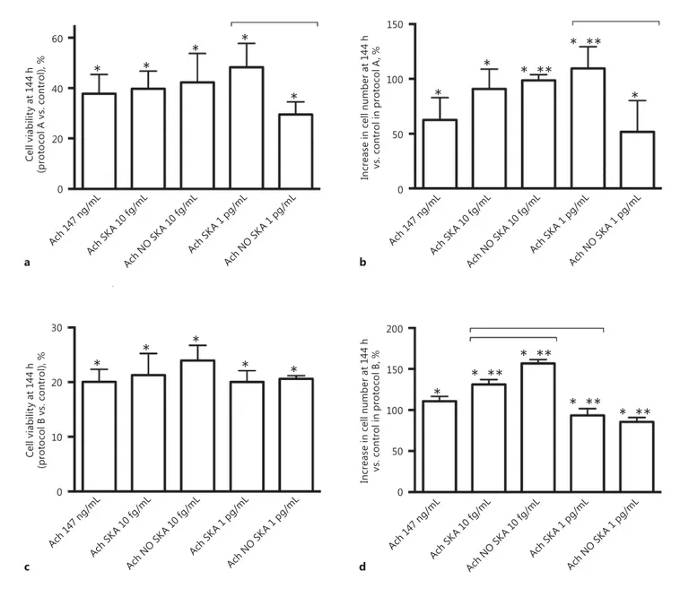
appropriate, using GraphPad Prism 5 (GraphPad Software, La Jolla, CA, USA). p < 0.05 was considered statistically significant. All data from the densitometric analysis were normalized to con-trol values (defined as 1). All other data from each experimental protocol were normalized to control values in percent (defined as 0%). 40 20 60 0 Ce ll v ia bi lit y a t 1 44 h (p ro to co l A vs. c on tro l), % * * * * * * * * * * * * * * ** * ** * ** * ** * ** * ** * Ach 14 7 ng/ mL Ach S KA 1 0 fg/ mL Ach N O SKA 1 0 fg/ mL Ach S KA 1 p g/mL Ach N O SKA 1 p g/mL Ach 14 7 ng/ mL Ach S KA 1 0 fg/ mL Ach N O SKA 1 0 fg/ mL Ach S KA 1 p g/mL Ach N O SKA 1 p g/mL Ach 14 7 ng/ mL Ach S KA 1 0 fg/ mL Ach N O SKA 1 0 fg/ mL Ach S KA 1 p g/mL Ach N O SKA 1 p g/mL Ach 14 7 ng/ mL Ach S KA 1 0 fg/ mL Ach N O SKA 1 0 fg/ mL Ach S KA 1 p g/mL Ach N O SKA 1 p g/mL 150 100 50 0 In cr ea se in c el l n um be r a t 1 44 h vs . c on tr ol i n p ro to co l A , % 20 10 30 0 150 100 50 200 0 Ce ll v ia bi lit y a t 1 44 h (p ro to co l B vs. c on tro l), % In cr ea se in c el l n um be r a t 1 44 h vs . c on tr ol i n p ro to co l B , % a b c d
Fig. 1. Cell viability and cell counts after 144 h of stimulation with
different formulations of Ach. a , b Results obtained by MTT and
crystal violet staining in NHEK treated only at T0 with different
formulations of highly diluted Ach and maintained for 144 h. c ,
d The same agents were added every 24 h for 144 h, and then
NHEK cell viability and proliferation were measured. The results of the MTT test are expressed as means ± SD (%) of 6 biological
replicates normalized to the control. In the crystal violet evalua-tion, cell counts are expressed as means ± SD (%) of 6 biological replicates. * p < 0.05 vs. control. * * p < 0.05 vs. 147 ng/mL Ach. Bars indicate significance between the activated and nonactivated forms and between the 2 activated forms. Ach, acetylcholine; SKA, sequential kinetic activation; NO SKA, nonsequential kinetic acti-vation.
Results
Proliferation and Cell Viability
NHEK cells were treated using 2 protocols (i.e., A and B) for 144 h to verify the effects of highly diluted Ach
com-pared to 147 ng/mL Ach on cell viability and proliferation. As shown in Figure 1 a and b, in protocol A, the formu-lation of sequentially kinetically activated Ach 1 pg/mL was able to improve cell viability (48.31 ± 9.4% compared to the control) compared to the nonactivated form (29.49
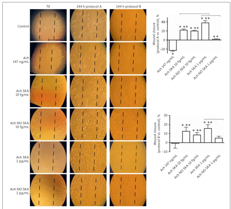
T0 144 h protocol A 144 h protocol B Control Ach 147 ng/mL Ach SKA 10 fg/mL Ach NO SKA 10 fg/mL Ach SKA 1 pg/mL Ach NO SKA 1 pg/mL 20 0 40 –20 40 20 10 0 30 –10 W oun d closur e (p ro to co l A vs. c on tro l), % * ** * ** * ** * ** * ** * ** * * * Ach 14 7 ng/ mL Ach S KA 1 0 fg/ mL Ach N O SKA 1 0 fg/ mL Ach S KA 1 p g/mL Ach N O SKA 1 p g/mL Ach 14 7 ng/ mL Ach S KA 1 0 fg/ mL Ach N O SKA 1 0 fg/ mL Ach S KA 1 p g/mL Ach N O SKA 1 p g/mL W oun d closur e (p ro to co l B v s. c on tr ol ), %
Fig. 2. Involvement of different formulations of Ach in NHEK
wound closure. On the left are representative pictures of wound healing with each treatment at T0, after 144 h in protocol A, and after 144 h in protocol B taken through microscopy at an original magnification of ×20. Scale bar, 50 μm for all images. On the right, the wound closure area calculated by measuring the diminution of the wound bed surface by the time using ImageJ software in
pro-tocol A and B is shown. The results are expressed as means ± SD (%) of wound closure in 5 biological replicates normalized to each T0. * p < 0.05 vs. control. * * p < 0.05 vs. 147 ng/mL Ach. Bars in-dicate significance between the activated and nonactivated forms and between the 2 activated forms. Ach, acetylcholine; SKA, se-quential kinetic activation; NO SKA, nonsese-quential kinetic activa-tion.
± 5.05% compared to the control), with 147 ng/mL Ach (37.8 ± 7.64% compared to the control) and with sequen-tially kinetically activated Ach 10 fg/mL (39.74 ± 7.01% compared to the control). This effect was statistically sig-nificant ( p < 0.05) in comparison to untreated cells. This increase was also observed with crystal violet staining ( Fig. 1 b), in which the greater number of counted cells was observed with the sequentially kinetically activated form of Ach 1 pg/mL (109.6 ± 9.67% compared to the control). However, the sequentially kinetically activated solutions (Ach SKA 10 fg/mL and Ach SKA 1 pg/mL) were able to induce effects similar to those observed on cell proliferation using 147 ng/mL Ach (90.8 ± 8.11%; 109.6 ± 9.67% compared to the control vs. 62.61 ± 10.3% compared to the control). On the contrary, in protocol B ( Fig. 1 c, d) nonsignificant changes in cell viability and proliferation were observed in the presence of the sequen-tially kinetically activated form (Ach SKA 1 pg/mL and Ach SKA 10 fg/mL). The maximum effects were observed in the presence of nonsequentially kinetically activated Ach 10 fg/mL (NO SKA). These results demonstrate the importance of dilution and mechanical activation in the treatment of cells during the longest period.
Effects of Activated Ach Solutions on NHEK Migration
In this step, the effects of highly diluted Ach in differ-ent formulations on NHEK migration in a wound healing assay were evaluated ( Fig. 2 ). Our results showed in-creased migration activity ( p < 0.05) in KC treated with sequentially kinetically activated Ach 10 fg/mL and 1 pg/ mL (both protocols A and B) compared to the control and 147 ng/mL Ach, and these effects were more evident in the presence of sequentially kinetically activated Ach 1 pg/mL (38 ± 4.92% in protocol A and 15.72 ± 4.12% in protocol B) after 144 h of treatment. The group treated with nonsequentially kinetically activated Ach 10 fg/mL in both protocols showed a greater effect than that treated with nonsequentially kinetically activated Ach 1 pg/mL, with a maximum effect in protocol A. On the contrary, nonsequentially kinetically activated Ach 1 pg/mL was ef-fective in protocol B only. These data suggest that the
se-quentially kinetically activated solutions of Ach (10 fg/ mL and 1 pg/mL) have an active role in wound closure, as shown in Figure 2 (left).
Effects of Highly Diluted Ach on ROS Production, Mitochondrial Membrane Potential, and Oxygen Consumption
In NHEK cells treated with 147 ng/mL Ach in both protocols an increase in ROS production of about 65.86 ± 7.69% (protocol A) and 67.5 ± 3.54% (protocol B) com-pared to the control was observed, but in the presence of sequentially kinetically activated Ach solutions (10 fg/mL and 1 pg/mL) this increase was significantly lower (13 ± 5.66 and 9.59 ± 2.65% in protocol A, respectively; 10 ± 1.41 and 8 ± 1.1% in protocol B, respectively) than what was observed with 147 ng/mL Ach and with the nonse-quentially kinetically activated form in both protocols. The data are shown in Figure 3 a and b. These data dem-onstrate the greater efficacy of the sequentially kinetically activated form in terms of normal KC health. As shown in Figure 3 c and d, the activated solutions in both proto-cols (A and B) were able to increase ( p < 0.05) the mito-chondrial membrane potential compared to 147 ng/mL Ach and the nonsequentially kinetically activated form, whereas the maximum effects were observed in the pres-ence of sequentially kinetically activated Ach 1 pg/mL (Ach SKA 1 pg/mL 79.01 ± 3.77% in protocol A and 81.6 ± 4.21% in protocol B compared to the control), indicat-ing the greater biochemical energy of the cells. In order to obtain a more complete description of mitochondrial me-tabolism and function, an oxygen consumption assess-ment was also performed. As shown in Figure 3 e and f, the rate of oxygen consumption decreased in all samples, indicating physiological activity of the mitochondria, and data were inversely proportional to mitochondrial poten-tial movements ( p < 0.05). It is noteworthy that the se-quentially kinetically activated solutions (10 fg/mL and 1 pg/mL) induced oxygen consumption similar to that observed in samples treated with 147 ng/mL Ach (–18.76 ± 1.38 and –7.49 ± 2.5%, respectively, in protocol A; –3.3 ± 0.61 and –9.08 ± 0.88%, respectively, in protocol B),
Fig. 3. ROS production ( a , b ), mitochondrial potential membrane
( c , d ), and oxygen consumption ( e , f ) in NHEK cells. a , b ROS
production is expressed as means ± SD (%) of 5 biological repli-cates normalized to control values and indicated as a percentage of cytochrome C reduced per microgram of protein with respect to
the control. c , d Evaluation of the mitochondrial membrane
po-tential in NHEK treated with different formulations of Ach; results are reported as means ± SD (%) of 5 biological replicates
normal-ized to control values. e , f Oxygen consumption under the same
conditions and treatments was assessed. The results are expressed as means ± SD (%). * p < 0.05 vs. control. * * p < 0.05 vs. 147 ng/mL Ach. Bars indicate significance between the activated and nonac-tivated forms and between the 2 acnonac-tivated forms. Ach, acetylcho-line; SKA, sequential kinetic activation; NO SKA, nonsequential kinetic activation.
60 40 20 80 0 60 40 20 80 0 RO S p ro du ct io n v s. c on tr ol (pro to co l A ), % 60 40 20 80 0 –10 –20 –30 0 0 –10 –20 –30 10 –40 –40 60 40 20 80 0 JC -1 ra tio ( 595 n m /5 35 n m ) vs. c on tro l (p ro to co l A ), % JC -1 ra tio ( 595 n m /5 35 n m ) vs. c on tro l (p ro to co l B ), % RO S p ro du ct io n v s. c on tr ol (pro to co l B ), % Ach 14 7 ng/ mL Ach S KA 1 0 fg/ mL Ach N O SKA 1 0 fg/ mL Ach S KA 1 p g/mL Ach N O SKA 1 p g/mL Ach 14 7 ng/ mL Ach S KA 1 0 fg/ mL Ach N O SKA 1 0 fg/ mL Ach S KA 1 p g/mL Ach N O SKA 1 p g/mL Ach 14 7 ng/ mL Ach S KA 1 0 fg/ mL Ach N O SKA 1 0 fg/ mL Ach S KA 1 p g/mL Ach N O SKA 1 p g/mL Ach 14 7 ng/ mL Ach S KA 1 0 fg/ mL Ach N O SKA 1 0 fg/ mL Ach S KA 1 p g/mL Ach N O SKA 1 p g/mL Ach 14 7 ng/ mL Ach S KA 1 0 fg/ mL Ach N O SKA 1 0 fg/ mL Ach S KA 1 p g/mL Ach N O SKA 1 p g/mL Ach 14 7 ng/ mL Ach S KA 1 0 fg/ mL Ach N O SKA 1 0 fg/ mL Ach S KA 1 p g/mL Ach N O SKA 1 p g/mL * ** * ** * ** * ** * ** * ** * ** * ** * ** * ** ** ** * * * * * * * * * * * * Inte ns ity v s. contr ol (pro to co l A ), % In te ns ity vs. c on tro l (pro to co l B ), % a c e b d f * ** * ** * ** * ** * ** 3
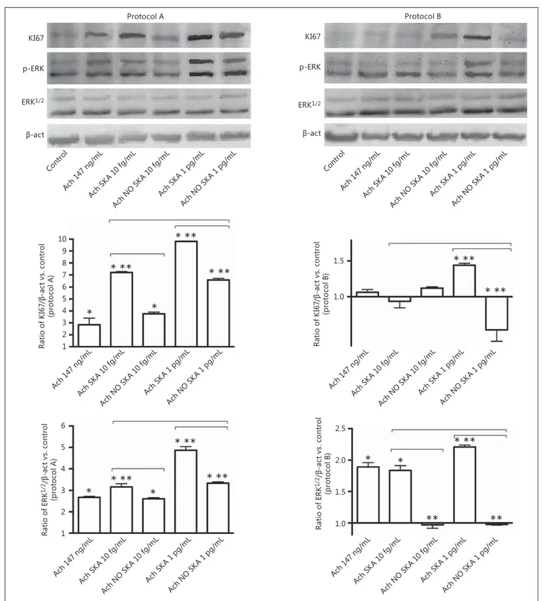
KI67 Protocol A p-ERK DŽ-act ERK1/2 Ach 14 7 ng/ mL Cont rol Ach S KA 1 0 fg/ mL Ach N O SKA 1 0 fg/ mL Ach S KA 1 p g/mL Ach N O SKA 1 p g/mL Protocol B KI67 p-ERK DŽ-act ERK1/2 Ach 14 7 ng/ mL Cont rol Ach S KA 1 0 fg/ mL Ach N O SKA 1 0 fg/ mL Ach S KA 1 p g/mL Ach N O SKA 1 p g/mL Ach 14 7 ng/ mL Ach S KA 1 0 fg/ mL Ach N O SKA 1 0 fg/ mL Ach S KA 1 p g/mL Ach N O SKA 1 p g/mL 9 8 7 6 5 4 3 2 10 1 Ra tio o f K I67 /DŽ -a ct v s. co nt ro l (pro to co l A ) * * Ach 14 7 ng/ mL Ach S KA 1 0 fg/ mL Ach N O SKA 1 0 fg/ mL Ach S KA 1 p g/mL Ach N O SKA 1 p g/mL * * 5 4 3 2 6 1 Ra tio o f ER K 1/2 /DŽ -a ct vs. c on tro l (pro to co l A ) Ach 14 7 ng/ mL Ach S KA 1 0 fg/ mL Ach N O SKA 1 0 fg/ mL Ach S KA 1 p g/mL Ach N O SKA 1 p g/mL 1.5 1.0 Ra tio o f K I67 /DŽ -a ct v s. co nt ro l (pro to co l B ) Ach 14 7 ng/ mL Ach S KA 1 0 fg/ mL Ach N O SKA 1 0 fg/ mL Ach S KA 1 p g/mL Ach N O SKA 1 p g/mL 2.0 1.5 2.5 1.0 Ra tio o f ER K 1/2 /DŽ -a ct v s. co nt ro l (pro to co l B ) * * ** ** * ** * ** * ** * ** * ** * ** * ** * ** * **
Fig. 4. Western blot and densitometric analysis of ERK/MAPK and
KI67 in NHEK cells. Protein extracts were analyzed by
immuno-blotting with specific antibodies against the indicated proteins. The pictures represent 5 biological replicates for each experimen-tal protocol (A and B). Data are expressed as means ± SD of 5
bio-logical replicates for each experimental protocol (A and B). * p < 0.05 vs. control. * * p < 0.05 vs. 147 ng/mL Ach. Bars indicate sig-nificance between the activated and nonactivated form and be-tween the 2 activated forms. Ach, acetylcholine; SKA, sequential kinetic activation; NO SKA, nonsequential kinetic activation.
20 10 0 30 –10 Ce ll v ia bi lit y a t 1 44 h (p ro to co l A v s. co ntr ol ), % * * Atro pine 7 .25 μg/ mL Chel 3 84 n g/mL H89 5 19 n g/mL H89 5 19 n g/mL +Ach 1 47 n g/mL H89 5 19 n g/mL +Ach S KA 1 0 fg/ mL H89 5 19 n g/mL +Ach N O SKA 1 0 fg/ mL H89 5 19 n g/mL +Ach S KA 1 p g/mL H89 5 19 n g/mL +Ach N O SKA 1 p g/mL Chel 3 84 n g/mL +Ach 1 47 n g/mL Chel 3 84 n g/mL +Ach S KA 1 0 fg/ mL Chel 3 84 n g/mL +Ach N O SKA 1 0 fg/ mL Chel 3 84 n g/mL +Ach S KA 1 p g/mL Chel 3 84 n g/mL +Ach N O SKA 1 p g/mL Atro pine 7 .25 μg/ mL+A ch 14 7 ng/ mL Atro pine 7 .25 μg /mL+ Ach S KA 1 0 fg/ mL Atro pine 7 .25 μg /mL+ Ach S KA 1 p g/mL Atro pine 7 .25 μg /mL+ Ach N O SKA 1 p g/mL Atro pine 7 .25 μg /mL+ Ach N O SKA 1 0 fg/ mL Atro pine 7 .25 μg/ mL Chel 3 84 n g/mL H89 5 19 n g/mL H89 5 19 n g/mL +Ach 1 47 n g/mL H89 5 19 n g/mL +Ach S KA 1 0 fg/ mL H89 5 19 n g/mL +Ach N O SKA 1 0 fg/ mL H89 5 19 n g/mL +Ach S KA 1 p g/mL H89 5 19 n g/mL +Ach N O SKA 1 p g/mL Chel 3 84 n g/mL +Ach 1 47 n g/mL Chel 3 84 n g/mL +Ach S KA 1 0 fg/ mL Chel 3 84 n g/mL +Ach N O SKA 1 0 fg/ mL Chel 3 84 n g/mL +Ach S KA 1 p g/mL Chel 3 84 n g/mL +Ach N O SKA 1 p g/mL Atro pine 7 .25 μg/ mL+A ch 14 7 ng/ mL Atro pine 7 .25 μg /mL+ Ach S KA 1 0 fg/ mL Atro pine 7 .25 μg /mL+ Ach S KA 1 p g/mL Atro pine 7 .25 μg /mL+ Ach N O SKA 1 p g/mL Atro pine 7 .25 μg /mL+ Ach N O SKA 1 0 fg/ mL 0 5 –5 In cr ea se in c el l n um be r a t 1 44 h vs . c on tr ol i n p ro to co l A , % a b * ** * ** * ** * ** * ** * ** * *** *** ** * **
Fig. 5. Cell viability and proliferation in the presence of M1, PKC, and PKA blockers.
Results obtained by MTT ( a ) and crystal
violet staining ( b ) in NHEK treated only
with protocol A. The results are expressed as a means ± SD (%) of 4 biological repli-cates normalized to the control. * p < 0.05 vs. control. * * p < 0.05 vs. atropine 7.25 μg/ mL. Bars indicate significance between the activated and nonactivated forms and be-tween the 2 activated forms in pretreated samples. Ach, acetylcholine; SKA, sequen-tial kinetic activation; NO SKA, nonse-quential kinetic activation; Chel, chelery-thrine.
whereas the nonactivated solutions showed a greater de-crease in oxygen consumption, thus indicating possible stress conditions of the mitochondria.
Analysis of Intracellular Pathways
To investigate the mechanisms activated by highly di-luted and 147 ng/mL Ach in cell proliferation, NHEK cells were treated with the same experimental protocols used before. As shown in Figure 4 , all of these agents were able to induce significant changes in the expressions of KI67 and ERK/MAPK. In particular, we used the KI67 protein as a cellular marker to investigate KC prolifera-tion as KI67 is known to be specifically expressed in nu-clei of proliferating cells during the cell cycle. As reported in Western blot and densitometric analyses ( Fig. 4 ), in both protocols sequentially kinetically activated Ach 1 pg/mL was able to induce a significant increase ( p < 0.05) in KI67 compared to Ach 10 fg/mL and Ach 147 ng/ mL, and these data confirmed the ability of sequentially kinetically activated Ach 1 pg/mL to improve cell prolif-eration in NHEK cells. However, Ach 10 fg/mL also showed a significant efficacy, in particular in protocol A, in which the increase in KI67 was higher compared to that with either 147 ng/mL Ach or the nonsequentially kinet-ically activated form. In addition, ERK, important mem-bers of the MAPK family regulating cell migration, were evaluated. In both protocols the activated solution of se-quentially kinetically activated Ach 1 pg/mL was able to increase the expression of ERK1/2, and this effect was more evident in protocol A compared to what was ob-served with nonsequentially kinetically activated 147 ng/ mL Ach and sequentially kinetically activated Ach 10 fg/ mL. However, the sequentially kinetically activated Ach 10 fg/mL solution was more effective than the nonse-quentially kinetically activated one in inducing phos-phorylation of ERK/MAPK. This is an important finding since ERK phosphorylation acts as a switch for other in-tracellular pathways and thus is a clear index of cellular health. All of these data demonstrate the greater influence of the highly diluted Ach on cell proliferation of KC com-pared to the ponderal Ach concentration.
Analysis of the Involvement of Muscarinic Receptor Subtypes and Calcium Signaling in Effects Induced by Ach
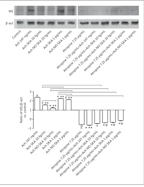
Since the effects induced by Ach are mediated through its muscarinic and nicotinic receptors via the calcium sig-naling cascade, some of these elements were also analyzed during protocol A. In particular, the muscarinic receptor subtype 1 (M1) was analyzed. As shown in Figure 5 , pre-treatment with atropine at 7.25 μg/mL (M1 inhibitor) was able to reduce cell viability and proliferation of NHEK cells. In particular, in the presence of different formula-tions of Ach a significant reduction ( p < 0.05) in the ben-eficial effects on cell viability and proliferation was ob-served and this effect was more evident in the presence of sequentially kinetically activated Ach 1 pg/mL. In par-ticular, a decrease of about 40% in cell viability and about 97% in cell proliferation compared to stimulation with-out pretreatment was observed.
In addition, the importance of the involvement of the M1 receptor in the mechanism activated by all forms of Ach was also clarified by the experiments on wound healing, in which pretreatment with atropine at 7.25 μg/ mL significantly reduced the area of closure alone and also in the presence of all forms of Ach ( Fig. 6 ). Finally, as reported in Figure 7 , sequentially kinetically activat-ed Ach 1 pg/mL and 10 fg/mL were able to induce a significant increase (Ach SKA, p < 0.05) compared to the control and to the corresponding not-activated form in a manner similar to that observed with 147 ng/ mL Ach. In order to verify this effect, NHEK cells were pretreated with atropine (7.25 μg/mL) alone and in the presence of the different formulations of Ach. This pre-treatment prevented the activation of M1, and succes-sive stimulation with different formulations of Ach was not able to restore this condition. These data confirm the involvement of muscarinic receptor in NHEK cells. The downstream signaling from the muscarinic and nicotinic classes of Ach receptors involves the second messenger pathways that control the expression and ac-tivity of effector molecules via the common signaling cascade Ca 2+ -CamKII-PKC-MEK-ERK. In this context
Fig. 6. Involvement of M1, PKC, and PKA blockers in NHEK
wound closure. a–d Representative pictures of wound healing with
each treatment at T0 and after 144 h with protocol A through mi-croscopy at an original magnification of ×20. Scale bar, 50 μm for
all microscopy pictures. e–g Wound closure area; areas were
cal-culated by measuring the decrease in the surface of the bed of the wound over time using the ImageJ software (protocol A). The
re-sults are expressed as means ± SD (%) of the wound closure of 4 biological replicates normalized to each T0. * p < 0.05 vs. control. * * p < 0.05 vs. a specific blocker. Bars indicate significance between the activated and nonactivated forms and between the 2 activated forms in pretreated samples. Ach, acetylcholine; SKA, sequential kinetic activation; NO SKA, nonsequential kinetic activation; Chel, chelerythrine.
a b c d e f g T0 Control Atropine 7.25 μg/mL Chel 384 ng/mL H89 519 ng/mL Ach 147 ng/mL Ach 10 fg/mL
SKA Ach 10 fg/mL NO SKA Ach 1 pg/mLSKA Ach 1 pg/mLNO SKA
Ach 147 ng/mL Ach 10 fg/mL
SKA Ach 10 fg/mL NO SKA Ach 1 pg/mLSKA Ach 1 pg/mLNO SKA
Ach 147 ng/mL Ach 10 fg/mL
SKA Ach 10 fg/mL NO SKA Ach 1 pg/mLSKA Ach 1 pg/mLNO SKA
Atropin e 7.25 μg/mL Atropin e 7.25 μg/mL +Ach 147 ng/ mL Atropin e 7.25 μg/m L+Ac h SKA 1 0 fg/ mL Atropin e 7.25 μg/m L+Ac h NO S KA 1 0 fg/ mL Atropin e 7.25 μg/m L+Ac h SKA 1 p g/mL Atropin e 7.25 μg/m L+Ac h NO S KA 1 p g/mL * * * * * * * * * * –5 –10 0 –15 W oun d closur e vs. c on tro l, % W oun d closur e vs. c on tro l, % W oun d closur e vs. c on tro l, % –5 –10 –15 –20 0 –25 H89 5 19 ng/ mL H89 5 19 ng/ mL+A ch 147 n g/mL H89 5 19 n g/mL +Ach S KA 1 0 fg/ mL H89 5 19 ng/ mL+A ch N O SKA 1 0 fg/ mL H89 5 19 n g/mL +Ach S KA 1 p g/mL H89 5 19 ng/ mL+A ch N O SKA 1 p g/mL Chel 3 84 n g/mL Chel 3 84 ng/ mL+A ch 147 n g/mL Chel 3 84 n g/mL +Ach S KA 1 0 fg/ mL Chel 3 84 n g/mL +Ach N O SKA 1 0 fg/ mL Chel 3 84 n g/mL +Ach S KA 1 p g/mL Chel 3 84 n g/mL +Ach N O SKA 1 p g/mL –5 –10 –15 0 –20 * ** * ** * ** * ** * ** * ** * ** * ** 6
PKC and PKA kinases were also analyzed in NHEK cells treated under the same conditions as reported before. As shown in Figure 5 , the specific blockers of PKC (che-lerythrine at 384 ng/mL) and PKA (H89 at 519 ng/mL) were able to prevent any increase in cell viability or cell proliferation, maintaining the cell at a basal-like level. In the presence of all forms of sequentially kinetically activated and nonsequentially kinetically activated Ach, these effects were not reverted. These data confirm the ability of these blockers to modulate specific intracel-lular mechanisms as reported in the literature. In addi-tion, similar data were also observed in wound healing experiments in which the blockers prevented the
mi-gration of cells and consequently closure of the lesion area. Finally, as shown in Figure 8 , all of the forms (se-quentially kinetically activated and nonse(se-quentially ki-netically activated) and 147 ng/mL Ach were able to in-duce the activation of PKC and PKA, indicating that Ach was able to involve the calcium intracellular path-way to exert its effects. In particular, 147 ng/mL Ach and sequentially kinetically activated Ach 10 fg/mL showed similar effects. However, all other formulations were able to induce the activation of these kinases ( p < 0.05) compared to the control. These findings were confirmed by the pretreatments with chelerythrine (384 ng/mL) and H89 (519 ng/mL) for PKC and PKA, M1 DŽ-act Cont rol Ach 14 7 ng/ mL Ach S KA 1 0 fg/ mL Ach N O SKA 1 0 fg/ mL Ach S KA 1 p g/mL Ach N O SKA 1 p g/mL Ach 14 7 ng/ mL Ach S KA 1 0 fg/ mL Ach N O SKA 1 0 fg/ mL Ach S KA 1 p g/mL Ach N O SKA 1 p g/mL Atro pine 7 .25 μg/ mL Atro pine 7 .25 μg/ mL+A ch 14 7 ng/ mL Atropin e 7.25 μg/m L+Ac h SKA 1 0 fg/ mL Atro pine 7 .25 μg /mL+ Ach N O SKA 1 0 fg/ mL Atropin e 7.25 μg/m L+Ac h SKA 1 p g/mL Atro pine 7 .25 μg /mL+ Ach N O SKA 1 p g/mL Atropin e 7.25 μg/mL Atro pine 7 .25 μg/ mL+A ch 14 7 ng/ mL Atropin e 7.25 μg/m L+Ac h SKA 1 0 fg/ mL Atropin e 7.25 μg/m L+Ac h NO S KA 1 0 fg/ mL Atropin e 7.25 μg/m L+Ac h SKA 1 p g/mL Atropin e 7.25 μg/m L+Ac h NO S KA 1 p g/mL 2 1 0 3 –1 Ra tio o f M 1/ DŽ -a ct vs. c on tro l * * ** * ** * ** * ** * ** * * * * * Ĵ Ĵ Ĵ
Fig. 7. Western blot and densitometric
analysis of the M1 receptor in NHEK cells. Protein extracts were analyzed by immu-noblotting with specific antibodies against the indicated proteins. The pictures repre-sent an example of 3 biological replicates for protocol A. Ach, acetylcholine; SKA, sequential kinetic activation; NO SKA, nonsequential kinetic activation. Data are expressed as means ± SD of 3 biological replicates. * p < 0.05 vs. control. * * p < 0.05
vs. 147 ng/mL Ach. φ p < 0.05 vs. 7.25 μg/
mL atropine (specific M1 inhibitor). Bars indicate significance among the different forms of Ach, between activated forms alone and in the presence of pretreatment with atropine.
respectively. These agents were analyzed alone and in combination with the different formulations of Ach. As evidenced by Western blot and densitometric analysis in both of these conditions, stimulation with Ach was not effective in inducing activation of the specific ki-nase. These findings confirm the data observed with ERK phosphorylation and demonstrate the great influ-ence of the highly diluted sequentially kinetically acti-vated Ach on KC functions.
Discussion
Burn injuries, deep erosions, chronic ulcers, diabetic wounds, and graft donor sites are a few of the clinical situ-ations [Chernyavsky et al., 2012] in which epithelialization is uncontrolled or incomplete. In this context, we decided to study novel approaches to facilitate epithelialization. In the last year, continuously increasing evidence for an ex-tremely high sensitivity of biological objects to chemical
Cont rol Ach 14 7 ng/ mL Ach S KA 1 0 fg/ mL Ach N O SKA 1 0 fg/ mL Ach S KA 1 p g/mL Ach N O SKA 1 p g/mL Ach 14 7 ng/ mL Ach S KA 1 0 fg/ mL Ach N O SKA 1 0 fg/ mL Ach S KA 1 p g/mL Ach N O SKA 1 p g/mL Chel 3 84 n g/mL Chel 3 84 n g/mL +Ach 1 47 n g/mL Chel 3 84 ng/ mL+A ch SKA 1 0 fg/ mL Chel 3 84 n g/mL +Ach N O SKA 1 0 fg/ mL Chel 3 84 ng/ mL+A ch SKA 1 p g/mL Chel 3 84 n g/mL +Ach N O SKA 1 p g/mL Chel 3 84 n g/mL Chel 3 84 n g/mL +Ach 1 47 n g/mL Chel 3 84 n g/mL +Ach S KA 1 0 fg/ mL Chel 3 84 n g/mL +Ach N O SKA 1 0 fg/ mL Chel 3 84 n g/mL +Ach S KA 1 p g/mL Chel 3 84 n g/mL +Ach N O SKA 1 p g/mL H89 5 19 n g/mL H89 5 19 n g/mL +Ach 1 47 n g/mL H89 5 19 ng/ mL+A ch SKA 1 0 fg/ mL H89 5 19 ng/ mL+A ch N O SKA 1 0 fg/ mL H89 5 19 ng/ mL+A ch SKA 1 p g/mL H89 5 19 ng/ mL+A ch N O SKA 1 p g/mL H89 5 19 ng/ mL H89 5 19 n g/mL +Ach 1 47 n g/mL H89 5 19 n g/mL +Ach S KA 1 0 fg/ mL H89 5 19 n g/mL +Ach N O SKA 1 0 fg/ mL H89 5 19 n g/mL +Ach S KA 1 p g/mL H89 5 19 n g/mL +Ach N O SKA 1 p g/mL Ach 14 7 ng/ mL Ach S KA 1 0 fg/ mL Ach N O SKA 1 0 fg/ mL Ach S KA 1 p g/mL Ach N O SKA 1 p g/mL Chel 3 84 n g/mL Chel 3 84 n g/mL +Ach 1 47 n g/mL Chel 3 84 n g/mL +Ach S KA 1 0 fg/ mL Chel 3 84 n g/mL +Ach N O SKA 1 0 fg/ mL Chel 3 84 n g/mL +Ach S KA 1 p g/mL Chel 3 84 n g/mL +Ach N O SKA 1 p g/mL H89 5 19 ng/ mL H89 5 19 n g/mL +Ach 1 47 n g/mL H89 5 19 n g/mL +Ach S KA 1 0 fg/ mL H89 5 19 n g/mL +Ach N O SKA 1 0 fg/ mL H89 5 19 n g/mL +Ach S KA 1 p g/mL H89 5 19 n g/mL +Ach N O SKA 1 p g/mL PKC p-PKA DŽ-act 2 1 0 2 –1 1 Ra tio o f PK C/ DŽ -a ct vs. c on tro l Ra tio o f PK C/ DŽ -a ct vs . c on tr ol * * ** * ** * *** *** ** * ** * * * * * * * * * * Ĵ Ĵ Ĵ Ĵ Ĵ Ĵ Ĵ ĴĴ ĴĴ ĴĴ ĴĴ * * * * * * * * ĴĴ ĴĴ *ĴĴ *Ĵ *Ĵ Ĵ Ĵ * ** * ** * ** * ** * ** * **
Fig. 8. Western blot and densitometric analysis of PKC and PKA
in NHEK cells. Protein extracts were analyzed by immunoblotting with specific antibodies against the indicated proteins using also a specific antagonist (384 ng/mL chelerythrine and 519 ng/mL H89, respectively). The pictures represents an example of 3 biological replicates for protocol A. Data are expressed as means ± SD of 3 biological replicates. Ach, acetylcholine; SKA, sequential kinetic
activation; NO SKA, nonsequential kinetic activation; Chel, chel-erythrine. * p < 0.05 vs. control. * * p < 0.05 vs. 147 ng/mL Ach.
φ p < 0.05 vs. 384 ng/mL chelerythrine. φφ p < 0.05 vs. 519 ng/mL
H89.Bars indicate significance among the different forms of Ach, between activated forms alone, and in the presence of pretreat-ment with specific blockers.
endogenous effects has been reported [Werkheiser et al., 2011; Marzotto et al., 2014; Landreneau et al., 2015]. Or-gans, tissues, and cells are capable of reacting to the pres-ence of peptides, hormones, and molecular messengers at very low concentrations. A pioneering study demonstrated that low doses of solutions of mechanically activated IL-12 and IFN-γ, codelivered via the oral route to experimental asthmatic mice, can revert this pathological condition, re-storing a normal balance between Th1- and Th2-derived cytokines and leading the animals to a healthy condition [Gariboldi et al., 2009]. Moreover, many clinical trials have tested highly diluted SKA drugs in various pathologies. Significant effectiveness has been reported in supportive therapy and pain relief in cancer patients [Rajendran, 2004; Sunila et al., 2009; Radice et al., 2014]. In addition, a reduc-tion of symptoms and an improved quality of life have been observed in patients with psoriasis or vitiligo [Witt et al., 2009; Roberti et al., 2014; Barygina et al., 2015] or in im-mune system imbalances [Kho et al., 2012].
In this work, normal human epithelial KC were used to study the effects of highly diluted and mechanically acti-vated Ach solutions (10 –12 –10 –14 ) in an incisional wound model to confirm the hypothesis that low-dose Ach could be a more physiological stimulus for KC viability, prolif-eration, and migration.
Two different protocols have been used in order to as-sess the best modality of stimulation of cultured KC. Pro-tocol A, characterized by a single stimulation, appears to be more effective than the protocol with repeated stimula-tions every 24 h (protocol B) on cell proliferation, viability, and migration. We investigated cell proliferation and cell migration in different experiments. These two biological functions were involved in two important physiological as-pects of skin regeneration – during the creation of a wound the cells migrate and after its closure the cells return to a normal turnover. It can be hypothesized that a single stim-ulation triggers a sequence of intracellular reactions with-out any negative effect. Moreover, in both protocols low-dose Ach induced a lower ROS production than high-con-centration Ach and the control.
One of the most compelling findings of this work is the difference in responses between sequentially kinetically ac-tivated and nonsequentially kinetically acac-tivated Ach solu-tions, with the former being much more effective than the latter. The observation of an increased effect induced by mechanical activation of an active principle is not com-pletely new and has been observed by other authors [Gari-boldi et al., 2009]. However, in the present work, enhanced effects due to mechanical activation have been demon-strated for the first time within the nonneuronal
choliner-gic system. The mechanisms underlying this increased ef-fectiveness are still unclear. Here only a mere observation of the increased efficacy of sequentially kinetically activat-ed versus nonsequentially kinetically activatactivat-ed low-dose Ach solutions is reported. More research will be necessary in the future in order to clarify this aspect.
Ach is present in all living cells [Hebb, 1962; Grando et al., 1993; Fania et al., 2012]. Human skin presents the high-est concentration of free Ach [Klapproth et al., 1997; Fania et al., 2012], which depends on its production and degrada-tion induced, respectively, by choline acetyltransferase and acetylcholinesterase. To exert its effects, Ach binds its mus-carinic and nicotinic receptors (the musmus-carinic ones are transmembrane glycoproteins applying their function through G proteins and leading to activation of the second messengers; the nicotinic ones are ligand-gated ion chan-nels that modify the flow of Na + , Ca 2+ , and K + ) [Fania et al., 2012]. The concentrations of Ach used in this study can be considered very similar to physiological release from human KC. Grando et al. [1993] reported that a single KC synthesizes a mean of 2 × 10 –17 moles and releases 7 × 10 –19 moles Ach/min. This fits very well with the amount of Ach administered to cultured cells during the experiments de-scribed in this work. Data available in the literature show significant improvements in mucocutaneous diseases in patients who received cholinergic agents as treatment for a concurrent disease or owing to the habitual use of nicotine products [Kuwahara et al., 2000; Mehta and Martin, 2000]. Those reports suggest that cholinergic agents have a huge potential for dermatological use.
Human KC possess cholinergic enzymes for Ach syn-thesis and degradation [Grando et al., 1993] and are also responsive to Ach, believed to act as a local hormone in the epidermis [Grando et al., 1993; Beck et al., 2006]. Ach acts via the calcium pathway, which funtions as a mediator of its effects on the epidermis [Grando et al., 2006] and then activates all of the intracellular pathways involved in calci-um-signaling transduction, including ERK/MAPK [Beck et al., 2006; Kühne et al., 2015]. It is important to consider the difference in activity which we observed between 147 ng/mL Ach and sequentially kinetically activated or non-sequentially kinetically activated forms on proliferation, viability, cell migration, ROS production, oxygen con-sumption, and the mitochondrial membrane potential of NHEK. In both protocols (A and B), the sequentially ki-netically activated Ach 1pg/ml formulation was able to im-prove cell viability compared to 147 ng/mL Ach and it in-duced effects similar to those of 147 ng/mL Ach on cell proliferation. In a minor manner, similar effects were also observed in the presence of sequentially kinetically
acti-vated Ach 10 fg/mL. Ach 147 ng/mL was chosen as the control because it was demonstrated, with a dose-response curve, to be the most effective concentration in a similar HaCaT (spontaneously transformed aneuploid immortal human keratinocyte cell line) experimental model [Metzger et al., 2005]. Data described in this work indicate for the first time the efficacy of activated low doses of Ach on cell culture and demonstrate a minor toxicity compared to 147 ng/mL. For this reason, low-dose Ach could be important for the development of new therapies for nonhealing wounds. As a matter of fact, nonhealing wounds are a ma-jor healthcare problem involving important social and eco-nomic aspects. Old age, immobility, and repeated trauma create conditions for the genesis and maintenance of in-flammatory processes leading to wound formation. In ad-dition, metabolic, neurologic, and circulatory factors play an important role in wound formation and contribute to chronicity. For this purpose, the ability of cells to migrate in the presence of the same agents and conditions used be-fore was also investigated in a wound healing assay model. Sequentially kinetically activated forms (1 pg/mL and 10 fg/mL) revealed a greater capacity for migration compared to 147 ng/mL Ach, and these effects were more evident af-ter a single administration. This is an important element because the response to doses of Ach may depend on its sensitivity to external perturbations. To verify these benefi-cial effects on wound closure under physiological condi-tions, ROS production, oxygen consumption, and mito-chondrial membrane potential were investigated. Sequen-tially kinetically activated forms showed a statistically significant decrease in ROS production accompanied by an increase in mitochondrial membrane potential and de-creased oxygen consumption in comparison with 147 ng/ mL Ach, indicating the better biochemical energy of the cells. Also in this case, protocol A showed greater effects, indicating that a single treatment is enough to induce ben-eficial effects. Finally, the involvement of ERK/MAPK (in-volved in the regulation of cell migration) and KI67 (ex-pressed in the nuclei of proliferating cells during the cell cycle) in the mechanism activated by Ach confirmed the better influence of the single treatment (protocol A) versus continuous treatment of the activated low doses on cell proliferation compared to 147 ng/mL Ach. Since protocol A showed the better efficacy, the other experiments on the M1 receptor and the calcium signaling cascade were per-formed only under this condition. Sequentially kinetically activated Ach 1 pg/mL confirmed its higher effectiveness compared to the sequentially kinetically activated Ach 10 fg/mL formulation in M1 receptor, PKC, and PKA activa-tion. These findings confirm that highly diluted Ach acts
through the same mechanisms as concentrated Ach and support the hypothesis of beneficial effects exerted by low doses without any negative effect (e.g., oxidative injury).
It is noteworthy that, although 147 ng/mL Ach showed some efficacy in cell proliferation, it may be harmful to the cells due to a significant increase in ROS production.
Our findings suggest that a single treatment of low dos-es (10 –12 –10 –14 ) of sequentially kinetically activated Ach triggers a cascade of dynamic events that results in better regulation of KC functions, cell viability, proliferation, and migration. These elements are very important for hypo-thetical possible fields of application in the clinical setting. In conclusion, low doses of sequentially kinetically ac-tivated Ach seem to play an active role in an in vitro mod-el of wound healing. Moreover, these data suggest that ad-ministration of Ach at doses in a physiological range may not only be effective but is also likely to be safe. An intrigu-ing aspect of this study is with regard to the difference in activity that was observed between sequentially kinetically activated and nonsequentially kinetically activated solu-tions, with the former being more effective than nonacti-vated solutions. The use of actinonacti-vated blends is common in different fields, including pharmaceutical technology, to obtain a high therapeutic potential with very low dosages.
Acknowledgments
The authors thank Dr. Mariangela Fortunato for her valuable help with language. GUNA S.p.a (Vincenzo Miranda) contributed to preparing and donating solutions.
This project was made possible by a scholarship funded by the Fondazione Giovanni Goria and the Fondazione CRT within the Project ‘Master dei Talenti della Società Civile – 2014.
Disclosure Statement
The authors declare that they have no conflict of interests.
References Avvakumov, E.G., S.V. Chizhevskaya, E.S. Stoya-nov, M.V. Povetkina, A.M. Chekmarev, V.L. Shafirov, O.B. Vinokurova (1999) Influence of the nature of components in mechanically ac-tivated mixture of zirconium and silicon ox-ides on solid-phase synthesis of zircon. Russ J Appl Chem 72: 1498–1503.
Barygina, V., M. Becatti, T. Lotti, S. Moretti, N. Taddei, C. Fiorillo (2015) Treatment with low-dose cytokines reduces oxidative-mediat-ed injury in perilesional keratinocytes from vitiligo skin. J Dermatol Sci 79: 163–170.
Beck, B., A. Zholos, V. Sydorenko, M. Roudbaraki, V. Lehen’kyi, P. Bordat, N. Prevarskaya, R. Skryma (2006) TRPC7 is a receptor-operated DAG-activated channel in human keratino-cytes. J Invest Dermatol 126: 1982–1993. Beckmann, J., K.S. Lips (2013) The non-neuronal
cholinergic system in health and disease. Pharmacology 92: 286–302.
Brod, S.A., M. Khan (1996) Oral administration of IFN-alpha is superior to subcutaneous admin-istration of IFN-alpha in the suppression of chronic relapsing experimental autoimmune encephalomyelitis. J Autoimmun 9: 11–20. Cappellano, G., F. Uberti, P.P. Caimmi, S.
Pietro-nave, D.A. Mary, C. Dianzani, E. Micalizzi, M. Melensi, R. Boldorini, G. Nicosia, E. Crosio, A. Chiocchetti, F. Aina, M. Prat, U. Dianzani, G. Vacca, C. Ariatti, E. Grossini (2013) Different expression and function of the endocannabi-noid system in human epicardial adipose tis-sue in relation to heart disease. Can J Cardiol 29: 499–509.
Chernyavsky, A.I., J. Arredondo, J. Qian, V. Gali-tovskiy, S.A. Grando (2009) Coupling of ionic events to protein kinase signaling cascades upon activation of alpha7 nicotinic receptor: cooperative regulation of alpha2-integrin ex-pression and Rho kinase activity. J Biol Chem 284: 22140–22148.
Chernyavsky, A.I., S. Marchenko, C. Phillips, S.A. Grando (2012) Auto/paracrine nicotinergic peptides participate in cutaneous stress re-sponse to wounding. Dermatoendocrinology 4: 324–330.
D’Amico, L., E. Ruffini, R. Ferracini, I. Roato (2012) Low dose of IL-12 stimulates T cell re-sponse in cultures of PBMCs derived from non-small cell lung cancer patients. J Cancer Ther 3: 337–342.
Fania, L, A. Zampetti, G. Guerriero, C. Feliciani (2012) Alteration of cholinergic system in ke-ratinocytes cells produces acantholysis: a pos-sible use of cholinergic drugs in pemphigus vulgaris. Antiinflamm Antiallergy Agents Med Chem 11: 238–242.
Gariboldi, S., M. Palazzo, L . Zanobbio, G.F. Dusio, V. Mauro, U. Solimene, D. Cardani, M. Man-tovani, C. Rumio (2009) Low dose oral admin-istration of cytokines for treatment of allergic asthma. Pulm Pharmacol Ther 22: 497–510. Grando, S.A., D.A. Kist, M. Qi, M.V. Dahl (1993)
Human keratinocytes synthesize, secrete, and degrade acetylcholine. J Invest Dermatol 101: 32–36.
Grando, S.A., M.R. Pittelkow, K.U. Schallreuter (2006) Adrenergic and cholinergic control in the biology of epidermis: physiological and clinical significance. J Invest Dermatol 126: 1948–1965.
Hebb, C.O., K. Krnjevic (1962) The physiological significance of acetylcholine; in Elliott, K.A.C., I.H. Page, J.H. Quester (eds): Neurochemistry, ed 2. Springfield, Thomas, pp 452–521. Kho, M.M., A.P. Bouvy, M. Cadogan, R.
Kraai-jeveld, C.C. Baan, W. Weimar (2012) The ef-fect of low and ultra-low dosages thymoglobu-lin on peripheral T, B and NK cells in kidney
transplant recipients. Transpl Immunol 26: 186–190.
Klapproth, H., T. Reinheimer, J. Metzen, M. Münch, F. Bittinger, C.J. Kirkpatrick, K.D. Höhle, M. Schemann, K. Racké, I. Wessler (1997) Non-neuronal acetylcholine, a signal-ling molecule synthezised by surface cells of rat and man. Naunyn Schmiedebergs Arch Pharmacol 355: 515–523.
Kühne, S., W. Ockenga, A. Banning, R. Tikkanen (2015) Cholinergic transactivation of the EGFR in HaCaT keratinocytes stimulates a flotillin-1 dependent MAPK-mediated tran-scriptional response. Int J Mol Sci 16: 6447– 6463.
Kurzen, H., I. Wessler, C.J. Kirkpatrick, K. Ka-washima, S.A. Grando (2007) The non-neuro-nal cholinergic system of human skin. Horm Metab Res 39: 125–135.
Kuwahara, R.T., R.B. Skinner, E.W. Rosenberg (2000) Nicotine gum for oral lichen planus. J Dermatol 27: 755.
Landreneau, J.P., M.R. Shurin, M.V. Agassandian, A.A. Keskinov, Y. Ma, G.V. (2015) Shurin Im-munological mechanisms of low and ultra-low dose cancer chemotherapy. Cancer Mi-croenviron 8: 57–64.
Liang, C.C., A.Y. Park, J.L. Guan (2007) In vitro scratch assay: a convenient and inexpensive method for analysis of cell migration in vitro. Nat Protoc 2: 329–333.
Marzotto, M., D. Olioso, M. Brizzi, P. Tononi, M. Cristofoletti, P. Bellavite (2014) Extreme sen-sitivity of gene expression in human SH-SY5Y neurocytes to ultra-low doses of Gelsemium
sempervirens. BMC Complement Altern Med
14: 104.
Mehta, J.N., A.G. Martin (2000) A case of pemphi-gus vulgaris improved by cigarette smoking. Arch Dermatol 136: 15–17.
Metzen, J., F. Bittinger, C.J. Kirkpatrick, H. Kilbin-ger, I. Wessler (2003) Proliferative effect of acetylcholine on rat trachea epithelial cells is mediated by nicotinic receptors and musca-rinic receptors of the M1-subtype. Life Sci 72: 2075–2080.
Metzger, M., L. Just, A. Boss, U. Drews (2005) Identification and functional characterization of the muscarinic receptor M3 in the human keratinocyte cell line HaCaT. Cells Tissues Organs 180: 96–105.
Radice, E., G. Bellone, V. Miranda (2015) En-hancement of the immunostimulatory func-tions of ex vivo generated dendritic cells from early stage colon cancer patients by consecu-tive exposure to low doses of sequential-kinet-ic-activated IL-4 and IL-12: a preliminary study. Transl Oncol 8: 327–338.
Radice, E., V. Miranda, G. Bellone (2014) Low-dos-es of sequential-kinetic-activated interferon-γ enhance the ex vivo cytotoxicity of peripheral blood natural killer cells from patients with ear-ly-stage colorectal cancer: a preliminary study. Int Immunopharmacol 19: 66–73.
Rajendran, E.S. (2004) Homeopathy as a support-ive therapy in cancer. Homeopathy 93: 99– 102.
Roberti, M.L., L. Ricottini, A. Capponi, E. Sclau-zero, P. Vicenti, E. Fiorentini, C. Savoia, G. Scornavacca, D. Brazioli, L. Gaio, R. Giannet-ti, C. Ignazzi, G. Meloni, L.M. Chinni (2014) Immunomodulating treatment with low dose interleukin-4, interleukin-10 and interleu-kin-11 in psoriasis vulgaris. J Biol Regul Ho-meost Agents 28: 133–139.
Sato, T., D. Chida, T. Iwata, M. Usui, K. Hatori, T. Abe, S. Takeda, T. Yoda (2010) Non-neuronal regulation and repertoire of cholinergic recep-tors in organs. Biomol Concepts 1: 357–366. Seo, M.D., T.J. Kang, C.H. Lee, A.Y. Lee, M. Noh
(2012) HaCaT keratinocytes and primary epi-dermal keratinocytes have different transcrip-tional profiles of cornified envelope-associat-ed genes to T helper cell cytokines. Biomol Ther (Seoul) 20: 171–176.
Sun, H.Y., N.P. Wang, F. Kerendi, M. Halkos, H. Kin, R.A. Guyton, J. Vinten-Johansen, Z.Q. Zhao (2005) Hypoxic postconditioning reduc-es cardiomyocyte loss by inhibiting ROS gen-eration and intracellular Ca 2+ overload. Am J
Physiol Heart Circ Physiol 288: H1900–H1908. Sunila, E.S., R. Kuttan, K.C. Preethi, G. Kuttan
(2009) Dynamized preparations in cell cul-ture. Evid Based Complement Alternat Med 6: 257–263.
Uberti, F., P.P. Caimmi, C. Molinari, D. Mary, G. Vacca, E. Grossini (2011) Levosimendan modulates programmed forms of cell death through K(ATP) channels and nitric oxide. J Cardiovasc Pharmacol 57: 246–258.
Uberti, F., F.D. Lattuada, V. Morsanuto, U. Nava, G. Bolis, G. Vacca, D.F. Squarzanti, C. Cisari, C. Molinari (2014) Vitamin D protects human endothelial cells from oxidative stress through the autophagic and survival pathways. J Clin Endocrinol Metab 99: 1367–1374.
Werkheiser, J.L., S. Sydserff, S.J. Hubbs, M. Ding, M.S. Eisman, D. Perry, A.J. Williams, J.S. Smith, L. Mrzljak, D.L. Maier (2011) Ultra-low exposure to α-7 nicotinic acetylcholine re-ceptor partial agonists elicits an improvement in cognition that corresponds with an increase in α-7 receptor expression in rodents: implica-tions for low dose clinical efficacy. Neurosci-ence 186: 76–87.
Wessler, I., H. Kilbinger, F. Bittinger, R. Unger, C.J. Kirkpatrick (2003) The non-neuronal cholinergic system in humans: expression, function and pathophysiology. Life Sci 72:
2055–2061.
Wessler, I., C.J. Kirkpatrick (2008) Acetylcholine beyond neurons: the non-neuronal choliner-gic system in humans. Br J Pharmacol 154: 1558–1571.
Wessler, I., T. Reinheimer, H. Kilbinger, F. Bit-tinger, C.J. Kirkpatrick, J. Saloga, J . Knop
(2003) Increased acetylcholine levels in skin biopsies of patients with atopic dermatitis. Life Sci 72: 2169–2172.
Witt, C.M., R. Lüdtke, S.N. Willich (2009) Ho-meopathic treatment of patients with psoriasis – a prospective observational study with 2 years follow-up. J Eur Acad Dermatol Vene-reol 23: 538–543.