G
LYCOSIDASES AND NANOVESICLES
:
NOVEL
BIOLOGICAL
TOOLS
FOR
BIOTECHNOLOGICAL APPLICATIONS
.
Federica De Lise
Dottorato in Biotecnologie – XXX ciclo
Dottorato in Biotecnologie – XXX ciclo Università di Napoli Federico II
G
LYCOSIDASES AND NANOVESICLES
:
NOVELS
BIOLOGICAL
TOOLS
FOR
BIOTECHNOLOGICAL APPLICATIONS
Federica De Lise
Dottorando: Federica De Lise
Co-Relatore: Prof. Ramaroson
Andriantsitohaina
Co-Relatore:
nnnnnn
Prof. Alberto Di Donato
nnn
Why do we fall, Bruce?
So we can learn to pick ourselves up.
1
INDEX
RIASSUNTO pag. 5 ABSTRACT ABBREVIATIONS pag. 11 pag. 13CHAPTER I – INTRODUCTION pag. 15
1.1 Biocatalysis and glycosidases pag. 17
1.2 α-L-rhamnosidases pag. 18
1.3 Enzyme immobilization pag. 20
1.3.1 Outer Membrane Vesicles pag. 23
1.3.2 Extracellular Vesicles pag. 25
1.4 Novosphingobium sp. PP1Y: a novel microbial source of OMVs and
glycosidases pag. 26
AIMS OF THE THESIS pag. 31
CAPTER II – MATERIALS AND METHODS pag. 33
2.1 Generals pag. 35
2.2 Cloning of orf PP1YRS05470 and construction of the pET22b(+)/rha-p
expression vector pag. 35
2.3 Construction of the pET22b(+)/rha-his expression vector pag. 35
2.4 Mutagenesis of the pET22b(+)/rha-his expression vector and
construction of rRHA-Phis single mutants pag. 36
2.5 rRHA-P recombinant expression pag. 36
2.6 rRHA-P purification pag. 37
2.7 rRHA-Phis and mutants recombinant expression pag. 37
2.8 rRHA-Phis and RHA-Phis mutants purification pag. 38
2.9 Enzyme activity assays pag. 39
2.10 TLC Analysis for substrate specificity pag. 40
2.11 Growth of Novosphingobium sp. PP1Y pag. 40
2.12 OMVs isolation and purification pag. 40
2.13 AFM analysis pag. 41
2.14 DLS and Zeta-potential measurements pag. 41
2.15 Scanning electron microscopy (SEM) pag. 41
2.16 Proteome Analysis pag. 41
2
2.18 Carbohydrates analysis pag. 42
2.19 Cell viability MTT assay pag. 42
2.20 Cell culture pag. 43
2.21 Production and isolation of EVs pag. 43
2.22 Western Blot analysis pag. 43
2.23 Nanoparticle tracking analysis pag. 44
2.24 Cytofluorimetric Annexin V binding analysis pag. 44
CHAPTER III – RESULTS SECTION I. A NOVEL Α-L-RHAMNOSIDASE
ACTIVITY FROM NOVOSPHINGOBIUM SP. PP1Y pag. 47
3.1.1 Cloning, recombinant expression and purification of recombinant
RHA-P pag. 49
3.1.2 Biochemical characterization of rRHA-P pag. 51
3.1.3 Optimization of rRHA-P purification: his-tag cloning pag. 54
3.1.4 Functional and structural characterization of rRHA-P: Alanine
scanning mutagenesis pag. 57
CHAPTER III– RESULTS SECTION II. EXTRACELLULAR VESICLES IN
BIOTECHNOLOGICAL PROCESSES pag. 63
3.2.1 Outer Membrane Vesicles (OMVs) from Novosphingobium sp. PP1Y pag. 65
3.2.1.1 Identification of OMVs from PP1Y pag. 65
3.2.1.2 OMVs purification pag. 67
3.2.1.3 OMVs characterization pag. 68
3.2.1.4 Biocompatibility pag. 72
3.2.2 Extracellular Vesicles (EVs) from macrophages pag. 73
3.2.2.1 Isolation of extracellular vesicles (EVs) from murine macrophages RAW
264.7 cell lines pag. 73
3.2.2.2 Characterization of EVs pag. 74
3.2.2.3 The role of isolated EVs in inflammatory processes pag. 77
CAPTER IV – DISCUSSION pag. 83
RERERENCES APPENDICES
pag. 95 pag. 105
APPENDIX I – List of publications pag. 107
APPENDIX II – Communications pag. 108
APPENDIX III –Experiences in foreign laboratories pag. 109
APPENDIX IV – Other papers pag. 110
5
RIASSUNTO
Base scientifica del progetto
Biocatalisi e glicosidasi
Le biocatalisi rappresentano uno strumento ampiamente utilizzato nel campo delle biotecnologie. Un valore aggiunto in tale ambito viene fornito dall’utilizzo di enzimi come catalizzatori, i quali possono aumentare la selettività delle reazioni, la resa dei prodotti desiderati ed il cui utilizzo comporta una diminuzione dell’utilizzo di sostanze chimiche inquinanti. Tra gli enzimi utilizzati, le glicosidasi risultano particolarmente interessanti, soprattutto nella produzione di detergenti (cellulasi ed amilasi), di bioetanolo a partire dalla biomassa vegetale, di soft-drinks ad alto contenuto zuccherino ecc.Tra le glicosidasi, negli ultimi anni grande attenzione è stata dedicata alle α-L-ramnosidasi (α-RHA), enzimi che catalizzanol’idrolisi di un residuo di ramnosio in un’ampia varietà di composti presenti in natura, come i flavonoidi, e che sono prodotte da tessuti di origine animale, da piante, funghi e batteri. Nell’ultimo decennio le α-RHA hanno attirato un grande interesse per il loro utilizzo come biocatalizzatori nell’industria alimentare, farmaceutica e nei processi di chimica industriale. In particolare, questi enzimi vengono utilizzati nell’industria alimentare per esaltare gli aromi dei vini attraverso l’idrolisi enzimatica dei glicosidi terpenici contenenti L-ramnosio, e per dolcificare e chiarificare i succhi di frutta ottenuti da agrumi. Nonostante le applicazioni finora descritte, ad oggi solo poche α-L ramnosidasi di origine batterica sono state caratterizzate e attualmente un numero estremamente esiguo di strutture tridimensionali è depositato in banca dati. Tutto questo rende le α-L ramnosidasi una classe di enzimi ancora poco caratterizzata e con un interessante potenziale biotecnologico ancora da esplorare.
L’immobilizzazione degli enzimi
L’utilizzo di enzimi in applicazioni industriali è spesso limitato dalla loro instabilità, dal loro elevato costo di purificazione e dall’impossibilità di disporne in grande quantità. Tutte queste problematiche trovano una loro soluzione nell’immobilizzazione degli enzimi su diversi supporti, approccio che può aumentare sia la stabilità di un enzima che la sua attività enzimatica, avvantaggiando l’avanzamento delle reazioni e aumentando la rese dei prodotti desiderati. Diverse tipologie di supporti per l’immobilizzazione sono state fino ad ora utilizzate e studiate ma, negli ultimi anni, di particolare interesse si è rivelato l’utilizzo di nanobiomateriali. In particolare, la ricerca si è recentemente focalizzata sull’isolamento di nanoparticelle di origine biologica quali le Outer Membrane Vesicles (OMVs) originate da batteri gram-negativi e le Extracellular Vesicles (EVs), secrete invece da cellule eucariotiche.
-Outer Membrane Vesicles (OMVs) e Extracellular Vesicles (EVs)
Le OMVs sono proteo-liposomi con un diametro di circa 20-200 nm, prodotte dai batteri gram-negativi durante il loro ciclo vitale. Sembra che la loro funzione possa essere quella di veicolare tossine, molecole segnale, nutrienti e attività enzimatiche di vario genere, e che siano quindi implicate in svariati processi. Queste nanostrutture possono essere interessanti in campo biotecnologico sia per l’immobilizzazione di enzimi, sia per creare nuovi supporti per il “drug-delivery”. Ad oggi le OMVs maggiormente
6
studiare derivano da batteri gram-negativi patogeni; al contrario, le OMVs secrete da batteri non patogeni, che dovrebbero essere più indicate in campo farmaceutico, risultano invece essere ancora poco studiate e caratterizzate. Per quanto riguarda le EVs, si tratta di particelle di grandezza variabile tra i 30 e i 2000 nm secrete da gran parte delle cellule eucariotiche, classificate, in base alla loro differente origine, in due principali categorie: esosomi e microparticelle (MP). Tali nanostrutture sono implicate in diversi processi fisiologici quali ad esempio la coagulazione, e sembrano avere un ruolo nell’insorgenza delle patologie cardiovascolari e del cancro [4,6,7 star]; in campo biotecnologico potrebbero avere un grande potenziale nell’ambito della progettazione di nuovi farmaci. Ciò che rende le EVs più idonee delle OMVs batteriche nel campo del drug-delivery deriva dal fatto che tali nanovescicole, essendo di origine eucariotica, dovrebbero essere meno immunogeniche. Le EVs possiedono inoltre uno specifico meccanismo di “cell-targeting”, che permetterebbe una veicolazione del farmaco mirata ad uno specifico tessuto.
Novosphingobium sp. PP1Y
Nel laboratorio in cui ho svolto il seguente progetto di dottorato, è stato recentemente isolato, dalle acque superficiali di una piccola baia all’interno del porto di Pozzuoli, un α-proteobatterio gram-negativo: Novosphingobium sp. PP1Y (N.sp. PP1Y). N.sp. PP1Y appartiene all’ordine degli sfingomonadali ed è in grado di utilizzare un numero sorprendentemente ampio di idrocarburi aromatici mono- e policiclici come unica fonte di carbonio e di energia, con un efficace adattamento alla crescita su miscele complesse di molecole aromatiche disciolte in fasi non polari (come il gasolio e la benzina). Considerate le sue peculiari caratteristiche, N.sp. PP1Y risulta essere molto interessante per l’isolamento sia di nuove attività enzimatiche sia di OMVs. L’interesse per l’isolamento di nuove glicosidasi di interesse biotecnologico da tale batterio nasce dalla considerazione che l’analisi del suo genoma, completamente annotato, ha evidenziato un’abbondanza unica di geni codificanti per glicosil-idrolasi (53 ORFs) e glicosil-trasferasi (57 ORFs); tra questi sono stati identificati 8 geni codificanti per proteine con attività ramnosidasica.
Altro aspetto interessante è il fatto che PP1Y mostra un complesso dimorfismo planctonico/sessile ed è in grado di colonizzare superfici idrofobiche o interfacce acqua/gasolio. Dati preliminari hanno inoltre mostrato la capacità di PP1Y di formare, in soluzione, flocculi amorfi o un biofilm strutturato. Queste caratteristiche morfologiche e metaboliche, oltre ad evidenziare la potenzialità dello stesso biofilm per applicazioni industriali nelle biotecnologie dei materiali e del biorisanamento, suggerirebbero la produzione di OMVs per l’acquisizione di nutrienti e la comunicazione cellulare.
Obiettivi del progetto di ricerca
Sulla base delle considerazioni descritte nella sezione precedente, le α-L-rhamnosidasi, le OMVs e le EVs sono considerate interessanti elementi per il miglioramento dei processi biocatalitici nelle industrie biotecnologiche.
Lo scopo di questa tesi è stato quello di: i) caratterizzare una nuova α-L-rhamnosidasi da Novosphingobium sp. PP1Y, un microorganismo gram-negativo recentemente isolato nel porto di Pozzuoli; ii) Isolare e caratterizzare OMVs da Novosphingobium sp. PP1Y ed EVs da macrofagi murini.
7
Risultati
Isolamento e caratterizzazione di una nuova α-L-rhamnosidasi da
Novosphingobium sp. PP1Y
Nel laboratorio in cui ho svolto il mio progetto di dottorato, è stata recentemente identificata un’attività α-L-ramnosidasica nell’estratto crudgrezzoo del batterio Novosphingobium sp. PP1Y, cresciuto in presenza di 0.3 mM naringina. Il gene
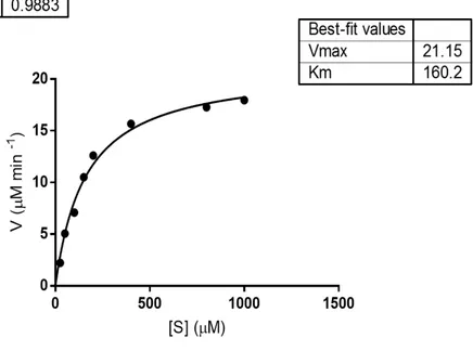
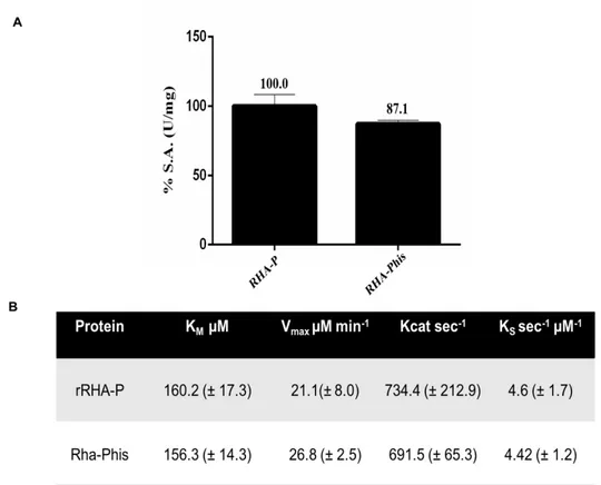
codificante per tale enzima è stato clonato in un vettore di espressione pET22b(+), e la proteina è stata espressa in maniera ricombinante in cellule di E. coli BL21(DE3). I primi tentativi di espressione hanno evidenziato la presenza della proteina nella sola porzione insolubile; di conseguenza, la temperatura di induzione è stata diminuita da 37 a 23 °C e la crescita batterica è stata effettuata in terreno LB ad alte concentrazioni di NaCl e in presenza di Betaina e Sorbitolo, due molecole che possono agire da “chaperon molecolari”, consentendo un miglioramento del folding della proteina stessa. La proteina è stata quindi espressa nelle condizioni ottimizzate e la purificazione è stata effettuata seguendo tre passaggi cromatografici, al termine dei quali la resa ed il fattore di purificazione risultavano però non soddisfacenti. La sequenza amminoacidica di rRHA-P, un monomero di 101,500 ± 5,000 Da, è stata verificata per spettrometria di massa. La successiva caratterizzazione dal punto di vista biochimico di rRHA-P, ha evidenziato una KM inferiore rispetto alle altre α-RHA
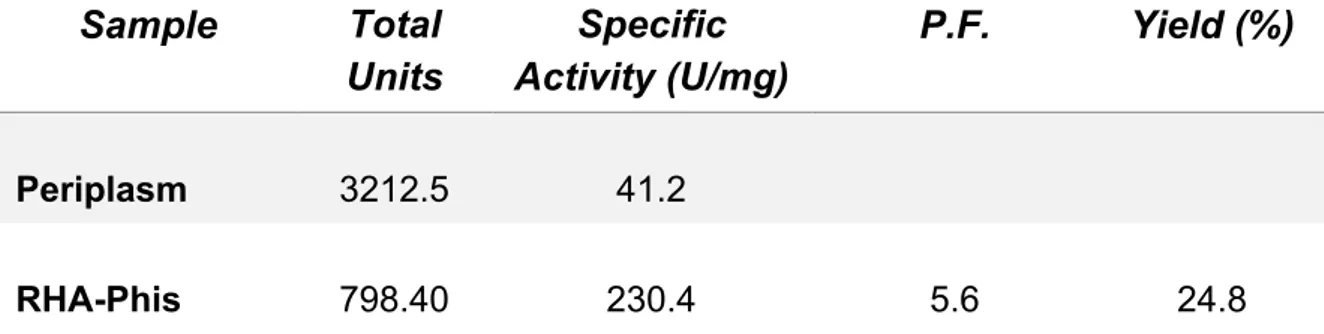
riportate in letteratura, suggerendo una maggiore affinità di rRHA-P per il substrato sintetico utilizzato. Per valutare l’effettivo potenziale biotecnologico di rRHA-P, sono state inoltre determinate le condizioni ottimali di attività enzimatica in diverse condizioni sperimentali. In particolare, rRHA-P presenta proprietà leggermente basofile con un’attività ottimale a pH 6.9 e a 40.9 °C, conservando una certa stabilità fino a 50°C. L’enzima possiede inoltre una moderata tolleranza ai solventi organici, quali etanolo e DMSO; i risultati ottenuti indicano che rRHA-P mostra diverse caratteristiche di interesse biotecnologico. È stata a tal proposito valutata la capacità di rRHA-P di idrolizzare alcuni flavonoidi naturali, la cui solubilità è più elevata in condizioni di pH basico, alte temperature e in presenza di solventi organici. I risultati ottenuti hanno mostrato che rRHA-P è in grado di convertire, dopo 3h di incubazione, di completamente la naringina nei corrispondenti prunina e ramnosio, e parzialmente rutina e neoesperidina. Tali dati supportano ulteriormente l’utilizzo dell’enzima in processi di bioconversione. Le analisi di spettrometria di massa ed il sequenziamento N-terminale effettuati sulla proteina rRHA-P purificata hanno evidenziato l’assenza dei primi 23 aminoacidi codificati dal gene, il che ha suggerito la presenza di un peptide segnale, successivamente allontanato in seguito a modifiche post-traduzionali della stessa. Tale peptide possiede delle caratteristiche peculiari e, da dati presenti in letteratura, sembra essere coinvolto nel “sorting” della proteina nello spazio periplasmatico. Tale ipotesi è stata confermata da esperimenti di espressione analitica, in cui la frazione periplasmatica evidenzia un’attività ramnosidasica 400 volte maggiore rispetto alla frazione citoplasmatica. Considerando la bassa resa di purificazione di rRHA-P, si è deciso di clonare e di esprimere in maniera ricombinante la proteina fusa con un his-tag, e di utilizzare, come primo step di purificazione, l’estrazione della frazione periplasmatica. Seguendo il protocollo appena descritto, è stato necessario un solo step di purificazione attraverso una cromatografia di affinità che ha permesso di ottenere, con una resa del 25%, una proteina molto pulita caratterizzata da una minima percentuale di contaminanti. Saggi di attività sulla proteina purificata non hanno mostrato una significativa influenza del tag di istidine nè sulla attività specifica, né sull’efficienza catalitica dell’enzima. Per implementare l’utilizzo biotecnologico di
8
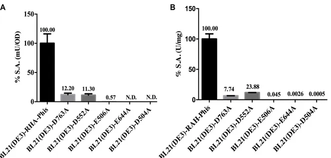
rRHA-P, sono stati effettuati esperimenti di mutagenesi per ottenere una preliminare identificazione degli ipotetici residui amminoacidici responsabili dell’attività catalitica dell’enzima. È stato quindi effettuato, in primo luogo, un allineamento della sequenza della proteina rRHA-P con sequenze di ramnosidasi della stessa famiglia GH106, già presenti in banca dati. Successivamente, individuati i 5 residui maggiormente conservati (D504, E506, D552, E644 e D763), si è proceduto con un approccio di mutagenesi sito specifica in cui i residui di interesse sono stati sostituiti con alanina ed è stata valutata la loro influenza sull’attività specifica di rRHA-P. Le mutazioni in posizione 504, 506 e 644 sembrano portare ad una totale inattivazione dell’attività catalitica, mentre le mutazioni in posizione 552 e 763 hanno mostrato una ritenzione dell’attività enzimatica di rRHA-P compresa tra il 6 e il 12%. Tali dati suggeriscono un
effetto differenziale dei residui amminoacidici individuati dall’allineamento e pongono le basi per futuri interventi di mutagenesi finalizzati al fine-tuning dell’attività enzimatica su substrati di interesse biotecnologico.
Isolameto di vescicole batteriche e eucariotiche
Per ottimizzare l’utilizzo degli enzimi come biocatalizzatori, oltre all’ingegnerizzazione, molto utile può essere la loro immobilizzazione su diversi tipi di supporti. In particolare, al fine di sviluppare supporti biocompatibili, sono stati recentemente studiati sistemi di immobilizzazione composti da membrane di origine biologica. Durante la seconda parte del mio periodo di dottorato, mi sono quindi occupata della caratterizzazione di due possibili sistemi di immobilizzazione, uno di origine batterica (Outer Membrane Vesicles OMVs) ed uno di derivazione eucariotica (Extracellular Vesicles EVs).
Isolamento e caratterizzazione di OMVs da Novosphingobium sp. PP1Y
Le OMVs sono state isolate da N. sp. PP1Y, un batterio gram-negativo non patogeno. Analisi di microscopia elettronica (SEM) e a forza atomica (AFM) hanno evidenziato, durante la crescita in terreno minimo, una significativa attività di vescicolazione in tarda fase esponenziale, evidenza che spiegherebbe la vescicolazione di N. sp. PP1Y come possibile strategia per l’acquisizione di nutrienti. Le OMVs identificate sono state quindi purificate attraverso una serie di ultracentrifugazioni e la loro analisi strutturale ha evidenziato una forma sferica e un diametro di ≈ 130-150 nm, in linea con le OMVs descritte in letteratura. Le OMVs da N. sp. PP1Y, risultano inoltre avere una peculiare composizione biochimica sia per quanto riguarda le proteine che per quanto concerne gli acidi grassi. L’analisi proteomica ha infatti evidenziato la presenza di una preponderanza di enzimi idrolitici rispetto alle cellule del ceppo PP1Y. Ancor più interessante risulta il fatto che la maggior parte delle proteine maggiormente presenti sulle OMVs, non lo è invece nelle cellule di PP1Y; in particolare le OMVs risultano avere, al contrario delle cellule, una elevata percentuale di proteine di membrana ed enzimi idrolitici ed una quasi totale assenza di enzimi deputati al metabolismo cellulare. Anche la composizione in acidi grassi sembra essere fortemente regolata nelle OMVs, che infatti presentano una grande percentuale di acidi grassi saturi, rispetto alla membrana delle cellule; questo aspetto suggerisce una specifica selezione degli acidi grassi durante la vescicolazione, che avverrebbe in zone della membrana batterica più “rigide” rispetto al resto della cellula.
I dati ottenuti incrementano fortemente l’interesse delle OMVs nel campo delle biotecnologie anche se un limite nel loro utilizzo è rappresentato dalla loro immunogenicità verso le cellule eucariotiche, dovuta alla presenza di LPS sulla membrana esterna. In questo contesto, N. sp. PP1Y sembra essere un buon candidato in quanto la sua membrane esterna non presenta LPS. Infatti, il trattamento di cellule
9
di cheratinociti umani con concentrazioni crescenti di OMVs, non ha mostrato una significativa diminuzione della vitalità cellulare rispetto al controllo; tale dato suggerisce una potenziale biocompatibilità delle OMVs per le cellule eucariotiche, che necessita chiaramente di ulteriori indagini effettuate su altre linee cellulari.
Isolamento e caratterizzazione di EVs da macrofagi
Una valida alternativa alle OMVs batteriche per il drug-delivery è rappresentata dalle vescicole isolate da cellule eucariotiche: EVs. Tali nanostrutture sono classificate, in base alla loro origine e dimensione, in Microparticelle (MPs), che hanno una grandezza di ≈ 400-2000 nm ed esosomi, di ≈ 50-200 nm. Tali vescicole originano da diversi tipi di cellule eucariotiche, il che le rende meno immunogeniche per le stesse e quindi più attraenti per il drug-delivery; inoltre, grazie alla presenza di specifici recettori di membrana, le EVs possono avere uno specifico cell-targeting che può aumentare l’efficienza di veicolazione di un farmaco. Nell’ultima parte del mio progetto di dottorato mi sono quindi occupata della caratterizzazione e della valutazione dell’effetto immunogenico di EVs isolate da macrofagi murini. Per l’isolamento delle EVs sono stati valutati differenti stimoli, tra cui l’utilizzo di LPS, acido oleico e acido palmitico, i quali incrementano la vescicolazione rispetto alle cellule non stimolate. Tali vescicole, dopo opportuna purificazione tramite ultracentrifugazione, sono risultate essere omogenee in taglia e conformi rispetto a quelle descritte in letteratura. La valutazione dell’effettivo utilizzo delle EVs in campo biotecnologico passa necessariamente attraverso la determinazione dell’effetto di tali nanostrutture sulle cellule eucariotiche. In particolare, negli ultimi anni è stata riscontrata una correlazione tra le EVs isolate da macrofagi e l’instaurazione di uno stato di infiammazione. Per tale motivo, nelle EVs isolate da cellule sottoposte a stimoli di vescicolazione, è stata valutata l’espressione di Nlrp3, un complesso proteico definito “infiammosoma” responsabile dell’attivazione della gran parte dei processi infiammatori. In particolare, la stimolazione dei macrofagi con LPS e acido palmitico aumenta significativamente l’espressione di Nlpr3, suggerendo un’effettiva differenza anche nell’effetto delle diverse EVs su altre cellule. In particolare, essendo le cellule vascolari le prime coinvolte nell’infiammazione, l’effetto pro-infiammatorio delle EVs, sia MPs che esosomi, è stato valutato su cellule muscolari lisce vascolari (VSMC). In questo modello sperimentale è stato evidenziato un significativo aumento dell’espressione della caspasi-1 attivata in seguito a trattamento delle VSMC con MPs ed esosomi isolate da macrofagi stimolati con LPS e acido palmitico, mentre si è osservato un aumento dell’espressione della caspasi-8 soltanto dopo trattamento con esosomi isolati da macrofagi stimolati con LPS e acido palmitico.
In conclusione, in questo lavoro di dottorato è stato messo a punto un sistema di espressione ricombinante e di purificazione di una nuova α-RHA, il cui potenziale biotecnologico è stato analizzato e discusso. Inoltre, questo progetto di dottorato ha permesso di caratterizzare due possibili sistemi di immobilizzazione e di veicolamento di attività enzimatiche e/o di piccole molecole organiche, di origine batterica (OMVs) ed eucariotica (EVs). Tutti i sistemi analizzati in questo progetto di dottorato risultano essere molto interessanti dal punto di vista biotecnologico e applicabili in diversi campi.
11
ABSTRACT
Biocatalysis represents a versatile and valuable tool for industrial biotechnologies. The use of enzymes as biocatalysts has reached its present industrial level, due to their optimal reaction selectivity, high reaction rates, high product purity, and a significant decrease in the generation of chemical waste. The use of enzymes in industrial applications has been limited by several factors, mainly the high cost of enzymes, their instability, and their availability in small amounts. To overcome these problems, the quest for new catalytic activities and the development of new technical approaches, such as enzyme immobilization, to improve their stability and practical applications, remain a central focus of the current biotechnological research. In this PhD thesis, the biotechnological potential of a novel bacterial glycosidase (α-RHA), and the characterization of two possible immobilization systems, bacterial OMVs and eukaryotic EVs is reported.
More in detail, an optimized expression and purification procedure allowed to characterize a novel α-RHA from the microorganism Novosphingobium sp. PP1Y, which resulted to be appealing from a biotechnological point of view for its interesting catalytic behaviour. Moreover, mutagenesis experiments, allowed a preliminary identification of the amminoacidic residues responsible for the catalytic activity of rRHA-P, which could be further mutagenized to fine-tune rRHA-P catalytic efficiency on selected substrates.
In addition, two different potential scaffolds from bacteria (OMVs) and from an eukaryotic cell line (EVs) were isolated and characterized. Both systems resulted to be appealing either for enzyme immobilization or for drug-delivery strategies. In particular, OMVs isolated from N. sp. PP1Y were characterized by a peculiar biochemical composition, which showed some differences with the originating whole cells. EVs isolated from human macrophages resulted to have differential effects on inflammation activation, and their potential as a valid alternative to bacterial OMVs for the development of novel delivery Biosystems is discussed.
13
ABBREVIATIONS
α-RHA = α-L-rhamnosidase ABS = absorbance
AFM = atomic force microscopy BSA = bovin serum albumin DLS = dynamic light scattering
DMEM = Dulbecco modified Eagle’s medium DMSO = dimethyl sulfoxide
EtOH = ethanol
EVs = extracellular vesicles
GC-MS = gas chromatography – mass spectrometry GFP = green fluorescent protein
GHs = glycosyl hydrolases GTs = glycosyl transferases
IPTG = isopropyl β-D-1-thiogalactopyranoside LB = Luria Bertani
LC-MS = liquid chromatography – mass spectrometry LPS = lipopolysaccharide
MOPS = morpholinopropansulphonic acid MPs = microparticles
MUFA = monounsaturated fatty acid NTA = nano tracking particles analysis OA = oleic acid
OD = optical density OM = outer membrane
OMPs = outer membrane proteins OMVs = outer membrane vesicles ORF = open reading frame
PA = palmitic acid
PAHs = polycyclic aromatic hydrocarbons PBS = phosphate sulphate buffer
PLs = phospholipids
pNPR = para-nitrophenyl rhamnopyranoside ppb = parts per billion
PPMM = potassium phosphate minimal medium PUFA = polyunsaturated fatty acid
RT = room temperature
SDS = sodium dodecyl sulphate SEM = scanning electron microscopy SFA = saturated fatty acid
TLC = thin layer chromatography TLR4 = Toll-like receptor 4 UFA = unsaturated fatty acid
15
CHAPTER I
INTRODUCTION
17
CHAPTER I
INTRODUCTION
1.1 Biocatalysis and glycosidases
Biocatalysis represents nowadays a versatile and valuable tool for industrial biotechnologies. The use of enzymes as biocatalysts gives invaluable advantages over conventional chemical technologies, for achieving high reaction selectivity, higher reaction rate, improved product purity, and a significant decrease in chemical waste generation. More in detail, in the fine chemical and pharmaceutical industries several advantages can be obtained from the use of biocatalytic processes, such as:
- reduction of processing steps (improved productivity and lower costs);
- transfer of processes from organic solvents to water (lower emissions, saving on raw materials and waste treatment);
- use of eco-friendly mild reaction conditions (avoiding very high or very low temperatures, and therefore heating/refrigeration costs).
Biocatalysts can be performed either with whole cells or purified enzymes, in solution or immobilized.
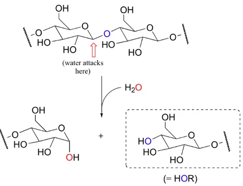
Glycosidases are a class of enzymes particularly suited for biotechnological applications. These enzymes catalyse the hydrolysis of the glycosidic linkage, leading to the formation of a sugar hemiacetal or hemiketal and the corresponding free aglycon [1] (Fig. 1).
Figure 1. Glycosidases reaction mechanism.
Glycosidases (GH) are present in almost all living organisms [2] where they play different roles. Based on the amino acid sequence and folding and the diversity of the reactions catalysed, glycosidases have been classified in many different ways.
18
Recently, a new type of classification was proposed based on the amino acid similarity within the proteins; this classification is available in the Carbohydrate-Active Enzymes database (CAZy - http://www.cazy.org/) [2] and provides a direct relationship between sequence and folding similarities, which can be found in 130 amino acid sequence-based families. In general, GHs belonging to the subfamilies of the same family share a common ancestor, a similar 3D structure and are characterized by an identical catalytic mechanism [2].
Despite all their natural functions, glycosidases are nowadays considered an attractive target for food, paper and pulp industries, as well as in organic chemistry, where glycosidases have proven to be efficient catalysts, able to hydrolize very stable glycosidic bonds in glycoconjugates, oligo- and poly-saccharides [3]. For this reason, glycosidases have wide and documented applications in several fields of biotechnology. Classical examples of industrial application of these enzymes include detergent formulations (cellulases and amylases) for the removal of glycan spots and fading of the denim color [4] pulp and paper bleaching (xylanases) [5], conversion of lignocellulosic plant biomass into bioethanol (cellulases, glucosidases, xylanases, etc. [6], production of High Fructose Corn Syrup for soft drinks (amylases and glucoamylases) [7], fruit juice processing (pectinases) [8], and many others. Glycosidases have recently attracted the attention of many pharmaceutical industries since they are involved in many biological processes such as cell-cell or cell-virus recognition, immune responses, cell growth, and viral and parasitic infections [9]. In addition to the hydrolytic ability, GHs can also be used under appropriate conditions for the reverse reaction, thus promoting the formation of glycosidic linkages. These reactions are called transglycosylations [10] and generally require high substrate concentration. In this case, the glycosyl donor can be a monosaccharide, an oligosaccharide or an activated glycoside. This specific action of glycosidases, which work either as hydrolases or in the glycosynthetic mode [11], are of great interest for the production of functional foods and drugs, whose biological activities might be improved by either the removal or the addition of specific glycan moieties [12]. In conclusion, glycosidases are biotechnologically attractive for the preparation of structurally well-defined oligosaccharides compared to chemical processes that require instead, complex protection and deprotection steps.
Continuous progress in the study of these enzymes and the application of molecular evolution and site-directed mutagenesis for better performance, is currently improving their potential use in oligosaccharide synthesis [13].
1.2 α-L-rhamnosidases
α-L-Rhamnosidases (α-RHAs) are a subset of GHs that have gained much attention in recent years. These enzymes catalyse the hydrolysis of a terminal L-rhamnose from a large number of natural products [14].
L- Rhamnose is widely distributed in plants as component of flavonoid glycosides, terpenyl glycosides, pigments, signalling molecules, and in cell walls as a component of complex heteropolysaccharides, such as rhamnogalacturonan and arabinogalactan-proteins [15-18]. In bacteria, L-rhamnose appears to be included in membrane rhamnolipids [19-20] and polysaccharides [21]. According to the GHs classification, α-RHAs are grouped in the CAZy (carbohydrate-active enzymes) database (www.cazy. org) into four different families: GH28, GH78, GH106, and NC (non-classified).
α-RHAs are produced by a large number of animal tissues, plants, fungi, bacteria and bacteriophages [16], but their physiological role is still not well understood. The
19
hypothesis can be advanced that the role of α-RHAs is linked to the broad distribution of L-rhamnose as a component in bacterial and plant cell walls, glycosides, biofilms and glycolipids [16,22].
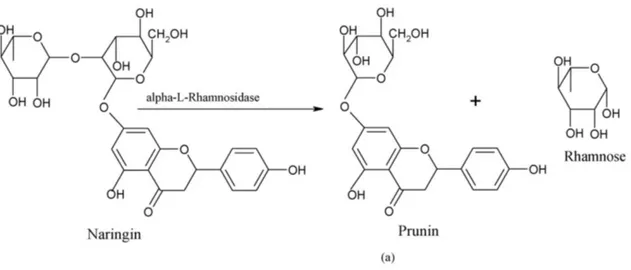
In the last decade, α-RHAs have attracted a great deal of attention due to their potential application as biocatalysts in a variety of industrial processes and in particular in the food industry [1]. Among α-RHAs substrates, particularly interesting results natural flavonoids (Fig. 2), polyphenolic compounds generally characterized by a three-ring structure, which consists of two phenyl rings (A and B) and a heterocyclic ring (C). These molecules are naturally produced in plants in glycosylated forms, showing the presence of either a rutinoside (6-α-l-rhamnosyl-β-d-glucose) or a neohesperidoside (2-α-l-rhamnosyl-β-d-glucose) disaccharidic unit bound in different positions. These molecules are intriguing due to their potential antioxidant, antitumor and anti-inflammatory properties [23-24]. In particular, naringin, hesperidin and rutin, flavanone glycosides found in grapefruit juices, lemons, sweet oranges and vegetables, have gained increasing recognition for their potential antioxidant, antitumor and anti-inflammatory properties [25-28]. In humans, endogenous β-glucosidases are responsible for removing in the small intestine glucose (or possibly arabinose or xylose) moiety from flavonoids thus allowing their effective absorption; these enzymes, however, are not able to cleave terminal rhamnose units, thereby limiting the bioavailability of rhamnosylated flavonoids that are further converted by the colon local microflora [29-30]. Therefore, enzymatic rhamnose removal from potentially bioactive flavonoids might be the key point to improve their intestinal absorption and their beneficial properties on human health [31-32]. In addition, the ability to hydrolyze glycosylated flavonoids has been used, in the industrial field, to mitigate the bitterness of citrus juices, which is primarily caused by naringin [33-34]. Moreover, the corresponding de-rhamnosylated compound, prunin, is endowed with antimicrobial properties [28], and shows an improved intestinal assimilation when compared to naringin. Other applications of α-RHAs are gaining popularity in the oenological industry, where these enzymes are used to hydrolyze terpenyl glycosides and enhance aroma in wine, grape juices and derived beverages [35-37].
Figure 2. Hydrolysis of naringin to prunin by α-L-rhamnosidase in the corrisponding product: prunin (a)
20
Moreover, in their glycosynthetic mode of action α-L-rhamnosides can “decorate” flavonoids in fruit juices and wine, antibiotic and antitumoral drugs [38].
It is worth to note that the use of an α-RHA for the synthesis of rhamnose-containing chemicals by reverse hydrolysis was recently reported, suggesting a yet unexplored potential of these class of enzymes in the chemical and pharmaceutical industry [39]. Despite their potential as industrial biocatalysts, only a limited number of bacterial rhamnosidases have been extensively studied and characterized [40-44]. In fact, commercial preparations of α–RHAs, naringinases and hesperidinases available and currently used in oenology, are all isolated from fungal sources (Aspergillus niger and
Penicillium decumbens) [45-47].
Different features of these two families of enzymes, especially pH optimum, suggest different applications for fungal and bacterial enzymes. As an example, fungal enzymes show more acidic pH optima compared to bacterial counterparts, for which neutral and alkaline optimal pH values have generally been reported [48-49]. This feature suggested that bacterial α-RHAs might be considered, for example, an alternative source of biocatalysts to use in basic conditions for the biotransformation of flavonoids such as naringin or hesperidin, whose solubility strongly increases at higher pH value [48-49]. Most importantly, few attempts have been described so far to engineer bacterial α-RHAs to unravel the molecular details concerning their catalytic mechanism, to modify their substrate specificity or to improve their catalytic efficiency towards different substrates [50]. A major stumbling is represented by the limited number of α-RHAs crystal structures currently available, with a consequent lack of structural data of these enzymes. To date, only two bacterial α-RHAs belonging to the GH106 family were described, one from Sphingomonas paucimobilis FP2001 [51] and the α-RHA BT0986 from Bacteroides thetaiotaomicron [52]. Moreover, only five crystal structures of bacterial RHA have been solved so far, which include the putative α-RHA BT1001 from Bacteroides thetaiotaomicron VPI-5482 [53], the α-α-RHA B (BsRhaB) from Bacillus sp. GL1[54], α-RHA (SaRha78A) from Streptomyces
avermitilis [55], and α-RHA (KoRha) from Klebsiella oxytoca [56]. All these enzymes
belong to the GH78 group. Only one example of a GH106 α-RHAs crystal structure was recently reported and in the α-RHA BT096 expressed by Bacteroides
thetaiotaomicron [52].
Therefore, it is evident that bacterial α-RHAs represent a yet unexplored reservoir of potential biocatalysts for which more functional and structural data are required.
1.3 Enzyme immobilization
The use of enzymes in industrial applications has been limited by several factors, mainly the high cost of the enzymes, their instability, and availability in small amounts. In addition, enzymes are soluble in aqueous media and it is difficult and expensive to recover them from reactor effluents at the end of the catalytic process. These limits the use of soluble enzymes to batch operations, followed by disposal of the spent enzyme-containing solvent [57]. Over the last few decades, intense research in the area of enzyme technology has provided many approaches that facilitate their practical applications. Among these, technological developments in the field of immobilized biocatalysts resulted to be a very powerful tool to improve almost all enzyme properties, such as stability, activity, specificity selectivity, and enzyme inhibition [58]. Enzyme immobilization offers the possibility of a wider and more economical exploitation of biocatalysts in industry, waste treatment, drug delivery, and bioprocess monitoring devices, like biosensors [57]. Besides the application in industrial processes, immobilization techniques could be the basis for the developing a large
21
number of biotechnological devices with applications in diagnostics and biosensors [59].
Immobilization means associating the biocatalysts with an insoluble matrix, so that it can be retained in a proper reactor geometry for its economic reuse under stabilized conditions (Fig.3) [60]. Immobilization helps in the development of continuous processes allowing more economic organization of the operations, automation, and investment/capacity ratio. Immobilized biocatalysts offer several other advantages such as, for example, the availability of the product in greater purity. Purity of the product is crucial, for example, in food processing and pharmaceutical industry since contamination could cause serious toxicological, sensory, or immunological problems [60].
Another major advantage includes greater control over enzymatic reaction, as well as high volumetric productivity, with lower residence time, which are of great significance in the food industry [57]. Moreover, compared to their free forms, immobilized enzymes are generally more stable and easier to handle [57]. In addition, reaction products are not contaminated with the enzyme (especially useful in the food and pharmaceutical industries), and in the case of proteases, the rate of the autolysis process can be dramatically reduced upon immobilization [61].
To date, more than one hundred techniques to immobilize enzymes have been developed, such as entrapment, adsorption, ionic binding, and disulphide bonds formation. Enzymes can be attached to the support by interactions, ranging from reversible physical adsorption and ionic linkages to stable covalent bonds (Fig.3) [62]. Ideal support properties include physical resistance to compression, hydrophilicity, inertness toward enzymes ease of derivatization, biocompatibility, resistance to microbial attack, and availability at low cost [62-64]. Supports can be classified as inorganic and organic according to their chemical composition, and organic supports can be subdivided into natural and synthetic polymers [65]. The most common are synthetic polymers, biopolymers (cellulose, starch, agarose, carragenans, and chitosan), inorganic supports (alumina, silica, zeolites), hydrogels and so on.
In this framework, nanobiotechnology is gaining much attention from the scientific community, and “Nanobiocatalysis” is one direct application of this growing field [66]. In early approaches to nanobiocatalysis, enzymes were immobilized on various nanostructured materials using conventional approaches, such as simple adsorption and covalent attachment. This approach was used by immobilizing enzymes onto nanostructured materials, such as nanoporous materials and magnetic nanoparticles [67]. Nanosized particles of noble metals, for example, were used due to their attractive electronic, optical, and thermal properties as well as catalytic properties and potential applications in the fields of physics, chemistry, biology, medicine, and material science [68]. Recently, new immobilization studies were performed by using, as immobilization systems, artificial or natural lipid bilayers of biological membranes. Lipid vesicles (liposomes) have been recently used for this purpose, despite the fact that the long-term stability of liposomes may be problematic [69]. Lipid vesicles, generally, are not thermodynamically stable and do not assemble ‘spontaneously’ (without input of external energy); they are only kinetically stable, and their physical properties may depend on how and under which conditions lipid vesicles have been prepared. To overcome all these limitations, a recent interest for the immobilization of enzymes and biocatalysts in general focuses on the isolation of bilayer membranes from both prokaryotic and eukaryotic sources.
22
http://www.easybiologyclass.com Figure 3. Enzyme immobilization examples.
In this context, extracellular nanostructures, such as bacterial Outer Membrane Vesicles (OMVs) and eukaryotic Extracellular Vesicles (EVs), proteoliposomes of 20-2000 nm diameter, have been recently investigated as biotechnological scaffolds alternative to liposomes [70-71].
23
1.3.1 Outer Membrane Vesicles
Outer membrane vesicles (OMVs) are nanoscale proteoliposomes of 20-200 nm
diameter, naturally derived from the surface of some Gram-negative and Gram-positive bacteria as part of their natural growth cycle. In Gram-negative bacteria, OMVs are initially formed as a bulge arising from the outer membrane (Fig 4) [70]. Thus, they are generally surrounded by a single phospholipidic bilayer derived from the outer membrane and are primarily composed of lipopolysaccharides, membrane phospholipids (PLs) and outer membrane proteins (OMPs) [70-73]. In addition, a portion of periplasmic space is often enclosed in these vesicles, which includes proteins, toxins or effectors that are delivered to the environment to accomplish several biological functions (Fig 4).
The production of OMVs has been reported from a large number of Gram-negative bacteria, [73-75] in a variety of environments, including planktonic cultures, fresh and salt water, biofilms, inside eukaryotic cells and within mammalian hosts [76-78]. Among OMV-producing Gram-negative bacteria, pathogenic microorganisms such as
Escherichia coli, Helicobacter pylori, Pseudomonas aeruginosa, have been so far
major targets for OMV studies [79-81]. The main interest towards pathogenic-OMVs was driven from their cargo proteins, identified as major virulence factors or agents of inflammatory responses. Some examples include E. coli cytolysin A (ClyA), E. coli heat labile enterotoxin (LT) [82]. However, OMVs functions are far from being limited to pathogenicity, and the role of these carrier structures has proven to be far more complex than what initially supposed.
http://genesdev.cshlp.org/content/19/22/2645/F1.large.jpg Figure 4. OMVs example. LPS: lipopolysaccharide; OM: outer membrane; Pp: periplasm; Cyt: cytosol;
IM: inner membrane.
Indeed, it is currently well known that OMVs are also involved in interspecies communication and competition, biofilm formation, DNA lateral gene transfer, antibiotic resistance and nutrient acquisition [79,83-87]. This system allows signalling proteins
24
or effectors to be delivered, even over great distances, at high concentration in close proximity of an acceptor site. Ultimately, the binding specificity between surface-exposed bacterial adhesins and environmental ligands or receptors guarantees a high target specificity. Several hypotheses on OMVs biogenesis have been proposed, although they are not fully comprehensive and often restricted to a few species of bacteria [74-75, 86, 88]. However, OMV production occurs at a constitutive level for a wide variety of bacteria, suggesting that this is a highly conserved process [74-75, 86]. Furthermore, vesiculation levels can be altered by factors such as temperature, nutrient availability, oxidation, quorum sensing and envelope- targeting antibiotics [86-87, 89]. So far, only few examples of OMVs isolated from non-pathogen gram-negative bacteria have been reported. Some examples of non-pathogenic, environmental bacteria from which OMVs have been isolated include the halophilic marine bacterium
Novosphingobium pentaromativorans US6-1, and the soil bacterium Pseudomonas putida K2440 [90-91]. These environmental OMV-producing bacteria are endowed with
the ability to use various aromatic compounds, including polycyclic aromatic hydrocarbons (PAHs) and several other pollutants, as major carbon and energy sources. Little is still known about the vesicles produced by these organisms, however two main roles of these OMVs have been suggested. The first one involves the use of OMVs as carriers of hydrolytic enzymes, such as proteases and glycosidases, which play a role in the acquisition of nutrients from the surrounding environment [18]. This system would improve nutrient acquisition and thus bacterial survival, particularly in starving growth conditions [71, 90- 93]. The second hypothesis suggests that these structures might be involved in i) the formation of the biofilm matrix [20], ii) the onset and maintaining of biofilm communities [71, 83, 88, 93] and iii) the antibiotic resistance expressed by the biofilm bacterial community. Beyond a role in biofilm onset, OMVs might mediate interactions within and external to the biofilm through OMV-associated quorum-signalling molecules [88, 93-94].
In recent years, a great deal of attention was also devoted to the study of OMVs for their potential use in biotechnological industry. Drug delivery, enzyme immobilization and construction of innovative biosensors are some of the major research fields of interest. Bacterial OMVs can be foreseen as platforms for the immobilization of proteins and enzymatic complexes and be employed as carriers for drugs and molecules, allowing these latter to be conveyed to a highly specific target site and in a protected environment [95-98]. By taking advantage of the unique feature to embed outer membrane proteins into OMVs, a wide range of functional proteins such as green fluorescence protein (GFP) and β-lactamase, have been genetically tethered to the surface of a hyper-vesiculating Escherichia coli strain- therefore to the corresponding OMVs- using the virulence factor cytotoxin ClyA as the surface anchor [95]. Unlike complex enzyme assembly onto liposomes or polymerosomes, these results indicate the feasibility of designing OMVs as synthetic nanoreactors, using only standard molecular biology techniques. This approach has been recently used for example, to immobilize three glycosidase activities on engineered E. coli OMVs [98]. A trivalent protein scaffold was genetically tethered onto the OMVs to enable the positional specific recruitment of three different cellulases. This work reported that the assembled enzyme complex, not only retained full activity, but also hydrolyzed cellulose 23-fold faster than non-complexed enzyme [98]. The biotechnological use of OMVs isolated from gram-negative bacteria, for example as drug delivery systems, is currently limited, among others, by the presence in their outer membrane of the immunogenic LPS. Several studies have in fact demonstrated how LPS is sensed by the Toll-like receptor 4 (TLR4) complex, triggering a pro-inflammatory response and playing an important
25
role in septic shock [99]. As a consequence, much interest is currently devoted to vesiculating non-pathogen strains that do not produce LPS.
1.3.2 Extracellular Vesicles
Extracellular vesicles (EVs) are membrane-derived particles with a diameter size ranging from 30 nm to 2,000 nm, surrounded by a (phospho)lipid bilayer and released by cells in the human body [100]. EVs s are classified, based on their cellular origin, biological function or biogenesis, in three main groups: exosomes, microvesicles and apoptotic bodies [101]. Distinct processes responsible for vesicle release from cells have been identified; in particular, exosomes are derived from the multivesicular endosomal cell compartment[71. 101-103] whereas EVs originate by direct budding from the cell plasma membrane [102-106] Both extracellular vesicle types contain cytoplasmic proteins, certain lipid raft-interacting proteins and RNAs but, owing to their highly regulated biogenesis, exosomes typically accommodate some additional defined components [71]. Apoptotic bodies represent another type of membrane-limited vesicle. These are larger than exosomes and EVs [108] and are formed exclusively during the late stage of apoptosis (Fig. 5).
Within the past decade, extracellular vesicles have gained attention as important mediators of intercellular communication [108].
Shedding of EVs is considered to be a physiological phenomenon that leads cell activation and growth. Many stimuli have been shown to increase vesicle secretion, including hypoxia, oxidative stress and inflammation [109-111]. These evidences
suggested that EVs are implicated in physiological responses such as immune surveillance [112], blood coagulation [113], tissue repair [114] or in pathological disorders, for example cancer [115] and cardiovascular diseases [116].
Yamamoto S, Azuma E, Muramatsu M, Hamashima T, Ishii Y, Sasahara M. (2016) Significance of extracellular vesicles: pathobiological roles in disease. Cell Struct Funct
26
EVs could also carry and deliver multiple information through lipids, proteins or nucleic acids transfer. Proteins sorting into shedding vesicles is selective; specific proteins may be included or excluded from membrane EVs, leading to the expression of proteins arrays that differ from those present on the surface of the cells from which they originated [117-118]. It is also possible that EVs may interact with specific target cells after their release. For example, EVs shed from platelets interact with macrophages and endothelial cells but not with neutrophils, [119] whereas EVs from neutrophils interact with platelets, macrophages, and dendritic cells [120-121]. Due to their specific targeting, the current biotechnological interest in EVs derives from their great potential as a new way, for innovative therapies, to deliver drugs to specific target cells [115]. Thus, EVs can be engineered to over-express different therapeutic proteins, mRNA or miRNA - by driving their synthesis from the relevant EV producing cells [122]. Indeed, EVs possess a lot of peculiarities that give them ideal drug-delivery features. More specifically, EVs are stable within the body. EVs do not induce (ii) tumour generation [123], or (iii) immune rejection after allogenic administration [124]. In addition, EVs carrying proteins and/or nucleic acids can be easily modulated to confer them specific pharmacological functions [125].
EVs have been also studied, in particular from macrophages, for the identification of new potential therapeutic targets associated to inflammatory response and for the development of a new drug delivery strategy. All these features suggest that EVs, as well as bacterial OMVs, could be used for biotechnological purposes. The interest in using eukaryotic EVs rather than OMVs, lies in their specific drug delivery application. Indeed, the fact that EVs derives from eukaryotic cells, could make these latter less immunogenic than OMVs. Moreover, their composition can be physiologically determined or driven by exogenous modulations either on their origin cells or directly on purified EVs populations [126]. Another advantage in using EVs as novel natural bio-carriers, lies in their specific cell-targeting, which has already been described in literature [127-129]. For this reason, EVs appear to be a suitable alternative to traditional delivery systems currently in development, limiting inflammation processes and improving target-specific delivery.
1.4 Novosphingobium sp. PP1Y: a novel microbial source of OMVs and glycosidases
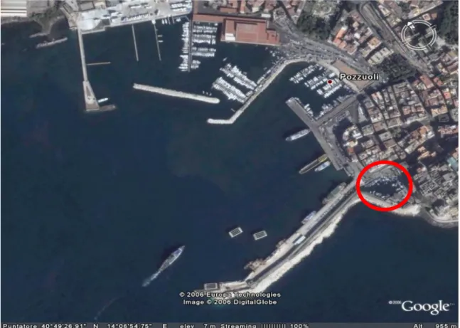
In the laboratory in which my PhD project has been developed, a novel gram-negative strain has been isolated and characterized, named Novosphingobium sp. PP1Y [99,120]. The microorganism was isolated from surface waters of a small dock bay in the harbour of Pozzuoli which is used for the storage of small boats and is characterized by a severe pollution of the water by mono-, poly- and heterocyclic aromatic hydrocarbons (Fig. 6)
The microbiological analysis of the bacterium indicated that the microorganism belongs to the order of Sphingomonadales, a class of bacteria whose outer membrane is characterized by the presence of glycosphingolipids, instead of the more common lipopolysaccharides.
This peculiarity makes the surface of their cells more hydrophobic than those of the other Gram-negative strains and has probably contributed to the ability of these microorganisms to degrade mono- and polycyclic aromatic hydrocarbons (PAHs). N.
sp. PP1Y, however, shows different characteristics if compared to other members of
the order, as it appears not only to be able to grow using, as the sole source of carbon and energy, a wide range of mono-and polycyclic aromatic substrates such as pyrene,
27
naphthalene and phenanthrene, but also to have evolved an effective adaptation to the growth on complex mixtures of aromatic molecules dissolved in non-polar phases such as diesel and gasoline.
Due to its peculiar natural environment and metabolism, this strain might be considered a valuable reservoir of novel biotechnological tools.
In particular, Sphingomonadales show the presence of a great abundance of both glycosyl hydrolases (GHs) and glycosyltransferases (GTs), activities that are probably involved in the biosynthesis of complex extracellular polysaccharides and microbial biofilms [130].
The interest for Novosphingobium sp. PP1Y carbohydrate-active enzymes has grown as the sequencing and annotation of the whole genome, recently completed, has revealed peculiar biochemical and biotechnological properties, namely some metabolic pathways specifically involved in the degradation of several aromatic hydrocarbons, and the resistance to toxic compounds. A great number of genes encoding for both GHs (53 orfs) and GTs (57 orfs) were identified in this microorganism [99]. In particular, the number of GHs is remarkable when compared, respectively, to the 16 GHs and the 32 GHs of the closely related strains of Sphingomonas wittichii and Sphingobium chlorophenolicum. The 53 GHs of strain PP1Y are distributed among 27 different families. The most represented families are GH3, GH13 and GH23 with 9, 5 and 9 members respectively. Among these, there are 8 genes encoding for α-RHAs. A novel α-RHA activity in N. sp. PP1Y crude extract was recently described, which was used, among others, for bioconversion experiments on substrates such as naringin, rutin and hesperidin [131].
Strain PP1Y shows a very high propensity to adopt the sessile phenotype and efficiently colonise several types of hydrophobic surfaces including water/oil interfaces (Fig. 7) [99]. In addition to these features, strain PP1Y has a surprisingly high tolerance to diesel oil, which allows its growth in biphasic diesel oil/water cultures containing more oil than water. To the best of our knowledge, such behaviour has never been reported for a Sphingomonas. Sphingomonas sp. Ant 17, the sole Sphingomonads known to use fuels as a sole carbon and energy source, has been reported to grow in 1:1000 (v/v) fuel/water biphasic systems [131]. Several details suggested also the production of OMVs in strain PP1Y, which might play a potential role in biodegradation and nutrient supply in natural habitats, as well as being involved in cell-cell communication and genetic transfer. N. sp. PP1Y can grow as either planktonic free cells or sessile-aggregated macroscopic “flocks” of 1-10 mm long, with a composition in fatty acids similar to the cell membrane (Izzo V., unpublished results); noteworthy, this behaviour has already been described for other Sphingomonads, which show the so-called “planktonic/sessile dimorphism” [132-133]. Most importantly, cell free supernatants from cultures of N. sp. PP1Y were able to effectively stabilize paraffin drops, obtained by shaking a paraffin/water biphasic system, which remained stable for more than 1 week. These last data in particular suggested that strain PP1Y produced an extracellular emulsifier, and it was shown that secretion was dependent upon the presence of neither aromatic hydrocarbon nor an oil phase in the growth medium [133]. Interestingly, culture supernatants showed a low total carbohydrate level and no detectable polysaccharides [134].
Thus, N. sp. PP1Y might be particularly appealing for OMVs isolation and characterization in order to shed light on vesiculation mechanisms related to environment adaptation. Noteworthy, the lack of LPS on the membrane surface of strain PP1Y, and the availability for this microorganism of a completely sequenced and
28
annotated genome [134], are extremely appealing to develop a novel molecular tool for drug delivery systems and enzyme immobilization.
During my PhD work, specific aspects of the biotechnological potential of N. sp. PP1Y were investigated, focusing the attention on the use of this microorganism to isolate and characterize novel examples of i) α-RHA activities and ii) OMVs for industrial applications and biotechnological purposes.
29
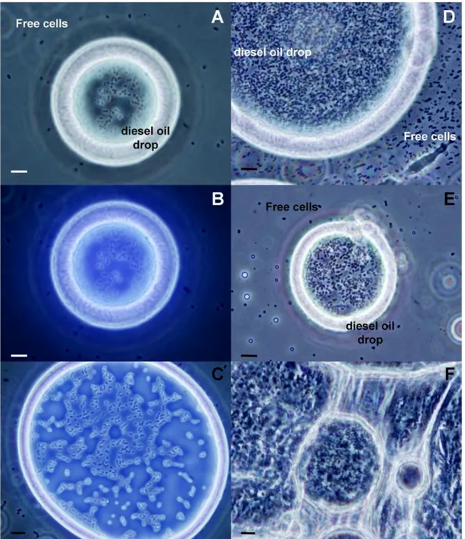
Figure 7. Phase contrast microscopy analysis of coated diesel oil drops isolated from a culture of
Novosphingobium sp. PP1Y grown using diesel oil as sole carbon and energy source. A) and B) Coated diesel oil drop after 2 days of incubation observed using visible light only or UV/visible light, respectively. C) Coated diesel oil drop after 2 days of incubation observed using UV/visible light. D) and E) Coated diesel oil drops after 10 days of incubation. F) Super-aggregates of coated drops. Bar = 10 μm.
31
AIMS OF THE THESIS
Both α-L-rhamnosidases and extracellular vesicles are elements of great interest for the development and implementation of novel biocatalysis processes of biotechnological importance. A great deal of attention is being devoted by the biotech community to the use of these two systems for applications in the food and pharmaceutical industries. Main aim of this PhD project has been to characterize the biotechnological potential of a bacterial α-L-rhamnosidase isolated from
Novosphingobium sp. PP1Y, and of novel examples of vesicles, Outer Membrane
Vesicles (OMVs) isolated from the same microorganism and Extracellular Vesicles (EVs) from macrophages.
This general aim has been pursued through the following activities:
1. Purification and characterization of a novel α-L-rhamnosidases from N. sp. PP1Y; 2a. Isolation, purification and characterization of OMVs from N. sp. PP1Y;
2b. Isolation, purification and characterization of Extracellular Vesicles (EVs) from human macrophages.
Tasks 1 and 2a were performed in the laboratory of Professor Alberto Di Donato, university of Naples, Federico II. The 2btask was developed, starting September 2016 through March 2017, at INSERM (University of Angers, France) in the laboratories of the “Stress oxidant et patologies mètaboliques U1063” of Prof. Ramaroson Andriantsitohaina.
33
CHAPTER II
35
CHAPTER II
MATERIALS AND METHODS
2.1 Generals
N. sp. PP1Y was isolated from polluted seawater in the harbor of Pozzuoli (Naples, Italy) as already described []. Bacterial growth was followed by measuring the optical density at 600 nm (OD/mL, from now on referred to as OD600). Nanosize analysis for
the determination of OMVs size was performed by IZON technical service using qNANO GOLD (http://www.izon.com). Protein quantification was performed using BCA kit assay purchased from Thermo Scientific.
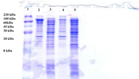
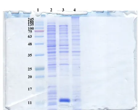
Protein concentration was measured using the Bio-Rad Pro- tein System [52] using bovine serum albumin (BSA) as standard. Polyacrylamide gel electrophoresis was carried out using standard techniques [53]. SDS–PAGE 15% Tris–glycine gels were run under denaturing conditions and proteins were stained with Coomassie brilliant blue G-250. “Wide range” (200–6.5 kDa) molecular weight standard was from Sigma (ColorBurstTM Electrophoresis Marker).
2.2 Cloning of orf PP1YRS05470 and construction of the pET22b(+)/rha-p expression vector.
Genomic DNA was extracted from a 50mL saturated culture of N. sp. PP1Y as described elsewhere [130]. OrfPP1Y RS05470 coding for the α-RHA activity was amplified in two contiguous fragments, owing to the considerable length of the orf (3441 bp). The first fragment, named rha-up (1816 bp), was amplified using an internal downstream primer, RHA-Intdw (5′- AGGCGGCCATGGGAATGT-3′), which included an internal NcoI site already present in orfPP1Y RS05470, and an upstream primer, RHA-up (5’-GGGAATTCCATATGCCGCGCCTTTCGCT-3’), designed to add a NdeI restriction site at 5’ of orf PP1Y RS05470. The second half to the gene, named rha-dw (1625 bp), was amplified using the upstream primer RHA-Intup (5’-ACATTCCCATGGCCGCCT-3’), complementary to RHA-Intdw, and the downstream primer RHA-dw (5’-AAAACCGAGCTCTCAATGCCCGCCCGTG-3’) that was intended to incorporate a SacI restriction site downstream of the amplified orf. The amplified fragments, rha-up and rha-dw, were purified from agarose gel, digested, respectively, with NdeI/NcoI and NcoI/SacI, and individually cloned in pET22b(+) vector previously cut with the same enzymes. Ligated vectors were used to transform E. coli, strain Top10 competent cells. The resulting recombinant plasmids, named pET22b(+)/rha-up and pET22b(+)/rha-dw, were verified by DNA sequencing. Next, the construction of complete rha-p gene in pET22b(+) was performed. First, both pET22b(+)/rha-dw and pET22b(+)/rha-up were digested with NcoI/SacI restriction endonu- cleases to obtain, respectively, fragment rha-dw and linearized pET22b(+)/rha-up. Digestion products were purified from agarose gel electrophoresis, eluted and ligated. Ligation products were used to transform E. coli Top10 competent cells and the resulting recombinant plasmid, named pET22b(+)/rha-p was verified by DNA sequencing.
2.3 Construction of the pET22b(+)/rha-his expression vector.
To obtain the pET22b(+)/rha-his expression vector, which expresses a recombinant
RHA-P fused to a C-terminus His-tag, a single point mutagenesis (TGA to CGA) on the STOP codon of the coding sequence of RHA-P, previously cloned, was performed. In this way, an arginine residue was inserted and the coding sequence was extended on the pET22b(+) plasmid, thus including a linker sequence and the coding region for
36
a 6-His-tag domain at 3’of the gene. The plasmid pET22b(+)/rha-dw, bearing only the C-terminus 1,625 bp-long fragment of the gene and already present in our laboratory, was chosen to perform the mutagenesis, in order to use a smaller plasmid for the amplification procedure. The mutagenesis experiment was done using specific
designed complementary primers named RHAmutUP
(5’ACCACGGGCGGGCATCGAGCCGTCGACAAGC3’) and RHAmutDW
(5’GCTTGTCGACGGCTCGATGCCCGCCCGTGGT3’) containing the desired mutated codon. Quickchange II site directed mutagenesis kit (Agilent technologies) was used for this experiment, following the manufacturer protocol. Mutagenesis was then verified by DNA sequencing. A fragment containing the mutated codon was identified between the single restriction sites AatII - NotI. The cassette was excised from the pET22b(+)/rha-dw plasmid, purified from agarose gel and subcloned in a pET22b(+)/rha-p total vector, by digesting both mutagenized fragment and pET22b(+)/rha-p with AatII / NotI restriction endonucleases. Digestion products were separated by agarose gel electrophoresis, purified and ligated. Ligation products were used to transform E. coli Top10 competent cells and the resulting recombinant plasmid, named pET22b(+)/rha-his was verified by DNA sequencing.
2.4 Mutagenesis of the pET22b(+)/rha-his expression vector and construction of rRHA-Phis single mutants.
A mutagenesis cassette containing all the residues to modify was identified in pET22b(+)/rha-his sequence, between the single restriction sites AatII and KpnI. The mutagenesis cassette was excised from pET22b(+)/rha-his vector, purified from agarose gel and ligated into a pGEM-3Z vector digested using the same enzymes. Ligation products were used to transform E. coli Top10 competent cells and the resulting recombinant plasmid, named pGEM-3Z/rha, was verified by DNA sequencing. The plasmid pGEM-3Z/rha was then used in five different mutagenesis experiments where for the identified conserved aminoacids D503, E506, D552, E644, and D763 were mutated into alanine codons. For D503, D552 and D763 the codon GAT was converted in GTC, for E506 the codon GAG was converted in GCG and for E644 the codon GAA was converted in GCA. Therefore, five couple of specific mutagenic primers were designed and the QuikChange II Site-Directed Mutagenesis Kit (Agilent Technologies) was used on pGEM-3Z/rha in five separate reactions, following the manufacturer protocol. Mutagenized fragments in pGEM-3Z/rha vector were then individually subcloned into pET22b(+)/rha-his vector using AatII/KpnI restriction endonucleases. Mutagenized clones were verified by DNA sequencing and named: pET22b(+)/D503A, pET22b(+)/E506A, pET22b(+)/D552A, pET22b(+)/E644A, and pET22b(+)/D763A (pET22b(+)/D-EXXXA).
2.5 rRHA-P recombinant expression. Protein expression was carried out in E. coli
BL21(DE3) strain transformed with pET22b(+)/rha-p plasmid. All the media described in this paragraph contained 100 g/mL of ampicillin.
-Analytical expression:
E. coli BL21(DE3) competent cells transformed with plasmid pET22b (+)/rha-p were inoculated in a sterile 50mL Falcon tube containing 12.5 mL of either LB or LB containing a final concentration of 0.5 M NaCl (LB-N). Cells were grown under constant shaking at 37 ◦ C up to 0.6–0.7 OD600. This preinoculum was diluted 1:100 in 12.5 mL
of either one of the four following media: LB, LB-N, LB supplemented with 1 mM of both betaine and sorbitol (LB-BS), or LB containing a final concentration of 0.5 M NaCl and 1 mM of both betaine and sorbitol (LB-NBS). Cells were grown under constant